
BASIC SCIENCE SECTION
Characterization of sodium channels in cultured human uterinesmooth muscle cells
Roger C. Young, MD, PhD, and Lea Herndon-Smith
Charleston, South Carolina
Voltage-clamp studies using the whole-cell patch clamp technique were performed on single cells ofcultured human uterine smooth muscle obtained from term pregnancies. The small size of the cells
allowed time resolution of transient ionic currents as short as 3 msec. A large, voltage-activated inwardcurrent was identified as a sodium channel conductance by the following critiera: (1) Removal of sodiumfrom the bath eliminated the current; (2) current was blocked by the sodium-channel-blocking agent
tetrodotoxin; (3) current was observed in the absence of calcium. The sodium current was large (maximalinward current 7.2 f-LA/cm2) and of short duration (decayed within 10 msec). The onset of activation of thiscurrent was -40 mV, with peak inward current at -10 mV. Steady-state voltage inactivation of this
channel demonstrated half-maximal inactivation at - 67 mV, indicating that this channel is largely
inactivated at normal resting potentials. (AM J OBSTET GVNECOL 1991 ;164:175-81.)
Key words: Human uterine smooth muscle, electrophysiology, voltage-activated sodiumchannel
Uterine contractions are thought to be regulatedlargely by the free intracellular concentration of calcium. 1 Depolarization of the membrane is responsiblefor opening voltage-activated calcium channels," whichallows for the influx of calcium from the extracellularspace. Any ion channel that modifies the active properties of the cell membrane potentially alters the capabilities of the cell to raise intracellular calcium concentrations.
Sodium conductance effects on action potentials wereseen in rat uterine tissue after exposure to neurotoxins'; however, voltage clamp observation of a sodiumcurrent under physiologic ion concentrations has onlyrecently been reported.' Sodium currents have notbeen previously reported in human uterine smoothmuscle. We report here on large, rapidly decaying sodium currents observed in cultured human uterine myocytes under physiologic ion concentrations that areexpressions of tetrodotoxin-sensitive, voltage-activatedsodium channels.
From the Department of Obstetrics and Gynecology, Medical University of South Carolina.Supported by the Department of Obstetrics and Gynecology, MedicalUniversity of South Carolina, and National Institute ofHealth grantNo. K08-HD-00827.Received for publication May 22, 1990,. revised August 17, 1990,.accepted August 24, 1990.Reprint requests: Roger C. Young, MD, PhD, Department of Obstetrics and Gynecology, 171 Ashley Ave., Charleston, SC 29425.6/1/25078
Material and methods
Cell culture. Small strips of pregnant human myometrium (0.5 x 0.5 x 2.0 cm) were excised from theupper margin of the uterine incision through the loweruterine segment at the time of cesarean delivery (Medical University of South Carolina Institutional ReviewBoard Approval No. 3605). Serosa and endometriumwere dissected free and discarded. The myometriumwas minced into 1 mm cubes and washed in calciummagnesium-free Hanks' solution (Sigma Chemical Co.,St. Louis). The washed myometrial pieces were placedinto a solution composed of calcium-magnesium-freeHanks' solution containing 0.2% (wt/vol) type III collagenase (Worthington Biochemical Corp., Freehold,N.J.), and maintained at 35° C for 30 to 120 minutesuntil free cells began to appear. The tissue and freecells were then centrifuged at 20 g for 5 minutes, thesupernatant was discarded, and the pellet was washedin Dulbecco's modified Eagle's medium (DMEM) sup
plemented with 10% fetal calf serum, penicillin (l00U Iml, Sigma), and streptomycin (l00 f.Lg/ml, Sigma).Aliquots of the suspension were plated into 25ml culture flasks (Corning Medical and Scientific, Medfield,Mass.). The cells were kept in a humidified incubatorgassed with 95% air plus 5% carbon dioxide at 37° Cin the medium described above. On reaching confluence (usually 7 to 14 days), cells were freed from theflask with 1 X trypsin, washed with culture media, anddivided into three or four fresh 25ml culture flasks.
175

176 Young and Herndon-Smith
Table I. Composition of solutions
Composition (mmol/L)
Solution No. Na+ Mg++
I 120 5 32 120 5 33 5 120 34 5 110 10 0.1
All solutions contain HEPES (5 mmollL) at pH 7.2, adjustedwith potassium hydroxide. Chloride is the counter ion forcations in the table. Solution 4 is the electrode-filling solutionand also contains I mmollL EGTA as a calcium buffer. Tetraethylammonium chloride (TEA +) functions as a potassiumchannel-blocking agent in solution 4 and as an inert cation forsodium substitution in solution 3.
Cells used for patch clamp studies were harvested between the third and sixth passage and had been maintained at confluence between 2 and 6 days.
Recording. The myometrial cells were freed fromthe bottom of the flask with I x trypsin and 1.5mmol/L ethylenediaminetetraacetic acid in calciummagnesium-free Hanks' solution. The cells werewashed in media once, followed by three washings witha solution composed of 120 mmol/L sodium chloride,S mmol/L potassium chloride, and 10 mmol/LHEPES, pH 7.2 ± 0.05. HEPES is 4-(2-hydroxyethyl)I-piperazine ethane sulfonic acid. An aliquot of thissolution was placed into the recording chamber and thecells were allowed to settle and adhere to the glass bottom. The recording chamber was then flooded with anelectrolyte bathing solution selected from Table I. Cellswere observed with a Zeiss Axiovert 35 (Carl Zeiss, Inc.,New York) inverted phase-contrast microscope.
Electrodes were pulled on a horizontal puller(Flaming-Brown, Sutter Instruments, San Rafael,Calif.) with borosilicate capillary tubes. Data reportedhere were obtained with seal resistances ofat least 5 GO.Electrode resistances ranged from 4 to 7 MO whenfilled with electrolyte solution (solution 4, Table I).
Tetraethylammonium chloride (10 mmol/L), a wellknown potassium-channel-blocking agent; was used inthe electrode solution to reduce outward currents. Ethylene bis(oxyethylenenitrolo)tetraacetic acid (EGTA)was used as a calcium buffer to maintain free intracellular calcium concentrations in the nanomolar range.
Patch clamp recordings6 were performed with theAxopatch I-B amplifier (Axon Instruments, FosterCity, Calif.). A I GO feedback resistor was used, anddata were filtered through a four-pole Bessel filter setat 2 kHz cutoff. After gigaohm seal formation betweenthe patch pipette and the cell membrane, intracellularaccess was accomplished with either moderate suctionon the pipette or high-voltage pulses (1.6 V for severalmilliseconds).
Data were digitized with the Axolab 1100 data ac-
January 1991Am J Obstet Gynecol
quisition system and analyzed with p-CLAMP software(Axon Instruments) on a Compaq 286 computer. Allrecordings were made at 20° to 25° C. Electrode andstray capacitance was electrically compensated. Electrical compensation of the charging currents of the cellmembrane capacitance was not used, as minor driftinduced large current artifacts in the I to 10 msec timeframe.
Voltage clamp data were obtained by applying voltage step or voltage ramp commands from the holdingpotential to test potentials while monitoring the membrane current. By standard convention, positive ionsflowing into the cell constitute inward current, whichis represented by a downward deflection of current.The magnitude and time course of membrane currentswere stable over time (up to I hour). Five seconds between test pulses was allowed for the cell to reequilibrate.
Current-voltage curves were obtained by plotting observed currents versus test potentials. Membrane currents were measured from the holding level and, unlessspecifically noted, were not corrected for leak. Data forinactivation curves were obtained with the double pulsetechnique-a varying conditioning potential followedimmediately by a constant test potential. These currents were corrected for leak before analysis. Curvefitting of the inactivation curves was performed witha Marquardt nonlinear regression with the aid ofSTATGRAPHICS software (STSC, Inc., Rockville,Md.). The equation fit was the Hodgkin-Huxley formulation 7 for inactivation:
Iin/Iin(max) = [I + exp«V - Vos)/kW1
Results
Cultured human uterine myocytes exhibit large inward currents under voltage clamp conditions (Fig.I, A). Outward current is reduced with the potassiumchannel-blocking agent' tetraethylammonium chloride(10 mmol/L) in the electrode. Residual outward currents are apparent (here, 67 pA for the voltage pulseto 0 mV); however, they are small enough to allow clearobservation of inward currents. In the presence of bothsodium and calcium, two types of inward current canbe identified (bath: solution I). The larger current is arapidly developing, rapidly decaying inward currentwith a current-voltage relationship showing the beginning of activation at - 30 mV and peak inward currentat - 10mV (Fig. I, B). The smaller, more slowing decaying current activates at - 40 mV. The peak inwardcurrent attributable to this conductance cannot be accurately determined under these conditions since thelarge fast transient dominates the tracing between - 30and +20 mY.
To determine the ion specificity of these conduct-

Volume 164I'lImlJer 1, I'an 1
Sodium channels in human myometrium 177
240 msec
200pA L3msec
A
l°OpAL
10 msec
-100mV----
1§ ..10mV
11~"'50mVi
600B
400
200
C- O.sc -200
~-400::l
u
-600
-800-100 -80 -60 -40 -20 0 20 40
voltage (mV)
B600
400
200C-.s 0
c~ -200::lu
·400
·600
·800·100 -80 -60 -40 ·20 0 20 40
vollage (mV)
Fig. 1. A, Inward current recorded from cultured humanmyometrium with whole-cell voltage-clamp technique. Bathing solution contains 120 mmol/L sodium and 3 mmol/L calcium (solution I). Lower tracing shows voltage paradigm.Holding potential is -100 mY. Four test pulses from holdingto - 30, - 20, - 10, and °mY are shown. B, Current-voltagerelationship for peak inward current (0) and inward currentthat persisted longer than 20 msec (-).
ances, ion substitution experiments were performed.Fig. 2, A, shows the current tracings obtained whenmagnesium is substituted for calcium (bath: solution 2).The large, fast transients persist, but the slower transients are not apparent. The current-voltage relationship of the peak inward current under these conditions(Fig. 2, B) is only slightly different than the currentvoltage relationship obtained in the presence ofcalcium(Fig. 1, B). The onset of activation is -40 mV and thepeak inward current is -10 mV in this calcium-freesolution.
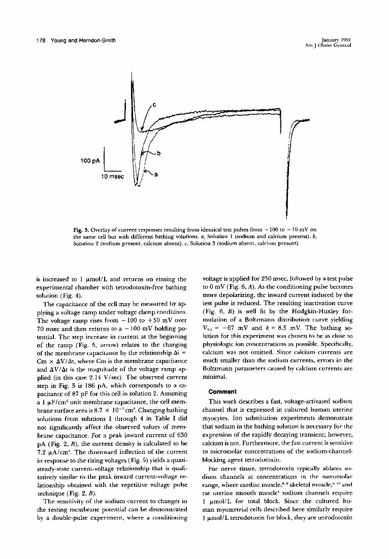
Fig. 3 is a comparison of current responses of thesame cell under differing bathing compositions. Froma holding potential of -100 mY, a test voltage of -10mV was applied after exchange of bathing solution.Tracing a was obtained in the presence of both sodiumand calcium (bath: solution 1) and is composed of a
Fig. 2. A, Inward sodium currents observed with calcium-freebathing solution (solution 2). B, Current-voltage relationshipfor peak inward currents.
fast and a slow component. Tracing b was obtainedafter exchange of bathing solution I with a solutionthat did not contain calcium (bath: solution 2). Underthese conditions, the fast transient persists, but the slowtransient is much reduced. The I msec difference intime-to-peak current is a result of a small loss of timeresolution that occurred during solution change. Exchange of the bathing solution with one that containscalcium but no sodium (bath: solution 3) yields tracingc, where the slow transient is observed but the fasttransient is not. This experiment demonstrates the dependence of the fast transient on the presence of sodium, but not on the presence of calcium, and impliesthat the fast transient is the expression of a sodium
channel.Confirmation of the fast transient as a sodium chan
nel current can be made by investigation of the sensitivity of the fast transient to tetrodotoxin, a specificsodium-channel-blocking agent! The rapidly decayinginward current is lost as the tetrodotoxin concentration
178 Young and Herndon-Smith
100pAL10 msec
b
IiII.,
January 1991Am J Obstet Gynecol
Fig. 3. Overlay of current responses resulting from identical test pulses from -100 to - 10 mV onthe same cell but with different bathing solutions. a, Solution 1 (sodium and calcium present). b,Solution 2 (sodium present, calcium absent). c, Solution 3 (sodium absent, calcium present).
is increased to 1 JLmol/L and returns on rinsing theexp~rimental chamber with tetrodotoxin-free bathingsolution (Fig. 4).
The capacitance of the cell may be measured by applying a voltage ramp under voltage damp conditions.The voltage ramp rises from - 100 to +50 mV over70 msec and then returns to a - 100 mV holding potential. The step increase in current at the beginningof the ramp (Fig. 5, arrow) relates to the chargingof the membrane capacitance by the relationship ~i =Cm X ~V/~t, where Cm is the membrane capacitanceand ~V/~t is the magnitude of the voltage ramp applied (in this case 2.14 V/sec). The observed currentstep in Fig. 5 is 186 pA, which corresponds to a capacitance of 87 pF for this cell in solution 2. Assuminga I JLF/cm2 unit membrane capacitance, the cell membrane surface area is 8.7 X 10- 7 cm2
• Changing bathingsolutions from solutions 1 through 4 in Table I didnot significantly affect the observed values of membrane capacitance. For a peak inward current of 630pA (Fig. 2, B), the current density is calculated to be7.2 JLA/cm2
• The downward inflection of the currentin response to the rising voltages (Fig. 5) yields a quasisteady-state current-voltage relationship that is qualitatively similar to the peak inward current-voltage relationship obtained with the repetitive voltage pulsetechnique (Fig. 2, B).
The sensitivity of the sodium current to changes inthe resting membrane potential can be demonstratedby a double-pulse experiment, where a conditioning
voltage is applied for 250 msec, followed by a test pulseto 0 mV (Fig. 6, A). As the conditioning pulse becomesmore depolarizing, the inward current induced by thetest pulse is reduced. The resulting inactivation curve(Fig. 6, B) is well fit by the Hodgkin-Huxley formulation of a Boltzmann distribution curve yieldingVO.5 = -67 mV and k = 8.5 mY. The bathing solution for this experiment was chosen to be as close tophysiologic ion concentrations as possible. Specifically,calcium was not omitted. Since calcium currents aremuch smaller than the sodium currents, errors in theBoltzmann parameters caused by calcium currents areminimal.
Comment
This work describes a fast, voltage-activated sodiumchannel that is expressed in cultured human uterinemyocytes. Ion substitution experiments demonstratethat sodium in the bathing solution is necessary for theexpression of the rapidly decaying transient; however,calcium is not. Furthermore, the fast current is sensitiveto micromolar concentrations of the sodium-channelblocking agent tetrodotoxin.
For nerve tissue, tetrodotoxin typically ablates sodium channels at concentrations in the nanomolarrange, where cardiac muscle,s.g skeletal muscle,S. 10 andrat uterine smooth muscle4 sodium channels require1 JLmol/L for total block. Since the cultured human myometrial cells described here similarly require1 JLmol/L tetrodotoxin for block, they are tetrodotoxin

Volume 164Number I, Part I
200 PAL4msec
Sodium channels in human myometrium 179
Fig. 4. Sodium current responses to increasing concentrations of tetrodotoxin, followed by washoutof bath with tetrodotoxin-free solution (a, 0; b, 10- 7 mol/L; C, 10-6 mol/L; d, washout). Bath: solution2. Test pulses are from holding potential of -100 to -10 mY.
-100 mV
50mV
t10 msec
Fig. 5. Under voltage clamp conditions, a 70 msec voltage ramp is applied from holding potential(- 100 mV) to + 50 mV. Arrow marks onset of voltage ramp. Current needed to fully charge membrane capacitance is 186 pA, yielding a membrarie capacitance of 87 pF (see text).
sensitive. Persistence of tetrodotoxin sensitivity of sodium channels in these cells, which have undergone atleast three passages in culture, is in contrast to reportson cultured cardiac9 and denervated skeletaPO muscle,where tetrodotoxin sensitivity was rapidly lost.
Recently Ohya and Sperelakis' reported a sodiumchannel in myocytes freshly isolated from uteri of pregnant rats. They reported a maximum current densityof 1.64 ,...,AIcm2 for the sodium channel in comparisonto 7.2 ,...,A/cm2 seen here for cultured human myocytes,The relatively large value for peak sodium current isactually a lower limit, since the time resolution of ourexperiment is at best 3 msec, and the sodium currenttransient has a half-life of 2 to 4 msec. In other respects
the similarity of the sodium channels is remarkable(Table II).
Inoue et al.,ll in freshly isolated myometrium fromhuman pregnancy, failed to observe a voltage-activatedsodium current. The time resolution of the currenttracings in the whole-cell voltage-clamp experiments isnot reported and may not have been sufficient for resolution of the fast sodium current. However, in thatreport the resting membrane potential did vary withexternal sodium concentration.
The physiologic role of the sodium channel is notimmediately apparent. The resting potential of freshlydispersed myometrial cells from human pregnancy hasbeen measured with intracellular microelectrode tech-

180 Young and Herndon-Smith
A
, 130 msec
J~-.If!- ----'l_----,
100pA L20 msec
January 1991Am J Obstet Gynecol
omv ..,.100 mV-~~~~~~~lO .. ·50 mV
B 1.0
0.8
~E 0.6
" 0.4
0.2
a·100 -90 -80 -70 -60
voltage (mV)
-so -40 ·30
Fig. 6. A, Current responses to test pulse to 0 mY after various conditioning pulses (see Methods).Bath: solution I. Conditioning pulse duration is 250 msec. B, Hodgkin-Huxley inactivation curveof peak inward current. Nonlinear regression of data (solid line) yields Yo; = - 67 mY, k = 8.5 mV.
Table II. Comparison of electrophysiologicparameters between sodium channels of rat4
and human myometrium (this work)
Activation (mV) Inactivation (mV)
Onset I Peak Vo., I k
Rat -40 0 -64 7.1Human -40 -10 -67 8.5
Data on rats were obtained on freshly dispersed uterinecells from pregnant rats, whereas data on human cells wereobtained from cultured uterine myocytes. Both sets of activation data were obtained in calcium-free solutions with inactivation data obtained in physiologic calcium solution.
niques to be around - 50 mV, although there was alarge amount of cell-to-cell variability. II. 12 At this potential and under physiologic ion concentrations (bath:solution 1), the sodium channels are significantly inactivated (Fig. 6, B). More important, the inactivationof the sodium channel required a conditioning pulseduration of only 250 msec (Fig. 6, A), which was specifically chosen to be of shorter duration than slow-wavemembrane potential changes. Thus, as a slow wave depolarizes the cell to the potential at which calcium channels begin to open (approximately - 40 mV), 11 the so-
dium channels will be largely inactivated and will notlikely contribute to the rising spike of the action potential. This reasoning is in agreement with our observation that substitution of half of the extracellular sodium with tetraethylammonium chloride fails to reducethe maximal rise of the action potential spike in freshlydispersed human uterine myocytes. 13 Thus sodium currents cannot be demonstrated to significantly contribute to the rising phase of the action potential in humanmyocytes.
Trypsin was used to release the cells from the cultureflasks and may have modified the activation or inactivation properties of the channel. 14 The close agreementof the electrical properties of the sodium channels ofthese human cells with those found for the sodiumchannels of rat uterine cells isolated with the use ofcollagenase4 argues against significant channel modification by enzymes.
Whenever possible, conditions were chosen to minimize deviations from physiologic conditions. For example, suppression of outward currents was found tobe adequate with the use of only 10 mmol/L tetraethylammonium chloride in the electrode solution, although harsher conditions such as total replacement ofintracellular potassium are more effective. The use of

Volume 164Number 1. Part 1
cesium (a potent potassium-channel-blocking agent)was attempted but abandoned because of cell destruction after approximately 10 minutes of experimentation.
We chose a 250 msec duration for the conditioningpulse in the double-pulse sodium-channel inactivationexperiment rather than the usual duration of severalseconds" to determine that inactivation occurs on atime scale that is shorter than that of uterine slow-wavedepolarization. Additionally, 3 mmollL calcium wasused in the sodium-channel inactivation experiment toprevent adverse effects of nonphysiologic calcium solutions. 15
Cultured human uterine cells seem to be an appropriate model system for the study of ion channels ofhuman myometrium. The improved time resolutioninherent to the smaller cell size allows quantitative characterization of the fast sodium channel by means ofsingle-electrode techniques.
REFERENCES
I. Anwer K, Sanborn BM. Changes 'in intracellular freecalcium in isolated myometrial cells: role of extracellular and intracellular calcium and possible involvementof guanine nucleotide-sensitive proteins. Endocrinology1989;124:17-23.
2. Honore E, Amedee T, Martin C, Dacquet C, MironneauC, Mironneau J. Calcium channel current and its sensitivity to (+ )isradipine in cultured pregnant rat myometrial cells. PRuegers Arch 1989;414:477-83.
3. Amedee T, Renaud JF, Jmari K, Lombet A, MironneauJ. Lazdunski M. The presence of Na+ channels in myometrial smooth muscle cells is revealed by specific neu-
Sodium channels in human myometrium 181
rotoxins. Biochem Biophys Res Commun 1986;137:67583.
4. Ohya Y, Sperelakis N. Fast a+ and slow Ca2 + channelsin single uterine muscle cells from pregnant rats. Am JPhysiol 1989;257(Cell Physiol 26):C408-12.
5. Stanfield PRo Tetraethylammonium ions and the potassium permeability of excitable cells. Rev Physiol BiochemPharmacoI1983;97:1-67.
6. Marty A, Neher E. Tight seal whole-cell recording. In:Sakmann B, Neher E, eds. Single channel recording.Plenum Press, New York: 1983 chap 7.
7. Hodgkin AL, Huxley AF. The dual effect of membranepotential on sodium conductance in the giant axon ofLoligo. J Physiol 1952;1l6:497-506.
8. Narahashi T. Chemicals as tools in the study of excitablemembranes. Physiol Rev 1974;54:813-89.
9. McLean MJ, Sperelakis N. Rapid loss of sensitivity to tetrodotoxin by chick ventricular myocyte cells after separation from the heart. Exp Cell Res 1974;86:351-64.
10. Harris JB, Thesleff S. Studies on tetrodotoxin resistantaction potentials on denervated skeletal muscle. ActaPhysiol Scand 1971 ;83:382-8.
II. Inoue Y, Nakao K, Okabe K, et al. Some electrical properties of human pregnant myometrium. AM J OBSTETGYNECOL 1990;162:1090-8.
12. Pressman EK, Tucker JAJr, Anderson NCJr, Young RC.Morphologic and electrophysiologic characterization ofisolated pregnant human myometrial cells. AM J OBSTETGYNECOL 1988; 159: 1273-9.
13. HeschelerJ, Trautwein W. Modification of L-type calciumcurrent by intracellularly applied trypsin in guinea-pigventricular myocytes. J Physiol 1988;404:259-74.
14. Yamamoto Y, Hu SL, Kao Cy' Inward current in singlesmooth muscle cells of the guinea pig taenia coli. J GenPhysiol 1989;93:521-50.
15. Young RC, Smith LH, Anderson NC Jr. Passive membrane properties and inward calcium current of pregnanthuman uterine smooth muscle cells. AM J OBSTET GyNECOL 1991 [In press].


