

Radiographic and Clinical Changes of the Tibial Tuberosity Following Tibial Plateau Leveling Osteotomy
David H. Kergosien, Matthew D. Barnhart, Christopher E. Kees1, Briana G. Danielson,Jeff D. Brourman, William D. DeHoff, and Eric R. Schertel
Department of Surgery, MedVet Associates Ltd., 300 E. Wilson Bridge Road, Worthington, OH 43085
1Center for Research in Scientific Computation, Department of Mathematics, NorthCarolina State University, Raleigh, NC 276958205
ABSTRACTObjective To investigate radiographic changes of the tibial tuberosity following TibialPlateau Leveling Osteotomy (TPLO) surgery and to identify clinical findings and riskfactors associated with such changes. Study Design Retrospective study.Sample population 186 clientowned dogs comprising 219 stifles that underwentTPLO surgery.Methods Patient data surveyed included radiographic changes of the tibial tuberosityduring reexamination, age, body weight, whether unilateral or single session bilateralsurgery had been performed, location of the antirotational pin, approximate tibialtuberosity area, and approximate average tibial tuberosity width. Results Fracture with resulting caudal displacement of the proximal tibial tuberosity (3of 219 or 1.4%) occurred less frequently than nondisplaced tibial tuberosity fractures(16 of 219 or 7.3%). Age, weight, average tibial tuberosity width , location of the antirotational pin, and single session bilateral surgery were identified as risks factors for thenondisplaced fracture. Weight divided by the square of the average tibial tuberositywidth, interpreted as an indicator of the stress on the tibial tuberosity, may be a strongerrisk factor than either weight or average tibial tuberosity width alone. Conclusions Dogs undergoing single session bilateral TPLO surgery are clearly atgreater risk for developing the nondisplaced tibial tuberosity fracture. The statisticalsignificance of the other risk factors is less clear. The nondisplaced tibial tuberosityfracture does not appear to adversely affect outcome or lead to tibial tuberosity avulsion. Significant risk factors for fracture of the proximal tibial tuberosity with caudaldisplacement are unknown, but placement of the antirotational pin distal to the insertion of the straight patellar tendon may weaken the bone. Clinical Relevance Factors including age, weight, tibial tuberosity thickness, andconditions which may enhance strain on the tibial tuberosity, such as single session bilateral procedures, may increase the risk of fractures.
1
INTRODUCTION Cranial cruciate ligament (CrCL) rupture is a common orthopedic problem in
dogs. Tibial Plateau Leveling Osteotomy (TPLO) is a surgical technique that treatsCrCL disease by providing functional stability of the stifle joint during weight bearingby decreasing cranial tibial thrust.13 While the TPLO technique has increased in popularity, few reports document postoperative findings or expectations.
One reported postoperative complication of TPLO surgery is avulsion of thetibial tuberosity.46 Tibial tuberosities cut thin during the osteotomy or tibial plateausthat require a large amount of rotation for leveling may produce a greater risk for avulsion.4 There have been few reports describing the frequency at which tibial tuberosityavulsions occur and none that specify risk factors which may lead to fracture and avulsion.
In two recent retrospective studies of TPLO surgeries, minimally displaced fractures of the tibial tuberosity occurred in 4% and 3.1% of the patients.5, 6 Nine of the 14cases in the first report had no acute exacerbation of lameness, and the fracture wasconsidered an incidental finding.5 The six cases reported in the second study healedwithout specific treatment.6
The observation of a radiographic lucency/fracture of the tibial tuberosity insome patients following TPLO surgery has produced questions as to what changes areclinically significant. The purpose of this study was to investigate radiographicchanges of the tibial tuberosity following TPLO surgery and to identify clinical findings and risk factors associated with such changes. Our hypothesis was that mechanicalcharacteristics of the tibial tuberosity leading to increased bone stresses are associatedwith tibial tuberosity fractures following TPLO surgery.
MATERIALS AND METHODSMedical records of 243 dogs that underwent TPLO surgery between March
2000 and February 2002 were reviewed. Fiftyseven cases were excluded from thestudy because reevaluation radiographs were not available. The resulting group of 186dogs included 219 stifles. Twentytwo of the dogs had single session bilateral TPLOsurgeries performed. Eleven of the dogs had unilateral TPLO surgeries performed atdifferent times for treatment of sequential cranial cruciate ligament disease.
A tibial plateau leveling osteotomy was performed on every stifle reviewed. Allsurgeries were performed by one surgeon according to the patented technique as described by Slocum.4
Reevaluation radiographs were examined for evidence of radiographic changesin the tibial tuberosity. Additional data surveyed included age (years), body weight atthe time of surgery (kg), whether unilateral or single session bilateral surgery had beenperformed, and the location of the antirotational pin.
Immediate postoperative lateral radiographic views of each hind limb from thehock to the stifle were examined in order to derive relevant structural characteristics of
2

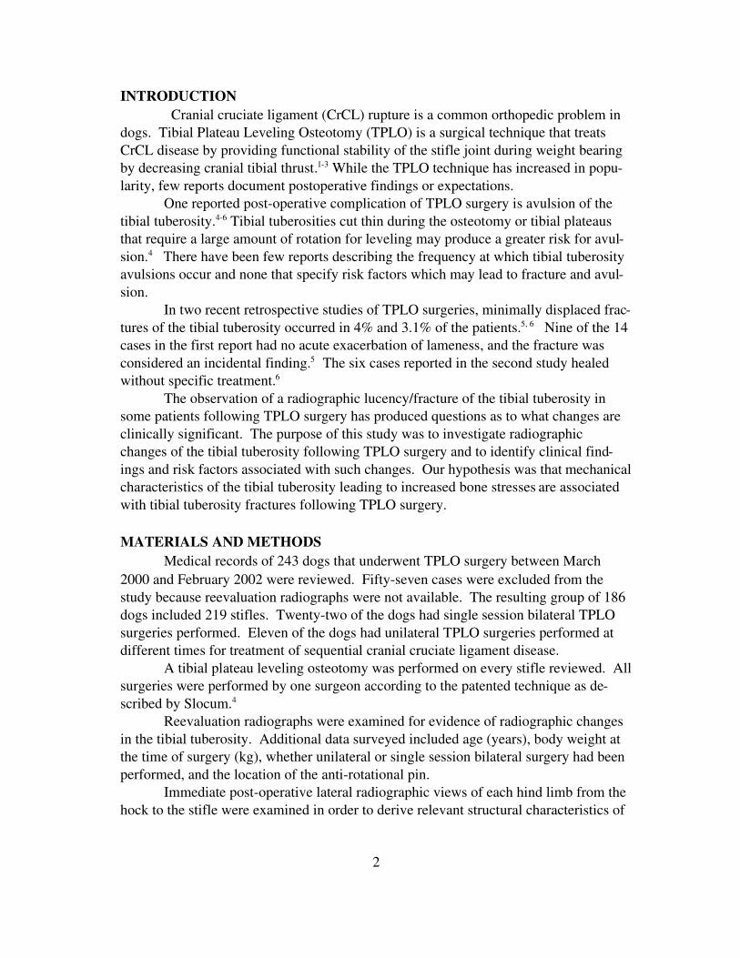
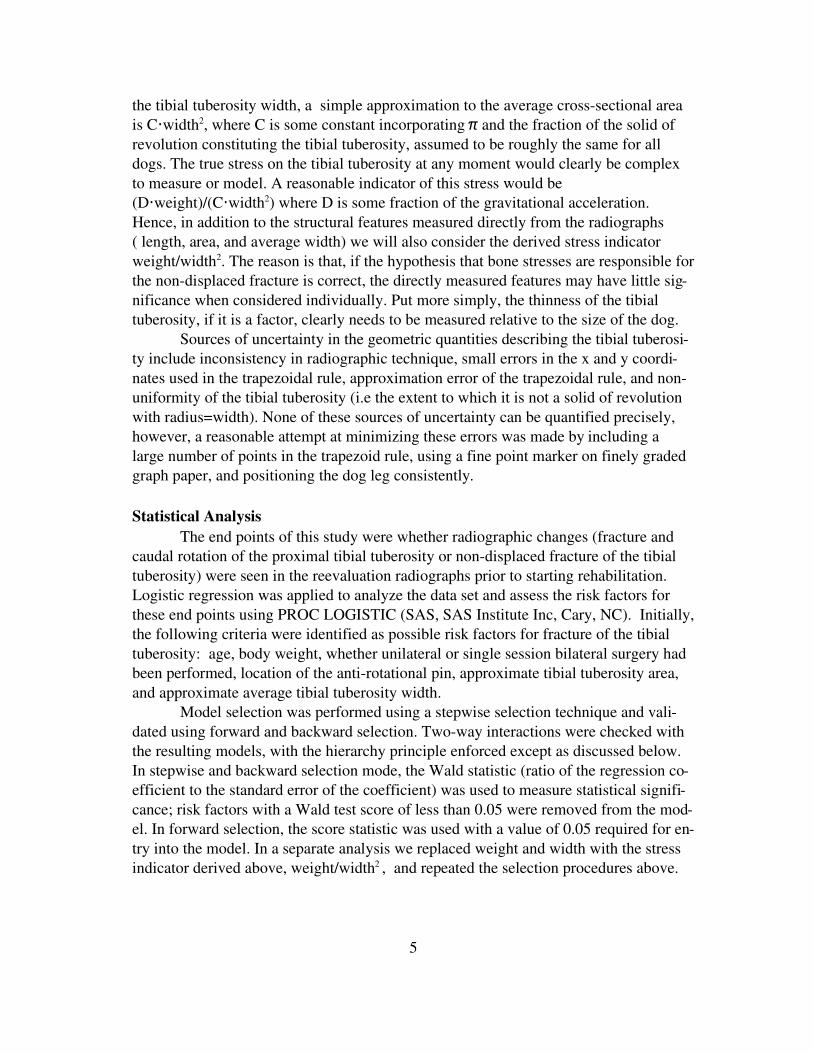
the tibial tuberosity. One surgeon identified each of the anatomic landmarks of the tibia used to define the long axis of the tibia (xaxis). The xaxis was the line drawn alongthe tibial long axis, proximally passing through a point that divided the medial and lateral intercondylar tubercles and a point distally passing through the center of the talus.The boundaries of the tibial tuberosity were then defined as follows (Fig 1). First, aline representing the yaxis at the distal most point of the osteotomy was drawn perpendicular to the tibial long axis. Next, the cranial boundary of the tibial tuberosity wasdefined as the cranial surface of the tibial tuberosity (line f) and the caudal boundary ofthe tibial tuberosity was defined as the radial osteotomy surface (line g) (Fig 1). The yaxis was perpendicular to the tibial xaxis, therefore determining the area bounded bythe yaxis, f, and g (the tibial tuberosity), reduced to finding the area under the curves fand g and then taking the difference (Fig 2).
3
Figure 1. Defining the Boundaries of the Tibial Tuberosity

The trapezoid rule was used to approximate these areas. The length of the tibialtuberosity was defined as the distance between the proximal most aspect of the tibialtuberosity and the distal most aspect of the radial osteotomy. The approximate averagetibial tuberosity width was determined by dividing the approximate tibial tuberosityarea by the length of the tibial tuberosity.
The curves f and g were determined by superimposing graphing paper with 1mm intervals (National Brand Engineering Forms, 10 millimeters to the centimeter)over the immediate postoperative radiographs. The tibial tuberosity was then tracedusing an ultra fine point 1 mm permanent marker. Graph points were recorded at 1 mmintervals for both the cranial boundary (line f) and the caudal boundary of the tibialtuberosity (line g).
Radiographs provide only twodimensional information about the tibial tuberosity. Given the stress on the tibial tuberosity generated by tension on the patellar ligament, the average crosssectional area perpendicular to the area measured above (i.e.going into the radiograph) is a more relevant quantity. Intuitively, the average crosssectional area is the average thickness of the tibial tuberosity. The only information inthe radiograph on the crosssectional area is the average tibial tuberosity width. Assuming the tibial tuberosity is some part of the solid of revolution with radius equal to
4
Figure 2. Measuring the Tibial Tuberosity
the tibial tuberosity width, a simple approximation to the average crosssectional areais C·width2, where C is some constant incorporating π and the fraction of the solid ofrevolution constituting the tibial tuberosity, assumed to be roughly the same for alldogs. The true stress on the tibial tuberosity at any moment would clearly be complexto measure or model. A reasonable indicator of this stress would be(D·weight)/(C·width2) where D is some fraction of the gravitational acceleration.Hence, in addition to the structural features measured directly from the radiographs( length, area, and average width) we will also consider the derived stress indicatorweight/width2. The reason is that, if the hypothesis that bone stresses are responsible forthe nondisplaced fracture is correct, the directly measured features may have little significance when considered individually. Put more simply, the thinness of the tibialtuberosity, if it is a factor, clearly needs to be measured relative to the size of the dog.
Sources of uncertainty in the geometric quantities describing the tibial tuberosity include inconsistency in radiographic technique, small errors in the x and y coordinates used in the trapezoidal rule, approximation error of the trapezoidal rule, and nonuniformity of the tibial tuberosity (i.e the extent to which it is not a solid of revolutionwith radius=width). None of these sources of uncertainty can be quantified precisely,however, a reasonable attempt at minimizing these errors was made by including alarge number of points in the trapezoid rule, using a fine point marker on finely gradedgraph paper, and positioning the dog leg consistently.
Statistical Analysis The end points of this study were whether radiographic changes (fracture and
caudal rotation of the proximal tibial tuberosity or nondisplaced fracture of the tibialtuberosity) were seen in the reevaluation radiographs prior to starting rehabilitation.Logistic regression was applied to analyze the data set and assess the risk factors forthese end points using PROC LOGISTIC (SAS, SAS Institute Inc, Cary, NC). Initially,the following criteria were identified as possible risk factors for fracture of the tibialtuberosity: age, body weight, whether unilateral or single session bilateral surgery hadbeen performed, location of the antirotational pin, approximate tibial tuberosity area,and approximate average tibial tuberosity width.
Model selection was performed using a stepwise selection technique and validated using forward and backward selection. Twoway interactions were checked withthe resulting models, with the hierarchy principle enforced except as discussed below.In stepwise and backward selection mode, the Wald statistic (ratio of the regression coefficient to the standard error of the coefficient) was used to measure statistical significance; risk factors with a Wald test score of less than 0.05 were removed from the model. In forward selection, the score statistic was used with a value of 0.05 required for entry into the model. In a separate analysis we replaced weight and width with the stressindicator derived above, weight/width2 , and repeated the selection procedures above.
5

RESULTSTwo radiographic changes were seen in the tibial tuberosity following TPLO
surgery. Fracture and subsequent caudal displacement of the proximal tibial tuberositywith the tibial plateau (3 of 219 or 1.4%) occurred less frequently than nondisplacedtibial tuberosity fractures (16 of 219 or 7.3%). The clinical findings associated witheach differed greatly.
Cases with caudal displacement of the fractured tibial tuberosity were readmitted to the hospital within the first two weeks following surgery for an acute exacerbation of lameness. Physical examination findings included marked lameness, joint effusion, softtissue swelling, and bruising of the proximal tibia. Of the three stifles withthis complication, two belonged to a grossly obese dog that had undergone single session bilateral TPLO surgery.
Radiographic findings in these cases included caudal displacement of the fractured proximal tibial tuberosity and caudal rotation of the proximal osteotomy segmentresulting in an increased tibial plateau angle. The osteotomy gap between the fracturedproximal tibial tuberosity and the proximal osteotomy segment was unchanged, hencethere was no displacement of the proximal tibial tuberosity relative to the proximal osteotomy segment (Fig 3). Each of these cases had antirotational pins placed throughthe midsection of the tibial tuberosity, at the same level that the fracture occurred. Dueto the small number of cases with this complication, statistically significant risks factors could not be determined.
6

Nondisplaced tibial tuberosity fractures were seen in 16 cases. In each case, radiographs were taken during a scheduled 6 or 8week reevaluation appointment andnone of these cases presented for an acute exacerbation of lameness. The fracture sitewas approximately onehalf way between the proximal most aspect of the tibialtuberosity and the distal most aspect of the osteotomy. Slight obliquity of the fracturewas seen with a craniodistal to caudoproximal orientation (Fig 4). The length of thefracture spanned the entire tibial tuberosity width on lateral radiographic projection.The width of the fracture line ranged from 1 to 5 millimeters, but no displacement ofthe tibial tuberosity or patella alta was observed. No clinical signs were associated withthese findings. In all cases, findings were deemed incidental and normal postoperativerehabilitation was begun.
7
Figure 3. Caudally Displaced Tibial Tuberosity Fracture with Concurrent Caudal Rotation of theProximal Osteotomy Segment

The stepwise selection procedure identified single session bilateral TPLOsurgery, age, weight, and the approximate average tibial tuberosity width as risk factorsfor the nondisplaced tibial tuberosity fracture occurring. Backward selection identifiedthe same set of risk factors while forward selection procedure also identified pin placement as a statistically significant risk factor. We chose to include the pin placement inthe model since it appeared to have some effect. No statistically significant interactionswere identified by any of the selection procedures while enforcing the model hierarchy.The model parameters, Wald score pvalues, standard errors, and 95% confidence intervals are presented in table 1. Some sample odds ratios representing the increase in riskpredicted by the model for several different changes in risk factors is provided alongwith 95% confidence intervals on these odds ratios is given in table 2 . For example, themodel predicts a 2.63 fold in crease in the risk of tibial tuberosity fracture for a 4 yearincrease in age.
8
Figure 4. NonDisplaced Tibial Tuberosity Fracture

In light of the significance of weight and the tibial tuberosity width as risk factors, one possible physiological cause of the nondisplaced tibial tuberosity fracture isstress on the tibial tuberosity via the patellar ligament. As discussed above, a moreappropriate risk factor might then be an approximation of the average and/or peak stresson the tibial tuberosity. The approximation of this stress discussed above would be proportional to weight/width2 . This new variable is an interaction between the initial riskfactors above. Since it has an independent physical significance, however, the
Term Estimate Standard
Error
Wald pvalue Confidence
IntervalIntercept 3.60 1.80 0.046 7.13, 0.06pin 1.17 0.63 0.064 0.07, 2.40age 0.24 0.12 0.046 0.004, 0.48width 0.43 0.18 0.014 0.78, 0.09weight 0.063 0.024 0.009 0.016, 0.11bilateral 2.45 0.71 0.000 1.26, 3.63Table 1. Model Parameters (Hierarchy PreservingModel)
Effect Unit Odds Ratio Confidence Intervalwidth 2 2.38 1.25, 5.05width 4 5.66 1.55, 25.48 width 6 13.47 1.93, 128.6age 2 1.62 1.02, 2.66age 4 2.63 1.03, 7.07age 8 6.94 1.06, 50.04bilateral 1 11.58 3.67, 41.43weight 5 1.37 1.09, 1.77weight 10 1.87 1.19, 3.12weight 20 3.51 1.42, 9.72weight 40 12.35 2.00, 94.58pin 1 3.22 0.93, 11.07Table 2. Odds Ratios (Hierarchy Preserving Model)
hierarchy principle in the selection procedures was not enforced. Replacing weight andwidth by weight/width2 and running the selection procedure above yields the risk factors weight/width2 , pin placement, and bilateral surgery as risk factors. Tables 3 and 4provide the same information for the stressbased model as given previously.
The approximate tibial tuberosity area was not a statistically significant risk factor for the nondisplaced tibial tuberosity fracture. Tibial tuberosity avulsion, as classically described, appears to be a rare complication of TPLO surgery but was not reportedin the study.
9

Term Estimate Standard Error Wald pvalue Confidence Intervalintercept 4.80 0.80 0.0001 6.59, 3.39weight/width2 2.50 1.15 0.029 0.43, 5.06pin 1.39 0.57 0.015 0.27, 2.52bilateral 2.19 0.56 0.000 1.08, 3.30Table 3. Model Parameters (Stressbased Model)
Effect Unit Odds Ratio Confidence Intervalweight/width2 0.2 1.65 1.05, 2.58weight/width2 0.4 2.72 1.11, 6.68weight/width2 0.8 7.39 1.23, 27.08pin 1 4.03 1.30, 12.47bilateral 1 8.93 2.95, 27.08Table 4. Odds Ratios (Stressbased Model)
DISCUSSIONTwo types of fractures were seen in the tibial tuberosity following TPLO
surgery. We observed a caudally displaced fracture of the proximal tibial tuberositywith concurrent caudal rotation of the proximal osteotomy segment, a finding that hasnot been previously reported. In each case the clinical findings were typical of bonefracture: acute exacerbation of lameness, pain, softtissue swelling, and bruising.
Placement of the antirotational pin at the site of the fracture may weaken thebone allowing for fracture to occur. Implant loosening was confirmed during revisionof each TPLO. These cases were repaired with open reduction and internal fixation following the removal and replacement of the TPLO plate and screws and the placementof a tension band wire in the tibial tuberosity. Each fracture healed and limb functionfollowing rehabilitation was good.
It is unclear whether fracture of the tibial tuberosity precedes caudal rotation ofthe plateau or not. We feel that it is more likely that fracture fixation failure of the osteotomy resulted in caudal rotation of the tibial plateau, and that the proximal tibialtuberosity fractured as a result. Two of the three tibial plateaus having this complicationbelonged to the same patient who was grossly obese and had undergone single sessionbilateral TPLO surgery. Placement of the antirotational pin proximal to the Sharpey’ sfibers of the straight patella ligament, the addition of a second TPLO bone plate, and/orstaged unilateral TPLO surgeries may prevent such complications.
The second type of fracture, a nondisplaced tibial tuberosity fracture, shouldnot be confused with an avulsion fracture. Whether this radiographic finding is a truefracture or an osseous lucency created by bony remodeling is not known. A true fracture would be expected to displace proximally due to patellar ligament tension and beassociated with clinical signs. An alternative explanation may be osseous resorption ofthe tibial tuberosity resulting in a focal lucency without an actual fracture occurring.
10

Osseous resorption may be caused by thermal necrosis during the osteotomy, stress remodeling due to changes in normal strain on the tibial tuberosity, or vascular compromise secondary to soft tissue dissection. It is unknown why proximal displacement ofthe tibial tuberosity did not occur in these cases. An incomplete fracture and/or strongsoft tissue attachments may prevent proximal displacement.
Assessment of weight bearing by physical examination for each of these dogswas determined to be appropriate for the stage of recovery. It is possible that a moreobjective assessment of weight bearing such as force plate analysis may identify decreased weight bearing in these patients, or that clinical signs associated with fracturehad resolved by the time of our reevaluation.
Possible causes of the nondisplaced tibial tuberosity fracture include excessivepull from the quadriceps mechanism, vascular compromise secondary to soft tissue dissection, osteotomy gap stress, and thermal damage that occurred during the osteotomy.The data and statistical models in this study provide evidence that the nondisplacedfracture may be due to stress on the tibial tuberosity during recovery. Dogs undergoing single session bilateral TPLO surgery must bear weight on the affected limbs following surgery, and the statistical models predicted 8.5 and 9.6 fold increases in risk forthese dogs. Indeed 10 of the 19 cases with nondisplaced fracture underwent bilateralsurgery while there were only 44 bilateral cases among the 219 cases in the study. Onedog that underwent bilateral single session TPLO surgery developed the lucency on onetibial tuberosity but not the other. The tibial tuberosity with the lucency had a significantly smaller approximate average tibial tuberosity width. Of the remaining 9 nondisplaced fracture cases with unilateral surgeries, 6 had weight/width2 ratios of larger thanthe mean weight/width2 of 0.47 (standard deviation 0.18) with a mean weight/width2
for these 6 cases of 0.70 (standard deviation 0.14). Of the remaining 3 cases, 2 occurred in dogs aged 10 and 11 years. Likewise, the logistic regression analysis suggestsa correlation between the nondisplaced fracture and weight, tibial tuberosity width (orweight/width2 ) and age. Other contributing factors to a stressrelated fracture could beagerelated degradation of the bone strength, faster healing in younger dogs, and weakness due to thermal damage. Immature animals are known to have faster bone healingwhen compared to a mature dog, but the differences in healing between mature andgeriatric patients is not known. Thermal damage and/or vascular compromise could alsohave been a direct mechanism for the nondisplaced fracture but none of the data collected contain any information on such processes.
In 5 of the 16 cases with the lucency, the antirotational pin was placed in themidregion of the tibial tuberosity. In all of these cases the radiographic lucency occurred proximal to the pin tract and a clear distinction between the two was present.Only in the backward selection procedure was the location of the pin identified as arisks factor for fracture, but the authors recommend placement of the antirotational pinproximal to the insertion of the patellar ligament on the tibial tuberosity (Sharpey’ sfibers).
11

Variations in tibial tuberosity length could explain why tibial tuberosity areawas not a risk factor. Based on the technique that we used to approximate area, it waspossible for a long thin tibial tuberosity to have an equal area to that of a short thick tibial tuberosity. Attempts to identify a “ minimally acceptable tibial tuberosity width”were unsuccessful, however the data supports the perception that a thicker tuberosity ismore resistant to fracture.
CONCLUSIONSFactors including age, weight, tibial tuberosity width, and conditions which may
enhance strain on the tibial tuberosity, such as single session bilateral procedures andpin placement, may increase the risk of fractures. Nondisplaced tibial tuberosity fractures do not appear to adversely affect outcome and do not require surgical fixation.
ACKNOWLEDGMENTSThe authors thank Tim Vojt for preparation of the figures, the SAS consulting
group in the Department of Statistics at North Carolina State University for help gettingstarted with the SAS software system, and Dennis Boos in the Department of Statisticsfor his suggestions on the statistical analysis.
REFERENCES:1. Slocum B, Devine T: Cranial tibial thrust: A primary force in the canine stifle.
J Am Vet Med Assoc 183:456459, 19832. Slocum B, Slocum TD: Tibial plateau leveling osteotomy for repair of cranial
cruciate ligament rupture in the canine. Vet Clin North Am 23:777795, 1993.3. Slocum B, Slocum TD: Tibial plateau leveling osteotomy for cranial cruciate
ligament rupture, in Bojrab MJ (ed 4): Current techniques in small animalsurgery. Philadelphia, PA, Lea and Febiger, 1997, pp 12091215
4. Tibial Plateau Leveling Osteotomy Course, November 2000 and 2002,Slocum Enterprises Inc, Eugene, OR
5. Pacchiana PD, Morris E, Gillings SL, et al: Surgical and postoperative complications associated with tibial plateau leveling osteotomy in dogs with cranialcruciate ligament rupture: 397 cases (19982001). J Am Vet Med Assoc222:184193, 2003
6. Priddy NH II, Tomlinson JL, Dodam JR, et al: Complications with and ownerassessment of the outcome of tibial plateau leveling osteotomy for the treatmentof cranial cruciate ligament rupture in dogs: 193 cases (19972001). J Am VetMed Assoc 222:17261732, 2003
12


