

J. Bio. & Env. Sci. 2015
96 | George et al.
RESEARCH PAPER OPEN ACCESS
Conservation of an endangered ornamental and medicinal
aquatic plant, Nymphoides macrosperma Vasudevan through in
vitro propagation
E. Sheeja George1, Aney kutty Joseph1*, Alphi Korath2
1Department of Marine Biology, Microbiology & Biochemistry, School of Marine Sciences, Cochin
University of Science and Technology, Fine Arts Avenue, Kochi, Kerala, India
2School of Management & Entrepreneurship, Kerala University of Fisheries and Ocean Studies,
Panangad, Kochi, Kerala, India
Article published on October 29, 2015
Key words: Nymphoides macrosperma, Aquatic ornamental plant, Endemic, Endangered, In vitro
micropropagation.
Abstract
Nymphoides macrosperma is a valuable aquatic plant from Kerala, South India which is at the risk of extinction
in the nearby future. A protocol for rapid multiple shoot proliferation from the basal buds of this plant was
developed successfully. Treating the explants with 15% concentrated solution of commercial bleach (Robin liquid
bleach, Reckitt Benckiser, India), followed by a quick dip in absolute alcohol was proved to be the best
sterilization procedure to obtain clean cultures. After proper in vitro stabilization on plant growth regulator-free
(PGR-free) Murashige and Skoog (MS) basal medium, earliest shoot bud regeneration was obtained on a medium
containing full strength MS inorganic salts, 100 mg/l myo-inositol, 0.1 mg/l thiamine-HCl, 3% sucrose, 0.8% agar
as gelling agent and supplemented with a combination of 0.5 mg/l 6- Benzyl aminopurine (BAP) and 1.0 mg/l 6-
furfurylaminopurine (kinetin). Maximum multiplication of the in vitro shoots was obtained on MS medium
supplemented with 1.0 mg/l BAP. Lowering the concentration of BAP from 1.0 mg/l favored shoot elongation and
maximum elongation of the in vitro developed shoots was obtained on MS medium supplemented with 0.5 mg/l
BAP as well as on PGR-free MS basal medium. MS medium supplemented with 0.15mg/l indole-3-butyric acid
(IBA) was the best medium for in vitro root formation. The in vitro developed plantlets were successfully
hardened and planted out in the aquarium. N. macrosperma can be mass propagated through the procedure
standardized here so that the plant can be saved from the danger of extinction while being exploited
commercially.
*Corresponding Author: Aneykutty Joseph [email protected]
Journal of Biodiversity and Environmental Sciences (JBES) ISSN: 2220-6663 (Print) 2222-3045 (Online)
Vol. 7, No. 4, p. 96-107, 2015
http://www.innspub.net
J. Bio. & Env. Sci. 2015
97 | George et al.
Introduction
Nymphoides sp., belonging to the family
Menyanthaceae, are valuable aquatic plants having
bright potential to become a multimillion-dollar
commodity in the aquarium trade. Nymphoides
macrosperma Vasudevan, is an endangered species
reported as endemic to Ernakulam in Kerala, South
India (Vasudevan, 1968). It is a dioecious
rhizomatous herb with petiole-like branches bearing
single floating leaves at the apex and flower clusters
at the nodes. The white fringed floral petals and the
light green floating leaves (Vasudevan, 1968) make
the plant attractive in water gardens. The plant is also
used in aquaria as fillers (Ansari and Jeeja, 2006).
Besides being ornamental, N. macrosperma has
medicinal importance too. In South India, a drug by
name ‘Granthika Tagara’ belonging to N.
macrosperma (Yoganarasimhan et al., 1979) is used
in several therapeutic preparations for the treatment
of various diseases such as anaemia, epilepsy, fever,
cough, jaundice and mental disorders (Murali et al.,
2007; Veesam et al., 2012).
N. macrosperma has growing demands in the
aquarium (Personal communication; Unnikrishnan S.
K., Oriental Aquatics, Singapore) and pharmaceutical
industries (Murali et al., 2007; Veesam et al., 2012)
due to its ornamental and medicinal properties. But
as the plant has been facing the challenges of habitat
destruction and water pollution for the past few
decades, it has become rare in its natural habitat and
has been enlisted as ‘endangered’ (Ansari and Jeeja,
2006). Therefore immediate measures should be
adopted for large scale propagation of this plant to
meet the market demands while conserving it in its
natural habitat. It has been reported that, most of the
aquatic plants are difficult to be propagated through
conventional methods as they show infrequent seed
production and slow rate of propagation through
rhizome (Kane et al., 1991; Dissanayake et al., 2007).
It is in this scenario, mass propagation of N.
macrosperma by using the technique of tissue culture
has become a necessity.
The technique of micropropagation offers
large scale production of plantlets within a very short
span of time irrespective of the climatic conditions.
Moreover, the plantlets produced through tissue
culture will be free from pests and diseases. In
microprogation, a plant part (explant) is cultured on a
medium containing optimum concentration of plant
growth regulators so as to induce direct or indirect
(through callus formation) shoot and root
organogenesis. But the exogenous hormone
requirement for shoot and root organogenesis varies
with the species and with the explant that is used for
culture initiation (Chawla, 2002). Moreover, a
standardized surface sterilization is also needed for
getting axenic cultures from the explant. Therefore,
standardization of an appropriate protocol is
necessary for the micropropagation a particular plant.
So far no reports are available on the
micropropagation of N. macrosperma nevertheless
Jenks et al., (2000) and Niranjan and Sudarshana
(2000) have published papers on the in vitro micro
propagation of two other species, Nymphoides indica
and Nymphoides cristatum respectively. The present
study was aimed at standardizing an efficient protocol
for the micropropagation of N. macrosperma.
Materials and methods
Explant sterilization and culture initiation
Mother plants (Figure 1) were collected from River
Periyar in Aluva, Kerala, India. Leaf clusters and roots
were removed from the stolon and the stolons were
washed under running tap water for 10 minutes,
soaked in 5% detergent solution (Cleansol, India) for
10 minutes and again washed thoroughly in running
tap water to remove the superficial dirt. Surface
sterilants tried to obtain axenic cultures included
0.1% mercuric chloride (HgCl2) and commercial
bleach containing 5.25% sodium hypochlorite as
active ingredient (Robin Liquid Bleach, Reckitt
Benckiser, India). To standardize the most suitable
surface sterilization procedure, basal bud explants
were treated with 0.1% HgCl2 for 5 different
durations (1 min, 2 min, 3 min, 4 min, 5 min) or with
six different dilutions of commercial bleach solution

J. Bio. & Env. Sci. 2015
98 | George et al.
(5%, 10%, 15%, 20%, 25% and 30 %) for six different
durations (1 min, 5 min, 10 min, 15 min, 20 min and
25 min). Each treatment was done with 10
replications and number of survival and formation of
healthy cultures with each treatment was recorded
after 4 weeks of explant inoculation. After discarding
the surface sterilant, explants were washed thrice
with sterile water, subjected to a quick dip in 70%
ethanol, and again washed thrice with sterile water.
Inside the laminar air chamber, the basal buds were
isolated and inoculated on medium containing full
strength Murashige and Skoog (MS) inorganic salts
(Murashige and Skoog, 1962), 100 mg/l myo-inositol,
0.1 mg/l thiamine-HCl, 3% sucrose, 0.8% agar as
gelling agent and devoid of any plant growth
regulators (PGRs). The pH of the media was adjusted
to 5.8 using 0.1 N HCl, dispensed in 150×25mm
culture tubes (15 ml medium per tube), and
autoclaved at 1210 C for 15 minute. Cultures were
maintained in a culture room at 25 ± 20C under a 16
hour photoperiod with light intensity of 35 μmol
photons m-2s-1 from Philips cool white fluorescent
tubes. The cultures were subcultured twice at an
interval of 4 weeks on fresh media with the same
media composition for attaining in vitro stabilization
and were then subcultured onto media containing
various PGRs at varying concentrations.
Media used
The media tried to get earliest culture initiation and
shoot regeneration included PGR-free MS basal
medium and MS media supplemented with 0.5, 1.0,
1.5 and 2.0 mg/l BAP or Kinetin or nine different
combinations of BAP and kinetin (Table 1). All the
media used for the in vitro propagation of N.
macrosperma contained MS mineral salts
(Murashige and Skoog, 1962), 100 mg/l myo-inositol,
0.1 mg/l thiamine-HCl, 3% sucrose and 0.8% agar.
Number of days taken for bud release with each
media was recorded; and the in vitro shoots from
each media were subjected to repeated subculture on
the same media to analyze the consistency in shoot
multiplication rate. Observations on shoot
multiplication, shoot elongation and root formation
on each medium were recorded after 4 weeks of third
subculture on the media. In addition to the above said
cytokinin-containing media, in vitro root formation
on MS media supplemented with 0.1, 0.15, 0.2, 0.25
or 0.3 mg/l Indole-3-acetic acid (IAA), IBA or
Naphthalene acetic acid (NAA) was also studied.
Data analysis
To analyze the effect of various concentrations of BAP
(0.5, 1.0, 1.5, 2.0 mg/l) and various concentrations of
kinetin (0.5, 1.0, 1.5, 2.0 mg/l) on culture initiation,
two experiments were set up separately in a
Completely Randomized Design (CRD), each with 5
treatments (including PGR-free MS basal as control)
and 6 replications. The experimental set up to analyze
the effect of various combinations of BAP and kinetin
on culture initiation was in CRD with 10 treatments
(including PGR-free MS basal as control) and 6
replications. Number of days taken for bud release on
each media was taken as the observation. In a similar
way, separate experiments were set up each in a CRD
to analyze the effect of various concentrations of BAP
(5 treatments and 10 replications), various
concentrations of kinetin (5 treatments and 10
replications) and various combinations of BAP and
kinetin (10 treatments and 10 replications) on various
aspects like shoot multiplication, shoot elongation
and in vitro rooting. To standardize the best medium
for shoot multiplication, number of shoots formed on
various media after 4 weeks of third subculture was
taken as the observation. The length (cm) of the in
vitro shoots and the number of in vitro roots per
shoot on each medium were taken as the observations
for standardizing the media for shoot elongation and
in vitro root formation respectively. Determination of
the most suitable media for in vitro root formation
was also based on three separate experiments which
analyzed the effect of different concentrations (0.1,
0.15, 0.2, 0.25 mg/l) of IAA, IBA and NAA on number
of roots formed per shoot. The observations were
taken after 4 weeks of third subculture on each media.
Data were subjected to square root transformation
(except the shoot elongation data) and analyzed by
Univariate Analysis of Variance using SPSS ver. 20.
J. Bio. & Env. Sci. 2015
99 | George et al.
Significant differences if any between the means were
compared by Tukey HSD (Zar, 1999).
Acclimatization of the plantlets
In the present study, hardening was done by culturing
the rooted plantlets on reduced concentration (1/10th)
of MS salts devoid of any plant growth regulators,
agar or sucrose for the first 4 weeks and then on
distilled water for the next 4 weeks under controlled
conditions of 24 ± 20 C under a 12-hours photoperiod
with light intensity of 35 µmol photons m-2s-1. The
plants were then potted in small plastic cups
containing Neopete (Strelings Farm Research
Services Pvt. Ltd., India) which were kept in small
glass tanks for another 4 weeks before their final
transfer to aquaria. The glass tanks contained tap
water reaching up to a height of ¼ of the pots.
Results and discussion
Standardization of Surface Sterilization Procedure
It was observed that, when the basal bud explants
were treated with 0.1% HgCl2, shorter treatment
durations (1.0 min) resulted in contaminated cultures
and longer treatment durations (above 1 minute)
resulted in scorching of the tissue. Only 2 out of 10
rhizome explants treated with 0.1% HgCl2 for 1
minute produced contamination-free healthy cultures
and none of the explants were survived when treated
for more than 1 minute. Explant sterilization using
0.1 to 0.8% HgCl2 have been reported in Piper
longum (Sonia and Das, 2002), Spilanthes acmella
(Haw and Keng, 2003) and Bacopa monnieri
(Sharma et al., 2010).
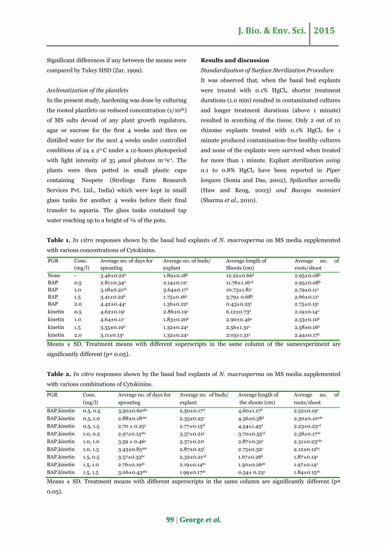
Table 1. In vitro responses shown by the basal bud explants of N. macrosperma on MS media supplemented
with various concentrations of Cytokinins.
PGR Conc.
(mg/l)
Average no. of days for
sprouting
Average no. of buds/
explant
Average length of
Shoots (cm)
Average no. of
roots/shoot
None - 3.46±0.22b 1.89±0.18b 12.22±0.66d 2.95±0.08b
BAP 0.5 2.81±0.34a 2.14±0.12c 11.76±1.16cd 2.95±0.08b
BAP 1.0 3.18±0.21ab 3.64±0.17d 10.73±1.81c 2.79±0.11a
BAP 1.5 3.41±0.22b 1.75±0.16b 3.79± 0.68b 2.66±0.11a
BAP 2.0 4.42±0.44c 1.36±0.22a 0.43±0.25a 2.75±0.15a
kinetin 0.5 4.62±0.19c 2.86±0.19c 6.12±0.73b 2.19±0.14a
kinetin 1.0 4.64±0.11c 1.83±0.20b 2.90±0.46a 2.53±0.10b
kinetin 1.5 3.55±0.19b 1.32±0.24a 2.56±1.31a 2.58±0.16b
kinetin 2.0 3.11±0.13a 1.32±0.24a 2.03±1.31a 2.44±0.17b
Means ± SD. Treatment means with different superscripts in the same column of the sameexperiment are
significantly different (p˂ 0.05).
Table 2. In vitro responses shown by the basal bud explants of N. macrosperma on MS media supplemented
with various combinations of Cytokinins.
PGR Conc.
(mg/l)
Average no. of days for
sprouting
Average no. of buds/
explant
Average length of
the shoots (cm)
Average no. of
roots/shoot
BAP,kinetin 0.5, 0.5 3.30±0.60abc 2.50±0.17d 4.60±1.17d 2.52±0.19e
BAP,kinetin 0.5, 1.0 2.88±0.18abc 2.35±0.25c 4.56±0.58d 2.30±0.10cde
BAP,kinetin 0.5, 1.5 2.70 ± 0.25a 2.77±0.15ef 4.54±1.45d 2.23±0.23cd
BAP,kinetin 1.0, 0.5 2.97±0.13abc 3.37±0.21g 3.70±0.55cd 2.38±0.17de
BAP,kinetin 1.0, 1.0 3.59 ± 0.46c 3.37±0.21g 2.87±0.32c 2.31±0.23cde
BAP,kinetin 1.0, 1.5 3.43±0.85abc 2.87±0.25f 2.75±0.32c 2.12±0.12bc
BAP,kinetin 1.5, 0.5 3.57±0.33bc 2.32±0.21cd 1.67±0.26b 1.87±0.14a
BAP,kinetin 1.5, 1.0 2.76±0.19ab 2.19±0.14bc 1.30±0.26ab 1.97±0.14a
BAP,kinetin 1.5, 1.5 3.06±0.43abc 1.99±0.17ab 0.54± 0.23a 1.84±0.15ab
Means ± SD. Treatment means with different superscripts in the same column are significantly different (p˂
0.05).
J. Bio. & Env. Sci. 2015
100 | George et al.
But in the present study, HgCl2 was found as not
suitable for producing axenic cultures from the
explants. Therefore, various concentrations of
commercial bleach for various durations were tried
for the purpose. Out of the various concentrations of
commercial bleach and various durations of
treatment tried to obtain axenic cultures, treatment
with 15% concentrated solution of commercial bleach
(0.79% Sodium hypochlorite as active ingredient) for
20 minutes followed by a quick dip in 70% ethanol
produced 80% contamination-free explants. When
the treatment duration and bleach concentration
decreased from the optimum level, rate of
contamination was steadily increased; whereas
scorching of the tissue was observed with increase in
bleach concentration and treatment duration. Reports
on explant sterilization using the appropriate
concentration of commercial bleach are available on
Ludwigia repens (Ozturk et al., 2004), Phyla
nodiflora (Ahmed et al., 2005), Rosa damascene
(Nikbakht et al., 2005) and Limnophila aromatica
(George et al., 2014). Present result also support the
use of alcohol in combination with commercial bleach
for surface sterilization of explants as advocated by
Bonga (1982), Jenks et al. (2000) and Maridass et al.
(2010).
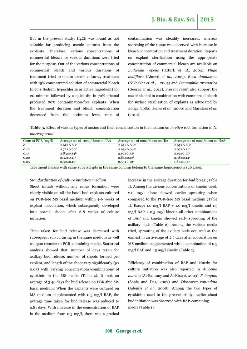
Table 3. Effect of various types of auxins and their concentration in the medium on in vitro root formation in N.
macrosperma.
Conc. of PGR (mg/l) Average no. of roots/shoot on IAA Average no. of roots/shoot on IBA Average no. of roots/shoot on NAA
0 2.95±0.08c 2.95±0.08bc 2.95±0.08d
0.10 2.72±0.09b 2.95±0.08bc 2.07±0.11b
0.15 2.83±0.14bc 3.10±0.32c 2.19±0.12b
0.20 2.32±0.11a 2.84±0.13b 2.38±0.14c
0.25 2.30±0.10a 2.55±0.10a 1.87±0.14a
Treatment means with same superscripts in the same column belong to the same homogenous sub group.
Standardization of Culture initiation medium
Shoot initials without any callus formation were
clearly visible on all the basal bud explants cultured
on PGR-free MS basal medium within 4-6 weeks of
explant inoculation, which subsequently developed
into normal shoots after 6-8 weeks of culture
initiation.
Time taken for bud release was decreased with
subsequent sub culturing in the same medium as well
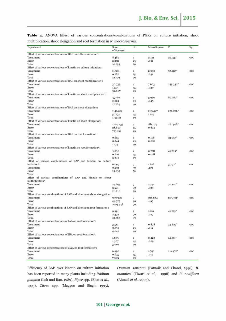
as upon transfer to PGR-containing media. Statistical
analysis showed that, number of days taken for
axillary bud release, number of shoots formed per
explant, and length of the shoot vary significantly (p˂
0.05) with varying concentrations/combinations of
cytokinin in the MS media (Table 4). It took an
average of 3.46 days for bud release on PGR-free MS
basal medium. When the explants were cultured on
MS medium supplemented with 0.5 mg/l BAP, the
average time taken for bud release was reduced to
2.81 days. With increase in the concentration of BAP
in the medium from 0.5 mg/l, there was a gradual
increase in the average duration for bud break (Table
1). Among the various concentrations of kinetin tried,
2.0 mg/l alone showed earlier sprouting when
compared to the PGR-free MS basal medium (Table
1). Except 1.0 mg/l BAP + 1.0 mg/l kinetin and 1.5
mg/l BAP + 0.5 mg/l kinetin all other combinations
of BAP and kinetin showed early sprouting of the
axillary buds (Table 2). Among the various media
tried, sprouting of the axillary buds occurred at the
earliest in an average of 2.7 days after inoculation on
MS medium supplemented with a combination of 0.5
mg/l BAP and 1.5 mg/l kinetin (Table 2).
Efficiency of combination of BAP and kinetin for
culture initiation was also reported in Avicenia
marina (Al-Bahrany and Al-Khayri, 2003), P. longum
(Sonia and Das, 2002) and Dioscorea rotundata
(Adeniyi et al., 2008). Among the two types of
cytokinins used in the present study, earlier shoot
bud initiation was observed with BAP-containing
media (Table 1).
J. Bio. & Env. Sci. 2015
101 | George et al.
Table 4. ANOVA Effect of various concentrations/combinations of PGRs on culture initiation, shoot
multiplication, shoot elongation and root formation in N. macrosperma.
Experiment Sum
of Squares
df Mean Square F Sig
Effect of various concentrations of BAP on culture initiation1:
Treatment
Error
Total
8.483
2.272
10.755
4
25
29
2.121
.091
23.333*
.000
Effect of various concentrations of kinetin on culture initiation1:
Treatment
Error
Total
11.961
0.767
12.729
4
25
29
2.990
.031
97.423*
.000
Effect of various concentrations of BAP on shoot multiplication1:
Treatment
Error
Total
30.733
1.354
32.087
4
45
49
7.683
.030
255.352*
.000
Effect of various concentrations of kinetin on shoot multiplication1:
Treatment
Error
Total
15.760
2.024
17.784
4
45
49
3.940
.045
87.587*
.000
Effect of various concentrations of BAP on shoot elongation:
Treatment
Error
Total
1141.989
50.131
1192.12
4
45
49
285.497
1.114
256.276*
.000
Effect of various concentrations of kinetin on shoot elongation:
Treatment
Error
Total
1724.295
28.897
753.192
4
45
49
181.074
0.642
281.978*
.000
Effect of various concentrations of BAP on root formation1:
Treatment
Error
Total
0.631
0.544
1.175
4
45
49
0.158
0.012
13.057*
.000
Effect of various concentrations of kinetin on root formation1:
Treatment
Error
Total
3.030
0.816
3.846
4
45
49
0.758
0.018
41.785*
.000
Effect of various combinations of BAP and kinetin on culture
initiation1:
Treatment
Error
Total
6.099
0.272
15.033
9
50
59
1.678
.179
3.792*
.000
Effect of various combinations of BAP and kinetin on shoot
multiplication1:
Treatment
Error
Total
24.695
3.521
28.216
9
90
99
2.744
.039
70.140*
.000
Effect of various combinations of BAP and kinetin on shoot elongation:
Treatment
Error
Total
959.973
44.575
1004.548
9
90
99
106.664
.495
215.361*
.000
Effect of various combinations of BAP and kinetin on root formation1:
Treatment
Error
Total
9.991
2.392
12.383
9
90
99
1.110
.027
41.775*
.000
Effect of various concentrations of IAA on root formation1:
Treatment
Error
Total
3.512
0.535
4.047
4
45
49
0.878
.012
73.825*
.000
Effect of various concentrations of IBA on root formation1:
Treatment
Error
Total
1.693
1.307
3.001
4
45
49
0.423
.029
14.571*
.000
Effect of various concentrations of NAA on root formation1:
Treatment
Error
Total
6.990
0.675
7.665
4
45
49
1.748
.015
116.478*
.000
Efficiency of BAP over kinetin on culture initiation
has been reported in many plants including Psidium
guajava (Loh and Rao, 1989), Piper spp. (Bhat et al.,
1995), Citrus spp. (Maggon and Singh, 1995),
Ocimum sanctum (Patnaik and Chand, 1996), B.
monnieri (Tiwari et al., 1998) and P. nodiflora
(Ahmed et al., 2005).

J. Bio. & Env. Sci. 2015
102 | George et al.
Fig. 1. Mother plants of Nymphoides macrosperma
Fig. 2. Multiple shoot formation on MS medium
containing 1.0 mg/l BAP.
Standardization of the medium for shoot
multiplication
In the present study, BAP was more effective than
kinetin on shoot multiplication also. Among the
various media tried, shoot multiplication was highest
on MS medium supplemented with 1mg/l BAP with
an average of 3.64 shoots per explant (Table 1; Figure
2); but with the same level of kinetin in the MS
medium, the average shoot number was 1.83 only.
Among the various concentrations of kinetin tried,
maximum shoot multiplication was observed with 0.5
mg/l kinetin with an average of 2.86 shoots per
explant (Table 1). Even though, BAP and kinetin
combinations produced better multiplication when
compared to the shoot multiplication obtained with
0.5 to 2.0 mg/l kinetin alone and 0.5 mg/l and 1.5
mg/l BAP alone, 1.0 mg/l BAP in combination with
0.5 to 2.0 mg/l kinetin was not better than 1.0 mg/l
BAP alone on shoot multiplication. Among the
various combinations of BAP and kinetin tried, shoot
multiplication was maximum and the same (an
average of 3.3.7 shoots per explant) on 1.0 mg/l BAP
in combination with 0.5 mg/l kinetin and 1.0 mg/l
BAP in combination with 1.0 mg/l kinetin (Table 2).
BAP promotion of shoot multiplication has been
reported in several plants such as P. guajava (Loh
and Rao, 1989), Piper spp. (Bhat et al., 1995), Citrus
spp. (Maggon and Singh, 1995), O. sanctum (Patnaik
and Chand, 1996) and B. monnieri (Tiwari et al.,
1998). Superiority of BAP over other cytokinins for
multiple shoot formation has also been reported in
many fruit plants (Lundergan and Jainic, 1980),
Acorus calamus (Anu et al., 2001) and Myriophyllum
aquaticum (Smitha et al., 2005). However, further
increase in the concentration of BAP or kinetin above
the optimum levels (1.0 mg/l for BAP and 0.5 mg/l
for kinetin) reduced the rate of shoot multiplication
(Table 1). Similar observations on the inhibition of
shoot multiplication by cytokinins beyond the
optimum level have been reported by Ahuja et al.
(1982) in Catharanthus roseus, Tiwari et al. (1998) in
B. monniera, Wang et al. (2004) in Scripus robustus,
Espinosa et al. (2006) in Prunus serotina and
Pandeya et al. (2010) in Clitoria ternatea.
Fig. 3. Shoot elongation on PGR-free MS medium.
Shoot elongation
Analysis of variance (Table 4) showed that, the
average shoot length varies significantly with various
concentrations of BAP or kinetin or various
combinations of BAP and kinetin in the media (p˂
0.05). Multiple shoots formed on MS media
J. Bio. & Env. Sci. 2015
103 | George et al.
containing 0.5 and 1.0 mg/l BAP elongated
considerably (11.76 cm and 10.73 cm respectively) in
the same media (Table 1). It was observed that, the
average length of the in vitro shoots (primary branch
bearing terminal leaf and secondary branches from
the node) decreased with increase in the
concentration of BAP in the media (Table 1). On MS
medium supplemented with 0.5 mg/l kinetin, average
length of the in vitro shoots was 6.12 cm; and a
gradual decrease in average shoot length was
observed with increase in the concentration of kinetin
also. Even though the shoots produced were smaller
when compared to those formed on media containing
0.5 mgl-1 BAP or 1.0 mgl-1 BAP alone, shoot
elongation was satisfactory on media containing 0.5
mgl-1 BAP in combination with 0.5 to 1.5 mgl-1 kinetin
(4.60 to 4.54 cm; Table 2). Therefore, the present
observation is contradictory to the findings of Preece
et al. (1987), Pijut et al. (1991) and Ozturk et al.
(2004) who have observed pronounced inhibition in
elongation and growth of shoots in woody plants,
Pinus strobus and L. repens respectively on media
Thidiazuron (TDZ) and BAP. Among the various
media used in the present study, greatest elongation
of the in vitro shoots was observed on plant growth
regulator-free MS basal medium with an average
length of 12.22 cm (Table 1; Figure 3). This
observation supports the opinion of Singha and
Bhatia (1988), Fasolo et al. (1989), Preece and Imel
(1991) and Ozturk et al. (2004) that, presence of
growth regulators is not essential for the growth of in
vitro developed shoots.
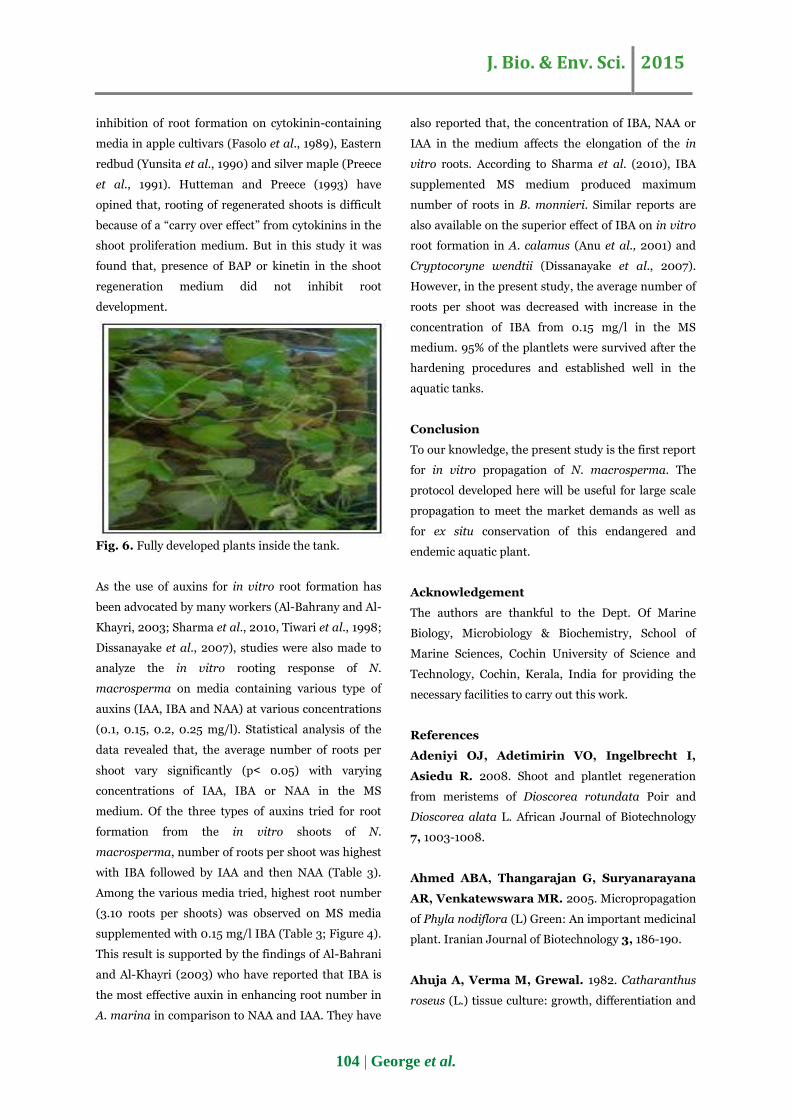
Fig. 4. In vitro root formation on MS medium
containing 0.15mg/l IBA.
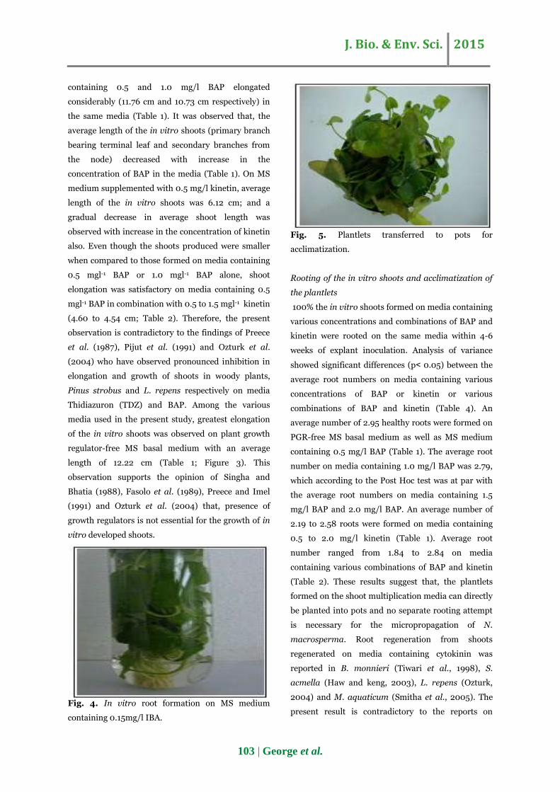
Fig. 5. Plantlets transferred to pots for
acclimatization.
Rooting of the in vitro shoots and acclimatization of
the plantlets
100% the in vitro shoots formed on media containing
various concentrations and combinations of BAP and
kinetin were rooted on the same media within 4-6
weeks of explant inoculation. Analysis of variance
showed significant differences (p˂ 0.05) between the
average root numbers on media containing various
concentrations of BAP or kinetin or various
combinations of BAP and kinetin (Table 4). An
average number of 2.95 healthy roots were formed on
PGR-free MS basal medium as well as MS medium
containing 0.5 mg/l BAP (Table 1). The average root
number on media containing 1.0 mg/l BAP was 2.79,
which according to the Post Hoc test was at par with
the average root numbers on media containing 1.5
mg/l BAP and 2.0 mg/l BAP. An average number of
2.19 to 2.58 roots were formed on media containing
0.5 to 2.0 mg/l kinetin (Table 1). Average root
number ranged from 1.84 to 2.84 on media
containing various combinations of BAP and kinetin
(Table 2). These results suggest that, the plantlets
formed on the shoot multiplication media can directly
be planted into pots and no separate rooting attempt
is necessary for the micropropagation of N.
macrosperma. Root regeneration from shoots
regenerated on media containing cytokinin was
reported in B. monnieri (Tiwari et al., 1998), S.
acmella (Haw and keng, 2003), L. repens (Ozturk,
2004) and M. aquaticum (Smitha et al., 2005). The
present result is contradictory to the reports on
J. Bio. & Env. Sci. 2015
104 | George et al.
inhibition of root formation on cytokinin-containing
media in apple cultivars (Fasolo et al., 1989), Eastern
redbud (Yunsita et al., 1990) and silver maple (Preece
et al., 1991). Hutteman and Preece (1993) have
opined that, rooting of regenerated shoots is difficult
because of a “carry over effect” from cytokinins in the
shoot proliferation medium. But in this study it was
found that, presence of BAP or kinetin in the shoot
regeneration medium did not inhibit root
development.
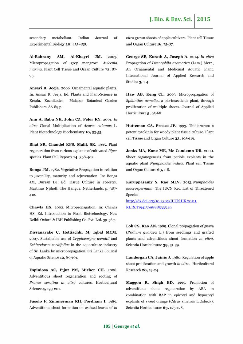
Fig. 6. Fully developed plants inside the tank.
As the use of auxins for in vitro root formation has
been advocated by many workers (Al-Bahrany and Al-
Khayri, 2003; Sharma et al., 2010, Tiwari et al., 1998;
Dissanayake et al., 2007), studies were also made to
analyze the in vitro rooting response of N.
macrosperma on media containing various type of
auxins (IAA, IBA and NAA) at various concentrations
(0.1, 0.15, 0.2, 0.25 mg/l). Statistical analysis of the
data revealed that, the average number of roots per
shoot vary significantly (p˂ 0.05) with varying
concentrations of IAA, IBA or NAA in the MS
medium. Of the three types of auxins tried for root
formation from the in vitro shoots of N.
macrosperma, number of roots per shoot was highest
with IBA followed by IAA and then NAA (Table 3).
Among the various media tried, highest root number
(3.10 roots per shoots) was observed on MS media
supplemented with 0.15 mg/l IBA (Table 3; Figure 4).
This result is supported by the findings of Al-Bahrani
and Al-Khayri (2003) who have reported that IBA is
the most effective auxin in enhancing root number in
A. marina in comparison to NAA and IAA. They have
also reported that, the concentration of IBA, NAA or
IAA in the medium affects the elongation of the in
vitro roots. According to Sharma et al. (2010), IBA
supplemented MS medium produced maximum
number of roots in B. monnieri. Similar reports are
also available on the superior effect of IBA on in vitro
root formation in A. calamus (Anu et al., 2001) and
Cryptocoryne wendtii (Dissanayake et al., 2007).
However, in the present study, the average number of
roots per shoot was decreased with increase in the
concentration of IBA from 0.15 mg/l in the MS
medium. 95% of the plantlets were survived after the
hardening procedures and established well in the
aquatic tanks.
Conclusion
To our knowledge, the present study is the first report
for in vitro propagation of N. macrosperma. The
protocol developed here will be useful for large scale
propagation to meet the market demands as well as
for ex situ conservation of this endangered and
endemic aquatic plant.
Acknowledgement
The authors are thankful to the Dept. Of Marine
Biology, Microbiology & Biochemistry, School of
Marine Sciences, Cochin University of Science and
Technology, Cochin, Kerala, India for providing the
necessary facilities to carry out this work.
References
Adeniyi OJ, Adetimirin VO, Ingelbrecht I,
Asiedu R. 2008. Shoot and plantlet regeneration
from meristems of Dioscorea rotundata Poir and
Dioscorea alata L. African Journal of Biotechnology
7, 1003-1008.
Ahmed ABA, Thangarajan G, Suryanarayana
AR, Venkatewswara MR. 2005. Micropropagation
of Phyla nodiflora (L) Green: An important medicinal
plant. Iranian Journal of Biotechnology 3, 186-190.
Ahuja A, Verma M, Grewal. 1982. Catharanthus
roseus (L.) tissue culture: growth, differentiation and
J. Bio. & Env. Sci. 2015
105 | George et al.
secondary metabolism. Indian Journal of
Experimental Biology 20, 455-458.
Al-Bahrany AM, Al-Khayri JM. 2003.
Micropropagation of grey mangrove Avicenia
marina. Plant Cell Tissue and Organ Culture 72, 87-
93.
Ansari R, Jeeja. 2006. Ornamental aquatic plants.
In: Ansari R, Jeeja, Ed. Plants and Plant-Science in
Kerala. Kozhikode: Malabar Botanical Garden
Publishers, 86-89 p.
Anu A, Babu NK, John CZ, Peter KY. 2001. In
vitro Clonal Multiplication of Acorus calamus L.
Plant Biotechnology Biochemistry 10, 53-55.
Bhat SR, Chandel KPS, Malik SK. 1995. Plant
regeneration from various explants of cultivated Piper
species. Plant Cell Reports 14, 398-402.
Bonga JM. 1982. Vegetative Propagation in relation
to juvenility, maturity and rejuvenation. In: Bonga
JM, Durzan DJ, Ed. Tissue Culture in Forestry.
Martinus Nijhoff: The Hangue, Netherlands, p. 387-
412.
Chawla HS. 2002. Micropropagation. In: Chawla
HS, Ed. Introduction to Plant Biotechnology. New
Delhi: Oxford & IBH Publishing Co. Pvt. Ltd. 39-56 p.
Dissanayake C, Hettiachhi M, Iqbal MCM.
2007. Sustainable use of Cryptocoryne wendtii and
Echinodorus cordifolius in the aquaculture industry
of Sri Lanka by micropropagation. Sri Lanka Journal
of Aquatic Science 12, 89-101.
Espiniosa AC, Pijut PM, Micher CH. 2006.
Adventitious shoot regeneration and rooting of
Prunus serotina in vitro cultures. Horticultural
Science 4, 193-201.
Fasolo F, Zimmerman RH, Fordham I. 1989.
Adventitious shoot formation on excised leaves of in
vitro grown shoots of apple cultivars. Plant cell Tissue
and Organ Culture 16, 75-87.
George SE, Korath A, Joseph A. 2014. In vitro
Propagation of Limnophila aromatica (Lam.) Merr.,
An Ornamental and Medicinal Aquatic Plant.
International Journal of Applied Research and
Studies 3, 1-4.
Haw AB, Keng CL. 2003. Micropropagation of
Spilanthes acmella., a bio-insecticide plant, through
proliferation of multiple shoots. Journal of Applied
Horticulture 5, 65-68.
Hutteman CA, Preece JE. 1993. Thidiazuron: a
potent cytokinin for woody plant tissue culture. Plant
cell Tissue and Organ Culture 33, 105-119.
Jenks MA, Kane ME, Mc Condemn DB. 2000.
Shoot organogenesis from petiole explants in the
aquatic plant Nymphoides indica. Plant cell Tissue
and Organ Culture 63, 1-8.
Karuppasamy S, Rao MLV. 2013. Nymphoides
macrospermum. The IUCN Red List of Threatened
Species
http://dx.doi.org/10.2305/IUCN.UK.20111.
RLTS.T194159A8885335.en
Loh CS, Rao AN. 1989. Clonal propagation of guava
(Psidium guajava L.) from seedlings and grafted
plants and adventitious shoot formation in vitro.
Scientia Horticulturae 39, 31-39.
Lundergan CA, Jainic J. 1980. Regulation of apple
shoot proliferation and growth in vitro. Horticultural
Research 20, 19-24.
Maggon R, Singh BD. 1995. Promotion of
adventitious shoot regeneration by ABA in
combination with BAP in epicotyl and hypocotyl
explants of sweet orange (Citrus sinensis L.Osbeck).
Scientia Horticulturae 63, 123-128.
J. Bio. & Env. Sci. 2015
106 | George et al.
Maridass M, Mahesh R, Raju G, Benniamin A,
Muthuchelian K. 2010. In vitro propagation of
Dendrobium nanum through rhizome bud culture.
International Journal of Biotechnology 1, 50-54.
Murali A, Sudha C, Madhavan V,
Yoganarsimhan SN. 2007. Anticonvulsant and
sedative activity of Tagara (Nymphoides
macrosperma). Pharmceutical Biology 45, 408-410.
Murashige T, Skoog F. 1962. A revised medium
for rapid growth and bioassays with tobacco tissue
cultures. Physiologia plantarum 15, 473-497.
Nikhbakht A, Kafi M, Mirmosoumi M. 2005.
Micropropagation of Damask rose (Rosa damascene)
cvs. Azran and Ghamsar. International Journal of
Agricutlural Biology 8530, 4-7.
Niranjan MH, Sudarshana MS. 2000. In vitro
plant regeneration in Nymphoides cristatum (Roxb)
Kuntze. Phytomorphology 50, 343-344.
Ozturk M, Khawar KM, Atar HH, Sancak C,
Ozcan S. 2004. In vitro Micropropagation of the
aquarium plant Ludwigia repens. Asia pacific Journal
of Molecular Biology 12, 21-28.
Pandeya K, Tiwari KN, Singh J, Varma JP,
Dubey SD. 2010. In vitro propagation of Clitoria
ternatea L: A rare medicinal plant. Journal of
Medicinal Plants Research 4, 664-668.
Patnaik S, Chand PK. 1996. In vitro propagation
of the medicinal herbs Ocimum americanum L. syn.
O. canum Sims. (hoary basil) and Ocimum sanctum
L. (holy basil). Plant Cell Report 15, 846-850.
Pijut PM, Michler CH, Voelker TM. 1991. Effects
of embryo explant orientation, Thidiazuron and agar
on Eastern white pine (Pinus strobus) adventitious
shoot initiation. In: Proc. Intl. Symp. Applications of
Biotechnology tree culture, protection and utilization.
Columbus, OH, 126-127.
Preece JE, Huetteman CA, Puello CA, Neuman
MC. 1987. The influence of Thidiazuron on in vitro
culture of woody plants. Horticultural Science 22,
10-12.
Preece JE, Huetteman CA, Ashby WC, Roth
PL. 1991. Micro and cutting propagation of silver
maple: Results with adult and juvenile propagule.
Horticultural Science 166, 142-148.
Preece JE, Imel MR. 1991. Plant regeneration from
the leaf explants of Rhododendron Hybrids. Scientia
Horticulturae 48, 159-170.
Sharma S, Kamal B, Rathi N. 2010. In vitro rapid
and mass multiplication of highly valuable medicinal
plant Bacopa monnieri (L.) Wettst. African Journal of
Biotechnology 9, 8318-8322.
Singha S, Bhatia SK. 1988. Shoot proliferation of
pear cultures on medium containing Thidiazuron and
Benzylaminopurine. Horticultural Science 23, 802-
806.
Smitha PS, Nazeem PA, Thressiamma J,
Mohan MV, Sherif PM. 2005. Micropropagation
of the aquarium plant-parrot feather milfoil-
Myriophyllum aquaticum (Velloso) Verdcourt,
Contributory paper 38. National Symposium on
Biotechnological Interventions for Improvement of
Horticultural crops: Issues and Strategies. Trissur:
Kerala Agricultural University.
Sonia EV, Das MR. 2002. In vitro
micropropagation of Piper longum- an important
medicinal plant. Plant cell Tissue and Org Culture 70,
325-327.
Tiwari V, Singh DB, Tiwari NK. 1998. Shoot
regeneration and somatic embryogenesis from
different explants of Brahmi [Bacopa monniera
(L.)Wettst.]. Plant Cell Report 17, 538-543.
Vasudevan R. 1968. A new species of Nymphoides
J. Bio. & Env. Sci. 2015
107 | George et al.
(Menyanthaceae) from South India. Kew Bulletin 22,
101-106.
Veesam GK, Devi RD, Vedhahari BN. 2012. In-
vitro Anthelmintic and Anti-Arthritic activity of
Alcoholic extract of Nymphoides macrosperma
Vasud. International Journal of PharmTech Research.
4, 506-509.
Wang J, Seliskar DM, Gallagher JL. 2004. Plant
regeneration via somatic embryogenesis in the
brackish wetland monocot Scripus robustus. Aquatic
Botany 79, 163-174.
Yoganarasimhan SN, Mary Z, Shetty JKP. 1979.
Identification of a market sample of Granthika
Tagara- Nymphoides macrospermum Vasudevan.
Current Science 48, 734-735.
Yusnita S, Geneve RL, Kester ST. 1990.
Micropropagation of white flowering eastern redbud
(Cercis Canadensis var alba L). Journal of
Environmental Horticulture 8, 177-179.
Zar J. 1999. Biostatistical Analysis. Pearson
Education Pte Ltd, Singapore. 98-112 p.


