

CRRT
Review and Refresh Pam Waters, RN
Acute Field Mentor-West US
Region
Baxter-Gambro Renal
Los Angeles, CA
June 11, 2014
Prismaflex 5.1 SW only
Course Objectives
By the end of the Gambro CRRT training course the participant will be able to:
• Discuss the basic CRRT principles
• Discuss the basic principles of the solute transport mechanisms
• Identify the clinical indications for administering CRRT, including an overview of
patient selection and therapy application
• Discuss evidence based practice and supporting research
• Describe the CRRT machine’s safety management features, pressure monitoring
and fluid balance principles.
© 2013, Gambro Lundia AB 2

Continuous Renal Replacement
Therapy (CRRT)
Any extracorporeal blood
purification therapy intended to
substitute for impaired renal
function over an extended period
of time and applied for or aimed
at being applied for 24 hours/day.
Bellomo R., Ronco C., Mehta R, Nomenclature for Continuous Renal
Replacement Therapies, AJKD, Vol 28, No. 5, Suppl 3, Nov 1996
“
”
© 2013, Gambro Lundia AB 3

Why CRRT? CRRT closely mimics the native kidney in treating AKI and fluid overload
• Removes large amounts of fluid and
waste products (urea, creatinine) over
time
• Re-establishes electrolyte and
pH balance
• Tolerated well by hemodynamically
unstable patients
© 2013, Gambro Lundia AB 4

Anatomy of a Hemofilter
• 4 External ports
• Blood and dialysis fluid
• Potting material
• Support structure
• Hollow fibers
• Semi-permeable membrane
• Outer casing
© 2013, Gambro Lundia AB 5

Hemofilter: Semi-permeable membrane Allows solutes (molecules or ions) up to a certain size to pass through
© 2013, Gambro Lundia AB 6

CRRT Transport Mechanisms

CRRT Modes of Therapy
• SCUF: Slow Continuous
Ultrafiltration. Primary goal is to
remove patient fluid
• CVVH: Continuous Veno-Venous
Hemofiltration. Primary goal is to
achieve small, medium and large
molecule clearance, remove
patient fluid
• CVVHD: Continuous Veno-
Venous HemoDialysis. Primary
goal is to achieve small molecule
clearance, remove patient fluid
• CVVHDF: Continuous Veno-
Venous HemoDiaFiltration.
Primary goal is to achieve highly
effective small, medium and large
molecule clearance, remove
patient fluid
All modes will assist in maintaining hemodynamic stability due to the gentle and
gradual fluid removal as tolerated by the patient MAP.
© 2013, Gambro Lundia AB 8

Flow Control Unit – Pumps
© 2013, Gambro Lundia AB 9

Molecular Weights
* Filter has a 50K cut off
© 2013, Gambro Lundia AB 10

Ultrafiltration The movement of fluid through a semi-permeable membrane driven by a
pressure gradient (hydrostatic pressure)
© 2013, Gambro Lundia AB 11

Diffusion = Hemodialysis The movement of solutes only from an area of higher concentration to an
area of lower concentration
* Filter has a 50K cut off
© 2013, Gambro Lundia AB 12

Major factors affecting diffusion
Solute removal by diffusion depends on:
• Concentration gradient blood / dialysis
• Dialysate flow rate
• Molecular size – diffusion clears small molecules
• Permeability of the membrane
© 2013, Gambro Lundia AB 13

How does diffusion work???
© 2013, Gambro Lundia AB 14

Convection “Solute drag”= hemofiltration The forced movement of fluid with dissolved solutes
(the fluid will drag the solutes)
© 2013, Gambro Lundia AB 15

Major factors affecting convection
Solute removal by convection depends on:
• High Membrane permeability
• Molecular size
• Degradation of filter membrane (can decrease performance)
• Replacement fluid flow rate (pressure gradient)
© 2013, Gambro Lundia AB 16

How Pre or Post Replacement
works! Post Replacement
• The replacement “fluid volume” will
be removed by the effluent pump.
• Blood will be concentrated ↑Hct.
• Post-filter replacement solution will
deliver replacement solution to
“replace” the removed “volume” and
replenish lost electrolytes.
Pre Replacement
• Pre-filter replacement solution will
deliver into the blood flow at set rate.
• Blood will be diluted ↓Hct.
• The replacement “fluid volume” will
be removed by the effluent pump.
© 2013, Gambro Lundia AB 17

Pre-filter replacement
Reference #3
© 2013, Gambro Lundia AB 18

Post-filter replacement
Reference #3
© 2013, Gambro Lundia AB 19

Why do we need to monitor Filtration
Fraction percentage (FF%)? • A FF > 25% can lead to
premature filter degradation
• To decrease the FF%,
prescribed fluid delivery
strategies may need to be
initiated such as a mix of pre-
and post-dilution.
• For accurate Filtration Fraction
percentage monitoring, the
patient’s hematocrit should be
updated once a day.
© 2013, Gambro Lundia AB 20
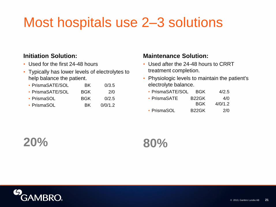
Most hospitals use 2–3 solutions
Initiation Solution:
• Used for the first 24-48 hours
• Typically has lower levels of electrolytes to
help balance the patient.
• PrismaSATE/SOL BK 0/3.5
• PrismaSATE/SOL BGK 2/0
• PrismaSOL BGK 0/2.5
• PrismaSOL BK 0/0/1.2
Maintenance Solution:
• Used after the 24-48 hours to CRRT
treatment completion.
• Physiologic levels to maintain the patient's
electrolyte balance.
• PrismaSATE/SOL BGK 4/2.5
• PrismaSATE B22GK 4/0
BGK 4/0/1.2
• PrismaSOL B22GK 2/0
80% 20%
© 2013, Gambro Lundia AB 21

Considerations for solution choice
Which mode of therapy?
• CVVH: PrismaSOL
• CVVHD: PrismaSATE
• CVVHDF: Both Solutions
• Be aware: 0.9% saline average Ph is 5.0 to 5.6. frequent bag changes
Which anticoagulant prescribed?
• Systemic, regional or none
What else is happening to the patient?
• Ventilatory settings, vasoactive drugs etc
© 2013, Gambro Lundia AB 22

Adsorption Molecular adherence to the surface or interior of the membrane.
© 2013, Gambro Lundia AB 23
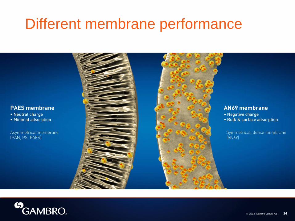
Different membrane performance
© 2013, Gambro Lundia AB 24

Questions
© 2013, Gambro Lundia AB 25

Who should be treated with CRRT?
© 2013, Gambro Lundia AB 26

AKI patient conditions
Reference #16
© 2013, Gambro Lundia AB 27

Definition of AKI
2.1.1: Acute kidney injury (AKI) is defined as any of the following:
• Increase in SCr by ≥ 0.3 mg/dl within 48 hours; or
• Increase in SCr to ≥ 1.5 times baseline, which is known or presumed to
have occurred within prior 7 days; or
• Urine volume <0.5 ml/kg/h for 6 hours
Reference #13
© 2013, Gambro Lundia AB 28

RIFLE and AKIN Criteria
Reference #4
© 2013, Gambro Lundia AB 29

Fluid overload A biomarker for treatment initiation?
Fluid overload with AKI was independently associated with mortality.
Reference #10
© 2013, Gambro Lundia AB 30

How to calculate % FO
Reference #10
© 2013, Gambro Lundia AB 31

Chapter 3.1: Prevention and Treatment of AKI
3.4.1: We recommend not using
diuretics to prevent AKI (1B)
Reference #13
© 2013, Gambro Lundia AB 32

Chapter 5.6: Modality of RRT for Patients with AKI
5.6.1: Use continuous and intermittent RRT as complementary
therapies in AKI patients. (Not Graded)
5.6.2: We suggest using CRRT rather than standard intermittent
RRT, for hemodynamically unstable patients. (2B)
5.6.3: We suggest using CRRT, rather than intermittent RRT, for
AKI patients with acute brain injury or other causes of
increased intracranial pressure or generalized brain edema.
(2B)
Reference #13
© 2013, Gambro Lundia AB 33

Comparison of IHD, SLEDD
and CRRT
Intermittent Hemodialysis SLEDD CRRT
Duration = 4 hours Duration = 6–12 hours Duration = 24 hours
Blood Flow = around 400 ml/min Blood Flow = 150–300 ml/min Blood Flow = 150–250 ml/min
Fluid used = Dialysate only Fluids used = Dialysate only
Fluids used = Dialysate &
Replacement solutions
Fluid Rates = 500–800 ml/min
Fluid Rates =
100-300 ml/min
Fluid Rates =
34–68 ml/min (2–4 L/hr)
Non Sterile Dialysate Non Sterile Dialysate
Sterile Dialysate &
Replacement solutions
Typical Net Fluid Removal =
0–1000 ml/hr
Typical Net Fluid Removal =
0–500 ml/hr
Typical Net Fluid Removal =
0–200 ml/hr
© 2013, Gambro Lundia AB 34

Correction of Fluid Overload: CRRT vs IHD
PICARD Study group
says CRRT is better
than IHD for fluid
removal!
Reference #10
© 2013, Gambro Lundia AB 35

Timing of RRT initiation: Starting RRT early may be associated with
improved outcomes!
Reference #14, 15
“Early” initiation of RRT has been associated with better
outcomes for AKI patients.
The published studies assessing the effect of timing of
RRT initiation are largely observational and have used
variable definitions of “early” vs. “late.” Nevertheless, two
meta-analyses involving critically ill AKI patients treated
with RRT showed that “early” RRT initiation was associated
with significantly reduced mortality risk compared to “late”
initiation.
© 2013, Gambro Lundia AB 36

Timing of RRT Initiation: Meta-Analysis
Karvellas et al, Crit Care 2011
Pooled OR of 0.45 for early start
© 2013, Gambro Lundia AB 37

© 2013, Gambro Lundia AB 38

KDIGO Clinical Practice Guideline
Chapter 5.8: Dose of RRT in AKI 5.8.4: We recommend delivering an effluent volume of 20-25
ml/kg/hr for CRRT in AKI (1A). This will usually require a
higher prescription of effluent volume. (Not Graded)
Reference #13
© 2013, Gambro Lundia AB 39

Prescribed vs Delivered
5.8.4: We recommend delivering an effluent volume of 20–25
ml/kg/hr for CRRT in AKI (1A). This will usually require a
higher prescription of effluent volume. (Not Graded)
5.8.1: The dose of RRT to be delivered should be prescribed
before starting each session of RRT.(Not Graded). We
recommend frequent assessment of the actual delivered
dose in order to adjust the prescription. (1B)
5.8.2: Provide RRT to achieve the goals of electrolyte, acid-base,
solute, and fluid balance that will meet the patient’s needs.
(Not Graded)
Reference #13
© 2013, Gambro Lundia AB 40

Key Take-aways
• Ensure your CRRT dose prescription is delivered!
• Urea is a traditional marker for chronic dialysis efficacy, CRRT
provides benefits above and beyond urea clearance
• Major contributors to under-delivery of CRRT dose can be
patient or treatment related
• CRRT provides slow, continuous and gentle replacement of
renal function…as close to native kidney function as possible!
© 2013, Gambro Lundia AB 41

Prescription screen on set up
© 2013, Gambro Lundia AB 42

Case study
• Patient: 82kg Female
• ICU LOS: 3 days
• Previously fit and well with no comorbidity
• Diagnosis: Pneumonia & Sepsis
• Labs
• Creatinine 1.1 mg/dL
• BUN = 67 mg/dL
• K+ = 5.9 mEq/L
• WBC’s = 31,000
• Intake in last 48 hrs = 11, 545 ml
• Output in last 48 hrs = 1,350 mls
• Ventilated
• MAP of 59 mmHg on
• Norepinephrine at 12 mcgs/min
• Dopamine at 20 mcgs/kg/min
• Urine output 0.2 mls/hr/kg with lasix
Questions:
Is the patient hemodynamically stable?
NO. Patient is on vasopressors and MAP is still low, which means the patient is hemodynamically unstable. KDIGO suggests using CRRT, rather than intermittent RRT, for hemodynamically unstable patients (2B)
What is % FO?
11.545L – 1.350L = 10.1.95 / 82kg = 0.12432927 X
100% = 12.4% FO which is associated with increased
mortality per PICARD
Is staging of AKI appropriate? Yes, because with increased stage of AKI, the risk of
death and need for renal replacement therapy (such as
CRRT) increases.
Can we remove fluid in this patient?
Yes, if done over 24 hours per KDIGO
What dose of CRRT should the patient be given?
82kg X 25mls/hr/kg effluent (minimum) = 2050mls/hr of
replacement and dialysate combined.
Add in estimated downtime of 20% = 2050mls/hr +
410mls/hr = 2460mls/hr = 30mls/hr/kg effluent dose.
Reference #10, 13
© 2013, Gambro Lundia AB 43

PACE: Applying what we know Patient, Access, Circuit, Equipment
The patient is a
part of the circuit
© 2013, Gambro Lundia AB 44

Check the patient!
Cardiac status Patient weight
Patient temperature Patient labs
Hemodynamic status – vital signs Sedation Level
Intravascular volume Chest tubes
Ventilator Status - Mode, reverse I:E
ratio, Positive pressure ventilation,
oscillator
Abdominal Pressure
Patient position – HOB 30º, prone,
rotation etc
Intra-aortic Balloon pump
Compartment syndrome
© 2013, Gambro Lundia AB 45

PACE: Applying what we know Patient, Access, Circuit, Equipment
Vascular Access Catheter
© 2013, Gambro Lundia AB 46

Vascular access: Location A veno-venous double or two single lumen venous catheters
Internal Jugular Vein
• Lower risk of complication
• Simplicity of catheter insertion
Femoral Vein
• Optimal site for immobilized patient
• Easiest site for insertion
Subclavian Vein
• Higher risk of pneumo/hemothorax
• Associated with central venous stenosis
© 2013, Gambro Lundia AB 47

Vascular access catheter: Important considerations
Desired characteristics:
see KDIGO guideline pg 101
• Size: Adults 11 french or larger
• Adequate Length
• Optimal Placement
Number ONE Circuit Management
Issue
Refer to and follow the hospital protocol
for specific guidelines
Vascular Access recommendations:
• Aspirate and discard anticoagulant before
flushing
• 10ml to 30ml syringe to assess patency
• Check for kinks/ clamps
Reference #13
© 2013, Gambro Lundia AB 48

CRRT Blood Flow Rate
Blood Flow Rate:
Machine Limits:
10ml/min - 450ml/min
Recommended: adults:
• Minimum 100 ml/min
• Preferred: Blood flow rate must be adequate
for the fluid removal rate
Considerations:
Vascular Access
• Size and patency
Hemofilter selection
Anticoagulation
Patent catheter
Blood flow rate, choice of filter and vascular
access site / size
should all compliment each other
Reference #12
© 2013, Gambro Lundia AB 49

PACE: Applying what we know Patient, Access, Circuit, Equipment
Hemofilter Set
© 2013, Gambro Lundia AB 50

Anticoagulation strategies
• Therapeutic on coumadin/warfarin
• Coagulopathy from various reasons – sepsis, liver failure
• Systemic anticoagulant – Heparin
• If HIT+ then consider regional or none
• Regional anticoagulant - Citrate
© 2013, Gambro Lundia AB 51

Why?
Aim of anticoagulation during CRRT is to prevent clotting of the
circuit in order to:
• preserve filter performance
• increase circuit survival
• minimize loss of blood due to increased circuit changes
Impact of filter clotting:
• CRRT is only continuous if anticoagulation is adequate
• decrease in clearance
• increase in filter changes
• wasted nursing time
• increase in cost
• Patient blood loss – may be MDR reportable!
© 2013, Gambro Lundia AB 52

Filter viability Trans-membrane Pressure (TMP)
• Pressure exerted on filter
membrane during operation
• Reflects pressure difference
between fluid and blood
compartments of filter
• Calculated by Prismaflex
software
© 2013, Gambro Lundia AB 53

Trans-Membrane Pressure (TMP)
Calculated and automatically
recorded:
• Entering Run mode - blood
flow is stabilized
• Blood flow rate is changed
• Patient fluid removal rate is
changed
• Replacement solution rate is
changed
© 2013, Gambro Lundia AB 54

Filter viability Filter Pressure Drop (ΔP Filter)
• Change of pressure from
blood entering filter and
leaving filter
• Determines pressure
conditions inside hollow fibers
• Calculated and automatically
recorded:
• Entering Run mode
• Blood flow rate is changed
• Calculated by Prismaflex
software
© 2013, Gambro Lundia AB 55

PACE: Applying what we know Patient, Access, Circuit, Equipment
CRRT machine delivers prescribed
therapies and solutions
© 2013, Gambro Lundia AB 56

Modality
Choose modality based on patient needs and desired outcomes
© 2013, Gambro Lundia AB 57

Questions
© 2013, Gambro Lundia AB 58
Questions
Thank you!

References
1) Bellomo R., Ronco C., Mehta R, Nomenclature for Continuous Renal
Replacement Therapies, AJKD, Vol 28, No. 5, Suppl 3, Nov 1996
2) Beginning and Ending Supportive Therapy for the Kidney. (2005, August
17). Acute renal failure in critically ill patients: a multinational, multicenter
study. JAMA, 7, 813-818.
3) Cerda, J. & Ronco , C. (2010). Choosing a RRT in AKI. In J. A. Kellum, R.
Bellomo, & C. Ronco (Eds.), Continuous Renal Replacement Therapy (pp.
79 - 92). New York, USA: Oxford University Press
4) DOse REsponse Multicentre International Collaborative Initiative. (2009,
April 15). Delivered dose of renal replacement therapy and mortality in
critically ill patients with acute kidney injury. Critical Care, 1-14.
http://dx.doi.org/doi:10.1186/cc7784
5) Karvellas, C. J., Farhat, M. R., Sajjad, I., Mogenson, S. S., Lueng, A. A.,
Wald, R., & Bagshaw, S. M. (2011, Feb 25). A comparison of early versus
late initiation of renal replacement therapy in critically ill patients with acute
kidney injury: a systematic review and meta-analysis. Critical Care, 15.
6) Prismaflex Tutorial Version 5.1 DVD
7) Prismaflex Operators manual. Version 5.10 of the Prismaflex software
contains the "libdmtx" library ("the Library"), Copyright © 2008, 2009 Mike
Laughton, Copyright © 2011 Gambro Lundia AB, released under the GNU
Lesser General Public License Version 2.1 ("the License"). A copy of the
License is attached to the manual. The user may obtain code in accordance
with section 6(c) of the License by contacting Gambro Lundia AB, Legal and
Intellectual Property Department.
8) Prismasate Specifications sheet
9) Prismasol Specifications sheet
10) Program to Improve Care in Acute Renal Disease. (2009, May 13). Fluid
accumulation, survival and recovery of kidney function in critically ill patients
with acute kidney injury. Kidney International, 76, 422-427.
11) Ricci, Z., Baldwin, I., & Ronco, C. (2010). Alarms and Troubleshooting. In J.
A. Kellum, R. Bellomo, & C. Ronco (Eds.), Continuous Renal Replacement
Therapy (pp. 121-128). New York, USA: Oxford University Press
12) Ricci, Z., Baldwin, I., & Ronco, C. (2010). Nonanticoagulation stratagies. In
J. A. Kellum, R. Bellomo, & C. Ronco (Eds.), Continuous Renal
Replacement Therapy (pp. 129-140). New York,
USA: Oxford University Press
13) Schlondorff, D., Ross, M., & Al-Awqati, Q. (Eds.). (2012). KDIGO Clinical
Practice Guideline for Acute Kidney Injury [Special issue]. Kidney
International, 2(1).
14) Sepsis Occurrence in Acutely Ill Patients. (2008, June 4). A positive fluid
balance is associated with a worse outcome in patients with acute renal
failure. Critical Care, 12(3), 1-7.
15) The RENAL Replacement Therapy Study Investigators. (2009, Octonber
22). Intensity of Continuous Renal-Replacement Therapy in Critically Ill
Patients. The New England Journal of Medicine, 361, 1627 – 1238.
16) 16) Uchino S, Kellum JA, Bellomo R, et al. Acute Renal Failure in Critically
Ill Patients: A Multinational, Multicenter Study. JAMA. 2005;294(7):813-818.
doi:10.1001/jama.294.7.813.
17) Zarbrock, A., & Singbartl, K. (2010). Vascular access for continuous renal
replacement therapy. In J. A. Kellum, R. Bellomo, & C. Ronco (Eds.),
Continuous Renal Replacement Therapy (pp. 93-99). New York, USA:
Oxford University Press.
18) http://www.fistulafirst.org/LinkClick.aspx?fileticket=GN8QYytKHFo%3d&tab
id=39
19) http://www.fistulafirst.org/Home.aspx
20) http://www.accessdata.fda.gov/cdrh_docs/pdf5/K052719.pdf
21) http://www.accessdata.fda.gov/cdrh_docs/pdf5/K051727.pdf
22) http://labtestsonline.org/understanding/analytes/urinalysis/tab/test
© 2013, Gambro Lundia AB 59


