

Iraqi Journal of Agricultural Sciences –2021:52(1):170-188 Jameel & Haider
170
DETERMINATION OF THE OPTIMUM CONDITIONS FOR
BIOSURFACTANT PRODUCTION BY LOCAL ISOLATE OF
LACTOBACILLUS PLANTARUM AND EVALUATE ITS ANTIMICROBIAL
ACTIVITY A .A. Jameel N. H. Haider
Researcher Prof.
Depart. of Biotech, Coll. Sci, University of Baghdad - Iraq
Email: [email protected]
ABSTRACT
Eighty five local isolates of Lactobacillus sp. which were isolated from different sources and
identified by biochemical test then subjected to the primary and secondary screening
processes to select the active Lactobacillus sp. isolate for biosurfactant production. Among the
isolates screened, twenty six isolates with maximum for tests in primary screening were
selected for secondary screening. It has been found that Lactobacillus sp. ADK2 had the
highest productivity of the biosurfactant. The selected isolate with highest level of
biosurfactant activity was identified as Lactobacillus plantarum ADK2 according to PCR
technique. The optimum conditions of biosurfactant production by isolate Lactobacillus
plantarum ADK2 using submerged fermentation were obtained in the synthetic mineral salt
medium (MSM) and natural BCDFTM medium the best production medium separately, 1.5%
(lactose and egg) as the best carbon source, 2% meat extract and 3.5% Pease as nitrogen
source, temperature 30 °C for two media and pH 5 with pH 3 in MSM and BCDFTM
respectively, after 96 hr and 72 hr in MSM and BCDFTM respectively of incubation period.
KEYWORDS: Emulsification, lipopeptide, BCDFTM medium.
جميل وحيدر 188-170(:1 (52: 2021-مجلة العلوم الزراعية العراقية
وتقييم LACTOBACILLUS PLANTARUMالمحلية بكترياال من اتيالمستحلب الحي لإنتاجتحديد الظروف المثلى فعاليته المايكروبية
علياء عبد الحسين جميل ناظم حسن حيدر باحث استاذ
جامعة بغداد -كلية العلوم -الاحيائية قسم التقنيات مستخلصال
من مصادر مختلفة وشخصت باختبارات كيموحيوية ثم .Lactobacillus spتم عزل خمسة وثمانون عزلة من بكتريا الغربلتين اظهرت ستة .عرضت الى غربلة اولية وثانوية لاختيار افضل عزلة لإنتاج المستحلب الحياتي من بين العزلات
وعشرون عزلة افضل انتاج للمستحلب الحياتي من الغربلة الاولية واختيرت للغربلة الثانية وجدت من خلال النتائج بان العزلة ADK2 Lactobacillus ثم تشخيص العزلة التي اظهرت اعلى انتاجية للمستحلب .لها اعلى انتاجية للمستحلب الحياتي
ثم تحديد الظروف المثلى لإنتاج . PCRبالاعتماد على تقنيات تفاعل البلمرة L.plantarum ADK2الحياتي يانها باستخدام عمليات تخمير المغمور في وسط الاملاح المعدنية L.plantarum ADK2المستحلب الحياتي من قبل العزلة
من اللاكتوز %1.5افضل وسط كاربوني كان باستخدام .بشكل منفصل (BCDTTM)المصنع وفي الوسط الطبيعي افضل رقم ,للوسطين اعلاه %30افضل حرارة كان ,من البزاليا كمصدر نتروجيني %3.5% مستخلص اللحم و 2 ,والبيض
المصنع الاملاح المعدنية طساعة من التخمير في وس 96في الوسط الطبيعي بعد 3لوسط الاملاح المعدنية و 5هيدروجيني .ساعة في الوسط الطبيعي 72و
BCDFTM سط انتاجو ،الببتيد الدهني استحلاب،الكلمات المفتاحية: Received:16 /2/2020, Accepted:17/5/2020

Iraqi Journal of Agricultural Sciences –2021:52(1):170-188 Jameel & Haider
171
INTRODUCTION Biosurfactants are surface active agents with
wide range of properties including reduction of
surface and interfacial tensions of liquids,
Surface active compounds produced by
microorganisms are of two main types; first,
that reduce surface tension at the air water
interface (biosurfactants) and second, that
reduce interfacial tension between immiscible
liquids, or at the solid-liquid interface
bioemulsifier (45). Surfactants are extensively
used for industrial, agricultural, food, cosmetic
and pharmaceutical applications. Most of these
surfactants are chemically synthesized and are
potentially toxic to the environment . (44)
Biosurfactants usually display emulsifying
capacity but bioemulsifier do not necessarily
reduce surface tension .Biosurfactants have
important advantages relative to chemically
synthesized surfactants, such as higher
biodegradability, low toxicity, greater
environmental compatibility, better foaming
properties and stable at extreme pH, salinity
and temperature.(32) Microbial surfactants are
considered to be secondary metabolites, play
important role for the survival of biosurfactant
producing microorganisms by facilitating
nutrient transport or microbe-host interactions
or by acting as biocide agents(26,29), bacterial
pathogenesis and biofilm formation.(8,9)
Bacteria are the main group of biosurfactant-
producing microorganisms, although they are
also produced by some yeasts and filamentous
fungi ,These compounds can be synthesized by
microorganisms growing on water-immiscible
hydrocarbons, as well as on water-soluble
compounds such as glucose, sucrose, glycerol,
or ethanol, and can be excreted or remain
attached to the cell wall (33). Diversity
existing among the biosurfactant-producing
microorganisms suggests that their production
represents an important survival strategy and
appears to have evolved in an independent, yet
parallel fashion (30). A number of studies have
reported the potential of lactobacilli as
biosurfactant producers (43). Biosurfactants
isolated from several lactobacilli have been
characterized as multicomponent mixtures,
consisting of protein and polysaccharides , in
other cases, the surface active compounds
were identified as glycolipids (53).The
chemical structure of the biosurfactants
produced by lactobacilli was examined from
different bacterial species: the L.helveticus
derived biosurfactant is mainly constituted by
lipid and sugar fractions; the L.pentosus,
L.lactis and L.paracasei biosurfactants are
glycoproteins or glycolipopeptides, while the
L.plantarum biosurfactants are of glycolipidic
or glycoproteic nature (14). Currently, the
main factor that prevents the widespread use
of biosurfactants is the process economics, and
many strategies have been developed to reduce
its production costs and make fermentation
competitive with chemical synthesis (39). The
use of inexpensive substrates like agro-
industrial wastes, medium and culture
conditions optimization, development of
efficient recovery process, and the engineering
of the producer microorganisms can contribute
to make their production more economically
attractive through the development of cheaper
and efficient processes (41,48). Future
biosurfactant research should, therefore, be
more focused on the economics of the
production processes, particularly through the
use of alternative low-cost fermentative media
(22). The antimicrobial activity of two
biosurfactants obtained from probiotic
bacteria, Lactococcus lactis 53 and
Streptococcus thermophilus A, have been
investigated against a variety of bacterial and
yeast strains isolated from explanted voice
prostheses and it was found that both the
biosurfactants have a high antimicrobial
activity even at low concentration (39).
Probiotics have long been known for their
antimicrobial activity and for the capacity to
interfere with the adhesion and formation of
biofilms of pathogens to epithelial cells of
urogenital and intestinal tracts, catheter
materials and voice prostheses and the
mechanisms of this interference have been
demonstrated to include, among others, the
release of biosurfactants (40). The Current
study was aimed to collect and identify of
local isolates of Lactobacillus sp., as well as
screening and evaluation the ability of local
isolates for production of biosurfactant,
determination the optimum conditions for
biosurfactant production from selected isolate
by using synthetic and neutral medium.

Iraqi Journal of Agricultural Sciences –2021:52(1):170-188 Jameel & Haider
172
MATERIALS AND METHODS
Chemicals and new natural medium MRS agar, and all other reagent grand
chemicals were purchased from Oxoid, Hi-
Media and Sigma - Aldrich, India, and natural
products, fruits and vegetable were obtained
from local market.
Samples collection and bacterial isolation One hundred fifteen samples were collected
from different sources as follow: (humans,
dairy products, fermented fruits, vegetables,
salted shrimp, pickles and al-sadder honey)
(Table 1). One gm. of (fermented fruits,
vegetables and salted shrimp) and one ml of
(dairy products, pickles and al-sadder honey)
were added to 9 ml of MRS broth and
incubated at 37˚C for 48 hr. in the presence of
3-5% CO2 by using Candle Jar, then one ml of
sample was added to 9 ml of 0.1% peptone
water in test tubes and dilution steps when
carried out until 10-6
were done. Then samples
were cultured on MRS agar medium and
incubated at 37˚C for 48 hr. in the presence of
3-5% CO2 by using Candle Jar (13).To prevent
the growth of fungal in cultures, 0.1%
antifungal (Nystatin) was added to cultures.
The isolates were purified by sub culturing on
MRS- agar as a selective media (12), then the
purified colonies were maintains on the same
media, thereafter gram staining ,biochemical
tests, grown on blood, chocolate agar. The
highest production isolates were identified by
PCR , these isolates were prepared for
screening experiments for biosurfactant
production according to method described by
Anandaraj and Thivakaran (4).
Screening the Lactobacillus sp. isolates for
biosurfactant production
Primary screening (semi-quantitative
screening)
Screening of isolates in blood agar plate
medium (Hemolysis test): Eighty five isolates
of Lactobacillus sp. were screened to select
higher producing isolates for biosurfactant
production by plate assay using blood agar
medium. One hundred microliter of bacterial
culture previously activated in MRS broth was
filled into the each well made in blood agar
medium using cork borer, and then incubated
at 37°C for 24 hr. in different aeration
conditions (aerobic and anaerobic using
anaerobic Jar). growth zone around the wells
was indication of biosurfactant secretion. The
radius of a zone was measured using electronic
ruler in mm (4).
Table 1. Different sources were collected for Lactobacillus isolation Samples No. of samples Source
Humans
Vaginal swabs 40 Were obtained from
healthy premenopausal
women in Medical City
Hospital, Baghdad
Mouth cavity 10 healthy people from
male and female with
ages between (20-30)
years
Salvia 10
Colostrum 10 During breastfeeding
Feces new born 13 10-40 days
Dairy products
Goat milk 4 Locally
Goat cheese 4 Turkey
Canoon 4 Iraq
Activia 4 Iraq-Irbil
Fermented Fruits and vegetables
Lemon 2 Locally
Orange 2 Locally Orange mandarin 2 Locally
Banana 2 Locally
Tomato 2 Locally
Other sources
Salted shrimp 2 Turkey
Pickles 2 Locally
Al–Sadder honey 2 Locally
Iraqi Journal of Agricultural Sciences –2021:52(1):170-188 Jameel & Haider
173
Screening of isolates in phenol red plate
medium: All 64 isolates were grown on
phenol red agar medium containing (g/l)
(Agar-Agar 15 gm, FeSO4. 7H2O 0.00028 gm,
K2HPO4 4.4gm, KCl 1.1gm, KH2PO4 3.4gm,
MgSO4. 7H2O 0.5, NaCl 1.1gm, NaNO3 15gm,
Peanut oil1.0 % (w/v), Yeast extract 0.5gm,
with adding 0.2 of Phenol red gave dark blue
zone. Then the clear zone that ferreted were
measured by electronic ruler.
Screening of isolates in blue agar plate
medium: All 45 isolates that grown on
previous media gave the clear zone were
culture on CTAB/ methylene blue agar
medium containing (g/l) (Agar-Agar 15g,
CTAB 0.2g, Glucose 5g, Methylene blue
0.005g, Peptone10g, and Yeast extract0.5g, pH
was adjusted to 7.3). One hundred microliter
of bacterial culture previously activated in
MRS broth was loaded into the each well
prepared in MSM with CTAB/methylene blue
using cork borer, and then incubated at 37°C
for 48 hr. in different aeration conditions
(aerobic and anaerobic by using anaerobic
Jar). A dark blue halo zone appearance
around the culture was an indication of
biosurfactant secretion. The radius of
inhibition zone was measured using electronic
ruler in mm.
Drop collapse assay In this method all 42 isolated tested, the
interfacial tension between the drop containing
the surfactant and the parafilm surface is
reduced which results in the spread of the
drop. Twenty five microliter of fresh bacterial
culture which activated in MRS broth
previously conditions, were pipetted as a
droplet on the parafilm. Distilled water was
used as negative control. The flattening of
droplet and spreading of the droplet on the
parafilm surface was observed. safranin was
added for staining purpose (55).
Penetration assay In this assay, the cavities of a 96 well
microplate were filled with 150 μl of a
hydrophobic paste made up of oil and silica
gel. The paste was covered with 20 μl of oil.
10 μl of a red staining solution (safranin) was
added to 90 μl of the bacterial culture for all
36 isolates. The coloured bacterial culture was
then placed on the surface of the paste (31).
Oil spreading test: All 9 isolates tested,
twenty ml of distilled water was added to a
petri plate then 20 μl of engine oil were add to
the surface of water. Twenty microliter of
bacterial culture, were placed onto the center
of oil membrane. Diameter of clearly oil
displaced circles was measured (33).
Screening of isolates in crystal violet plate
medium : All 8 isolates that gave a positive
results on the previous test were selected and
cultured on crystal violet agar medium
containing previous media (phenol red plate
above) with addition 100 µl of crystal violet
replacement phenol red. The isolate were
screened as in previous experiment above.
Biuret test
The Biuret test was used to detect the presence
of lipopeptide biosurfactants for six isolates.
Two ml of the bacterial culture which growing
in different aeration conditions (aerobic and
anaerobic in anaerobic Jar), were heated at 70
ºC before mixing with 1M NaOH solution.
Then, drops of 1% CuSO4 were slowly added
to observe any color change violet or pink ring
(15).
Secondary screening (quantitative
screening)
Biosurfactant production in liguid media:
Tow isolates with maximum productivity
based from primary screening were selected
and cultivated on MSM containing (g/l) (CaCl2
. 2H2O 0.1g, FeSO4. 7H2O 0.05g, K2HPO4
1.0g, KCl 1.1g, KH2PO4 0.5g, MgSO4.
7H2O0.6g, MnSO4 . 7H2O 0.03g , Na2MoO4.
2H2O 0.001g, NaCl5.0g, NH4NO3 1.0g, Peanut oil 2.0 % (w/v), pH was adjusted to 6.2
). A 250 ml flasks containing 50 ml of mineral
salt medium was inoculated with 1.0 ml (1x10⁸
cell/ml) of 48 hr. cultures of isolates. The
flasks were incubated under shaking (150 rpm)
at 37 ᵒC for 72 hr. in different aeration
conditions (aerobic and anaerobic by using N2
gas (flashing system). Then, the cultures were
centrifuged at 4 ᵒC, 8000 rpm, for 10 min. The
clear supernatant was considered as
extracellular biosurfactant production, the cells
were washed twice with distilled water and
resuspended in 10 ml of phosphate buffer
saline. The cells were then incubated at room
temperature for 4 hrs. with gentle stirring for
biosurfactant production. Thereafter, the broth
was centrifuged at 8000g for 10 min. The

Iraqi Journal of Agricultural Sciences –2021:52(1):170-188 Jameel & Haider
174
supernatant was considered as intracellular
biosurfactant. The supernatant was then
filtered through sterile 0.22 mm pore size filter
(Millipore) (52).
Measurement of surface tension
The surface tension of an aqueous solution
was measured by the Wilhelmy platinum plate
with a QBZY-2 Tensiometer (China). Twenty
ml of supernatant was poured into 50 ml glass
beaker and put onto the tensiometer platform.
The measurement was carried out at 25±1 ºC
after dipping the plate in the solution until
monitoring the value of supernatant surface
tension following the procedure of
measurement written in the manual of the
instrument. Between each measurement, the
Wilhelmy plate was rinsed with acetone and
burned by alcohol burner. For more accurate
value, the average of three records was used in
the study (45).
Determination of emulsification activity (E
24 :%( Two ml of cell free supernatant was
added to 2 ml of toluene (equal volumes v/v),
mixed with vortex for 2 min., and left for 24
hrs. at room temperature, the height of
emulsifier layer was measured. The
emulsification index was given as a percentage
of the height of the emulsified layer (mm) to
the total height of the liquid column (mm)
multiplied by 100 (10).
Bacterial adhesion to hydrocarbons
(BATH): The hydrophobicity of the cells can
be measured by BATH assay. Absorbance of
the suspension was measured at 600 nm (A0),
A one hundred μl of motor oil was mixing
with 2ml of cell suspension and was vortex
shaken for 3 min in test tubes. After mixing,
crude oil and aqueous phase were allowed to
separate for 1hour. The aqueous phase was
carefully removed. OD of the aqueous phase
was then measured at 600nm (A1) in a
spectrophotometer (28). Hydrophobicity is
expressed as the percentage of cell adherence
to crude oil and was calculated as follows:
H% = (1-A/A0) *100
Identification of Lactobacillus spp
The higher production Lactobacillus isolate of
biosurfactant was identified by using
sequences of the 16S ribosomal RNA, DNA
extraction, polymerase chain reaction (PCR).
The universal bacterial primer set was used to
amplify 16S rRNA from the genomic. The
PCR-amplified 16S rRNA fragments were
amplified using two universal primers, 27F: 5-
AGAGTTTGATCCTGGCTCAG 3- and
1492R:5-
CGGTTACCTTGTTACGACTT3-
.Primers. Solutions which used is X TAE
buffer, loading dye, DNA ladder marker,
Ethidium bromide (10mg / ml). The PCR
reaction mixture was prepared as (Table 2).
Table 2. Master mix components of PCR
The PCR cycling conditions for the set were a
touchdown approach of 30 cycles as follows: 1
cycle of genomic DNA was initial denatured at
95°C for 1 min, followed by 30 cycles of 95°C
for 30 sec, 60°C for 1 min, and 72°C for 1 min
with a final extension step I cycle of 72°C for
7 min. A 10 min incubation at 10°C was added
to the end of PCR program. PCR product
were send for Sanger sequencing using
ABI3730XL, automated DNA sequencer, by
Macrogen Corporation – Korea. The results
were received by email then analyzed using
genious software.
Optimum conditions of biosurfactant
production
Effect of fermentation media: The influence
of different fermentation media on the
production of biosurfactant was examined by
cultivation the selected isolate Lactobacillus
sp. (ADK2) in different culture media include
synthetic media (MSM and MRS medium) and
new natural media (Whey medium and
BCDFT medium containing (g/l) Banana 5g,
Corn 7g, Date 3g, Fig 3g, Tomato 4g).
Erlenmeyer flasks 250 ml containing 50 ml of
each tested medium in duplicates were
Master mix Components Stock Unit Final Unit Volume 1 Sample
Master Mix 2 X 1 X 12.5
Forward primer 10 μM 1 μM 1
Reverse primer 10 μM 1 μM 1
Nuclease Free Water 8.5
DNA 10 ng/μl 10 ng/μl 2
Total volume 25
Aliquot per single rxn
23 μl of Master mix per tube and
add
2μl of Template

Iraqi Journal of Agricultural Sciences –2021:52(1):170-188 Jameel & Haider
175
sterilized and inoculated with 1% (1x10⁸
cell/ml) of overnight culture of the isolate.
The flasks were incubated in a shaker
incubator (150 rpm) at 37 ºC for 72 hr. in
anaerobic conditions by using N2 gas (flashing
system). After incubation, the cultures were
centrifuge at 8000 rpm for 10 min. The cells
from each flask were washed twice in distilled
water and resuspended in 10 ml of phosphate
buffer saline. The cells were then incubated at
room temperature for 4 hrs. , with gentle
stirring for biosurfactant production. After 4
hrs, the broth was centrifuged at 8000g for 20
min.The supernatant was taken for
determination the emulsification activity and
surface tension in all the following
experiments.
Effect of pH Erlenmeyer flasks (250 ml) containing 50 ml
of the selected fermentation media (new
natural media BCDFTM and synthetic media
MSM) were adjusted using 0.1N HCL or 0.1N
NaOH to obtain different pH values (3, 4, 5, 6,
7, 8 and 9) ,150 rpm at 37 ºC for 72 hr, then
the culture medium was inoculated with 1% of
overnight culture of the selected isolate
(1x10⁸cell/ml) as previous experiment above.
Effect of temperature Biosurfactant production was achieved at
different temperatures (15, 30, 44, 53, and
60°C) . Before sterilization the media, the pH
was adjusted to (3.0 in natural media and 5.0
in synthetic media), then sterilized and
inoculated with 1% of overnight culture (1x10⁸
cell/ml) 150 rpm of the selected isolate
(ADK2) as in previous experiment above.
Effect of agitation speed Different rpm values (120, 140, 180, 200 and
220 rpm) were examined to determine the
optimum shaking required to obtain the high
biosurfactant activity. The synthetic with pH
5.0 and natural media with pH 3.0 at 30°C
were prepared and inoculated with of selected
isolate then incubated at selected rpm as
above.
Effect of carbon sources Different carbon sources were used in MSM
include (fructose, sucrose, glycerol, starch, and
lactose) , while boiled rice water, frying oil,
beef, cheese, and egg (47) were added
separately in new natural medium respectively
to determine the optimum carbon source for
biosurfactant production, each of these sources
was added to the medium in (1g/100 ml).
Then, pH was adjusted to 5.0 in MSM and 3.0
in new natural medium at 30°C 120 rpm, and
inoculated with 1% of overnight culture (1x10⁸
cell/ml) of the selected isolate (ADK2) as
previous experiment above.
Effect of carbon source concentration Different concentrations (1.5, 2, 2.5, 3 and 3.5
%) of (egg and lactose) as a carbon sources
were used in synthetic and neutral media
respectively to grow the bacterium in order to
determine the optimum concentration of
selected carbon sources for biosurfactant
production. pH was adjusted to 3.0 and 5.0 at
30°C in 120 rpm respectively. The flasks were
inoculated with 1% of overnight culture (1x10⁸
cell/ml) of the selected isolate (ADK2) as
previous experiment above.
Effect of nitrogen sources To determine the effect of nitrogen source on
biosurfactant production medium, synthetic
fermentation media supplemented with
(1g/100ml) of different nitrogen sources (
yeast extract, meat extract, urea, peptone, and
NaNO3) in MSM, at pH 5.0, while in new
natural medium using (peas, chickpeas, oat,
potato, peach), pH 3.0 at 30°C in 120 rpm.
After sterilization, the flasks were inoculated
with 1% of overnight culture (1x10⁸
cell/ml) of
the selected isolate (ADK2) as in previous
experiment above.
Effect of nitrogen source concentration The optimal nitrogen sources (peas and meat
extract) were added in gradual
concentration(1.5, 2, 2.5, 3 and 3.5 %) to the
new natural media and mineral salt medium
respectively. pH was adjusted to 3.0 and 5.0 at
30°C in 120 rpm respectively, then inoculated
with 1% of overnight culture (1x10⁸
cell/ml) of
the selected isolate (ADK2) as previous
experiment above.
Effect of incubation period In order to determine the optimum incubation
time for biosurfactant production, the time
course for biosurfactant production was
followed 0, 4, 16, 24, 48, 72, 96, 120 and 144
hr, pH was adjusted to 3.0 in natural media
and 5.0 in MSM. The two media were
prepared and inoculated with 1% of overnight
culture (1x10⁸
cell/ml) of the selected isolate

Iraqi Journal of Agricultural Sciences –2021:52(1):170-188 Jameel & Haider
176
(ADK2) incubated in different time at 30°C in
120 rpm.
Antimicrobial activity of crude
biosurfactant
A twenty ml Muller Hinton Agar was
prepared for petriplates each. All the
petriplates were swabbed with pathogenic
isolates (Staphylococcus aureus and
Pseudomoas aeruginosa). Fifty microliter of
cell free supernatant was loaded into the each
well prepared in MHA with using cork borer,
and then incubated at 37°C for 48 hr. After
incubation, the plates were checked for the
appearance of zone of inhibition. The radius of
inhibition zone was measured using electronic
ruler in mm (59).
RESULTS AND DISCUSSION
Bacterial isolation and identification : One
hundred fifteen samples were collected from
different sources. The samples were primarily
grown onto MRS agar plates as selective
media for isolation and incubated at 37 °C for
48 hr. with the presence of (3-5 %) CO2 by
using Candle Jar. The results were showed that
only eighty five isolates were found belongs to
genus Lactobacillus which subjected to
morphological, microscopy, and biochemical
tests in order to confirm their identification.
The isolates were identified as related to the
genus Lactobacillus by their small (2-5 mm),
convex, smooth, glistening colonies, and
opaque without pigment on MRS, no
hemolysis on blood, chocolate agar as Figure
1-A, 1-B, and 1-C. Microscopically, the
bacteria appeared under oil immersion lens
(100x) as gram positive bacilli, arranged
singly, pairs or short chains as shown in Figure
1-D .While the use biochemical test as
compared with identification schematic
diagram of Kotzamanidis et al, (28). Also the
results were revealed that all isolates were
negative for oxidase tests, catalase tests, and
indole tests.
Figure 1. Microscopically and morphology
examination of the Lactobacillus sp.
bacterial isolates under large objective lens
and different plates agar (A) grown on
MRS agar (B) grown on blood agar (C)
grown on chocolate agar and (D) visualized
at (100x) under microscope.
Screening of Lactobacillus spp. isolates for
biosurfactant production:
Primary screening (semi-quantitative
screening)
Screening of isolates in blood agar plate
medium (Hemolysis test : Hemolytic activity
assay is a primary method for screening a
biosurfactant producer. All eighty five isolates
were screened on blood agar plates. Sixty four
isolates showed positive results for haemolytic
activity by formation of a clear zone around
the colonies, with a diameter ranged from
(9.18-26.14) mm. The results also showed that
the isolate (ADK2) revealed the highest clear
zone (26.14 mm) under anaerobic condition
(intracellular) Figure 2, this result indicated
that the isolate Lactobacillus was able to
produce biosurfactants. The blood agar
method, is widely used to screen for
biosurfactant production. Rodrigues and
Teixeira (39) were showed that the culture of
Lactobacillus species producing beta
haemolysis was able to produce biosurfactants.
Rodrigues et al, (40) showed that the size of
the clear zone developed is in proportion to the
amount of the produced biosurfactant.
A
D C
B

Iraqi Journal of Agricultural Sciences –2021:52(1):170-188 Jameel & Haider
177
Figure 2. (A and B). Biosurfactant
producing isolate ADK2 on blood agar
medium (C): control using only distal water
Screening of isolates in phenol red plate
medium: Forty five isolates only were
biosurfactant producer through the formation
of clear inhibition zone of reduction around the
phenol plate well Figure 3. These isolates
gained different clearance zone ranged from
(15.20-27.66) mm. The isolate ADK2 showed
the highest clear zone (27.66 mm) under
anaerobic condition. phenol red or otherwise
called Phenolsulfonphthalein is a pH indicator
commonly used in cell biology laboratories. Is
contain salt (sodium salt) this salt inhibits most
bacteria dependent type of microorganisms, It
is found in most culture media such as
mannitol salt agar (MSA), modified oxford
agar (MOX), XLT-4 Agar and HardyCHROM
A agar (19).
Figure 3. (A and B). Biosurfactant
producing by ADK2 isolate on phenol red
agar medium (C) control using only distal
water
Screening of isolates in blue agar plate
medium: Blue agar plate method is a semi
quantitative agar plate method that is based on
the formation of an insoluble ion pair of
anionic surfactants with the cationic surfactant
CTAB and the basic dye methylene blue. The
results were revealed that the isolates had a
positive activity on CTAB agar by formation
dark blue halos in forty two isolates as in
Figure 4, that indicating of biosurfactant
production. The isolate ADK2 showed the
highest clear zone (29.21mm) under anaerobic
condition. CTAB agar plate method is a semi
quantitative assay for the detection of
extracellular glycolipids or other anionic
surfactants only (15). In the previous study
revealed nearly 9.38% of isolates as positive
for CTAB agar plate test, (36) recorded that
52.8% of isolates give positive results in
CTAB.
Figure 4. (A and B). Biosurfactant
producing by ADK2 isolate on CTAB agar
medium (C) : control using only distal
water
Drop collapse assay This assay relies on the destabilization of
liquid droplets by surfactants. Therefore, drops
of a cell suspension or of culture supernatant
were placed on an oil coated, solid surface. If
the liquid does not contain surfactants, the
polar water molecules were repelled from the
hydrophobic surface and the drops remain
stable. If the liquid contains surfactants, the
drops spread or even collapse because the
force or interfacial tension between the liquid
drop and the hydrophobic surface was
reduced. The stability of drops is dependent on
surfactant concentration and correlates with
surface and interfacial tension. Thirty six
isolates gave positive results for drop collapse
test Figure 5. Erum et al, (14) suggested that
positive cultures for collapse of the oil drop
resulted in better biosurfactant producer and
certainly been involved in lowering the surface
and interfacial tension between oil and water.
The drop-collapse method is a sensitive and
easy to perform method and has several
advantages in requiring a small volume of
samples, being rapid and easy to carry out, and
not requiring specialized equipment (47).
Figure 5. (A). Biosurfactant producing by
ADK2 isolate by drop collapse test(B)
control using only distal water
A
B
C
A B
C
A B
C
B A

Iraqi Journal of Agricultural Sciences –2021:52(1):170-188 Jameel & Haider
178
Penetration assay Out of the thirty six isolates selected for the
screening studies, only nine bacterial isolates
gave a positive result for penetration assay.
The best result was obtained from isolate
ADK2 Figure 6. Joshi et al, (26) were
developed an assay suitable for high
throughput screening for biosurfactant
production called the penetration assay. This
assay relies on the fact that if biosurfactant is
present, the hydrophilic liquid will cross the
oil layer and result in change in color from red
to cloudy white. Pseudomonas aeruginosa
was screened for biosurfactant production
using penetration assay (35). Similar results
were obtained by (39).
Figure 6. (A). Penetration assay for
Lactobacillus(ADK2) producing
biosurfactant, (B) nagetive control using
silica gel with oil
Oil spreading test For oil spreading test, the bacterial culture was
added in to engine oil containing plate. The
biosurfactant producing organism would
displace oil and form a clear zone in the center
of the plate which indicates the ability of
isolated organism to displace the oil. Only
eight isolates showed the clear zone by being
able to displace the oil around the colony
indicating biosurfactant production were
ranged from (8.94-47.87) mm, the isolate
ADK2 showed high surface activity by
showing oil displacement in 47.87mm
diameter in anaerobic condition Figure 7. No
clear zone was observed with control. Persson
et al, (38) showed that the size of the clear
zone developed is in proportion to the amount
of the produced biosurfactant, the isolate
L.rhamnosus showed the highest surface
activity with oil displacement diameter (10
mm) at24h of growth under anaerobic
condition, (8 mm) both at 48 h of growth
under aerobic and anaerobic condition, with
the lower values at 72 h.
Figure 7. (A). Oil displacement assay for
lactobacillus sp. ADK2 producing
biosurfactant, (B) negative control using
only oil
Screening of isolates in crystal violet plate
medium : For more detection and selection of
efficient bacterial isolates to biosurfactant
production, the crystal violet solid medium
were used by estimating the clear zone
diameter around the plate wells as Figure 8.
All 8 isolates were screened, the results were
revealed that the six isolates were have a
positive results for biosurfactant production.
The clear zone of isolates were aranged from
(10.75-22.15) mm. The isolate ADK2 showed
the highest clear zone (22.15 mm) under
anaerobic condition. Crystal violet has an
antibacterial action against microorganisms.
The effect of the dye, measured as minimum
inhibitory concentration or retardation of
growth, increases as the pH rises from 6 to 8.
The mode of action put forward by (49) that
the action of crystal violet is due to the
formation of an unionized complex of bacteria
with dye, is supported. (17) suggested that
crystal violet might block important biological
mechanisms, possibly connected with
oxidation processes.
Figure 8. (A and B). Biosurfactant
producing by ADK2 isolate on crystal
violete agar medium(C) control using only
distal water
Biuret test
Biuret reagent was used to detect the presence
of lipopeptide biosurfactants in the sample. A
negative result was observed, no color change
A B
A B
A
B
C

Iraqi Journal of Agricultural Sciences –2021:52(1):170-188 Jameel & Haider
179
to violet, when crude biosurfactant extract was
dissolved in Biuret reagent. All 6 isolates were
screened for test, only two isolates have
positive results (ADK2, ADK17) as Figure 9.
Figure 9. (A). nagetive control (B)
Lactobacillus sp. ADK2 producing
lipopeptide biosurfactant (C) non-
producing isolate (D) Lactobacillus sp.
ADK2 producing glycolipid biosurfactant
Secondary screening (quantitative
screening): For more detection and meticulous
selection of efficient bacterial isolate to
produce biosurfactant, two of Lactobacillus
spp. Were selected from primary screening
methods and screened again for biosurfactant
production by cultivated them in MSM
containing 1% peanut oil as a carbon source to
induce them to produce biosurfactant, in
different condition extracellular and
intracellular.
Determination surface tension The results of surface tension was measured
by tensiometer and showed a high
biosurfactant activity, ranging between (23.27
– 33.87 mN/m in anaerobic condition) while (
28.87-37.71 mN/m in aerobic condition).
ADK2 isolate revealed a higher reduction of
surface tension (23.27 mN/M) in anaerobic
conditions. Notice the detection of important
surface activity of biosurfactants recovered
from cells (cell-bound biosurfactant) or from
culture supernatant (excreted biosurfactant).
Rodrigues et al, (41) suggests that at least for
isolate L. rhamnosus L61, the existence of a
mixture or several compounds with surface
active properties. Similar aspects were
detected for L.paracasei biosurfactants. The
low molecular weight biosurfactant are able to
reduce the surface tension below 40 mN/m,
while the high molecular weight
bioemulsifiers can form and stabilize
emulsions without remarkable surface tension
reduction (41). After 120 h of incubation at
30ºC with steady favorite conditions, the
surface tension of solution of RL reached its
minimum value (27.2 mN/m) with
emulsification activity 67% and biomass 2.7
g/l by Alshaikh Faqri et al, (3).
Determination of emulsification activity (E
24%(: The emulsification activity of
biosurfactant produced extracellular and
intracellular by selected lactobacilli was
measured by toluene with bacterial
supernatant. The results showed that the
isolates were able to produce biosurfactant
with a variable emulsification activity ranging
(21%-66% in anaerobic condition) while
(12%-53% in aerobic condition. The isolates
(ADK2) showed the highest E24 activity 66 %
in anaerobic and cell bound conditions as
Figure 10. Organisms with high emulsifying
activity are promising microbial candidates for
biosurfactant production., the highest value of
emulsification index was found in L.
plantarum compared to other isolates. This
reveals that it is an emulsifier and has the
ability to reduce surface tension (42). For
another studies L.rhamnosus and L. fermentum
showed high emulsifying ability (43).
Figure 10. Emulsification index (E24%) of
isolate in toluene (A): isolate ADK2
producing E24% (B): staining with crystal
violet. (C): control
Bacterial adhesion to hydrocarbons Cell adherence with hydrophobic compounds
like diesel oil is considered as an indirect
method to screen bacteria for biosurfactant
production, because cells attach themselves
with oil droplets by producing surface active
compounds called biosurfactants. The two
isolates used in the present study were found
to be positive for the BATH assay, which is
indicative the affinity of the bacterial cells
towards hydrophobic substrate. Cell adherence
found for above positive isolates with motor
oil was in the range of (2% - 89 %. In
anaerobic condition) while (2% - 63%
in(aerobic conditions). ADK2 isolate
supernatant contain a higher cell adherence of
89% with motor oil in anaerobic and cell
bound conditions. BATH assay results
A B C D
A B

Iraqi Journal of Agricultural Sciences –2021:52(1):170-188 Jameel & Haider
180
revealed that a high cell adherence of 93.2 ±
1.2% was found for L. delbrueckii cells with
crude oil, which directly correlated with the
biodegradation potential observed in this study
for this strain. Similar high cell hydrophobicity
and degradation reported by Sauvageau (44)
for P. aeruginosa support the results obtained
in this study.
Identification Lactobacillus spp. Isolate In order to undertake the molecular analysis,
DNA was extracted from the putative
Lactobacillus sp. ADK2 isolate. In studies of
bacterial populations associated with genomic
pollution, Lactobacillus derived DNA, which
is readyiyl amplified with general bacterial
primer sets, can be easily detected following
DNA extraction and subsequent PCR
amplification. Here, using such general
primers (27F /1492R) band were detected
confirming the identity of the putative isolate
as member of the genus Lactobacillus therefor
the isolate ADK2was designated as
Lactobacillus plantarum and selected for the
remaining studied as in Figure 11. Analysis on
sequences and confirmation of
microorganism's homogenic data using rRNA
database (NCBI) after amplification of
Bacteria RNA ribosomal result indicate as
(Table 3).
Table 3. Data analysis of Lactobacillus species ADK2 on NCBI by using general gene 16s
ribosomal RNA gene
Figure 11. Agarose gel electrophoresis of
Lactobacillus isolate with 27F and 1492R
primers set (the first line are positive
Lactobacillus plantarum isolate, M: marker)
visualized under UV staining with
ethedium promide (agarose con. 1% and
run with 5V/cm
Optimum conditions for biosurfactant
production
Effect of fermentation media : The influence
of different fermentation media on the
production of biosurfactant was tested by
cultivating the isolate L. plantarum ADK2 in
four different media, namely A: synthetic
media (Mineral Salt, and MRS media) B: New
natural media (BCDFTM and whey). After
incubation, emulsifying activity and surface
tension of supernatant were determined.
Among the four media used, the new natural
media (BCDFTM) was found to be the best
medium for biosurfactant production as
indicated by the results of emulsifying activity
and surface tension. The highest emulsifying
activity (63%) and lowest surface tension
(22.08 mN/m) were observed with BCDFTM ,
while in synthetic media (MSM) the highest
emulsifying activity (60%) and lowest surface
tension (24.90 mN/m) were observed. The
other media give variable results as show in
Figure 12.Therefore, this medium (BCDFTM
and MSM) was selected to determin the
optimum conditions for other fermentation
parameters.
Figure 12. Effect of different fermentation
media on biosurfactant produced by L.
plantarum (ADK2), pH 6.2, at 37 ºC in
shaker incubator 150 rpm after 3 days in
anaerobic conditions by using N2 gas
Description Max
Score
Total
Score
Query
cover
E
value
Ident Accession
Lactobacillus plantarum
strain CAU7087 16S
ribosomal RNA gene, partial
sequence
1725
1725
99%
0
99%
MF4
24544.1

Iraqi Journal of Agricultural Sciences –2021:52(1):170-188 Jameel & Haider
181
Effect of Ph To investigate the effect of initial pH medium
on biosurfactant production by L. plantarum
ADK2, mineral salt medium and new natural
media which selected in previous study were
adjusted to different pH values. The obtained
results in Figure 13 indicate that the highest
emulsifying activity (64%) and lowest surface
tension (21.18 mN/m) occurred with pH 3
using new natural media, while in synthetic
media (MSM) the highest emulsifying activity
(58%) and lowest surface tension (25.08
mN/m) observed with pH 5. It was also shown
that a good activity was recorded with pH
values between 4 to 9 by using new natural
media compared with synthetic media. The
synthesis of the biosurfactant decreased
without the pH control, indicating the
importance of maintaining it throughout the
fermentation process (45). Environmental
factors and growth conditions such as pH
effect on biosurfactant production through
their effects of cellular growth or activity.
When the pH is maintained at 5.5, the
production of glycolipids reaches a maximum
by Candida antarctica , C. apicola, and
rhamnolipid production by Pseudomonas spp.
reached its maximum at a pH 7.(46)
Figure 13. Effect of pH values of
biosurfactant production by L. plantarum
(ADK2), at 37 ºC in shaker incubator 150
rpm after 3 days, Synthetic media and
Natural BCDFTM media in anaerobic
conditions by using N2 gas
Effect of temperature The temperature is one of the most important
parameters affecting on the production of
biosurfactant, so different incubation
temperatures were used. Results in Figure 14
show that the optimal temperature for
biosurfactant production was 30 ºC with an
emulsification activity 80 % and a surface
tension 20.91 mN/ m when natural media
used, while the synthetic media recorded
higher biosurfactant production at 30 ºC with
an emulsification activity 59 % and a surface
tension 23.28 mN/ m, the results also showed
the capability of isolate L. plantarum ADK2 to
grow and produce biosurfactant at wide range
of temperatures include 15, 44, 53 and 60°C.
Tthe constancy of production between 30 °C
and 37 ºC, while these was decreasing were at
both sides of this range (37). The 60 ᵒC was
the optimum temperature for biosurfactant
production by Geobacillus. pallidus (22).
Temperature is one of the important factors for
biosurfactant production. Temperature
influencing the rates of biochemical reactions
either by inducing or repressing enzyme
production. temperature causes alteration in
the composition of the biosurfactant produced
(25).
Figure 14. Effect of incubation temperature
on biosurfactant produced by L.plantarum
(ADK2), in shaker incubator 150 rpm after
3 days, Synthetic media pH 5 and Natural
BCDFTM pH 3 in anaerobic conditions by
using N2 gas .
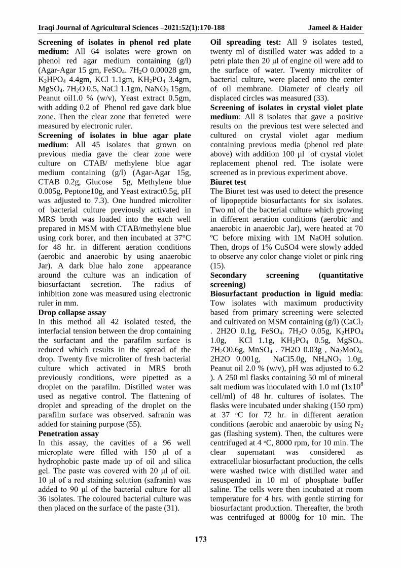
Effect of agitation speed The agitation represents another important
factor influencing on the biosurfactant
production. To evaluate the effect of the
agitation, the cultures were incubated at
different agitation speed (rpm) values ranging
between 120-220 rpm. The results in Figure
15 illustrated that maximum E24% (81%) with
reduction in surface tension (20 mN/m) and
E24% (59%) with reduction in surface tension
(22.80 mN/m) were obtained at 120 rpm for
natural and synthetic media .Dastgheib et al,
(11) who noticed that the optimum agitation
speed was 200 rpm when the biosurfactant was
produced by thermophillic Geobacillus
pallidus. The biosurfactant production by
Bacillus subtilis and P. aeruginosa was
Iraqi Journal of Agricultural Sciences –2021:52(1):170-188 Jameel & Haider
182
optimized in a shaker operating at 120 rpm
(24). The effect of rotation velocity (agitation)
on the biosurfactant concentration and surface
tension reduction was tested at 150 and 200
rpm. considered an important factor for cell
growth and biosurfactant production. It may
also be linked to the physiological function of
microbial emulsifier, it has been suggested that
the production of bioemulsifiers can enhance
the solubilization of water insoluble substrates
and consequently facilitate nutrient transport
to microorganisms (58)..
Figure 15. Effect of rpm values on
biosurfactant production by L.plantarum
(ADK2) grown in MSM, pH 5, and
BCDFTM pH 3, at 30ºC in shaker
incubator after 3 days in anaerobic
conditions by using N2 gas
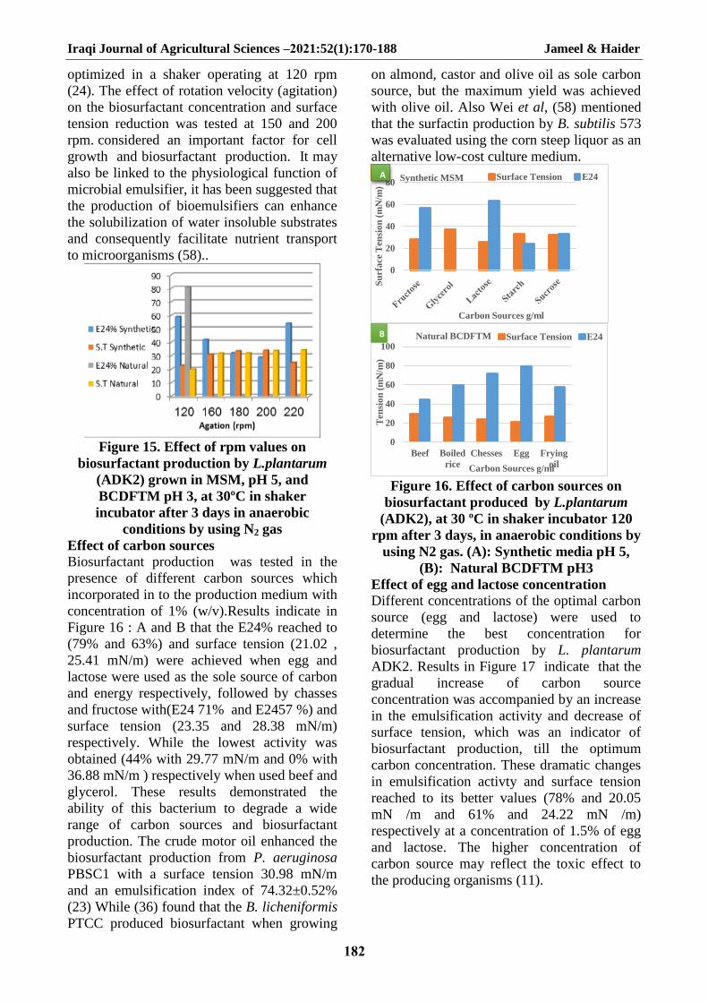
Effect of carbon sources Biosurfactant production was tested in the
presence of different carbon sources which
incorporated in to the production medium with
concentration of 1% (w/v).Results indicate in
Figure 16 : A and B that the E24% reached to
(79% and 63%) and surface tension (21.02 ,
25.41 mN/m) were achieved when egg and
lactose were used as the sole source of carbon
and energy respectively, followed by chasses
and fructose with(E24 71% and E2457 %) and
surface tension (23.35 and 28.38 mN/m)
respectively. While the lowest activity was
obtained (44% with 29.77 mN/m and 0% with
36.88 mN/m ) respectively when used beef and
glycerol. These results demonstrated the
ability of this bacterium to degrade a wide
range of carbon sources and biosurfactant
production. The crude motor oil enhanced the
biosurfactant production from P. aeruginosa
PBSC1 with a surface tension 30.98 mN/m
and an emulsification index of 74.32±0.52%
(23) While (36) found that the B. licheniformis
PTCC produced biosurfactant when growing
on almond, castor and olive oil as sole carbon
source, but the maximum yield was achieved
with olive oil. Also Wei et al, (58) mentioned
that the surfactin production by B. subtilis 573
was evaluated using the corn steep liquor as an
alternative low-cost culture medium.
Figure 16. Effect of carbon sources on
biosurfactant produced by L.plantarum
(ADK2), at 30 ºC in shaker incubator 120
rpm after 3 days, in anaerobic conditions by
using N2 gas. (A): Synthetic media pH 5,
(B): Natural BCDFTM pH3
Effect of egg and lactose concentration Different concentrations of the optimal carbon
source (egg and lactose) were used to
determine the best concentration for
biosurfactant production by L. plantarum
ADK2. Results in Figure 17 indicate that the
gradual increase of carbon source
concentration was accompanied by an increase
in the emulsification activity and decrease of
surface tension, which was an indicator of
biosurfactant production, till the optimum
carbon concentration. These dramatic changes
in emulsification activty and surface tension
reached to its better values (78% and 20.05
mN /m and 61% and 24.22 mN /m)
respectively at a concentration of 1.5% of egg
and lactose. The higher concentration of
carbon source may reflect the toxic effect to
the producing organisms (11).
0
20
40
60
80
Su
rfa
ce T
en
sio
n (
mN
/m)
Carbon Sources g/ml
Synthetic MSM Surface Tension E24A A
0
20
40
60
80
100
Beef Boiled
rice
Chesses Egg Frying
oil
Ten
sio
n (
mN
/m)
Carbon Sources g/ml
Natural BCDFTM Surface Tension E24B

Iraqi Journal of Agricultural Sciences –2021:52(1):170-188 Jameel & Haider
183
Figure 17. Effect the concentration of
carbon sources on biosurfactant produced
by L. plantarum (ADK2), at 30 ºC in shaker
incubator 120 rpm after 3 days, Synthetic
media pH 5 (lactose) and Natural
BCDFTM pH 3 (egg). in anaerobic
conditions by using N2 gas .
Effect of nitrogen sources: In order to
determine the effect of different types of
nitrogen sources on biosurfactant production
by L.plantarum. ADK2, different in nitrogen
surces were tested. Results in Figure 18 : A
and B showed that the production of
biosurfactant varies with different nitrogen
sources. The highest E24% (64%) with
lowering the surface tension of (22.18 mN/m)
were obtained when peas was used as nitrogen
source, while the highest E24% (59%) with
minimum surface tension of (26.3 mN/m)
observed when meat extract was used. While
the lowest emulsification activity and surface
tension observed with chickpeas and urea
(51% and 27.30 mN/m and 42% and 31.20
mN/m ) respectively, compared with other
nitrogen sources. The bacteria require nitrogen
to complete its metabolic pathways and it is
essential for the microbial growth as protein
and enzyme syntheses depend on it (2). The
ammonium salts and urea were preferred
nitrogen sources for biosurfactant production
by Arthrobacter paraffineus, whereas nitrate
supported the maximum surfactant production
by P. aeruginosa and Rhodococcus sp (23).
However, the potassium nitrate support the
maximum production of biosurfactant by the
yeast Rhodotorula glutinis IIP30 (11).
Figure 18. Effect of nitrogen sources on
biosurfactant produced by L.plantarum
(ADK2), at 30 ºC in shaker incubator 120
rpm after 3 days, in anaerobic conditions by
using N2 gas. (A): Synthetic media pH 5,
(B): Natural media BCDFTM pH 3.
Effect of peas and yeast extract
concentration Different concentrations of peas and meat
extract were used as nitrogen source to
determine the optimum concentration for
biosurfactant production by L. plantarum
ADK2. Results in Figure 19 illustrated that the
maximum E24% 66% and 58% and
minimum surface tension 23.03 and 28.30 mN
/ m, were obtained when peas and meat extract
were added in a concentration of 3.5% and 2
% (w/v) respectively. The results also showed
a reduction in emulsification activity and
increases in surface tension when the
concentration of peas or meat extract were
below or above 3.5% and 2 %.
. The best
nitrogen source for emulsifier production by
Bacillus licheniformis was 2% of sodium
nitrate (24). While Vandana and Peter (56)
detected that the production of biosurfactant
often occurs when the nitrogen source is
depleted in the culture medium, during the
stationary phase of cell growth, as an example
the biosurfactant production increased by P.
aeruginosa, Candida tropicalis IIP-4 and
Nocardia strain SFC-D due to the nitrogen
limitation.
0
20
40
60
80
Meat
extract
NaNO3
Peptone
Urea Yeast
extract
Su
rfa
ce T
en
sio
n (
mN
/m)
Nitrogen Sources g/ml
Synthetic MSM Surface Tension E24(%)A
0
20
40
60
80
Su
rfa
ce T
en
sio
n (
mN
/m)
Nitrogen sources g/ml
Natural BCDFTM Surface Tension E24 (%)B

Iraqi Journal of Agricultural Sciences –2021:52(1):170-188 Jameel & Haider
184
Figure 19. Effect of concentration of
nitrogen sources on biosurfactant produced
by L. plantarum ADK2, at 30 ºC in shaker
incubator 120 rpm after 3 days, Synthetic
media pH 5 (meat extract) and Natural
BCDFTM pH 3 (peas). in anaerobic
conditions by using N2 gas
Effect of incubation period Different incubation periods (0-144 hr.) were
examined to detect the optimal periods of
bacterial growth and biosurfactant production
by L. plantarum ADK2. Results in Figure 20
showed that the maximum E24% (88%) and
the lowest surface tension (19.09 mN/m) were
obtained during 72 hr. of incubation using
natural media, while the E24% (72%) with
reduction in surface tension (20.09 mN/m) was
obtained after 96 hr. using synthetic media.
Whereas after 72 and 96 hrs. of incubation, the
emulsification activity was decreased and with
an increasing the surface tension values with
increasing the incubation time. This may be
due to the change in the culture conditions
along this periods such as diminishing of an
anaerobic conditions, nutrients and
accumulating of toxic metabolites which
inhibit the bacterial growth. The result in the
current study pointed that biosurfactant
produced by Lactobacillus plantarum ADK2
increased with incubation period and the
production starded at early stationary phase
(48h) and reached maximum at 96 and 72 h for
synthetic and natural media beyond above
incubation time both growth and biosurfactant
production decrase. Bonilla et al, (7)
mentioned that the biosurfactant biosynthesis
stopped, probably due to the production of
secondary metabolites which could interfere
with emulsion formation and the adsorption of
surfactant molecules at the oil–water interface.
A maximum emulsan production by
Acenitobacter calcoaceticus RAG-1 during the
stationary growth phase (39). While Vandana
and Peter (56) were showed that the
biosurfactant biosynthesis using olive oil
occurred predominantly during the exponential
growth phase, suggesting that the biosurfactant
was produced as a primary metabolite
accompanying cellular biomass formation
(growth-associated kinetics). The RL
production was increased with time until it
reaches the maximum level after 108 h of
incubation where 10.6 g/L was obtained by
Alshaikh Faqri et al, (2)
Figure 20. Effect of incubation period on L.
plantarum (ADK2) grown in synthetic
(MSM) pH 5, and natural (BCDFTM) pH 3,
at 30ºC in shaker incubator 120 rpm after 3
days. in anaerobic conditions by using N2
gas
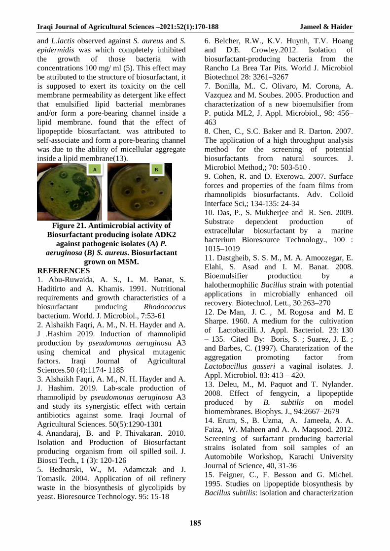
Antimicrobial activity of crude
biosurfactant: To evaluate the antimicrobial
effects of biosurfactants produced by selected
isolate, L. plantarum ADK2 was screened for
inhibitory activity against pathogenic bacteria.
Results in Figure 21 show that the
biosurfactants had an inhibitory effect against
some of pathogenic bacteria such as S. aureus
and P. aeruginosa. Antimicrobial activity of
supernatant from ADK2 isolate had a high
inhibitory effect against S. aureus and P.
aeruginosa with 34.18, 38.43 mm
respectively. This effect may be attributed to
the structure of biosurfactant, it is supposed to
exert its toxicity on the cell membrane
permeability as detergent like effect that
emulsified lipid bacterial membranes and/or
form a pore-bearing channel inside a lipid
membrane. The biosurfactants produced by
Streptococcus thermophilus and L. lactis
showed significant antimicrobial activity
against several bacterial and yeast strains
isolated from explanted voice prostheses (52).
The antimicrobial activity of the crude
biosurfactant isolated from S. thermophilus
Iraqi Journal of Agricultural Sciences –2021:52(1):170-188 Jameel & Haider
185
and L.lactis observed against S. aureus and S.
epidermidis was which completely inhibited
the growth of those bacteria with
concentrations 100 mg/ ml (5). This effect may
be attributed to the structure of biosurfactant, it
is supposed to exert its toxicity on the cell
membrane permeability as detergent like effect
that emulsified lipid bacterial membranes
and/or form a pore-bearing channel inside a
lipid membrane. found that the effect of
lipopeptide biosurfactant. was attributed to
self-associate and form a pore-bearing channel
was due to the ability of micellular aggregate
inside a lipid membrane(13).
Figure 21. Antimicrobial activity of
Biosurfactant producing isolate ADK2
against pathogenic isolates (A) P.
aeruginosa (B) S. aureus. Biosurfactant
grown on MSM.
REFERENCES 1. Abu-Ruwaida, A. S., L. M. Banat, S.
Haditirto and A. Khamis. 1991. Nutritional
requirements and growth characteristics of a
biosurfactant producing Rhodococcus
bacterium. World. J. Microbiol., 7:53-61
2. Alshaikh Faqri, A. M., N. H. Hayder and A.
J .Hashim 2019. Induction of rhamnolipid
production by pseudomonas aeruginosa A3
using chemical and physical mutagenic
factors. Iraqi Journal of Agricultural
Sciences.50 (4):1174- 1185
3. Alshaikh Faqri, A. M., N. H. Hayder and A.
J. Hashim. 2019. Lab-scale production of
rhamnolipid by pseudomonas aeruginosa A3
and study its synergistic effect with certain
antibiotics against some. Iraqi Journal of
Agricultural Sciences. 50(5):1290-1301
4. Anandaraj, B. and P. Thivakaran. 2010.
Isolation and Production of Biosurfactant
producing organism from oil spilled soil. J.
Biosci Tech., 1 (3): 120-126
5. Bednarski, W., M. Adamczak and J.
Tomasik. 2004. Application of oil refinery
waste in the biosynthesis of glycolipids by
yeast. Bioresource Technology. 95: 15-18
6. Belcher, R.W., K.V. Huynh, T.V. Hoang
and D.E. Crowley.2012. Isolation of
biosurfactant-producing bacteria from the
Rancho La Brea Tar Pits. World J. Microbiol
Biotechnol 28: 3261–3267
7. Bonilla, M.. C. Olivaro, M. Corona, A.
Vazquez and M. Soubes. 2005. Production and
characterization of a new bioemulsifier from
P. putida ML2, J. Appl. Microbiol., 98: 456–
463
8. Chen, C., S.C. Baker and R. Darton. 2007.
The application of a high throughput analysis
method for the screening of potential
biosurfactants from natural sources. J.
Microbiol Method,; 70: 503-510 .
9. Cohen, R. and D. Exerowa. 2007. Surface
forces and properties of the foam films from
rhamnolipids biosurfactants. Adv. Colloid
Interface Sci,; 134-135: 24-34
10. Das, P., S. Mukherjee and R. Sen. 2009.
Substrate dependent production of
extracellular biosurfactant by a marine
bacterium Bioresource Technology., 100 :
1015–1019
11. Dastgheib, S. S. M., M. A. Amoozegar, E.
Elahi, S. Asad and I. M. Banat. 2008.
Bioemulsifier production by a
halothermophilic Bacillus strain with potential
applications in microbially enhanced oil
recovery. Biotechnol. Lett., 30:263–270
12. De Man, J. C. , M. Rogosa and M. E
Sharpe. 1960. A medium for the cultivation
of Lactobacilli. J. Appl. Bacteriol. 23: 130
– 135. Cited By: Boris, S. ; Suarez, J. E. ;
and Barbes, C. (1997). Charaterization of the
aggregation promoting factor from
Lactobacillus gasseri a vaginal isolates. J.
Appl. Microbiol. 83: 413 – 420.
13. Deleu, M., M. Paquot and T. Nylander.
2008. Effect of fengycin, a lipopeptide
produced by B. subtilis on model
biomembranes. Biophys. J., 94:2667–2679
14. Erum, S., B. Uzma, A. Jameela, A. A.
Faiza, W. Maheen and A. A. Maqsood. 2012.
Screening of surfactant producing bacterial
strains isolated from soil samples of an
Automobile Workshop, Karachi University
Journal of Science, 40, 31-36
15. Feigner, C., F. Besson and G. Michel.
1995. Studies on lipopeptide biosynthesis by
Bacillus subtilis: isolation and characterization
B A
Iraqi Journal of Agricultural Sciences –2021:52(1):170-188 Jameel & Haider
186
of iturin, surfactin mutants. FEMS
Microbiology Letters 127: 11-15
16. Fernandes, P.A.V., I.R. Arruda, A.F.B.
Santos, A.A. Araujo, A.M.S. Maior and
E.A.Ximenes.2007.Antimicrbial activity of
surfactants produced by Bacillus subtilis R14
against multidrug resistant bacteria. Braz. J.
Microbiol,; 38: 704-709
17. Fischer, E. and R. Munoz.1947. J. Bact.,
53: 381-388
18. Ghribi, D., L. Abdelkefi-Mesrati, I. Mnif,
R. Kammoun, I.Ayadi,, I. Saadaoui,
S.Maktouf and S. Chaabouni-Ellouze. 2012.
Investigation of Antimicrobial Activity and
Statistical Optimization of Bacillus subtilis
SPB1 Biosurfactant Production in Solid-State
Fermentation. Journal of Biomedicine and
Biotechnology. 2012: 12
19. Gudina, E.J., J.A.,V. Rocha, L.R. Teixeira
and Rodrigues. 2010. Antimicrobial and
antiadhesive properties of a biosurfactant
isolated from Lactobacillus paracasei ssp.
paracasei A20, Lett. Appl.Microbiol. 50(4):
p.419-424
20. Hanen, B.A., J. Nawel, M. Hana, B.
Ahmed, and H. Noomen. 2015. Enhancement
of solubilization and biodegradation of
dieseloil by biosurfactant by Bacillus
amyloliquefaciens An6. Int Biodeterior
Biodegradation;99:8-14
21. Holt, J. C. , N. R. Krieg, A. Sneath, J. T.
Staley, and S. T. Williams. 1994. Bergeyۥ s
manual of systemic bacteriology. 9th ed.
Williams and Wilkins, Baltimore, London
22. Hugas, M., M. Garriga, T. Aymerich and
J.M. Monfort.1993. Biochemical
characterization of lactobacilli from dry
fermented sausages. Int. J. Food Microbiol;
18: 107-113
23. Johnson, V., M. Singh and V. S. Saini.
1992. Bioemulsifier production by an
oleaginous yeast Rhodotorula glutinis IIP-30.
Biotechnology Letters., 14: 487-490
24. Joice, P. A. and R .Parthasarathi.2014.
Optimization of biosurfactant production from
P. aeruginosa PBSC1. Int. J. Curr. Microbiol.
App. Sci., 3(9):140-151
25. Joice, P. A. and R. Parthasarathi. 2014.
Optimisation of biosurfactant production from
Pseudomonas aeruginosa PBSC1. Int. J. Curr.
Microbiol. App. Sci., 3:140-151
26. Joshi, S.J., H. Suthar, A.K. Yadav, K.
Hingurao and A. Nerurkar. 2013. Occurrence
of biosurfactant producing Bacillus sp. in
diverse habitats. ISRN Biotechnology; 1-6
27. Kalyani, A. L. T., S. G. Sireesha, A. K. G.
Aditya and G. G. Sankar.2014. Isolation and
antimicrobial activity of rhamnolipid
biosurfactant from oil-contaminated soil
sample using humic-acid salts-vitamin agar.
International Journal of Research in
Engineering and Technology. 3(5):357-365
28. Kotzamanidis,C., A. Kourelis, E.
Litopoulou-Tzanetaki, N.Tzanetakis and M.
Yiangou. 2010. Evaluation of adhesion
capacity,cell surfacetraits and
immunomodulatory activity of presumptive
probiotic Lactobacillus strains. Int.
J.FoodMicrobiol. 140, 154–
163.doi:10.1016/j.ijfoodmicro.2010. 04.004
29. Kuiper, I., L. Ellen, R.P.Lagendijk, P.D.
Jeremy, E.M.L. Gerda, E.T. Jane, J.J.L. Ben
and V.B. Guido .2004. Characterization of two
Pseudomonas putida lipopeptide
biosurfactants, putisolvin I and II, which
inhibit biofilm formation and break down
existing biofilms. Mol Microbiol,; 51(1): 97-
113
30. Langer, O., O. Palme, V. Wray, H. Tokuda
and S.Lang. 2006. Production and
modification of bioactive biosurfactants,
Process Biochemistry, vol. 41, no. 10, pp.
2138–2145
31. Maczek J., S. Junne and P. Götz. 2007.
Examining biosurfactant producing bacteria—
an example for an automated search for natural
compounds. Application Note CyBio AG.
32. Madhu,N. N. and A. S. G. Prapulla. 2014.
Evaluation and Functional Characterization of
a Biosurfactant Produced by Lactobacillus
plantarum CFR 2194.
Appl.Biochem.Biotechnol., 172(4), 1777-1789
33. Morikawa, M., Y. Hirata and T. Imanaka.
2000. A study on the structure-function
relationship of lipopeptide biosurfactants.
Biochimica et Biophyica Acta. 1488: 211-218
34. Mukherjee, S., P. Das and R. Sen. 2006.
Towards commercial production of microbial
surfactants. Trends in Biotechnology, vol. 24,
no. 11, pp. 509–515
35. Mulligan, C.N. 2005. Environmental
applications for biosurfactants. Environ
Pollution 133(2):183–198

Iraqi Journal of Agricultural Sciences –2021:52(1):170-188 Jameel & Haider
187
36. Noudeh, G. D., M. H. Moshafi, P.
Khazaeli and F. Akef. 2007. Studies on
bioemulsifier production by B. licheniformis
PTCC 1595. American Journal of
Pharmacology and Toxicology. 2 (4): 164-169
37. Okoliegbe, I. N. and O. O. Agarry. 2012.
Application of microbial surfactant (areview).
Scholarly Journals of Biotechnology. 1(1):15-
23
38. Persson, A., E. Oesterberg and M.
Dostalek. 1988. Biosurfactant production by
Pseudomonas fluorescens378: Growth and
product characteristics. Appl. Microbiol.
Biotechnol., 29(1):1–4
39. Rodrigues, L. R. and J. A. Teixeira. 2008.
Biosurfactants production from cheese whey,”
in Advances in CheeseWhey Utilization, M. E.
Cerd´an, M. Gonz´alez-Siso, and M. Becerra,
Eds., pp. 81– 104, Transworld Research
Network.
40. Rodrigues, L., A. Moldes, J. Teixeira and
R. Oliveira. 2006. Kinetic study of
fermentative biosurfactant production by
Lactobacillus strains. J Biochem Eng,; 28:
109-116
41. Rodrigues, L., A. Moldes, J. Teixeira and
R. Oliveira.2006b. Kinetic study of
fermentative biosurfactant production by
Lactobacillus strains. Biochemical
Engineering Journal., 28: 109–116
42. Saharan, B. S., R. K. Sahu and D. Sharma.
2011. A Review on biosurfactants:
fermentation, Current Developments and
Perspectives. J. of Genetic Engineering and
Biotechnol., 29
43. Saravanakumari, P. and K. Mani. 2010.
Structural characterization of a novel xylolipid
biosurfactant from Lactococcus lactis and
analysis of antibacterial activity against multi-
drug resistant pathogens, Bioresource
Technology, vol. 101, no. 22, pp. 8851– 8854
44. Sauvageau, J., J. Ryan, K. Lagutin, I. M.
Sims, B. L. Stocker and M. S. Timmer.
2012.Isolation and structural characterization
of the major glycolipids from Lactobacillus
plantarum, Carbohydrate Res. 357, 151-156.
45. Sekhon, K.K., S. Khanna and S.S.
Cameotra. 2012. Biosurfactant Production and
Potential Correlation with Esterase Activity. J.
Pet. Environ Biotechnol. 3,133
46. Sekhon, R. S., H. Lin, K. L. Childs, C. N.
Hansey, C. R. Buell, N. de Leon and S. M.
Kaeppler. 2011. Genome-wide atlas of
transcription during maize development. Plant
J., 66(4):553-63
47. Shoeb, E., N.Ahmed, J. Akhter, U. Badar,
I. K. Siddiqu, F.A. Ansari, M. Waqar, S.
Imtiaz, N. Akhtar, Q.A. Shaikh, R. Baig, S.
Butt, S. Khan, S. Hussain, B. Ahmed and M.A.
Ansari 2015. Screening and characterization of
biosurfactant-producing bacteria isolated from
the Arabian Sea coast of Karachi, Turk J.
Biol., 39, 210-216.
48. Simoes, M., L.C. Simoes and M.J. Vieira.
2010. A review of current and emergent
biofilm control strategies. LWT- Food Sci
Technol,; 43(4): 573-583
49. Stearn, E. W. and A. E. Stearn. 1928.
Univ. Missouri Studies, 3, 51
50. Stoyanova, L.G , N. L. Olsinskaya and L.
I. Vorobjeva. 1979. The ability of
propionbacteria to grow in liquid eggs and to
ferment all the carbohydrates of egg white in
24 h.ISBN. 0-7923-5884-8 .
51. Sumathip, R. and N. Yogananth. 2016.
Isolation and identification of Biosurfactant
producing Pseudomonas aeruginosa from
marine sediment samples and its Antimicrobial
Properties. Int. J. Adv. Res. Biol. Sci.3:200-12
52. Tahmourespour, A., R. Salehi and R. H.
Kermanshahi. 2011. Lactobacillus
acidophilus-derived biosurfactant effect on
gtfb and gtfc expression level in streptococcus
mutans biofilm cells. Brazilian Journal of
Microbiology 42: 330-339
53. Thavasi, R., S. Jayalakshmi and I.M.
Banat. 2011. Effect of biosurfactant and
fertilizer on biodegradation of crude oil by
marine isolates of Bacillus megaterium,
Corynebacterium kutscheri and Pseudomonas
aeruginosa. Bioresour. Technol. 102 (2), 772–
778
54. Thavasi,R., S. Jayalakshmi, and I.M.
Banat. 2011.Application of biosurfactant
produced from peanut oil cake by
Lactobacillus delbrueckii in biodegradation of
crude oil,” Bioresource Technology, vol. 102,
no. 3, pp. 3366–3372
55. Tugrul, T. and E. Cansunar. 2005.
Detecting surfactant producing
microorganisms by drop-collapse test. World
Journal of Microbiology and Biotechnology
21: 851-853

Iraqi Journal of Agricultural Sciences –2021:52(1):170-188 Jameel & Haider
188
56. Vandana, P. and J.K. Peter. 2014. Partial
purification and characterization of
biosurfactant from Pseudomonas aeruginosa
(C). Int. J. Eng. Sci. Res Technol;3:45-50
57. Vollbrecht, E., R. Heckmann, V. Wary, M.
Nimtz and S. Lang. 1998. Production and
structure elucidation of di- and oligosaccharide
lipids (biosurfactant) from Tsukamurella sp.
Appl. Microbial Biotechnol. 50: 530-537
58. Wei, Y.H., C. L. Chou and J.S. Chang.
2005. Rhamnolipid production by indigenous
pseudomonas aeruginosa J4 originating from
petrochemical wastewater. Biochemical
Engineering Journal. 27:146-154
59. Yalçin, E. and A. Ergene. 2009. Screening
the antimicrobial activity of biosurfactants
produced by microorganisms isolated from
refinery waste waters. Journal of Applied
Biological Sciences. 3 (2): 148 – 153
60. Zheng, C., J. He, Y. Wang, M. Wang and
Z. Huang. 2011. Hydrocarbon degradation and
bioemulsifier production by thermophilic G.
pallidus strains. Bioresource technology.
102:9155- 9161.


