

Developing an Evidence Based
Referral Protocol for Early
Diagnosis of Vestibular
Schwannomas
Jessica Scott, AuD
JDVAC Conference
February 23, 2010
• The views expressed
in this presentation
are those of the
author and do not
reflect the policy of
the Department of the
Army, Department of
Defense, or US
Government.
Introduction• Vestibular Schwannoma (VS)
Slow growing benign tumor
Develops on Vestibulocochlear Nerve (CN VIII)
Overproduction of Schwann Cells
• Wrap around nerve fibers
• Help support and insulate for increased conduction
Common Symptoms (Cummings et al., 2005)
• Hearing loss, Tinnitus, Vestibular, Aural Fullness, Otalgia
• CN VII (Facial) or CN V (Trigeminal)
• Labyrinthine artery
• Incidence of 1 in 100,000 (NIDCD, 2004)

Introduction• Tests that can potentially indicate the presence of a
VS Audiologic (Pure tone thresholds, speech discrimination testing,
acoustic reflex thresholds, acoustic reflex adaptation (decay))
Vestibular (ENG/VNG, rotary chair testing, VEMP)
Evoked Potentials (ABR, ECochG)
Radiologic (MRI, CT Scan)
• Most definitive test for diagnosing VSs Contrast-enhanced MRI
Contrast (typically gadolinium) - provides a greater contrast
between normal and abnormal tissue (Cummings et al., 2005)
Literature Review
• Previous Referral Criteria
Obholzer et al. (2004)
• Unilateral hearing loss: ≥15 dB HL at two adjacent
frequencies
• Bilateral hearing loss: ≥20 dB HL at two adjacent
frequencies
Sheppard et al. (1996)
• ≥15 dB HL asymmetry
• Unilateral tinnitus in the presence of normal
hearing

Data Collection• All data were collected retrospectively
Age/Gender
Previous Hx of Hazardous Noise Exposure
Hx of hearing loss (newly identified, stable, progressive, none)
Who referred them for a MRI (Audiology, ENT, Neurology, etc.)
Patient’s presenting symptoms
Audiologic test results• Pure tone thresholds (excluded if air-bone gaps)
• Speech discrimination scores
• Tympanometry* (excluded if abnormal)
• Acoustic reflex thresholds
• Acoustic reflex adaptation (decay)
• Only demographic exclusion criteria: No patients < 18 years of age
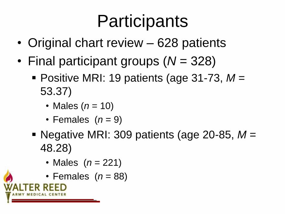
Participants• Original chart review – 628 patients
• Final participant groups (N = 328)
Positive MRI: 19 patients (age 31-73, M =
53.37)
• Males (n = 10)
• Females (n = 9)
Negative MRI: 309 patients (age 20-85, M =
48.28)
• Males (n = 221)
• Females (n = 88)
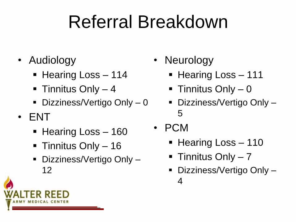
Referral Breakdown
• Audiology
Hearing Loss – 114
Tinnitus Only – 4
Dizziness/Vertigo Only – 0
• ENT
Hearing Loss – 160
Tinnitus Only – 16
Dizziness/Vertigo Only –
12
• Neurology
Hearing Loss – 111
Tinnitus Only – 0
Dizziness/Vertigo Only –
5
• PCM
Hearing Loss – 110
Tinnitus Only – 7
Dizziness/Vertigo Only –
4
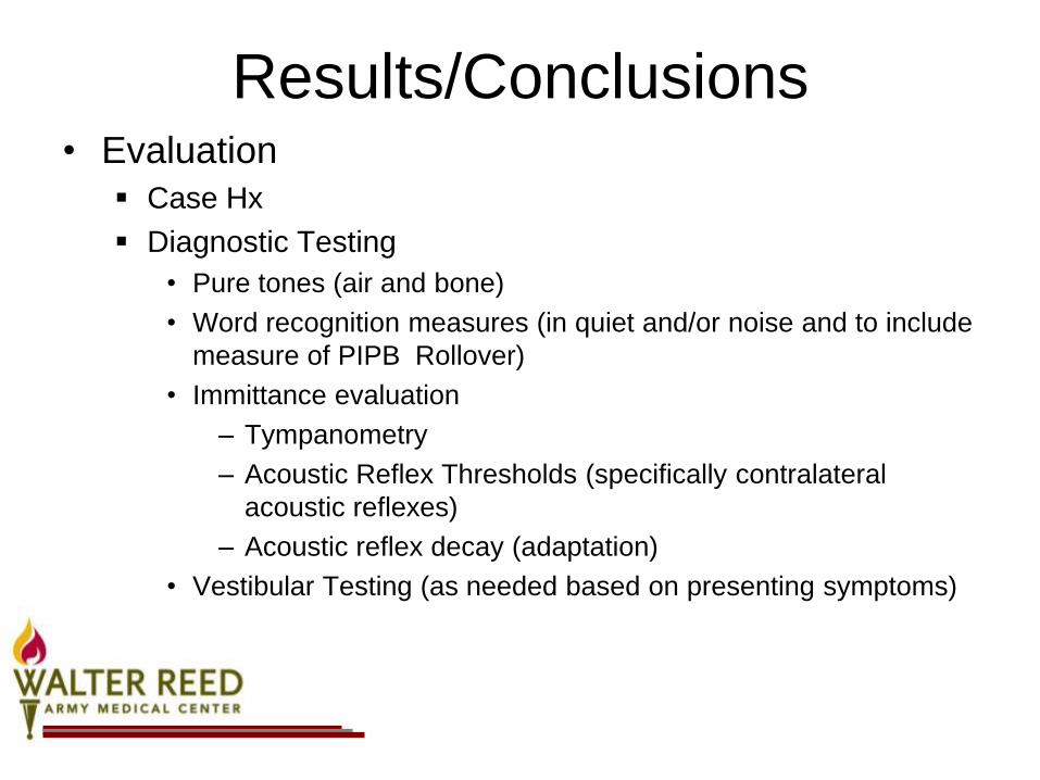
Results/Conclusions• Evaluation
Case Hx
Diagnostic Testing
• Pure tones (air and bone)
• Word recognition measures (in quiet and/or noise and to include
measure of PIPB Rollover)
• Immittance evaluation
– Tympanometry
– Acoustic Reflex Thresholds (specifically contralateral
acoustic reflexes)
– Acoustic reflex decay (adaptation)
• Vestibular Testing (as needed based on presenting symptoms)
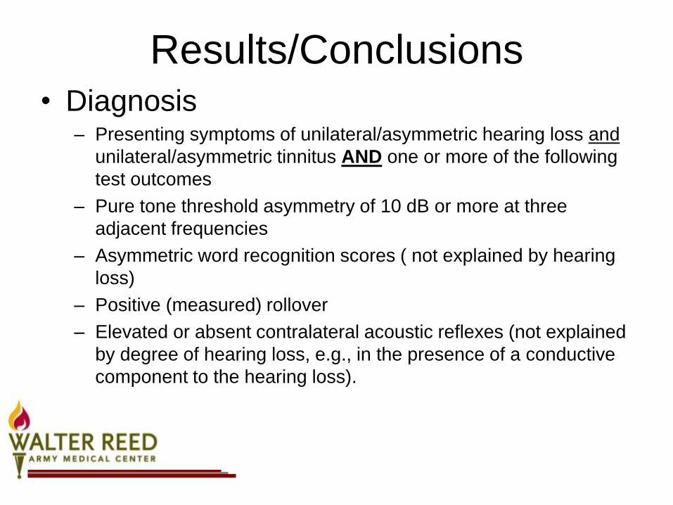
Results/Conclusions• Diagnosis
– Presenting symptoms of unilateral/asymmetric hearing loss and
unilateral/asymmetric tinnitus AND one or more of the following
test outcomes
– Pure tone threshold asymmetry of 10 dB or more at three
adjacent frequencies
– Asymmetric word recognition scores ( not explained by hearing
loss)
– Positive (measured) rollover
– Elevated or absent contralateral acoustic reflexes (not explained
by degree of hearing loss, e.g., in the presence of a conductive
component to the hearing loss).
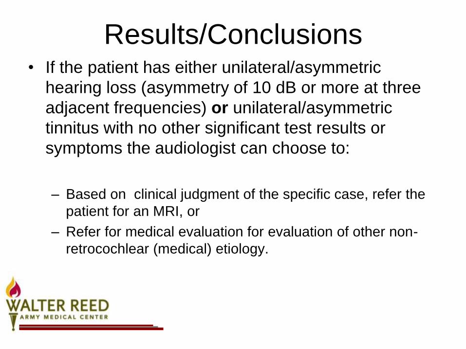
Results/Conclusions• If the patient has either unilateral/asymmetric
hearing loss (asymmetry of 10 dB or more at three
adjacent frequencies) or unilateral/asymmetric
tinnitus with no other significant test results or
symptoms the audiologist can choose to:
– Based on clinical judgment of the specific case, refer the
patient for an MRI, or
– Refer for medical evaluation for evaluation of other non-
retrocochlear (medical) etiology.
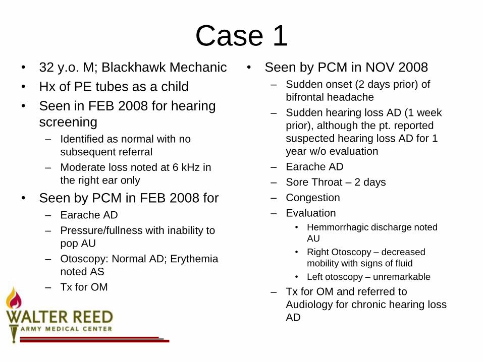
Case 1• 32 y.o. M; Blackhawk Mechanic
• Hx of PE tubes as a child
• Seen in FEB 2008 for hearing
screening
– Identified as normal with no
subsequent referral
– Moderate loss noted at 6 kHz in
the right ear only
• Seen by PCM in FEB 2008 for
– Earache AD
– Pressure/fullness with inability to
pop AU
– Otoscopy: Normal AD; Erythemia
noted AS
– Tx for OM
• Seen by PCM in NOV 2008
– Sudden onset (2 days prior) of
bifrontal headache
– Sudden hearing loss AD (1 week
prior), although the pt. reported
suspected hearing loss AD for 1
year w/o evaluation
– Earache AD
– Sore Throat – 2 days
– Congestion
– Evaluation
• Hemmorrhagic discharge noted
AU
• Right Otoscopy – decreased
mobility with signs of fluid
• Left otoscopy – unremarkable
– Tx for OM and referred to
Audiology for chronic hearing loss
AD
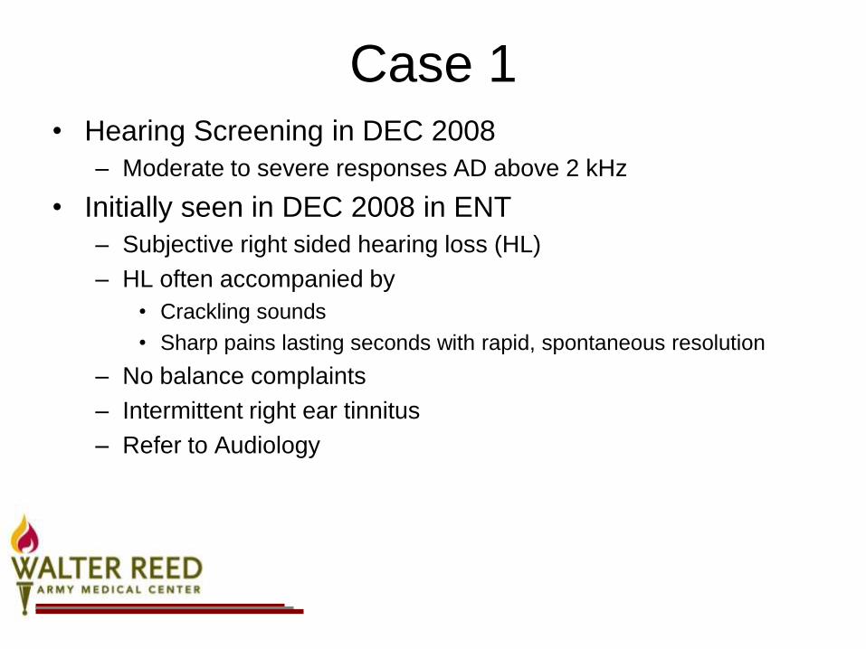
Case 1• Hearing Screening in DEC 2008
– Moderate to severe responses AD above 2 kHz
• Initially seen in DEC 2008 in ENT
– Subjective right sided hearing loss (HL)
– HL often accompanied by
• Crackling sounds
• Sharp pains lasting seconds with rapid, spontaneous resolution
– No balance complaints
– Intermittent right ear tinnitus
– Refer to Audiology
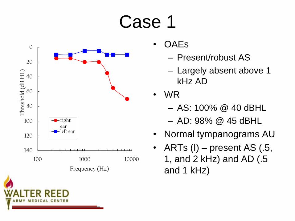
Case 1• OAEs
– Present/robust AS
– Largely absent above 1
kHz AD
• WR
– AS: 100% @ 40 dBHL
– AD: 98% @ 45 dBHL
• Normal tympanograms AU
• ARTs (I) – present AS (.5,
1, and 2 kHz) and AD (.5
and 1 kHz)
0
20
40
60
80
100
120
140
100 1000 10000
Th
resh
old
(d
B H
L)
Frequency (Hz)
right earleft ear
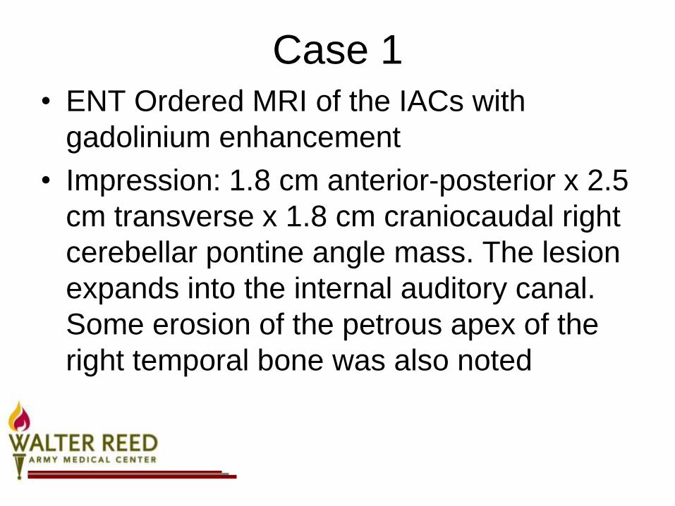
Case 1• ENT Ordered MRI of the IACs with
gadolinium enhancement
• Impression: 1.8 cm anterior-posterior x 2.5
cm transverse x 1.8 cm craniocaudal right
cerebellar pontine angle mass. The lesion
expands into the internal auditory canal.
Some erosion of the petrous apex of the
right temporal bone was also noted
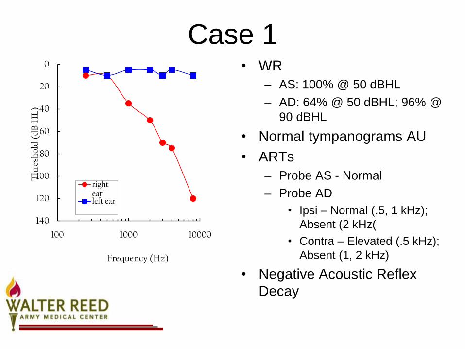
Case 1• WR
– AS: 100% @ 50 dBHL
– AD: 64% @ 50 dBHL; 96% @
90 dBHL
• Normal tympanograms AU
• ARTs
– Probe AS - Normal
– Probe AD
• Ipsi – Normal (.5, 1 kHz);
Absent (2 kHz(
• Contra – Elevated (.5 kHz);
Absent (1, 2 kHz)
• Negative Acoustic Reflex
Decay
0
20
40
60
80
100
120
140
100 1000 10000
Th
resh
old
(d
B H
L)
Frequency (Hz)
right earleft ear
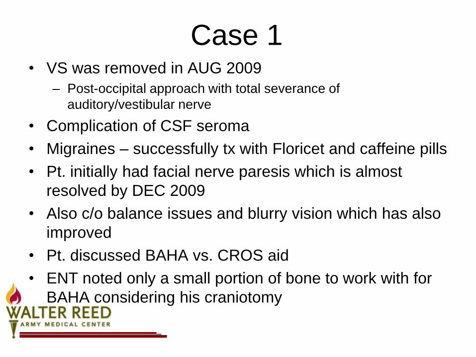
Case 1• VS was removed in AUG 2009
– Post-occipital approach with total severance of
auditory/vestibular nerve
• Complication of CSF seroma
• Migraines – successfully tx with Floricet and caffeine pills
• Pt. initially had facial nerve paresis which is almost
resolved by DEC 2009
• Also c/o balance issues and blurry vision which has also
improved
• Pt. discussed BAHA vs. CROS aid
• ENT noted only a small portion of bone to work with for
BAHA considering his craniotomy

Case 2• 60 y.o. M initially seen in Audiology in
June 1987
– Presented with
• Dysequilibrium problems
• Hearing loss AS>AD
• Distortion AS
• Intermittent pressure in his head
• Tinnitus AS
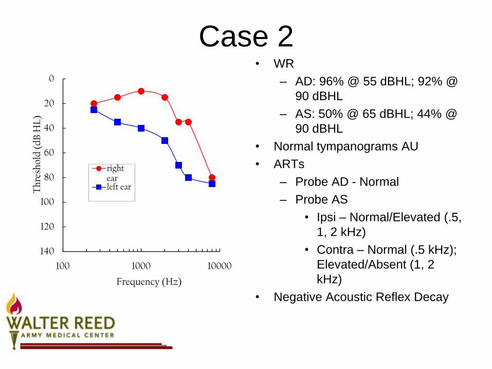
Case 2• WR
– AD: 96% @ 55 dBHL; 92% @
90 dBHL
– AS: 50% @ 65 dBHL; 44% @
90 dBHL
• Normal tympanograms AU
• ARTs
– Probe AD - Normal
– Probe AS
• Ipsi – Normal/Elevated (.5,
1, 2 kHz)
• Contra – Normal (.5 kHz);
Elevated/Absent (1, 2
kHz)
• Negative Acoustic Reflex Decay
0
20
40
60
80
100
120
140
100 1000 10000
Th
resh
old
(d
B H
L)
Frequency (Hz)
right earleft ear

Case 2• ABR – June 1987
– AD
• Absolute wave latencies were delayed for Waves
III and V.
• Morphology was considered fair
– AS
• Absolute latencies were delayed for Waves III and
V.
• Comparison between ears indicated significant
delay for Wave V in the left compared to the right
• Morphology was considered poor
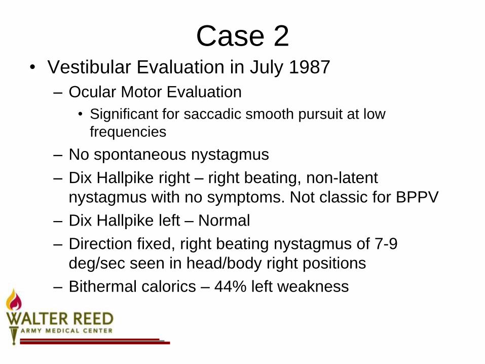
Case 2• Vestibular Evaluation in July 1987
– Ocular Motor Evaluation
• Significant for saccadic smooth pursuit at low
frequencies
– No spontaneous nystagmus
– Dix Hallpike right – right beating, non-latent
nystagmus with no symptoms. Not classic for BPPV
– Dix Hallpike left – Normal
– Direction fixed, right beating nystagmus of 7-9
deg/sec seen in head/body right positions
– Bithermal calorics – 44% left weakness

Case 2• Stable SNHL since
JUN 1987
• WR
– AD: 100% @ 50 dBHL
– AS: 60% @ 60 dBHL
0
20
40
60
80
100
120
140
100 1000 10000
Th
resh
old
(d
B H
L)
Frequency (Hz)
right earleft ear
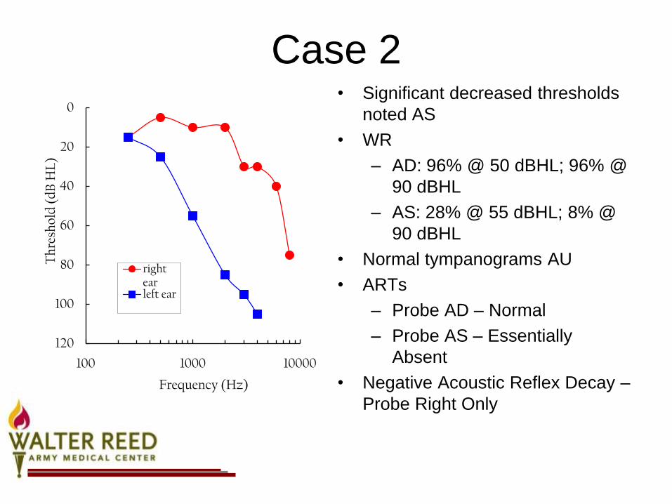
Case 2• Significant decreased thresholds
noted AS
• WR
– AD: 96% @ 50 dBHL; 96% @
90 dBHL
– AS: 28% @ 55 dBHL; 8% @
90 dBHL
• Normal tympanograms AU
• ARTs
– Probe AD – Normal
– Probe AS – Essentially
Absent
• Negative Acoustic Reflex Decay –
Probe Right Only
0
20
40
60
80
100
120
100 1000 10000
Th
resh
old
(d
B H
L)
Frequency (Hz)
right earleft ear

Case 2• Referred for MRI by ENT
– Noted a 1 x 1.5 cm acoustic neuroma on the
left side
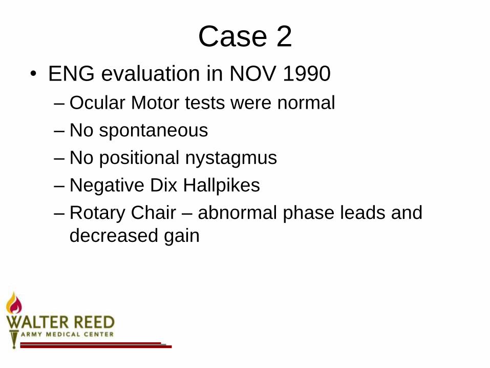
Case 2• ENG evaluation in NOV 1990
– Ocular Motor tests were normal
– No spontaneous
– No positional nystagmus
– Negative Dix Hallpikes
– Rotary Chair – abnormal phase leads and
decreased gain
Case 2• Removal of VS in NOV 1990.
• Audio was unchanged AD with an
anacoustic AS.
• Pt. reports no significant hearing
difficulties
Case 3• 73 y.o. M
• Presented to Audiology in DEC 2008
• Previous Noise Exposure
• Fm hx: Father and brother have both had different
brain tumors
• Presentation:
– Hearing loss
– Difficulty understanding speech
– No earache
– No tinnitus
– No balance difficulties

Case 3• WR
– AS: 100% @ 90 dBHL
– AD: 84% @ 75 dBHL; 48%
@ 90 dBHL
• Normal tympanograms AU
• ARTs
– Probe AS – Normal, except
Absent at 2 kHz in contra
condition
– Probe AD –Absent
0
20
40
60
80
100
120
140
100 1000 10000
Th
resh
old
(d
B H
L)
Frequency (Hz)
right earleft ear
Case 3• Negative MRI of the IACs
• Pt. was fit with hearing aids in both ears
Case 4• 42 y.o. F
• Pt. initially presented to Audiology in OCT
2005 for a second opinion on a Meniere’s
Dx
• Presentation
– No subjective hearing loss
– Intermittent tinnitus
– Vertigo, noted since Spring 2005
– No ear pain
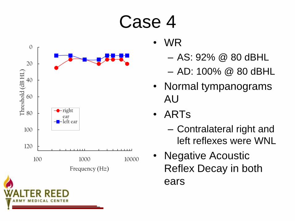
Case 4• WR
– AS: 92% @ 80 dBHL
– AD: 100% @ 80 dBHL
• Normal tympanograms
AU
• ARTs
– Contralateral right and
left reflexes were WNL
• Negative Acoustic
Reflex Decay in both
ears
0
20
40
60
80
100
120
100 1000 10000
Th
resh
old
(d
B H
L)
Frequency (Hz)
right earleft ear
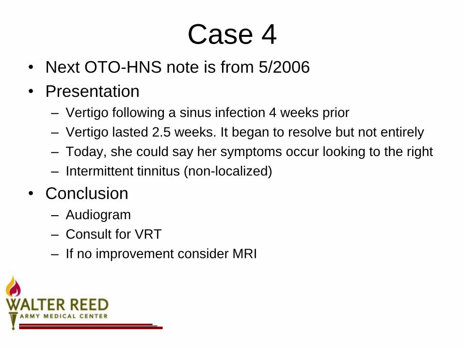
Case 4• Next OTO-HNS note is from 5/2006
• Presentation
– Vertigo following a sinus infection 4 weeks prior
– Vertigo lasted 2.5 weeks. It began to resolve but not entirely
– Today, she could say her symptoms occur looking to the right
– Intermittent tinnitus (non-localized)
• Conclusion
– Audiogram
– Consult for VRT
– If no improvement consider MRI

Case 4• Seen in Audiology in May 2006
– Presentation
• Subjective hearing loss AD
• Muffled sound AD
• Pressured sensation AD
• Tinnitus AD only

Case 4
• Stable hearing
• Excellent high level
word recognition in
both ears
• Normal
tympanograms and
ARTs
0
20
40
60
80
100
120
140
100 1000 10000
Th
resh
old
(d
B H
L)
Frequency (Hz)
right earleft ear
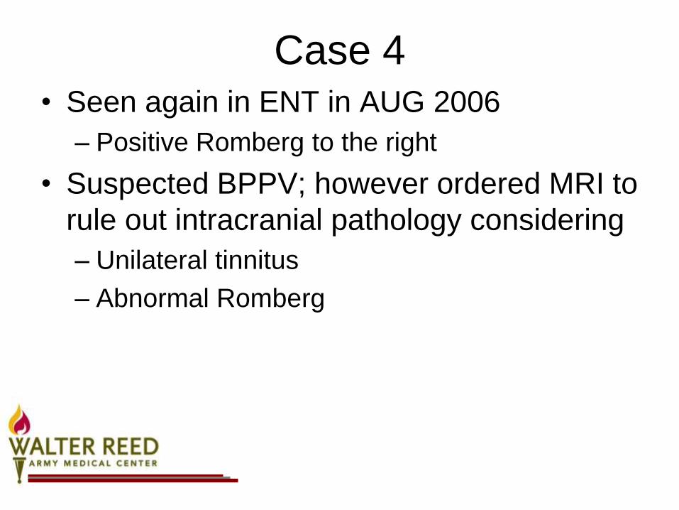
Case 4• Seen again in ENT in AUG 2006
– Positive Romberg to the right
• Suspected BPPV; however ordered MRI to
rule out intracranial pathology considering
– Unilateral tinnitus
– Abnormal Romberg
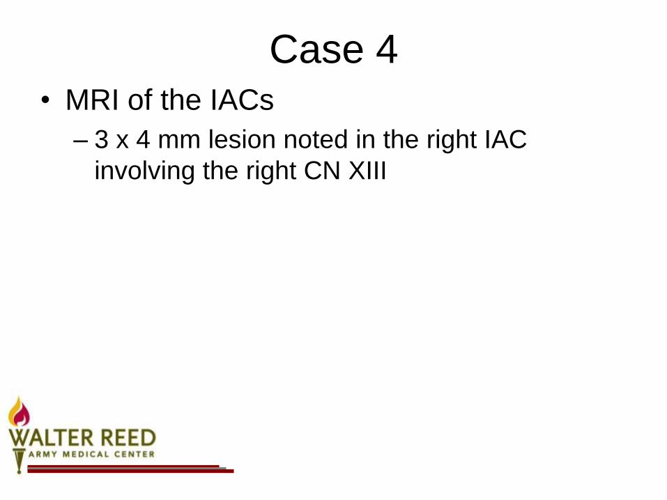
Case 4• MRI of the IACs
– 3 x 4 mm lesion noted in the right IAC
involving the right CN XIII
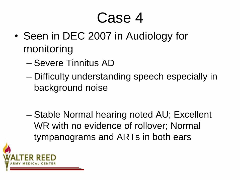
Case 4• Seen in DEC 2007 in Audiology for
monitoring
– Severe Tinnitus AD
– Difficulty understanding speech especially in
background noise
– Stable Normal hearing noted AU; Excellent
WR with no evidence of rollover; Normal
tympanograms and ARTs in both ears
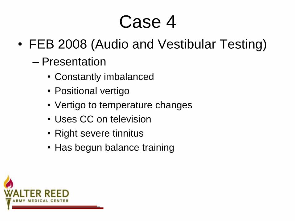
Case 4• FEB 2008 (Audio and Vestibular Testing)
– Presentation
• Constantly imbalanced
• Positional vertigo
• Vertigo to temperature changes
• Uses CC on television
• Right severe tinnitus
• Has begun balance training
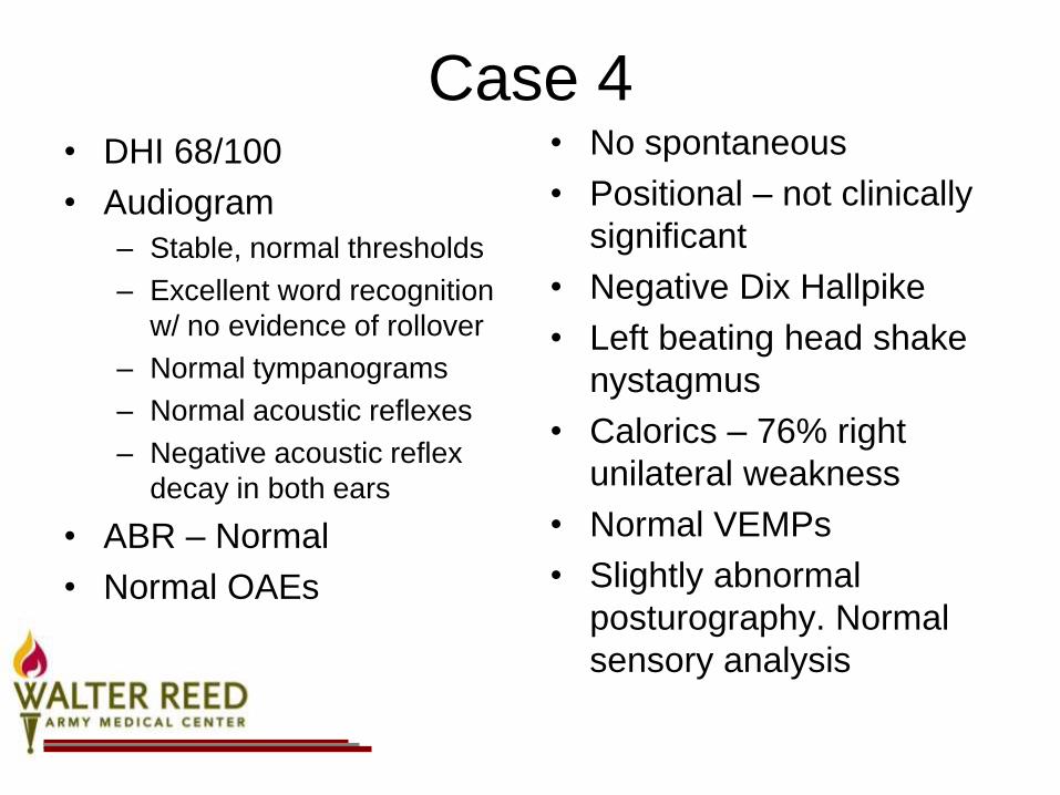
Case 4• DHI 68/100
• Audiogram
– Stable, normal thresholds
– Excellent word recognition
w/ no evidence of rollover
– Normal tympanograms
– Normal acoustic reflexes
– Negative acoustic reflex
decay in both ears
• ABR – Normal
• Normal OAEs
• No spontaneous
• Positional – not clinically
significant
• Negative Dix Hallpike
• Left beating head shake
nystagmus
• Calorics – 76% right
unilateral weakness
• Normal VEMPs
• Slightly abnormal
posturography. Normal
sensory analysis

Case 4• Removal of VS in APR 2008 via a middle
fossa approach
– Persistent tinnitus
– Mild imbalance that is improving
– Intermittent echo sound AD with occasional
tinny quality
– Some right ear pain behind the right pinna

Case 4• MRIs in 2009 have indicated a 7mm lesion in the right IAC
– Residual lesion vs. granulation tissue
• Newest Audio in SEPT 2009 was essentially normal
• ENT in SEPT 2009
– Right eye is “slow”
– Pain in the medial side of R eyebrow
– Mildly improved tinnitus
– Dry eyes
– Difficulty with taste on the right side of her tongue
– Mild preauricular numbness and pain AD
– Twitch in right eye tx with botox that is more tolerable

Case 5• 37 y.o. M
• Presnted to Audiology Clinic in JAN 2010
• C/o
– Intermittent ear pain AS
• Episodic, sharp pain
• Mostly AS, but rarely noted AD
• Pain for 10-15 seconds
• Randomly several times a week
– No hearing loss
– No tinnitus
– No dizziness
– Ears frequently feel like there is cotton in them

Case 5• WR
– AS and AD: 100% @
50 and 90 dBHL
• Normal tympanograms
AU
• ARTs - WNL
• Negative Acoustic
Reflex Decay in both
ears
0
20
40
60
80
100
120
100 1000 10000
Th
resh
old
(d
B H
L)
Frequency (Hz)
right earleft ear

Case 5• Initially had CT scan which was normal in
DEC 2009
• Sent for MRI of the IACs for chronic left
ear pain in JAN 2010
• Impression: 1.5 mm right intracanicular VS
References
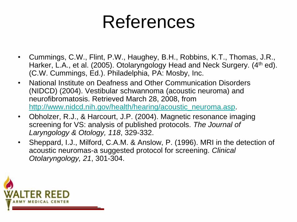
• Cummings, C.W., Flint, P.W., Haughey, B.H., Robbins, K.T., Thomas, J.R., Harker, L.A., et al. (2005). Otolaryngology Head and Neck Surgery. (4th ed). (C.W. Cummings, Ed.). Philadelphia, PA: Mosby, Inc.
• National Institute on Deafness and Other Communication Disorders (NIDCD) (2004). Vestibular schwannoma (acoustic neuroma) and neurofibromatosis. Retrieved March 28, 2008, from http://www.nidcd.nih.gov/health/hearing/acoustic_neuroma.asp.
• Obholzer, R.J., & Harcourt, J.P. (2004). Magnetic resonance imaging screening for VS: analysis of published protocols. The Journal of Laryngology & Otology, 118, 329-332.
• Sheppard, I.J., Milford, C.A.M. & Anslow, P. (1996). MRI in the detection of acoustic neuromas-a suggested protocol for screening. Clinical Otolaryngology, 21, 301-304.
Thank You!!


