
University of Nebraska - LincolnDigitalCommons@University of Nebraska - Lincoln
Papers in Veterinary and Biomedical Science Veterinary and Biomedical Sciences, Department of
2018
Development of septic polysynovitis and uveitis infoals experimentally infected with Rhodococcus equiLaura HuberUniversity of Georgia
Steeve GiguèreUniversity of Georgia, [email protected]
Londa J. BerghausUniversity of Georgia
Amanda HanafiUniversity of Georgia
Sarah Vitosh-SillmanUniversity of Nebraska-Lincoln, [email protected]
See next page for additional authors
Follow this and additional works at: https://digitalcommons.unl.edu/vetscipapers
Part of the Biochemistry, Biophysics, and Structural Biology Commons, Cell and DevelopmentalBiology Commons, Immunology and Infectious Disease Commons, Medical Sciences Commons,Veterinary Microbiology and Immunobiology Commons, and the Veterinary Pathology andPathobiology Commons
This Article is brought to you for free and open access by the Veterinary and Biomedical Sciences, Department of at DigitalCommons@University ofNebraska - Lincoln. It has been accepted for inclusion in Papers in Veterinary and Biomedical Science by an authorized administrator ofDigitalCommons@University of Nebraska - Lincoln.
Huber, Laura; Giguère, Steeve; Berghaus, Londa J.; Hanafi, Amanda; Vitosh-Sillman, Sarah; and Czerwinski, Sarah L., "Developmentof septic polysynovitis and uveitis in foals experimentally infected with Rhodococcus equi" (2018). Papers in Veterinary and BiomedicalScience. 289.https://digitalcommons.unl.edu/vetscipapers/289

AuthorsLaura Huber, Steeve Giguère, Londa J. Berghaus, Amanda Hanafi, Sarah Vitosh-Sillman, and Sarah L.Czerwinski
This article is available at DigitalCommons@University of Nebraska - Lincoln: https://digitalcommons.unl.edu/vetscipapers/289

RESEARCH ARTICLE
Development of septic polysynovitis and
uveitis in foals experimentally infected with
Rhodococcus equi
Laura Huber1, Steeve Giguère1*, Londa J. Berghaus1, Amanda Hanafi1, Sarah Vitosh-
Sillman2, Sarah L. Czerwinski3
1 Department of Large Animal Medicine, College of Veterinary Medicine, University of Georgia, Athens,
Georgia, United States of America, 2 School of Veterinary Medicine and Biomedical Sciences, University of
Nebraska, Lincoln, Nebraska, United States of America, 3 Department of Small Animal Medicine and
Surgery, College of Veterinary Medicine, University of Georgia, Athens, Georgia, United States of America
Abstract
Rhodococcus equi is one of the most important causes of disease in foals. Infection is typi-
cally characterized by pyogranulomatous pneumonia although extrapulmonary infections
occur occasionally. Uveitis and polysynovitis have been reported in foals naturally infected
with R. equi and are thought to be the result of an immune-mediated process. However, the
pathogenesis of these conditions is poorly understood. The objectives of this study were to
document the occurrence of uveitis and polysynovitis after experimental infection with R.
equi and to determine if these disorders are the direct result of infection at these sites. Foals
between 3 and 4 weeks of age were infected intratracheally with virulent R. equi using inoc-
ula of 1×10⁸ CFU (high inoculum; n = 16) or 1×10⁷ CFU (low inoculum; n = 12). Foals were
monitored twice daily and necropsy was performed 14 days post-infection. Aqueous humor
and synovial fluid were collected aseptically and the percentage of affected lung was calcu-
lated. The mean (± SD) percentage of affected lung was significantly higher with the high
inoculum (31.8 ± 14.6%) than with the low inoculum (14.4 ± 11.4%). Fourteen of 25 foals
developed uveitis and 20 of 28 foals developed polysynovitis. R. equi was cultured from the
aqueous humor of 11 foals and from the synovial fluid of 14 foals. The risk of development of
polysynovitis and protein concentration in the aqueous humor were significantly higher in
foals that received the high inoculum. These results indicate that polysynovitis and uveitis
are septic complications associated with the severity of lung disease.
Introduction
Rhodococcus equi is a Gram-positive facultative intracellular pathogen that has the ability to
survive and even replicate in macrophages. Infections caused by R. equi represent one of the
most important causes of disease in foals. R. equi is also as a common cause of opportunistic
infections in immunosuppressed people, particularly in individuals receiving chemotherapy or
infected with the human immunodeficiency virus [1–3]. Pneumonia is the most common
PLOS ONE | https://doi.org/10.1371/journal.pone.0192655 February 7, 2018 1 / 11
a1111111111
a1111111111
a1111111111
a1111111111
a1111111111
OPENACCESS
Citation: Huber L, Giguère S, Berghaus LJ, Hanafi
A, Vitosh-Sillman S, Czerwinski SL (2018)
Development of septic polysynovitis and uveitis in
foals experimentally infected with Rhodococcus
equi. PLoS ONE 13(2): e0192655. https://doi.org/
10.1371/journal.pone.0192655
Editor: Roy Martin Roop, II, East Carolina
University Brody School of Medicine, UNITED
STATES
Received: December 7, 2017
Accepted: January 26, 2018
Published: February 7, 2018
Copyright: © 2018 Huber et al. This is an open
access article distributed under the terms of the
Creative Commons Attribution License, which
permits unrestricted use, distribution, and
reproduction in any medium, provided the original
author and source are credited.
Data Availability Statement: All relevant data are
within the paper and its Supporting Information
files.
Funding: This work was supported by the Merck
Animal Health to SG and the Hodgson Equine
Research Endowment of the University of Georgia
to SG. The funders participated in study design but
had no role in data collection and analysis, decision
to publish, or preparation of the manuscript.

clinical manifestation of infections caused by R. equi in both species although numerous extra-
pulmonary disorders have been described [3, 4]. In a retrospective cohort of 150 foals with nat-
urally acquired infections caused by R. equi, at least 1 of 39 extrapulmonary disorders were
recognized in 74% of foals and detection of extrapulmonary disorders was associated with a
worse prognosis [4].
Polysynovitis, characterized by effusion of multiple synovial structures and absence of
lameness, occurs in approximately one fourth to one third of foals with naturally acquired
R. equi infections [4, 5]. Uveitis has also been described in foals naturally infected with R. equi[4]. Detection of immunoglobulins within the synovial membrane or iris by immunofluores-
cence in a small number of affected foals combined with the fact that R. equi is rarely isolated
from these sites at the time of diagnosis or necropsy [4–7] have led to the widespread belief
that polysynovitis and uveitis are immune-mediated disorders [6–8]. However, intrabronchial
challenge with a very high inoculum of virulent R. equi in one study resulted in polysynovitis
in all 4 foals [9]. Culture of the synovial fluid of affected foals within a few days of the onset
of synovial effusion yielded R. equi and histologic examination of the synovial membrane
revealed suppurative inflammation [9]. Therefore, an alternative hypothesis is that septic poly-
synovitis and uveitis results from systemic infection, but that the bacteria are eventually cleared
from these sites resulting in chronic non-septic inflammation at the time of diagnosis. Under-
standing the pathogenesis of polysynovitis and uveitis is important because immunosuppres-
sive agents indicated for the treatment of immune-mediated disorders might be detrimental if
these complications are the result of infection.
As a basis for this study, it was hypothesized that uveitis and polysynovitis are infectious
processes and that their occurrences are associated with the severity of pneumonia. The objec-
tives were to document the occurrence of uveitis and polysynovitis after experimental infection
with different inocula of R. equi and to determine if R. equi can be cultured from the synovial
fluid and aqueous humor.
Materials and methods
Preparation of R. equi for challenge
A strain of R. equi isolated from the lower respiratory tract of a foal with severe pneumonia
was used in this study. The strain was confirmed to contain the virulence plasmid by amplifica-
tion of vapA by PCR using primers described previously [10]. Bacteria were kept as frozen sta-
bilates in individual 10 mL aliquots. The live bacterial concentration was verified to be 1 × 10⁷
colony forming units (CFU)/mL after thawing.
Animals, intrabronchial challenge, and clinical monitoring
Twenty eight mixed breed foals were used in this study. The study was approved by the Institu-
tional Animal Care and Use Committee of the University of Georgia (approval number A2017
02-013-Y2-A3). Adequate transfer of passive immunity was confirmed in foals at approxi-
mately 24 h of age by measurement of plasma IgG concentration using glutaraldehyde
coagulation or a commercial immunoassay (Snap Foal IgG Test, Idexx Laboratories Inc.,
Westbrook, ME, USA). Foals were kept on pasture with their dams at a farm never used for
breeding horses previously and not used to house horses in the past 10 years. Mares and foals
were moved to individual stalls in an isolation facility 3 to 4 days prior to infection. Prior to
infection, foals were determined to be healthy based on a thorough physical examination that
included thoracic auscultation and ophthalmic examination. The mean (± SD) age at time of
infection was 23 ± 2 days (range 21 to 27 days). Prior to infection, foals were sedated with 0.5
mg/kg of xylazine hydrochloride and 0.05 mg/kg of butorphanol tartrate, intravenously. The
Polysynovitis and uveitis in foals infected with Rhodococcus equi
PLOS ONE | https://doi.org/10.1371/journal.pone.0192655 February 7, 2018 2 / 11
Competing interests: I have read the journal’s
policy and the authors of this manuscript have the
following competing interests: Steeve Giguere
acted as a paid consultant by Merck Animal Health
within the past 5 years. This does not alter our
adherence to PLOS ONE policies on sharing data
and materials.
upper third of the neck was prepared aseptically. After infiltrating 2 mL of 2% lidocaine subcu-
taneously, a small stab incision was made through the skin and a 12 gauge, 8.9 cm long catheter
was inserted in the lumen of the trachea. After removing the stylet, a 5 French polypropylene
tube was cut to a length of 20 cm and inserted through the catheter for delivery of the inocu-
lum in the trachea well rostral to the division of the 2 main bronchi. The first 16 foals received
10 mL of the inoculum (high inoculum = total dose of 1 × 10⁸ CFU) and the following 12
foals received 1 mL of the inoculum in 9 mL of sterile phosphate buffered saline (PBS) (low
inoculum = total dose of 1 × 10⁷ CFU). The day of infection was designated as day 0. Baseline
values for heart rate, respiratory rate, and temperature, were obtained on day 0, prior to seda-
tion. Animals were assessed throughout the study based on twice daily complete physical
examination by experienced veterinarians. At least once daily, the physical examination was
performed by a veterinarian board-certified in large animal internal medicine. The physical
examinations included visual inspection and palpation of the joints and heart rate, respiratory
rate, and temperature recording. Criteria for euthanasia prior day 14 post-infection were:
1-decreased milk consumption for 24 hours; 2- unable or unwilling to rise with minimal vocal
stimulation when entering the stall; and 3- respiratory distress for 24 hours. Respiratory dis-
tress was defined as inappropriate degree of effort to breathe based on a combined assessment
of respiratory rate, rhythm, and character. Milk consumption was considered decreased if the
mare’s udder was full and painful or if the foal was not observed to nurse for 24 h. One foal
was euthanized one day early because of suspected decreased milk consumption. Polysynovitis
was defined as presence of intra-articular effusion in more than one joint as identified by visual
inspection and palpation. An ophthalmic examination was repeated on day 14 post-infection.
Uveitis was defined as presence of aqueous flare, hypopyon, or hyphema.
Post-mortem examination and sample processing
Euthanasia was performed on day 14 post-infection by intravenous administration of a lethal
dose of pentobarbital and phenytoin sodium. All organs were examined macroscopically and
digital images of the dorsal and ventral aspects of the lungs were obtained. The entire lung
field was palpated and then cut in sections in an attempt to detect lesions not visible from the
surface. Given that lesions in all affected foals were visible from the surface, images were
uploaded to Photoshop (Adobe Systems Inc., San Jose, CA) for measurement of the total area
of the lungs and of the area of affected lung. For each foal, results were expressed as the mean
percentage of affected lungs. Lung and synovial membrane tissue from a tarsocrural joint were
collected and fixed in 10% buffered formol-saline. The fixed tissues were embedded in paraf-
fin, sectioned at 4 μm, stained with hematoxylin and eosin (H&E) and examined histologically
by a pathologist unaware of inoculum size and clinical findings. Aqueous humor was collected
aseptically from both eyes and synovial fluid was collected aseptically from both tarsocrural
joints. Fluid samples were frozen at -80˚C until processed within 2 months of collection. After
thawing, aliquots (50 μL) were placed in 1 mL of brain heart infusion broth and incubated at
37˚C for 24 h. Cultures were centrifuged at 16,000 × g for 5 minutes. The pellet was resus-
pended in 100 μL of PBS, plated on tryptic soy agar plates, and incubated 37˚C for 48 h. R. equiwas identified by colony morphology and PCR amplification of the choE gene of selected colo-
nies from each positive culture using previously published primers [11]. Presence of the viru-
lence plasmid was assessed by PCR amplification of vapA. Aqueous humor samples from a
given foal were considered positive if R. equi was cultured from at least one eye. Similarly,
synovial fluid samples from a given foal were considered positive if R. equi was cultured from
at least one joint. Protein concentrations in aqueous humor were measured using a turbidi-
metric method (TPCU3, Roche Diagnostics, Indianapolis, IN, USA) and protein
Polysynovitis and uveitis in foals infected with Rhodococcus equi
PLOS ONE | https://doi.org/10.1371/journal.pone.0192655 February 7, 2018 3 / 11
concentrations in synovial fluid were measured using a colorimetric assay (TP2, Roche Diag-
nostics) on a chemistry analyzer (Cobas c501, Roche Diagnostics) in a commercial diagnostic
laboratory (Veterinary Medical Center, University of Georgia, Athens, GA, USA). Samples
with protein concentrations above the analytical range were diluted as necessary. For a given
foal, protein concentrations from both eyes and joints were averaged for data analysis.
Statistical analysis
Normality of the data was assessed based on examination of histograms and normal quantile
plots of residuals and using the Shapiro-Wilk test. Constant variance of the data was assessed
by plotting residuals against predicted values and using Levene’s test. Heart rate, respiratory
rate and temperature data were analyzed using linear mixed-effects modeling with foal mod-
eled as a random effect and inoculum (high vs. low), day of the study, and interaction between
inoculum and day modeled as fixed nominal effects. Model fit was assessed using Akaike’s
information criterion values. Multiple pairwise comparisons were performed using the
method of Sidak to control for family-wise type I error rates. The Student t test was used to
compare continuous variables between 2 groups. Corrections for unequal variances were
applied when necessary. Fisher’s exact test was used to compare dichotomous variables. Spear-
man correlation coefficient on rank was used to determine if there was an association between
the percentage of affected lung and aqueous or synovial fluid protein concentration. Cox pro-
portional hazard regression was used to compare the rate of development of polysynovitis
between inocula. The proportional hazard assumption was tested graphically using log-log
plots and statistically on the basis of Schoenfeld residuals. Kaplan–Meier curves were gener-
ated to display development of polysynovitis over time for the 2 inocula (high vs low). For all
analyses, significance was set at P< 0.05.
Results
There was a significant effect of inoculum (P < 0.001), day of post-infection (P < 0.001) and
a significant interaction between inoculum and day of infection (P = 0.001) on rectal temper-
ature. Beginning 9 days post-infection, foals infected with the high inoculum developed sig-
nificantly higher rectal temperatures compared to their baseline values and to that of foals
infected with the low inoculum (Fig 1). Heart rate and respiratory rate did not increase sig-
nificantly relative to baseline and were not significantly different between the high and low
inocula. Twenty of 28 foals (71%) developed polysynovitis of the fetlocks, stifles, carpi and
tarsocrural joints. There was no apparent lameness in affected foals. The risk of development
of polysynovitis was reduced by approximately 66.4% (hazard ratio = 0.336; 95% CI = 0.138
to 0.817; P = 0.016) in foals infected with the low inoculum relative to foals infected with the
high inoculum (Fig 2). Fourteen of 25 foals (56%) subjected to an ophthalmic examination
on day 14 post-infection had uveitis as defined by the presence of aqueous flare. Two of
the 14 foals also had bilateral hypopyon. There was a strong and statistically significant
(P < 0.001) association between polysynovitis and uveitis with all 14 foals with uveitis also
having polysynovitis. Only 3 foals had polysynovitis without clinically detectable uveitis.
Other clinical signs detected occasionally included intermittent cough (n = 4) and mild self-
limiting diarrhea (n = 9).
Twenty-six of 28 foals had pneumonia ranging from small discrete granulomas or abscesses
to severe consolidation of the cranio-ventral and accessory lobes (Fig 3A). On cut section, the
consolidated parenchyma comprised multiple coalescing, tan-colored nodular lesions some of
which containing central areas of caseous necrosis (Fig 3B). Two foals had normal lungs mac-
roscopically. The bronchial lymph nodes of all foals with pulmonary lesions were enlarged.
Polysynovitis and uveitis in foals infected with Rhodococcus equi
PLOS ONE | https://doi.org/10.1371/journal.pone.0192655 February 7, 2018 4 / 11

Fig 1. Mean (±SD) rectal temperature of foals experimentally infected with virulent R. equi. Foals were infected
with a high inoculum (1 × 10⁸ CFU; n = 16) or a low inoculum (1 × 10⁷ CFU; n = 12). �Indicates a significantly higher
rectal temperature relative to day 0 and relative to the temperature of foals infected with the low inoculum.
https://doi.org/10.1371/journal.pone.0192655.g001
Fig 2. Kaplan-Meier curve of the cumulative probability of developing polysynovitis in foals infected
intratracheally with a high inoculum (1 × 10⁸ CFU; n = 16) or a low inoculum (1 × 10⁷ CFU; n = 12) of virulent R.
equi.
https://doi.org/10.1371/journal.pone.0192655.g002
Polysynovitis and uveitis in foals infected with Rhodococcus equi
PLOS ONE | https://doi.org/10.1371/journal.pone.0192655 February 7, 2018 5 / 11

Histopathology of affected lungs revealed severe, diffuse, chronic, pyogranulomatous pneumo-
nia with macrophages and giant cells containing numerous bacteria. The mean (± SD) per-
centage of affected lung in foals infected with the high inoculum (31.8 ± 14.6%; range 0 to
51.1%) was significantly (P = 0.002) higher than that of foals infected with the low inoculum
(14.4 ± 11.4%; range 0 to 33.5%). The percentage of affected lung was significantly higher for
foals with clinically detectable polysynovitis and uveitis relative to that of unaffected foals
(Fig 4).
Culture of aqueous humor yielded R. equi positive for vapA in 11 of 25 foals (5 with uveitis
and 6 without). Culture of the synovial fluid yielded R. equi positive for vapA in 14 of 28 foals
(12 with polysynovitis and 2 without). Aqueous humor and synovial fluid samples from the 2
foals with normal lungs did not yield R. equi. Histopathology of the synovial membrane was
available for 22 foals. Of those, 6 were normal or had only mild hyperplasia of synoviocytes
and all 6 samples were from foals without polysynovitis. Samples from 16 foals revealed syno-
vitis characterized by marked hyperplasia of synoviocytes, variable proliferation of stromal
cells in the synovium, moderate edema, and diffuse lymphohistiocytic and suppurative infil-
trates. Fibrinosuppurative material was occasionally adhered to the synovial membrane. Mac-
rophages containing many intracytoplasmic, 2 x 3 μm coccobacilli were identified in synovial
membrane samples from 10 of the 16 foals (Fig 5). All 10 foals had polysynovitis but only 5 of
these 10 foals had positive culture for R. equi in the synovial fluid.
Aqueous humor total protein concentration was significantly higher in foals with clinically
detectable uveitis and infected with the high inoculum than in foals without clinically detect-
able uveitis or infected with the low inoculum, respectively (Table 1). There was a strong posi-
tive correlation (rs = 0.80; P< 0.001) between the percentage of affected lung and aqueous
humor total protein concentration. Aqueous humor total protein concentration was signifi-
cantly higher in foals with uveitis than in foals without uveitis (Table 1). There was a moderate
Fig 3. Macroscopic pathological findings in a foal infected intratracheally with of virulent R. equi (1 × 10⁸ CFU)
and euthanized 14 days post-infection. (A) Severe bilateral consolidation of the cranio-ventral lungs. (B) Cross
section of a cranio-ventral lung lobe showing multiple coalescing nodular areas of pulmonary consolidation and
purulent exudate.
https://doi.org/10.1371/journal.pone.0192655.g003
Polysynovitis and uveitis in foals infected with Rhodococcus equi
PLOS ONE | https://doi.org/10.1371/journal.pone.0192655 February 7, 2018 6 / 11
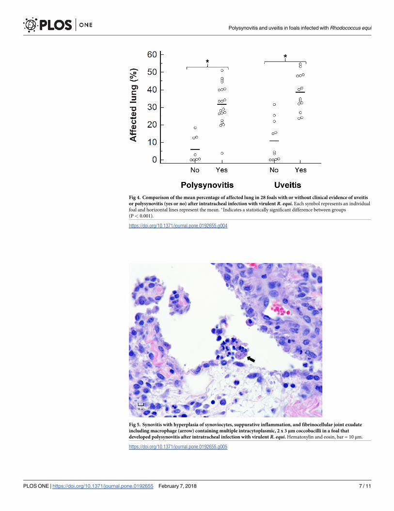
Fig 4. Comparison of the mean percentage of affected lung in 28 foals with or without clinical evidence of uveitis
or polysynovitis (yes or no) after intratracheal infection with virulent R. equi. Each symbol represents an individual
foal and horizontal lines represent the mean. �Indicates a statistically significant difference between groups
(P< 0.001).
https://doi.org/10.1371/journal.pone.0192655.g004
Fig 5. Synovitis with hyperplasia of synoviocytes, suppurative inflammation, and fibrinocellular joint exudate
including macrophage (arrow) containing multiple intracytoplasmic, 2 x 3 μm coccobacilli in a foal that
developed polysynovitis after intratracheal infection with virulent R. equi. Hematoxylin and eosin, bar = 10 μm.
https://doi.org/10.1371/journal.pone.0192655.g005
Polysynovitis and uveitis in foals infected with Rhodococcus equi
PLOS ONE | https://doi.org/10.1371/journal.pone.0192655 February 7, 2018 7 / 11

but statistically significant positive correlation (rs = 0.55; P = 0.003) between the percentage of
affected lung and synovial fluid total protein concentration.
Discussion
The present study documents the occurrence of polysynovitis and uveitis in a high proportion
of foals experimentally infected with virulent R. equi. The culture of virulent R. equi from aque-
ous humor and synovial fluid indicates that these extrapulmonary disorders are septic and not
immune-mediated processes. Multiple studies have described development of pneumonia in
foals after experimental infection with 103 to 1010 CFU of virulent R. equi by instillation into
the airways or by nebulization. However, uveitis had not been described in foals experimen-
tally infected with R. equi and polysynovitis was only described in 4 foals after infection with
109 CFU [9]. The reasons for the lack of detection or reporting of polysynovitis and uveitis in
prior experimental infections are unknown but these complications would be easily missed
without thorough ocular examination and palpation of the joints. In the present study, foals
with polysynovitis did not have apparent lameness and foals with uveitis did not have obvious
signs of ocular disease such as blepharospasm or ocular discharge. The clinical diagnosis of
polysynovitis and uveitis is somewhat subjective and might vary depending on the experience
or expertise of the clinician. Therefore we relied also on synovial fluid and aqueous humor
protein concentration and histopathology by a blinded pathologist to more objectively detect
and quantify inflammation at these sites.
In this study the association between clinically detectable polysynovitis and positive culture
for R. equi was not perfect since eight foals with polysynovitis had negative cultures. Histopa-
thology of the synovial membrane was performed in 6 of these 8 foals and suppurative inflam-
mation with intracellular coccobacilli was observed in 5 foals. Failure to culture bacteria from
synovial fluid in cases of septic arthritis is common in horses. In a retrospective study of horses
with naturally occurring septic arthritis culture was negative in approximately 24% of cases
[12]. Similarly, 9 of 14 foals with uveitis had negative culture. The 2 foals with the most chronic
uveitis as evidenced by hypopyon and fibrin in the anterior chamber had negative cultures.
Similarly, R. equi is rarely isolated from the aqueous humor of naturally infected foals which
typically have chronic disease at the time of sampling [4]. These findings suggest that culture
might be less likely to be positive in more chronic cases of uveitis. Although R. equi is fairly
resilient to temperature changes, the possibility that the sensitivity of culture was decreased by
prior freezing of the samples cannot be excluded. Sensitivity of detection of R. equi in aqueous
humor and synovial fluid might have been improved by PCR amplification. In adult horses
with recurrent uveitis, PCR for amplification of Leptospira DNA in aqueous humor is much
more sensitive than culture [13]. Similarly PCR is more sensitive than culture for the detection
Table 1. Comparison of mean (± SD) total protein concentration in aqueous humor and synovial fluid between inocula (high vs low) and presence or absence of
clinically detectable polysynovitis or uveitis in 28 foals experimentally infected with R. equi.
Sample Uveitis/polysynovitis Inoculum
Present Absent High Low
Aqueous humor (mg/dL) 1,578 ± 707a 103 ± 95 1,423 ± 971c 393 ± 432
Synovial fluid (g/dL) 3.0 ± 0.5b 1.7 ± 0.4 2.8 ± 0.8 2.5 ± 0.7
a Significantly higher than in foals without uveitis (P < 0.001).b Significantly higher than in foals without polysynovitis (P < 0.001).c Significantly higher than in foals infected with the low inoculum (P = 0.003).
https://doi.org/10.1371/journal.pone.0192655.t001
Polysynovitis and uveitis in foals infected with Rhodococcus equi
PLOS ONE | https://doi.org/10.1371/journal.pone.0192655 February 7, 2018 8 / 11

of bacteria in synovial fluid samples from horses with clinical evidence of septic arthritis [14].
In the present study we elected to rely only on culture to detect the presence of live bacteria.
Conversely, 2 foals without clinical evidence of polysynovitis and 6 foals without clinically
apparent uveitis had positive cultures for R. equi. Some of these foals likely had subclinical
uveitis as evidenced by increased protein concentrations in aqueous humor. Aqueous humor
protein concentration has been positively correlated to aqueous flare measurements using
flaremetry [15, 16], which would have been a more sensitive and objective indicator of uveitis
when compared to clinical examination. It is also possible that some foals were euthanized
prior to development of uveitis or polysynovitis. Finally, it is also possible that a small propor-
tion of foals with positive cultures for R. equi in synovial fluid or aqueous humor might clear
the bacteria without necessarily developing uveitis or polysynovitis.
The detection of R. equi in joint fluid and aqueous humor from multiple foals indicates
hematogenous dissemination of R. equi. Intermittent or persistent bacteremia with R. equimight be more common than previously recognized given that blood cultures are not routinely
performed in foals with pneumonia caused by R. equi. In one study, 6 of 10 foals with naturally
acquired pneumonia had positive blood cultures [17]. More recently, R. equi was isolated from
the blood of 11 of 19 foals and foals with positive blood culture results were less likely to sur-
vive than foals that were culture-negative [4]. Blood culture is also the most sensitive means of
diagnosis in people infected with this R. equi [2]. Unfortunately, blood cultures were not per-
formed in this study. Therefore, it is not possible to know when bacteremia occurred relative
to infection and to development of polysynovitis or uveitis.
One of the limitations of this study is the lack of an uninfected control group. Foals raised
at farms endemic for infections caused by R. equi commonly have subclinical pulmonary
disease. The inclusion of a large number of uninfected control foals would have been helpful
to document absence of naturally acquired pulmonary lesions and R. equi in lung tissue.
The nature of the lesions observed (severe ventral consolidation with a clear demarcation
between healthy and affected tissue), the high frequency of uveitis and polysynovitis, and
the consistent incubation period are all consistent with a single exposure to a relatively
heavy inoculum of R. equi and would be unheard of for natural infection especially at a farm
never used to breed horses and raise foals in the past. Ultimately, the novel and important
finding of this study is that polysynovitis and uveitis associated with pneumonia caused by
R. equi are septic in nature and not just the result of an immune mediated process. Even in
the unlikely hypothetical scenario where one or more foals were naturally infected with R.
equi while at pasture, the conclusions that development of uveitis and polysynovitis is asso-
ciated with the severity of pulmonary disease and septic in nature would be unchanged.
Therefore, it was decided that the sacrifice of multiple healthy foals was not justified for eth-
ical reasons.
The strong association between the presence and severity of uveitis and polysynovitis and
the severity of lung lesions emphasize the need to look for a primary infectious process in foals
diagnosed with these conditions. Knowledge of the pathogenesis of polysynovitis and uveitis
has important implications regarding therapy. The use of systemic corticosteroids has been
recommended for foals with polysynovitis and uveitis due to the belief that these extrapulmon-
ary disorders are non-septic and immune-mediated [8, 18]. The septic nature of uveitis and
polysynovitis and their strong association with the severity of pulmonary disease indicates that
additional studies are required before systemic corticosteroids are recommended for the treat-
ment of these conditions. The ability to reproduce these extrapulmonary disorders experimen-
tally in a high proportion of foals would make assessment of various therapies possible in
future studies.
Polysynovitis and uveitis in foals infected with Rhodococcus equi
PLOS ONE | https://doi.org/10.1371/journal.pone.0192655 February 7, 2018 9 / 11

Supporting information
S1 Table. Mean daily rectal temperature, heart rate and respiratory rate of foals experi-
mentally infected with virulent R. equi. Foals were infected with a high inoculum (1 × 10⁸
CFU; n = 16) or a low inoculum (1 × 10⁷ CFU; n = 12).
(PDF)
S2 Table. Occurrence of uveitis or polysynovitis, percentage of affected lung, culture
results, and fluid protein concentrations. Foals were infected with a high inoculum (1 × 10⁸
CFU; n = 16) or a low inoculum (1 × 10⁷ CFU; n = 12) of virulent R. equi.(PDF)
Acknowledgments
We thank Emily Hart and Jennifer Schaffer for technical assistance.
Author Contributions
Conceptualization: Laura Huber, Steeve Giguère.
Data curation: Laura Huber, Steeve Giguère, Londa J. Berghaus.
Formal analysis: Laura Huber, Steeve Giguère.
Funding acquisition: Steeve Giguère.
Investigation: Laura Huber, Steeve Giguère, Londa J. Berghaus, Amanda Hanafi, Sarah
Vitosh-Sillman.
Methodology: Laura Huber, Steeve Giguère, Londa J. Berghaus, Amanda Hanafi, Sarah
Vitosh-Sillman, Sarah L. Czerwinski.
Project administration: Steeve Giguère, Londa J. Berghaus.
Resources: Steeve Giguère.
Supervision: Steeve Giguère, Londa J. Berghaus.
Validation: Laura Huber, Steeve Giguère, Sarah Vitosh-Sillman, Sarah L. Czerwinski.
Visualization: Steeve Giguère, Sarah L. Czerwinski.
Writing – original draft: Laura Huber, Steeve Giguère.
Writing – review & editing: Laura Huber, Steeve Giguère, Londa J. Berghaus, Amanda Han-
afi, Sarah Vitosh-Sillman, Sarah L. Czerwinski.
References1. Arlotti M, Zoboli G, Moscatelli GL, Magnani G, Maserati R, Borghi V, et al. Rhodococcus equi infection
in HIV-positive subjects: a retrospective analysis of 24 cases. Scand J Infect Dis. 1996; 28(5):463–467.
PMID: 8953675
2. Donisi A, Suardi MG, Casari S, Longo M, Cadeo GP, Carosi G. Rhodococcus equi infection in HIV-
infected patients. AIDS. 1996; 10(4):359–362. PMID: 8728038
3. Yamshchikov AV, Schuetz A, Lyon GM. Rhodococcus equi infection. Lancet Infect Dis. 2010; 10
(5):350–359. https://doi.org/10.1016/S1473-3099(10)70068-2 PMID: 20417417
4. Reuss SM, Chaffin MK, Cohen ND. Extrapulmonary disorders associated with Rhodococcus equi infec-
tion in foals: 150 cases (1987–2007). J Am Vet Med Assoc. 2009; 235(7):855–863. https://doi.org/10.
2460/javma.235.7.855 PMID: 19793018
Polysynovitis and uveitis in foals infected with Rhodococcus equi
PLOS ONE | https://doi.org/10.1371/journal.pone.0192655 February 7, 2018 10 / 11

5. Sweeney CR, Sweeney RW, Divers TJ. Rhodococcus equi pneumonia in 48 foals: response to antimi-
crobial therapy. Vet Microbiol. 1987; 14(3):329–336. PMID: 3672875
6. Kenney DG, Robbins SC, Prescott JF, Kaushik A, Baird JD. Development of reactive arthritis and resis-
tance to erythromycin and rifampin in a foal during treatment for Rhodococcus equi pneumonia. Equine
Vet J. 1994; 26(3):246–248. PMID: 8542848
7. Madison JB, Scarratt WK. Immune-mediated polysynovitis in four foals. J Am Vet Med Assoc. 1988;
192(11):1581–1584. PMID: 2970450
8. Wilkes EJA, Hughes KJ, Kessel AE, Raidal SL. Successful management of multiple extrapulmonary
complications associated with Rhodococcus equi pneumonia in a foal. Equine Vet Educ. 2016; 28
(4):184–192.
9. Giguère S, Hondalus MK, Yager JA, Darrah P, Mosser DM, Prescott JF. Role of the 85-kilobase plas-
mid and plasmid-encoded virulence-associated protein A in intracellular survival and virulence of Rho-
dococcus equi. Infect Immun. 1999; 67(7):3548–3557. PMID: 10377138
10. Giguère S, Lee E, Williams E, Cohen ND, Chaffin MK, Halbert N, et al. Determination of the prevalence
of antimicrobial resistance to macrolide antimicrobials or rifampin in Rhodococcus equi isolates and
treatment outcome in foals infected with antimicrobial-resistant isolates of R equi. J Am Vet Med Assoc.
2010; 237(1):74–81. https://doi.org/10.2460/javma.237.1.74 PMID: 20590498
11. Ladron N, Fernandez M, Aguero J, Gonzalez ZB, Vazquez-Boland JA, Navas J. Rapid identification of
Rhodococcus equi by a PCR assay targeting the choE gene. J Clin Microbiol. 2003; 41(7):3241–3245.
https://doi.org/10.1128/JCM.41.7.3241-3245.2003 PMID: 12843070
12. Schneider RK, Bramlage LR, Moore RM, Mecklenburg LM, Kohn CW, Gabel AA. A retrospective study
of 192 horses affected with septic arthritis/tenosynovitis. Equine Vet J. 1992; 24(6):436–442. PMID:
1459056
13. Faber NA, Crawford M, LeFebvre RB, Buyukmihci NC, Madigan JE, Willits NH. Detection of Leptospira
spp. in the aqueous humor of horses with naturally acquired recurrent uveitis. J Clin Microbiol. 2000;
38(7):2731–2733. PMID: 10878072
14. Elmas CR, Koenig JB, Bienzle D, Cribb NC, Cernicchiaro N, Cote NM, et al. Evaluation of a broad
range real-time polymerase chain reaction (RT-PCR) assay for the diagnosis of septic synovitis in
horses. Can J Vet Res. 2013; 77(3):211–217. PMID: 24101798
15. Krohne SG, Krohne DT, Lindley DM, Will MT. Use of laser flaremetry to measure aqueous humor pro-
tein concentration in dogs. J Am Vet Med Assoc. 1995; 206(8):1167–1172. PMID: 7768737
16. Rankin AJ, Krohne SG, Glickman NW, Glickman LT, Stiles J. Laser flaremetric evaluation of experimen-
tally induced blood-aqueous barrier disruption in cats. Am J Vet Res. 2002; 63(5):750–756. PMID:
12013479
17. Leadon D, Farrelly B, Fogarty U, Buckley T. Platelet counting in diagnosis of Rhodococcus equi. Vet
Rec. 1988; 123(10):279.
18. Cohen ND. Treating foals with Rhodococcus equi infection: what do you recommend? Compend Contin
Educ Pract Vet. 2006; 1(1):14–18.
Polysynovitis and uveitis in foals infected with Rhodococcus equi
PLOS ONE | https://doi.org/10.1371/journal.pone.0192655 February 7, 2018 11 / 11


