
HAL Id: hal-00557354https://hal.archives-ouvertes.fr/hal-00557354
Submitted on 19 Jan 2011
HAL is a multi-disciplinary open accessarchive for the deposit and dissemination of sci-entific research documents, whether they are pub-lished or not. The documents may come fromteaching and research institutions in France orabroad, or from public or private research centers.
L’archive ouverte pluridisciplinaire HAL, estdestinée au dépôt et à la diffusion de documentsscientifiques de niveau recherche, publiés ou non,émanant des établissements d’enseignement et derecherche français ou étrangers, des laboratoirespublics ou privés.
Diagnostic accuracy of microbial keratitis with in vivoscanning laser confocal microscopy
Scott Hau, John Dart, Minna Vesaluoma, Dipak Parmar, Ilse Claerhout,Kanom Bibi, Frank Larkin
To cite this version:Scott Hau, John Dart, Minna Vesaluoma, Dipak Parmar, Ilse Claerhout, et al.. Diagnostic accuracy ofmicrobial keratitis with in vivo scanning laser confocal microscopy. British Journal of Ophthalmology,BMJ Publishing Group, 2010, 94 (8), pp.982. �10.1136/bjo.2009.175083�. �hal-00557354�
1
Diagnostic accuracy of microbial keratitis with in vivo scanning laser
confocal microscopy
Scott C Hau,1 John K G Dart,1 Minna Vesaluoma,1 Dipak N Parmar,2 Ilse Claerhout,3
Kanom Bibi,1 Daniel F P Larkin1
1 NIHR Biomedical Research Centre in Ophthalmology, Moorfields Eye Hospital and
Institute of Ophthalmology, London, UK, EC1V 2PD
2 Dept of Ophthalmology, Whipps Cross Hospital, London, UK, E11 1NR
3 Ghent University Hospital, De Pintelaan 185, 9000 Gent, Belgium
Word count: 2500
Corresponding author: Scott Hau, Research fellow, Moorfields Eye Hospital NHS
Foundation Trust, 162 City Road, London, UK, EC1V 2PD
Email: [email protected]
Tel: +447905 295888
Fax: +44207 5662471
Key words: Confocal microscopy, diagnostic accuracy, acanthamoeba keratitis, fungal
keratitis, Microsporidia, Nocardia
2
The Corresponding Author has the right to grant on behalf of all authors and does
grant on behalf of all authors, an exclusive licence (or non-exclusive for government
employees) on a worldwide basis to the BMJ Publishing Group Ltd and its Licensees
to permit this article (if accepted) to be published in British Journal of Ophthalmology
and any other BMJPGL products to exploit all subsidiary rights, as set out in our
licence
3
ABSTRACT
Aims
To determine the accuracy of diagnosing microbial keratitis by masked medical and
non-medical observers using the Heidelberg Retina Tomograph II / Rostock Cornea
Module in vivo confocal microscope.
Methods
Confocal images were selected for 62 eyes with culture or biopsy proven infections.
The cases comprised of 26 Acanthamoeba, 12 fungus, 3 Microsporidia, 2 Nocardia,
and 19 bacterial infections (controls). The reference standard for comparison was a
positive tissue diagnosis. These images were assessed on two separate occasions by 4
observers who were masked to the tissue diagnosis. Diagnostic accuracy indices,
Kappa (κ) statistic and percentage agreement values were calculated. The Spearman
correlation coefficient (rs) was calculated for the number of correct diagnoses versus
duration of disease.
Results
The highest sensitivity and specificity values were 55.8% and 84.2%, and the lowest
sensitivity and specificity values were 27.9% and 42.1%. The highest positive and
negative likelihood ratios were 2.94 and 0.59, respectively. Agreement values were:
fair to moderate (κ, 0.22-0.44) for reference standard versus observer diagnosis,
moderate to good in intra-observer variability (repeatability, κ 0.56-0.88), and poor to
moderate in inter-observer variability (reproducibility, κ, 0.15-0.47). The correct
4
diagnosis was associated with duration of disease for Acanthamoeba keratitis (rs = 0.60,
p = 0.001).
Conclusions
The diagnostic accuracy of microbial keratitis by confocal microscopy is dependent
on observer experience. Intra-observer repeatability was better than inter-observer
reproducibility. Difficulty in distinguishing host cells from pathogenic organisms
limits the value of confocal microscopy as a stand-alone tool in diagnosing microbial
keratitis.
5
INTRODUCTION 1
2
Difficulties in clinical and microbiological diagnosis are one of the major problems in 3
the management of microbial keratitis particularly when caused by protozoa 4
(Acanthamoeba and Microsporidia), fungi or filamentary bacteria. Diagnosis of these 5
pathogens is difficult as they often take days or weeks to grow in culture and, in any 6
case, culture is insensitive with culture positive rates rarely exceeding 60%.[1] 7
Although culture is still the primary diagnostic tool in tertiary referral centres it is not 8
widely available to many patients because of limited resources. 9
10
The confocal microscope allows detailed in vivo analysis of normal[2] and 11
pathological corneas. In patients with presumed corneal infection, it is used in 12
diagnosis and in examination of the extent of involvement of tissue by infection and 13
associated inflammation. All published studies have been directed at diagnosis and a 14
number have shown both white light and laser confocal microscopy to be effective in 15
diagnosing Acanthamoeba,[3-5] fungal,[6-8] Nocardia[9] and Microsporidia 16
keratitis.[10] However, these studies only present case series or reports and there are 17
limited published data on evaluating the diagnostic accuracy of confocal microscopy. 18
Two recent studies have found high sensitivity and specificity values for diagnosing 19
fungal keratitis (FK) and Acanthamoeba keratitis (AK) with the Confoscan 3.0 (Nidek 20
Technology, Padova, Italy).[11,12] However, factors such as observer or selection 21
bias, the absence of masking the observers from the microbiological diagnosis, and 22
lack of appropriate controls may have resulted in overestimates of the sensitivity and 23
specificity values. Although experience in interpreting confocal keratitis images is 24
essential, the accuracy of diagnosing microbial keratitis by clinicians with differing 25
6
levels of confocal microscopy experience and the potential of using trained 26
technicians in interpreting images have not previously been assessed. These are 27
important considerations in evaluating this technique. The aim of this study was to 28
examine the diagnostic accuracy of microbial keratitis with the Heidelberg Retina 29
Tomograph II / Rostock Cornea Module (HRT II/RCM) in vivo confocal microscope, 30
as a stand-alone tool, by trained medical and non-medical observers with differing 31
confocal microscopy experience. 32
33
MATERIALS AND METHODS 34
35
Patients 36
This study was approved by the Research & Ethics Committee of Moorfields Eye 37
Hospital and it adhered to the tenets of Declaration of Helsinki. We retrospectively 38
reviewed the case notes of a consecutive series of microbial keratitis patients who had 39
had both corneal cultures or corneal biopsy and confocal microscopy (n=105) from 40
January 1, 2005 to January 4, 2008. These cases were both those refractory to 41
conventional treatment and those with unusual clinical features such as perineural 42
infiltrates and ring infiltrates. Patients were either referred from Moorfields 43
Emergency Department or from other institutions. Of the 105 cases, 62 culture or 44
biopsy positive cases (62 eyes) were identified: 26 Acanthamoeba, 11 fungus, 1 45
fungus and bacteria, 3 Microsporidia, 2 Nocardia, and 19 bacteria. Bacteria were used 46
as controls because they are normally too small to detect with confocal microscopy, 47
[9,13] therefore the case which was culture positive for both fungus and bacteria was 48
classified as a fungal keratitis for the purposes of the study. We did not classify 49
7
Nocardia as controls because they are filamentous bacteria and can form filamentous 50
structures that are large enough to be distinguished by confocal microscopy.[9] 51
Empirical treatments started prior to assessment in this study included topical 52
antimicrobial agents and topical steroids for presumed herpes, bacterial or keratitis of 53
unknown cause, respectively. Irrespective of the referring diagnosis, all patients had 54
undergone a full clinical examination by a corneal specialist and repeat corneal 55
scraping for culture and confocal microscopy on the same day. If the scraping was 56
culture negative, and the keratitis progressive, then a corneal biopsy was later 57
performed. Exclusion criteria were culture or biopsy negative keratitis cases, and 58
patients who declined to have confocal microscopy or a corneal culture as part of their 59
clinical investigation. The reference standard for this study was a diagnosis either by 60
isolation on culture of a corneal scraping or histological diagnosis on a corneal biopsy; 61
other ancillary culture sources such as contact lens case and solutions were not used. 62
The clinical outcomes were recorded for all the patients in the study and were 63
consistent with the diagnosis based on culture or histology therefore it is unlikely, but 64
possible, that there was unrecognised polymicrobial infections which may have been 65
identified on confocal but not by culture or biopsy. We followed the Standards for 66
Reporting of Diagnostic accuracy (STARD) initiative in conducting this study.[14] 67
68
Culture and biopsy methods 69
Corneal scrapings for microbial culture were inoculated on the following media: 70
blood agar, Sabouraud’s dextrose agar (fungi), Robertson’s cooked meat (anaerobic 71
bacteria), Escherichia coli-seeded non-nutrient agar (Acanthamoeba), brain heart 72
infusion (fastidious organisms, fungi) and Lowenstein-Jensen (mycobacteria, 73
Nocardia). Scrapings were smeared on sterile glass slides for Gram and Giemsa stains. 74
8
All microbiological investigations were undertaken independently in an external 75
laboratory. For biopsy a superficial lamellar disc of the affected cornea was trephined 76
under local anaesthetic to provide a further specimen for microbiology and 77
histopathological staining. 78
79
Confocal microscopy measurement protocol 80
In vivo confocal microscopy was performed on all 62 eyes by a single experienced 81
observer (SH) with the HRT II / RCM (Heidelberg Engineering GmbH, Dossenheim, 82
Germany) confocal microscope following a Standard Operating Procedure as follows. 83
A sterile Tomocap (Heidelberg Engineering GmbH, Dossenheim, Germany) was 84
mounted over the objective of the microscope (Zeiss, x 63), and Polyacrylic acid 0.2% 85
(Viscotears, Novartis) was used as a coupling agent between the cap and the lens 86
objective. Topical anaesthetic (Proxymetacaine hydrochloride 0.5%, Chauvin) and 87
Carmellose sodium 1% (Celluvisc, Allergan) was instilled into both eyes to provide 88
comfort and act as a coupling fluid between the front of the Tomocap and the cornea. 89
Options for image acquisition include section (a single image at a particular depth), 90
volume (a series of images over 60µm depth) and sequence scans (a video sequence at 91
a particular depth). The volume scan option was selected for image acquisition 92
because it allowed the capturing of large number of images over a short space of time. 93
The central region of the corneal ulcer or corneal infiltrate was scanned first followed 94
by the top, left, bottom and right margin of the lesion. At each point, the epithelial 95
layer of the affected area was scanned first and the focal plane of the microscope 96
adjusted until the whole depth of the ulcer or infiltrate had been scanned. When there 97
was more than one infiltrate, the same scanning sequence was repeated for each 98
infiltrate. The wavelength of the laser employed in the HRT II / RCM is 670 nm and 99
9
each standard 2 dimensional image consists of 384 x 384 pixels covering an area of 100
400 μm x 400 μm. The axial resolution is 7.6μm; compared to other instruments such 101
as the Tandem scanning microscope (9µm) and ConfoScan 4 (29µm).[15] 102
103
Image selection 104
The confocal images of all the scans were reviewed by two experienced confocal 105
microscopist (SH and JD). In diagnosing keratitis, a considerable amount of time is 106
often needed to find an image that would yield sufficient information to be able to 107
identify the organism. This is due to masking of the organisms by the cellular 108
inflammatory response and that they seldom distribute evenly within the cornea 109
during active infection. Therefore, to ensure all our observers had the maximum 110
likelihood in diagnosing the type of keratitis, the best quality 384 x 384 pixel 111
resolution digital image indicating clearly the culture proven pathogen from the 112
corneal ulcer or infiltrate was selected and exported onto Microsoft Power Point® 113
(Microsoft Corp., Redmond, WA, USA). These included those of Acanthamoeba - 114
round single or double walled hyper-reflective objects (~10-20 μm) consistent with 115
Acanthamoeba cysts,[4,5] fungus - linear irregular branching hyper-reflective objects 116
consistent with fungal hyphae,[6,7] Microsporidia - small round hyper-reflective 117
deposits (~ 2 μm) located in between keratocytes,[10] Nocardia - small branching 118
filamentous structures within the corneal stroma,[9] and bacteria (control) – a mixture 119
of inflammatory cells. 120
121
122
123
10
Intra- and inter-observer agreement 124
All digital images were assessed prospectively in the same standard fashion in the 125
Reading Centre at Moorfields Eye Hospital by 4 observers (3 ophthalmologists and 1 126
medical technician) with differing levels of experience in assessing keratitis on 127
confocal microscopy as follows. Of the 3 ophthalmologists, observer A had 6 years of 128
experience in assessing microbial keratitis with confocal microscopy, observer B, 10 129
years of experience in confocal microscopy but not keratitis, and observer C, 6 130
months of experience in assessing keratitis with confocal microscopy. Observer D 131
was a medical technician who had 2 years of experience in performing confocal 132
microscopy using the HRT II / RCM and analysing keratitis images but with no 133
experience in the clinical appearance and treatment of different types of keratitis. To 134
ensure each observer was familiar with the image appearance of different cell types 135
obtained from the HRT II / RCM confocal microscope, examples of both normal 136
cellular morphology and the standard images of different pathogens were shown in a 137
presentation before their assessment. In addition, a series of five recent articles on 138
diagnosing keratitis with the HRT II / RCM [4,5,7,9,10] were given to each observer 139
to read 2 weeks prior to their scheduled assessment date. 140
141
The confocal images were viewed in random order and assigned an identification 142
number from 1 to 62. To ensure that there was masking between observers, the order 143
of viewing the images were randomised by computer before being assessed by the 144
next observer on a different day. No clinical details regarding each case were made 145
available to the observers. Each observer assessed the series of images in a masked 146
fashion on slide show in Microsoft Powerpoint® and recorded the diagnosis 147
corresponding to one of the following categories: AK, FK, Microsporidia (MK), 148
11
Nocardia (NK) or bacterial keratitis (BK). A reference sheet showing the range of 149
sizes of resident and inflammatory cells including epithelium and macrophages, and 150
pathogenic cells e.g. diameter of Acanthamoeba cysts was given to each observer. 151
Intra-observer variability (repeatability) was evaluated by asking each observer to 152
reassess the images, randomised in a different order, three weeks later in the same 153
standard fashion. Inter-observer variability (reproducibility) was assessed by 154
determining the level of agreement in diagnosis between observers. Readings of all 155
the digital images were collected on a standard pro-forma and analysed. 156
157
Data analysis 158
Data analysis was performed with SPSS V14.0 (SPSS Inc, Chicago, USA). We 159
calculated sensitivity, specificity, positive and negative likelihood ratios (LR) for both 160
image set for each observer. Positive LR predicts the probability of a positive test 161
result in patients with disease compared to those who do not have the disease. 162
Negative LR predicts the probability of a negative test in those who have the disease 163
compare to those who do not. The level of agreement between the reference standard 164
and different observers, and both intra and inter-observer variability were determined 165
using Kappa (κ) statistic. The interpretation of κ statistic is as follows: ‘poor’ if κ ≤ 166
0.20, ‘fair’ if κ 0.21 – 0.40, ‘moderate’ if κ 0.41 – 0.60, ‘substantial’ if κ 0.61 – 0.80 167
and ‘good’ if κ > 0.80.[16] In addition, we also calculated percentage agreement 168
values between reference standard and observers, within-observers, and between 169
different observers. Spearman’s rank correlation coefficient (rs) was used to determine 170
the relationship between the number of correct diagnoses and the duration of disease 171
for AK, FK and BK respectively. The duration of disease was defined as the time 172
from symptom onset to presentation to the Corneal and External Disease Service at 173
12
Moorfields. A value of P < 0.05 was deemed statistically significant. MK and NK 174
were excluded from this analysis because the numbers were too small. 175
176
RESULTS 177
178
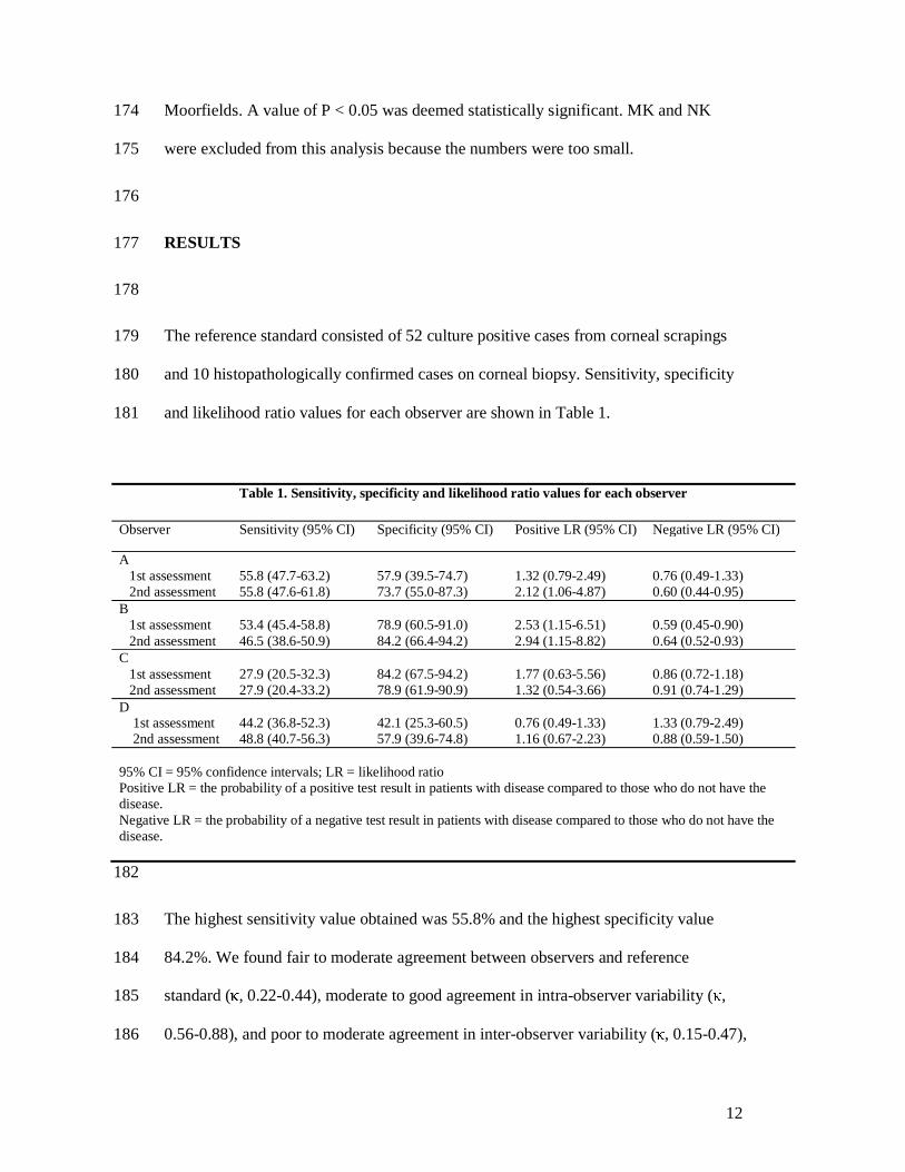
The reference standard consisted of 52 culture positive cases from corneal scrapings 179
and 10 histopathologically confirmed cases on corneal biopsy. Sensitivity, specificity 180
and likelihood ratio values for each observer are shown in Table 1. 181
182
The highest sensitivity value obtained was 55.8% and the highest specificity value 183
84.2%. We found fair to moderate agreement between observers and reference 184
standard (κ, 0.22-0.44), moderate to good agreement in intra-observer variability (κ, 185
0.56-0.88), and poor to moderate agreement in inter-observer variability (κ, 0.15-0.47), 186
Table 1. Sensitivity, specificity and likelihood ratio values for each observer
Observer Sensitivity (95% CI) Specificity (95% CI) Positive LR (95% CI) Negative LR (95% CI)
A 1st assessment 2nd assessment
55.8 (47.7-63.2) 55.8 (47.6-61.8)
57.9 (39.5-74.7) 73.7 (55.0-87.3)
1.32 (0.79-2.49) 2.12 (1.06-4.87)
0.76 (0.49-1.33) 0.60 (0.44-0.95)
B 1st assessment 2nd assessment
53.4 (45.4-58.8) 46.5 (38.6-50.9)
78.9 (60.5-91.0) 84.2 (66.4-94.2)
2.53 (1.15-6.51) 2.94 (1.15-8.82)
0.59 (0.45-0.90) 0.64 (0.52-0.93)
C 1st assessment 2nd assessment
27.9 (20.5-32.3) 27.9 (20.4-33.2)
84.2 (67.5-94.2) 78.9 (61.9-90.9)
1.77 (0.63-5.56) 1.32 (0.54-3.66)
0.86 (0.72-1.18) 0.91 (0.74-1.29)
D 1st assessment 2nd assessment
44.2 (36.8-52.3) 48.8 (40.7-56.3)
42.1 (25.3-60.5) 57.9 (39.6-74.8)
0.76 (0.49-1.33) 1.16 (0.67-2.23)
1.33 (0.79-2.49) 0.88 (0.59-1.50)
95% CI = 95% confidence intervals; LR = likelihood ratio Positive LR = the probability of a positive test result in patients with disease compared to those who do not have the disease. Negative LR = the probability of a negative test result in patients with disease compared to those who do not have the disease.
13
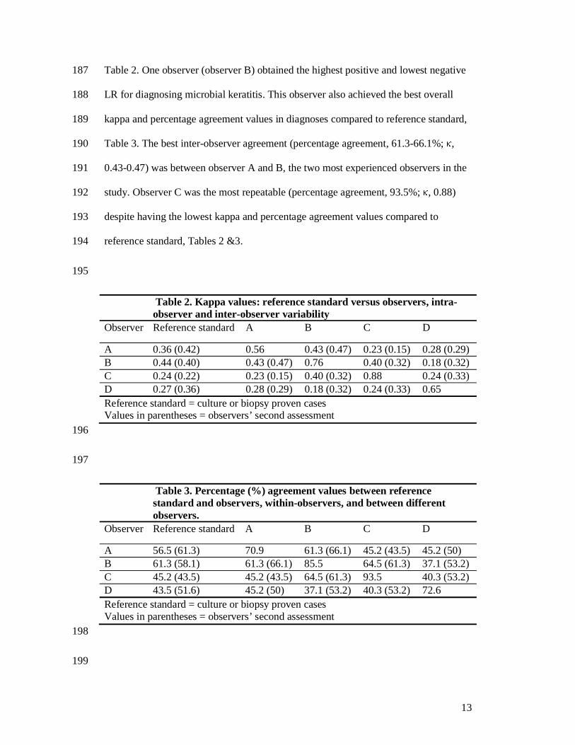
Table 2. One observer (observer B) obtained the highest positive and lowest negative 187
LR for diagnosing microbial keratitis. This observer also achieved the best overall 188
kappa and percentage agreement values in diagnoses compared to reference standard, 189
Table 3. The best inter-observer agreement (percentage agreement, 61.3-66.1%; κ, 190
0.43-0.47) was between observer A and B, the two most experienced observers in the 191
study. Observer C was the most repeatable (percentage agreement, 93.5%; κ, 0.88) 192
despite having the lowest kappa and percentage agreement values compared to 193
reference standard, Tables 2 &3. 194
195
Table 2. Kappa values: reference standard versus observers, intra-observer and inter-observer variability
Observer Reference standard A B C D
A 0.36 (0.42) 0.56 0.43 (0.47) 0.23 (0.15) 0.28 (0.29) B 0.44 (0.40) 0.43 (0.47) 0.76 0.40 (0.32) 0.18 (0.32) C 0.24 (0.22) 0.23 (0.15) 0.40 (0.32) 0.88 0.24 (0.33) D 0.27 (0.36) 0.28 (0.29) 0.18 (0.32) 0.24 (0.33) 0.65 Reference standard = culture or biopsy proven cases Values in parentheses = observers’ second assessment 196
197
Table 3. Percentage (%) agreement values between reference standard and observers, within-observers, and between different observers.
Observer Reference standard A B C D
A 56.5 (61.3) 70.9 61.3 (66.1) 45.2 (43.5) 45.2 (50) B 61.3 (58.1) 61.3 (66.1) 85.5 64.5 (61.3) 37.1 (53.2) C 45.2 (43.5) 45.2 (43.5) 64.5 (61.3) 93.5 40.3 (53.2) D 43.5 (51.6) 45.2 (50) 37.1 (53.2) 40.3 (53.2) 72.6 Reference standard = culture or biopsy proven cases Values in parentheses = observers’ second assessment 198
199
14
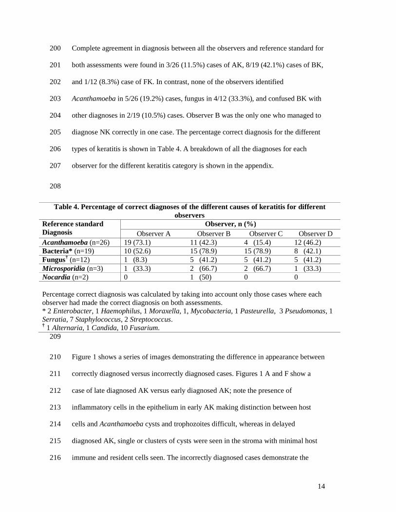
Complete agreement in diagnosis between all the observers and reference standard for 200
both assessments were found in 3/26 (11.5%) cases of AK, 8/19 (42.1%) cases of BK, 201
and 1/12 (8.3%) case of FK. In contrast, none of the observers identified 202
Acanthamoeba in 5/26 (19.2%) cases, fungus in 4/12 (33.3%), and confused BK with 203
other diagnoses in 2/19 (10.5%) cases. Observer B was the only one who managed to 204
diagnose NK correctly in one case. The percentage correct diagnosis for the different 205
types of keratitis is shown in Table 4. A breakdown of all the diagnoses for each 206
observer for the different keratitis category is shown in the appendix. 207
208
Table 4. Percentage of correct diagnoses of the different causes of keratitis for different observers
Reference standard Diagnosis
Observer, n (%) Observer A Observer B Observer C Observer D
Acanthamoeba (n=26) 19 (73.1) 11 (42.3) 4 (15.4) 12 (46.2) Bacteria* (n=19) 10 (52.6) 15 (78.9) 15 (78.9) 8 (42.1) Fungus† (n=12) 1 (8.3) 5 (41.2) 5 (41.2) 5 (41.2) Microsporidia (n=3) 1 (33.3) 2 (66.7) 2 (66.7) 1 (33.3) Nocardia (n=2) 0 1 (50) 0 0 Percentage correct diagnosis was calculated by taking into account only those cases where each observer had made the correct diagnosis on both assessments. * 2 Enterobacter, 1 Haemophilus, 1 Moraxella, 1, Mycobacteria, 1 Pasteurella, 3 Pseudomonas, 1 Serratia, 7 Staphylococcus, 2 Streptococcus. † 1 Alternaria, 1 Candida, 10 Fusarium.
209
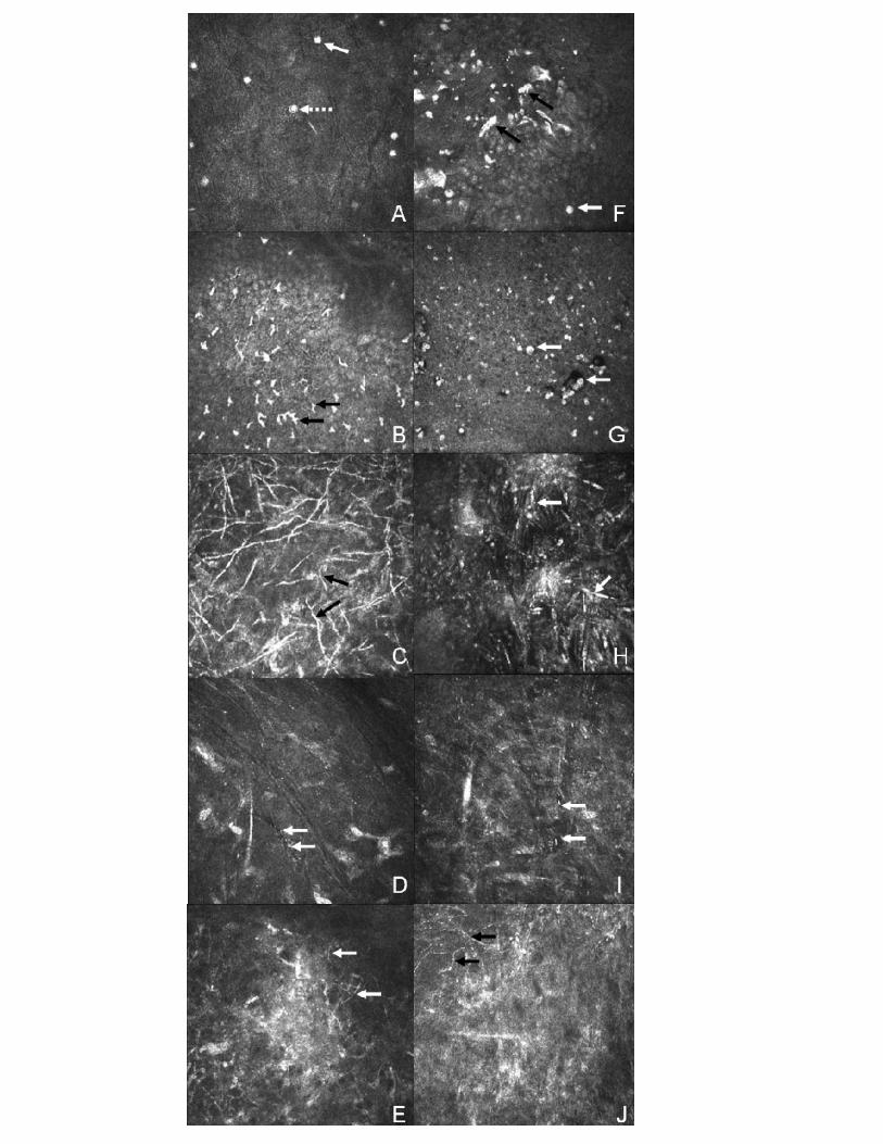
Figure 1 shows a series of images demonstrating the difference in appearance between 210
correctly diagnosed versus incorrectly diagnosed cases. Figures 1 A and F show a 211
case of late diagnosed AK versus early diagnosed AK; note the presence of 212
inflammatory cells in the epithelium in early AK making distinction between host 213
cells and Acanthamoeba cysts and trophozoites difficult, whereas in delayed 214
diagnosed AK, single or clusters of cysts were seen in the stroma with minimal host 215
immune and resident cells seen. The incorrectly diagnosed cases demonstrate the 216

15
difficulties in distinguishing host cells from pathogenic organisms, and Nocardia (Fig 217
1J) from FK because of their similarity in appearance on confocal microscopy. 218
219
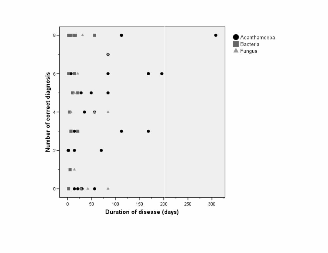
Figure 2 shows a plot between the number of correct diagnoses for Acanthamoeba, 220
bacteria and fungal keratitis versus the duration of disease (days). The graph shows a 221
moderate correlation between the number of correctly diagnosed cases and the 222
duration of disease for AK (rs = 0.60, p = 0.001), but not for BK (rs = 0.17, P = 0.49) or 223
FK (rs = -0.19, p = 0.57), respectively. Therefore, the longer the duration of AK, the 224
higher the likelihood that a correct diagnosis was made by the observers in grading 225
the confocal images. 226
227
DISCUSSION 228
229
Acanthamoeba and fungus are uncommon causes of corneal infection for which early 230
diagnosis is paramount because it yields better prognosis and reduces ocular 231
morbidity.[17,18] Although the current reference standard for diagnosing microbial 232
keratitis is corneal culture, the sensitivity varies because of numerous factors.[19] 233
234
The HRT II / RCM in vivo confocal microscope has been shown to be useful in 235
diagnosing a range of pathogens but validation studies of this new technology are few. 236
A recent review has reported the efficacy of diagnosing infections keratitis with 237
confocal microscopy to be inconclusive, with the possible exception of AK.[20] Our 238
results show moderate sensitivity and moderate to high specificity values in 239
diagnosing microbial keratitis with the HRT II / RCM confocal microscope, whereas 240

16
both Kanavi et al[11] and Tu et al[12] found very high sensitivity (>90%) in 241
diagnosing AK and FK respectively with the Confoscan 3. Tu et al,[12] using multi-242
test referencing standards, reported that when there are both clinical characteristics 243
and objective evidence of AK, the adjunctive usage of confocal microscopy exhibited 244
a sensitivity of 90.6% and specificity of 100%. In our study, we set out to evaluate the 245
diagnostic accuracy of confocal microscopy as a stand alone tool rather than a 246
supportive investigative technique, without the bias and influence of clinical findings. 247
Although assessing confocal images in the absence of clinical data does not reflect the 248
use of confocal microscopy in clinical settings, it is the only way to avoid bias when 249
analysing the images. Our inclusion criteria were based on culture positive cases 250
irrespective of confocal classification. Although we chose only one representative 251
image from each case this was the best available image for the organism that was 252
cultured from each case giving the observers the best opportunity to make a correct 253
confocal diagnosis; we believe that reviewing a series of images from each case 254
would either have made a correct confocal diagnosis more difficult or have had no 255
effect on the outcome. In addition, it allowed standardisation when viewing the 256
images so that all observers assessed the same number of images consecutively. The 257
absence of controls in the previous studies and the use of confocal ‘positive’ without 258
culture confirmation as a reference standard, or for the case definition[11,12], could 259
lead to selection bias and misdiagnosis resulting in an overestimation of sensitivity 260
values.[12,21,22] This is evident from our controls in which immune cells can often 261
be confused with AK cysts and vice versa leading to erroneous diagnosis. 262
Furthermore, ‘good’ confocal images have been illustrated in most published studies 263
to present findings without discussion of difficulties in analysing equivocal images. 264
We found fair to moderate agreement between reference standard and observer 265
17
diagnosis when a case mix of equivocal and unequivocal images were analysed by our 266
observers. The rigorous criteria in our study design in regard to the use of masked 267
observers and controls could explain why sensitivity values, even for the most 268
experienced observer, were lower. 269
270
Another explanation for the very high sensitivity values reported in one previous 271
study was the use of only one ophthalmology trained observer who, in addition to 272
being unmasked to the clinical findings, was experienced in the use of confocal 273
microscopy for keratitis diagnosis: this makes it difficult to extrapolate the results to 274
what might be expected outside their centres.[12] To evaluate the potential of using 275
this technology in clinics where an ophthalmologist with experience in confocal 276
microscopy may not be available, our graders included 2 experienced 277
ophthalmologists, an inexperienced ophthalmologist and an experienced technician. 278
We found a two-fold difference in sensitivity between the most experienced and the 279
least experienced observer indicating higher diagnostic accuracy with clinicians 280
experienced in confocal microscopy. Our results indicate the sensitivity value with a 281
trained technician, with no experience in the clinical appearance of different types of 282
microbial keratitis, was better than an inexperienced medical observer but with a 283
lower specificity value and positive LR. This raises the possibility of training non-284
medical personnel, in performing and analysing keratitis images. The highest positive 285
LR and lowest negative LR was achieved by observer B who was experienced in 286
confocal imaging of normal corneal anatomy and various pathological conditions 287
other than microbial keratitis, indicating experience gained in other aspects of 288
confocal microscopy improves the diagnostic outcome. 289
290
18
Intra-observer agreement (repeatability) was found to be moderate to good, indicative 291
of good observer repeatability in grading the images irrespective of the accuracy of 292
their diagnoses. Observer experience did not appear to improve intra-observer 293
repeatability as the observer with the lowest sensitivity had the highest repeatability 294
and vice versa. Inter-observer agreement (reproducibility) was poor to moderate 295
between different observers because of factors such as observer experience and 296
differences in techniques of classifying images by different observers. The two 297
observers who had the highest sensitivity values also had the best inter-observer 298
reproducibility, indicating experienced observers achieved a higher diagnostic 299
accuracy and reproducibility than less experienced observers. Therefore, to improve 300
reliability the same experienced operator should be employed if sequential imaging of 301
a patient is required. 302
303
Our observers were able to diagnose AK more accurately than any other type of 304
keratitis. The unique appearance of Acanthamoeba cysts on confocal microscopy and 305
the higher number of AK compared to other conditions in our study might explain this 306
outcome. However, AK was commonly confused with controls and vice versa because 307
of the diagnostic difficulty with some of the equivocal images. There was a marked 308
association between the accuracy of diagnosing AK and the duration of disease. 309
Previous case reports have mainly described the morphological features of cysts and 310
trophozoites in the epithelium and stroma during active infection,[4,5] but have not 311
related the number of cysts seen and the way they distribute with the different stages 312
of the disease process. In early disease, where the organism is mainly confined to the 313
epithelium, the presence of large numbers of inflammatory cells made diagnosing AK 314
more difficult because of the difficulty in distinguishing AK cysts and particularly 315

19
trophozoites from inflammatory cells.[5] Late presentation was associated with either 316
a greater number of Acanthamoeba cysts seen in the images or the fact that they were 317
easier to identify because of a reduction in the type and number of host cells seen. Our 318
experience, therefore, suggests that AK is easier to identify with confocal microscopy 319
in the later stages of infection. 320
321
The use of confocal microscopy in diagnosing FK has been widely reported in the 322
literature.[6-8] Filamentous fungal hyphae have characteristic linear hyper-reflective 323
lesions branching at 45 or 90 degrees angle,[7] whereas candida infection produces 324
pseudofilaments.[7] Despite these well described confocal appearances of FK in the 325
literature, the percentage of correct diagnosis in our series was low possibly due to 326
difficulties in differentiating other linear images from fungal hyphae.[23] 327
328
Nocardia and Microsporidia species are rare causes of microbial keratitis.[24] 329
Clinically, Nocardia may be misdiagnosed as mycotic or mycobacterial keratitis,[9,25] 330
whilst Microsporidia can be misdiagnosed as AK or herpes simplex keratitis. Despite 331
the rarity of these organisms, because of the unique appearance on confocal 332
microscopy with Microsporidia,[10] two observers managed to identify this organism 333
correctly in both of their assessments. Only observer B managed to obtain the correct 334
diagnosis in both assessments for diagnosing one case of Nocardia keratitis; the 335
unfamiliarity in interpreting confocal images of Nocardia, the similarity in appearance 336
of fungal hyphae and Nocardia filaments, and the small number of cases in our study 337
made diagnosing this organism difficult. The inclusion of both Nocardia and 338
Microsporidia cases might have reduced the overall sensitivity and specificity values 339
20
but as confocal findings of both organisms have been reported, we believe it was 340
appropriate to include them in the study. 341
342
In summary, to the best of our knowledge, this is the first study evaluating the 343
diagnostic accuracy of microbial keratitis using a single reference standard for 344
different masked observers with the HRT II / RCM confocal microscope. Although 345
confocal microscopy is non-invasive and can provide a rapid diagnosis for microbial 346
keratitis, (i) similarities between inflammatory and pathogenic cells, and (ii) difficulty 347
in interpreting equivocal images, limits its usefulness as a stand-alone tool in 348
diagnosing keratitis. Confocal microscopy is a useful adjunct in managing refractory 349
cases and we have shown that the diagnostic accuracy improves with clinician 350
experience. However, the diagnostic accuracy of confocal microscopy used in 351
isolation from the clinical assessment is still too low to be a substitute for tissue 352
diagnosis, particularly in patients with progressive disease. Improvement in clinician 353
training and experience, greater standardization of image interpretation, and the 354
development of new software in tandem with higher resolution imaging is likely to 355
improve the diagnostic accuracy of this technology in diagnosing microbial keratitis 356
in the future. 357
358
ACKNOWLEDGEMENTS 359
The authors would like to thank Dr Catey Bunce for her statistical advice and support. 360
361
COMPETING INTERESTS 362
None declared 363
364
21
FUNDING 365
None 366
367
368
REFERENCES 369
370
1. Yeh DL, Stinnett SS, Afshari NA. Analysis of bacterial cultures in infectious 371
keratitis, 1997 to 2004. Am J Ophthalmol 2006;142;1066-1068. 372
373
2. Jalbert I, Stapleton F, Papas E, et al. In vivo confocal microscopy of the human 374
cornea. Br J Ophthalmol 2003;87:225-236. 375
376
3. Parmar DN, Awward ST, Petroll WM, et al. Tandem scanning confocal corneal 377
microscopy in the diagnosis of suspected acanthamoeba keratitis. Ophthalmology 378
2006;113:538-47. 379
380
4. Matsumoto Y, Dogru M, Sato E, et al. The application of in vivo confocal scanning 381
laser microscopy in the management of acanthamoeba keratitis. Molecular Vision 382
2007;13:1319-26. 383
384
5. Kobayashi A, Ishibashi Y, Oikawa Y, et al. In vivo and ex vivo laser confocal 385
microscopy findings in patients with early-stage acanthamoeba keratitis. Cornea. 386
2008;27:439-45. 387
388
22
6. Avunduk AM, Beuerman RW, Varnell ED, Kaufman HE. Confocal microscopy of 389
Aspergillus fumigatus keratitis. Br J ophthalmol 2003;87:409-410. 390
391
7. Brasnu E, Bourcier T, Dupas B, et al. In vivo confocal microscopy in fungal 392
keratitis. Br J Ophthalmol. 2007;91:588-91 393
394
8. Tu EY, Park AJ. Recalcitrant Beauveria bassiana keratitis: confocal microscopy 395
findings and treatment with posaconazole (Noxafil). Cornea 2007;26:1008-10. 396
397
9. Vaddavalli PK, Garg P, Sharma S, et al. Confocal microscopy for Nocardia 398
keratitis. Ophthalmology 2006;113:1645-50. 399
400
10. Sagoo MS, Mehta JS, Hau S, et al. Microsporidium stromal keratitis: in vivo 401
confocal findings. Cornea 2007;26:870-3. 402
403
11. Kanavi MR, Javadi M, Yazdani S, Mirdehghanm S. Sensitivity and specificity of 404
confocal scan in the diagnosis of infectious keratitis. Cornea 2007;26:782-6. 405
406
12. Tu EY, Joslin CE, Sugar J, et al. The relative value of confocal microscopy and 407
superficial corneal scrapings in the diagnosis of Acanthamoeba keratitis. Cornea 408
2008;27:764-72 409
410
13. Petroll WM, Cavanagh HD, Jester JV. Clinical confocal microscopy. Curr Opin 411
Ophthalmol 1998;9:59-65. 412
413
23
14. Bossuyt PM, Reitsma JB, Bruns DE, et al. Towards complete and accurate 414
reporting of studies of diagnostic accuracy: the STARD initiative. Standards for 415
Reporting of Diagnostic Accuracy. Clin Chem 2003;49:1-6. 416
417
15. Zhivov A, Stachs O, Stave J et al. In vivo three-dimensional confocal laser 418
scanning microscopy of corneal surface and epithelium. Br J Ophthalmol 419
2009:93:667-672. 420
421
16. Petrie A, Sabin C. Medical statistics at a glance. 2nd ed, Oxford: Blackwell 422
Publishing Ltd 2005: 93-95. 423
424
17. Bacon AS, Dart JK, Ficker LA, et al. Acanthamoeba keratitis. The value of early 425
diagnosis. Ophthalmology 1993;100:1238-43. 426
427
18. Duguid IG, Dart JK, Morlet N, et al. Outcome of acanthamoeba keratitis treated 428
with polyhexamethyl biguanide and propamidine. Ophthalmology 1997;104:1587-92. 429
430
19. McLeod SD, Kolahdouz-Isfahani A, Rostamian K, et al. The role of smears, 431
cultures, and antibiotic sensitivity testing in the management of suspected infectious 432
keratitis. Ophthalmology 1996;103:23-8. 433
434
20. Labbé A, Khammari C, Dupas B, et al. Contribution of in vivo confocal 435
microscopy to the diagnosis and management of infectious keratitis. Ocul Surf 436
2009;7:41-52. 437
438
24
21. Gray R, Begg CB, Greenes RA. Construction of receiver operating characteristic 439
curves when disease verification is subject to selection bias. Med Decis Making 440
1984;4:151-64. 441
442
22. Choi BC. Sensitivity and specificity of a single diagnostic test in the presence of 443
work-up bias. J Clin Epidemiol 1992;45:581-6. 444
445
23. Chiou AG, Kaufman SC, Beuerman RW, et al. Differential diagnosis of linear 446
corneal images on confocal microscopy. Cornea 1999;18:63-6. 447
448
24. Sridhar MS, Sharma S, Reddy MK, et al. Clinicomicrobiological review of 449
Nocardia keratitis. Cornea 1998;17:17-22. 450
451
25. Lalitha P, Tiwari M, Prajna NV, et al. Nocardia keratitis: species, drug 452
sensitivities, and clinical correlation. Cornea 2007;26:255-9. 453
454 455
456
457
458
459
460
461 462
463
464
25
FIGURE LEGENDS 465
466
Figure 1 467
468
Confocal scans of correctly diagnosed versus incorrectly diagnosed cases. 469
Figures A to D demonstrate the characteristic features of inflammatory cells and 470
pathogenic organisms on confocal microscopy in which all the observers had made 471
the correct diagnoses. A, Acanthamoeba cysts (white arrow), some with double-472
walled appearance (dotted white arrow). B, Inflammatory cells (black arrows). C, 473
Fungal hyphae (black arrows). D, Microsporidia organisms (white arrows). 474
475
Figures E to J show a series of images of incorrectly diagnosed cases demonstrating 476
the difficulty in distinguishing host cells from pathogenic organisms and Nocardia 477
from fungal keratitis. E, Shows Nocardia filaments (white arrows) - only observer B 478
identified this correctly with all the other observers graded it as fungal hyphae. F, 479
Cultured Acanthamoeba - misdiagnosed as bacterial keratitis by observers B, C and D; 480
possible Acanthamoeba cysts (white arrows), and possible inflammatory cells (black 481
arrows). G, Cultured bacteria (Staphylococcus aureus) but was misdiagnosed as 482
Acanthamoeba by all the observers; multiple round lesions that could be identified as 483
inflammatory or Acanthamoeba cysts (white arrows). H, Cultured bacteria - 484
diagnosed as fungal keratitis by observers C & D; linear hyphae-like opacities that 485
were confused with fungal hyphae (white arrows). I, Cultured bacteria - diagnosed as 486
Microsporidia by all the observers; small hyper-reflective granules that appear similar 487
to Microsporidia organisms (white arrows). J, Cultured positive for Alternaria and 488
26
Staphylococcus aureus but diagnosed as Nocardia by observers A, B and D; hyphae 489
type lesions that appear similar to Nocardia filaments (black arrows). 490
491
Figure 2 492
493
Scattered plot showing the relationship between number of correctly diagnosed cases 494
and duration of disease (days) for Acanthamoeba, bacteria and fungal keratitis. 495
496


![A dot hybridization assay for the diagnosis of bacterial ... · Bacterial keratitis (BK) is the most common microbial keratitis in temperate countries [ 1,2] and is a leading cause](https://static.documents.pub/doc/80x56/5cea8a6c88c9934e128d5f12/a-dot-hybridization-assay-for-the-diagnosis-of-bacterial-bacterial-keratitis.jpg)

