

Diuretics
Excretion of Water and Electrolytes

Background
Primary effect of diuretics is to increase solute excretion, mainly as NaCl
Causes increase in urine volume due to increased osmotic pressure in lumen of renal tubule.
Causes concomitant decrease in extra-cellular volume (blood volume)
Certain disease states may cause blood volume to increase outside of narrowly defined limits Hypertension Congestive heart failure Liver cirrhosis Nephrotic syndrome Renal failure
Dietary Na restriction often not enough to maintain ECF and prevent edema diuretics needed

Review of Kidney Structure
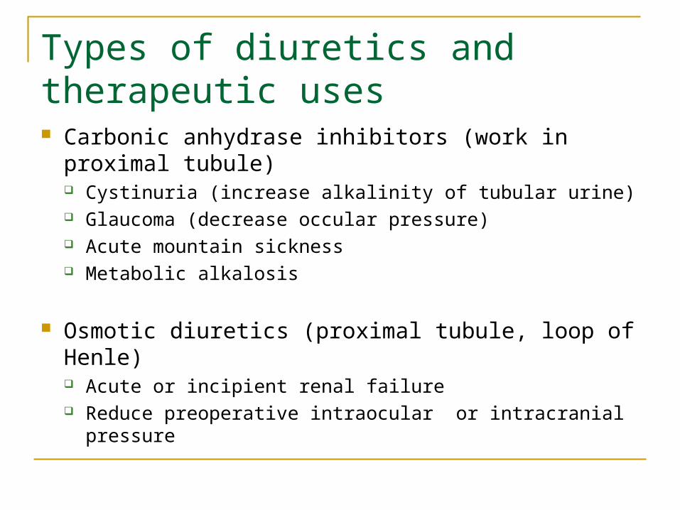
Types of diuretics and therapeutic uses Carbonic anhydrase inhibitors (work in proximal
tubule) Cystinuria (increase alkalinity of tubular urine) Glaucoma (decrease occular pressure) Acute mountain sickness Metabolic alkalosis
Osmotic diuretics (proximal tubule, loop of Henle) Acute or incipient renal failure Reduce preoperative intraocular or intracranial pressure

Types of diuretics and therapeutic uses Loop diuretics (ascending limb of loop)
Hypertension, in patients with impaired renal function
Congestive heart failure (moderate to severe) Acute pulmonary edema Chronic or acute renal failure Nephrotic syndrome Hyperkalemia Chemical intoxication (to increase urine flow)

Types of diuretics and therapeutic uses Thiazide diuretics (distal convoluted tubule)
Hypertension Congestive heart failure (mild) Renal calculi Nephrogenic diabetes insipidus Chronic renal failure (as an adjunct to loop
diuretic) Osteoporosis

Types of diuretics and therapeutic uses Potassium-sparing diuretics (collecting tubule)
Chronic liver failure Congestive heart failure, when hypokalemia is a problem
Osmotic agents (proximal tubule, descending loop of Henle, collecting duct) Reduce pre-surgical or post-trauma intracranial pressure Prompt removal of renal toxins One of the few diuretics that do not remove large amounts
of Na+
Can cause hypernatremia

Nephron sites of action of diuretics
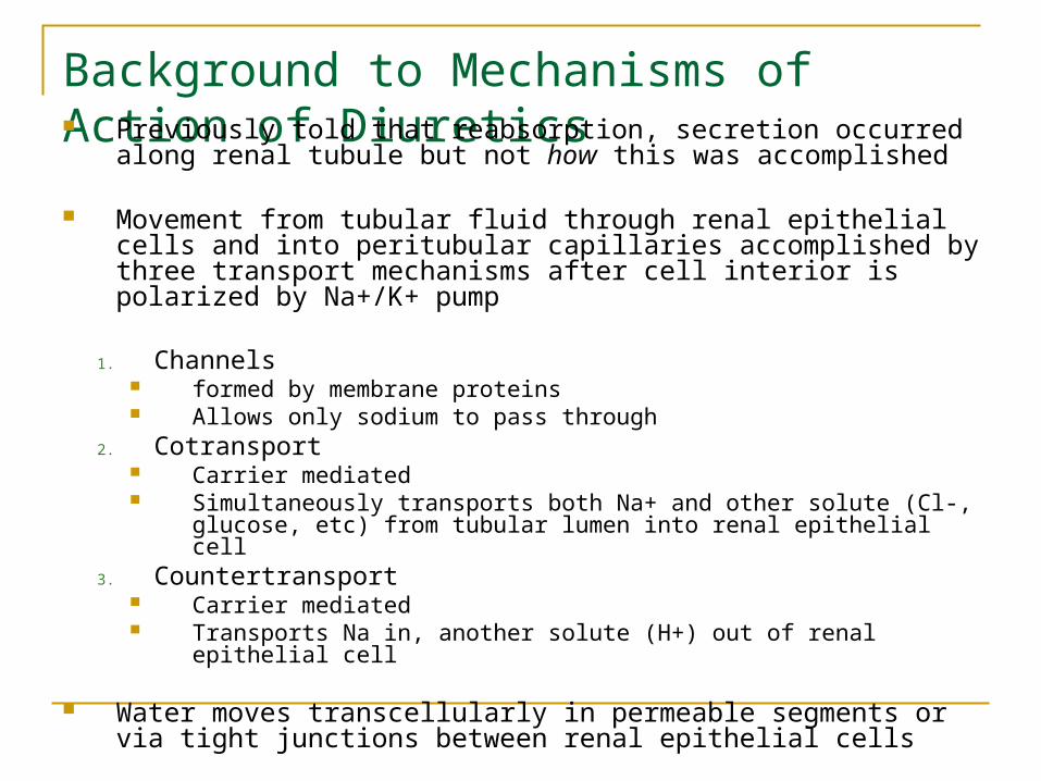
Background to Mechanisms of Action of Diuretics Previously told that reabsorption, secretion occurred along
renal tubule but not how this was accomplished
Movement from tubular fluid through renal epithelial cells and into peritubular capillaries accomplished by three transport mechanisms after cell interior is polarized by Na+/K+ pump
1. Channels formed by membrane proteins Allows only sodium to pass through
2. Cotransport Carrier mediated Simultaneously transports both Na+ and other solute (Cl-, glucose,
etc) from tubular lumen into renal epithelial cell3. Countertransport
Carrier mediated Transports Na in, another solute (H+) out of renal epithelial cell
Water moves transcellularly in permeable segments or via tight junctions between renal epithelial cells

Electrolyte Transport Mechanisms
Channel
Cotransport
Countertransport
Na+/K+ pump
X = glucose, amino acids, phosphate, etc.

Mechanisms of Action: Carbonic anydrase inhibitors
CAIs work on cotransport of Na+, HCO3- and Cl- that is coupled
to H+ countertransport Acts to block carbonic anhydrase (CA),
1. CA converts HCO3- + H+ to H2O + CO2 in tubular lumen
2. CO2 diffuses into cell (water follows Na+), CA converts CO2 + H2O into HCO3
- + H+ 3. H+ now available again for countertransport with Na+, etc)
4. Na+ and HCO3- now transported into peritubular capillary
CA can catalyze reaction in either direction depending on relative concentration of substrates

Site of Action of CAIs

Mechanisms of Action: Loop diuretics No transport systems in descending loop of Henle
Ascending loop contains Na+ - K+ - 2Cl- cotransporter from lumen to ascending limb cells
Loop diuretic blocks cotransporter Na+, K+, and Cl- remain in lumen, excreted along with water

Mechanisms of Action: Thiazide Diuretics in the Distal Convoluted Tubule Less reabsorption of water and electrolytes in the distal
convoluted tubule than proximal tubule or loop A Na+ - Cl- cotransporter there is blocked by thiazides

Mechanisms of Action: Collecting tubule and potassium-sparing diuretics Two cell types in collecting tubule1. Principal cells – transport Na, K, water
2. Intercalated cells – secretion of H+ and HCO3
3. Blocking Na+ movement in also prevents K+ movement out

Summary of sites of renal reabsorption of filtrate

Types and Names of DiureticsOsmotic agents Mannitol Proximal tubule
Descending loop
Collecting duct
Carbonic anydrase inhib.
Acetazolamide Proximal tubule
Thiazides Hydrochlorothiazide
Distal convoluted tubule
Loop diuretic Ethacrynic acid
Furosemide
Loop of Henle
Type Example Sites of Action
K+ - sparing Spironolactone
Amiloride
Collecting tubule

Structure of Classes of Diuretics

General Background of Diuretics Pattern of excretion of electrolytes (how
much of which type) depends on class of diuretic agent
Maximal response is limited by site of action
Effect of two or more diuretics from different classes is additive or synergistic if there sites or mechanisms of action are different

Osmotic diuretics
No interaction with transport systems All activity depends on osmotic pressure
exerted in lumen Blocks water reabsorption in proximal tubule,
descending loop, collecting duct Results in large water loss, smaller
electrolyte loss can result in hypernatremia

Carbonic anydrase inhibitors
Block carbonic-anhydrase catalyzation of CO2/ carbonic acid/carbonate equilibrium
Useful for treating glaucoma and metabolic alkalosis but can cause hyperchloremic metabolic acidosis from HCO3
- depletion

Loop diuretics
Generally cause greater diuresis than thiazides; used when they are insuffficient
Can enhance Ca2+ and Mg2+ excretion Enter tubular lumen via proximal tubular
secretion (unusual secretion segment) because body treats them as a toxic drug
Drugs that block this secretion (e.g. probenecid) reduces efficacy

Thiazide diuretics
Developed to preferentially increase Cl- excretion over HCO3
- excretion (as from CAIs)
Magnitude of effect is lower because work on distal convoluted tubule (only recieves 15% of filtrate)
Cause decreased Ca excretion hypercalcemia reduce osteoporosis

Comparison of loop and thiazide diuretics

Potassium-sparing diuretics
Have most downstream site of action (collecting tubule)
Reduce K loss by inhibiting Na/K exchange Not a strong diuretic because action is
furthest downstream Often used in combination with thiazide
diuretics to restrict K loss


