
Draft
Testosterone-related and seasonal changes in sebaceous
glands in the back skin of adult male brown bears (Ursus arctos)
Journal: Canadian Journal of Zoology
Manuscript ID cjz-2017-0028.R2
Manuscript Type: Article
Date Submitted by the Author: 06-Sep-2017
Complete List of Authors: Tomiyasu, Jumpei ; Obihiro University of Agriculture and Veterinary
Medicine, Laboratory of Theriogenology, Department of Applied Veterinary Medicine; Gifu University, United Graduate School of Veterinary Sciences Yanagawa, Yojiro; Hokkaido University, Laboratory of Theriogenology, Department of Veterinary Clinical Sciences Sato, Yoshikazu; Rakuno Gakuen University, Laboratory of Wildlife Ecology, Department of Environmental Symbiotic Science, College of Agriculture, Food and Environmental Sciences Shimozuru, Michito; Hokkaido University, Graduate School of Veterinary Medicine Nagano, Masashi; Hokkaido University, Laboratory of Theriogenology, Department of Veterinary Clinical Sciences Sasaki, Motoki; Obihiro University of Agriculture and Veterinary Medicine,
Laboratory of Veterinary Anatomy, Department of Basic Veterinary Science Sakamoto, Hideyuki; Noboribetsu Bear Park Matsumoto, Naoya; Noboribetsu Bear Park Kobayashi, Kohei; EnVision Conservation Office Kayano, Mitsunori; Obihiro University of Agriculture and Veterinary Medicine, Research Center for Global Agromedicine Haneda, Shingo; Obihiro University of Agriculture and Veterinary Medicine, Laboratory of Theriogenology, Department of Applied Veterinary Medicine Matsui, Motozumi; Obihiro University of Agriculture and Veterinary Medicine, Laboratory of Theriogenology, Department of Applied Veterinary Medicine; Gifu University, United Graduate School of Veterinary Sciences
Keyword: brown bear, sebaceous gland, testosterone, tree rubbing, back skin, scent
gland, <i>Ursus arctos</i>
https://mc06.manuscriptcentral.com/cjz-pubs
Canadian Journal of Zoology
Draft
1
Testosterone-related and seasonal changes in sebaceous glands in the back skin of adult male
brown bears (Ursus arctos)
Running Header: Sebaceous glands in back skin of male brown bears
J. Tomiyasu1, 2, Y. Yanagawa3, Y. Sato4, M. Shimozuru5, M. Nagano3, M. Sasaki2, 6, H. Sakamoto7, N.
Matsumoto7, K. Kobayashi8, M. Kayano9, S. Haneda1 and M. Matsui1, 2*
1Laboratory of Theriogenology, Department of Applied Veterinary Medicine, Obihiro University of
Agriculture and Veterinary Medicine, Obihiro, Hokkaido 080-8555, Japan 2United Graduate School of Veterinary Sciences, Gifu University, Gifu, Gifu 501-1193, Japan 3Laboratory of Theriogenology, Department of Veterinary Clinical Sciences, Graduate School of
Veterinary Medicine, Hokkaido University, Sapporo, Hokkaido 060-0818, Japan 4Laboratory of Wildlife Ecology, Department of Environmental Symbiotic Science, College of
Agriculture, Food and Environmental Sciences, Rakuno Gakuen University, Ebetsu, Hokkaido
069-0836, Japan 5Laboratory of Wildlife Biology and Medicine, Department of Environmental Veterinary Science
Graduate School of Veterinary Medicine, Hokkaido University, Sapporo, Hokkaido 060-0818, Japan 6Laboratory of Veterinary Anatomy, Department of Basic Veterinary Science, Obihiro University of
Agriculture and Veterinary Medicine, Obihiro, Hokkaido 080-8555, Japan 7Noboribetsu Bear Park, Noboribetsu, Hokkaido 059-0551, Japan 8EnVision Conservation Office, 5-2, Kita 9, Nishi 4, Kita-ku, Sapporo, Hokkaido 060-0809, Japan 9Research Center for Global Agromedicine, Obihiro University of Agriculture and Veterinary Medicine,
Obihiro, Hokkaido 080-8555, Japan
J. Tomiyasu, [email protected]; Y. Yanagawa, [email protected];
Y. Sato, [email protected]; M. Shimozuru, [email protected];
M. Nagano, [email protected]; M. Sasaki, [email protected];
H. Sakamoto, [email protected]; N. Matsumoto, [email protected];
K. Kobayashi, [email protected]; M. Kayano, [email protected];
S. Haneda, [email protected]; M. Matsui*, [email protected]
*Corresponding author
Motozumi Matsui, Tel: +81-155-49-5382, Fax: +81-155-49-5384
Email: [email protected]
Postal address: Nishi-2-11, Inada-cho, Obihiro, Hokkaido, 080-8555, Japan
Page 1 of 30
https://mc06.manuscriptcentral.com/cjz-pubs
Canadian Journal of Zoology

Draft
2
Testosterone-related and seasonal changes in sebaceous glands in the back skin of
adult male brown bears (Ursus arctos)
Jumpei Tomiyasu, Yojiro Yanagawa, Yoshikazu Sato, Michito Shimozuru, Masashi Nagano,
Motoki Sasaki, Hideyuki Sakamoto, Naoya Matsumoto, Kohei Kobayashi, Mitsunori
Kayano, Shingo Haneda and Motozumi Matsui
Abstract
Adult male brown bears (Ursus arctos; Linnaeus, 1758) display tree-marking behavior to
chemically signalize the dominance throughout the non-denning period, and this behavior
peaks during breeding season. Within the scent-marking sequence, back rub is one of a
core marking postures. The present study investigated 1) seasonal changes in sebaceous
glands in the back skin of brown bears and 2) the relationship between those changes and
testosterone levels. Back skin tissue samples and blood were collected from captive adult
intact and castrated males during pre-breeding, transitional, breeding and post-breeding
seasons, which were concurrent with back skin observations. In intact males, during the
transitional and breeding seasons, an oily secretion from the back skin was observed along
with enlarged sebaceous glands. The plasma testosterone concentrations during the
transitional and breeding seasons were increased compared with the pre- and post-breeding
seasons. Secretions and enlarged sebaceous glands were not found in castrated males, and
the plasma testosterone concentrations remained at baseline levels. Oily secretions of the
back skin glands that appear more abundant during breeding season are rubbed against
trees. Changes in size and volume of sebaceous glands, and thus their secreting capacity,
are likely testosterone-regulated.
Keywords: brown bear, sebaceous gland, testosterone, tree-rubbing, back skin, scent gland,
Ursus arctos
Page 2 of 30
https://mc06.manuscriptcentral.com/cjz-pubs
Canadian Journal of Zoology
Draft
3
Introduction
Olfactory communication is the primary form of communication between individuals
in many mammalian species, and it represents a key source of information (Gorman and
Trowbridge 1989; Müller-Schwarze 2006). Animals use feces, urine, and glandular
secretions from skin to communicate chemically (Müller-Schwarze 2006) without the need
for direct contact. Odors remain viable for long periods, even in the absence of the
producer (Margaret et al. 1980).
Brown bears (Ursus arctos; Linnaeus, 1758) are normally solitary with a large home
range (Dahle and Swenson 2003; Støen et al. 2005); therefore, scent is a valuable
mechanism of information transmission. Because the mating system of brown bears is
polygynous, adult males fight with each other for opportunities to mate with females
during the breeding season (May–July; Tsubota et al. 1985; Ishikawa et al. 2003). Young
male bears disperse from their natal areas, probably to avoid mating competition with older
bears (Zedrosser et al. 2007), and they are likely able to identify sexually mature male
brown bears by odors emitted from anal sacs (Rosell et al. 2011; Jojola et al. 2012).
Sub-adult male bears might store information derived from adult male odors for future
encounters to reduce the cost of conflict (Jojola et al. 2012). Therefore, brown bears seem
to use olfactory communication.
Brown bears often rub themselves against trees (Green and Mattson 2003; Karamalidis
et al. 2007; Clapham et al. 2014; Sato et al. 2014) placed along travel routes, perhaps as
landmarks (Green and Mattson 2003, Clapham et al. 2013). Adult male bears rub against
trees more often than other age and sex classes throughout the non-denning period, and
Page 3 of 30
https://mc06.manuscriptcentral.com/cjz-pubs
Canadian Journal of Zoology
Draft
4
adult males might utilize chemical signaling to impart dominance information (Clapham et
al. 2012). This behavior peaks during the breeding season (Green and Mattson 2003;
Clapham et al. 2012, 2014; Sato et al. 2014). A study of the motor activity of sent marking
in brown bears (Clapham et al. 2014), identified bipedal back rubbing as a core marking
posture. Elevated marking behavior might widely disperse information and increase
detectability (Albert et al. 1992), or convey competitive ability (White et al. 2002). We
speculated that male brown bears communicate chemically by rubbing the skin on the back
against surfaces during the breeding season, and that information derived from this
behavior is related to reproduction.
Wild brown bears seem to increase the amount of tree-rubbings during the breeding
season (Green and Mattson 2003; Clapham et al. 2012, 2014; Sato et al. 2014). We
hypothesized that male brown bears have sebaceous glands in back skin that become
enlarged by testosterone during the breeding season. This is because sebaceous gland
enlargement depends on testosterone levels in goats Capra hircus (L., 1758) (Iwata et al.
2000; Wakabayashi et al. 2000), golden hamsters Mesocricetus auratus (Waterhouse, 1839)
(Vandenbergh 1973), rat-like hamsters Cricetulus triton (de Winton, 1899) (Zhang et al.
2001), and musk rats Ondatra zibethica (L., 1766) (Lu et al. 2014). In order to investigate
the characteristic of sebaceous glands in skin from the back of brown bear, the present
study aimed 1) to define seasonal changes in sebaceous glands relative to breeding season
and: 2) to clarify the relationship between those changes and testosterone levels.
Materials and methods
Page 4 of 30
https://mc06.manuscriptcentral.com/cjz-pubs
Canadian Journal of Zoology
Draft
5
Experimental design
We investigated seasonal changes in sebaceous glands in skin from the backs of bears
as follows. We firstly visually assessed the skin on backs of anesthetized captive intact
bears and collected skin samples for histological analysis during the following seasons:
pre-breeding (February), transitional (April), breeding (May and June; Tsubota et al. 1985;
Ishikawa et al. 2003), and post-breeding (August and October), which is classified on the
basis of the previous study about testosterone of male brown bears (Tsubota and Kanagawa
1989). To examine whether or not sebaceous gland enlargement is specific to skin on the
back during the breeding season, we compared skin sampled from back and rump, because
back rubbing is a core posture, whereas rump rubbing is not (Clapham et al. 2014).
Moreover, to examine the relationship between those changes in sebaceous glands and
testosterone levels, we measured plasma testosterone concentrations and the size of the
sebaceous glands in intact and castrated male bears. The presence of androgen receptors
was immunohistochemically assessed.
Animals
We studied 11 captive Hokkaido male brown bears from the Noribetsu Bear Park,
Hokkaido, Japan (42ºN, 141ºE: Facility A; five individuals: four intact and one castrated),
and Sahoro Bear Mountain, Hokkaido Japan (43ºN, 142ºE: Facility B; six individuals: four
intact and two castrated). We used five of seven bears that spent the daytime outdoors in a
700 m2 open concrete space at Facility A. At night, four of them were placed in a 23 × 2.3
× 2.2 m3 (height × width × depth). Three others were placed in a 12 × 2.3 × 2.2 m3 (height
× width × depth). We used six of ten bears which were placed in an open, fenced
Page 5 of 30
https://mc06.manuscriptcentral.com/cjz-pubs
Canadian Journal of Zoology
Draft
6
mountainous area of 150,000 m2 during the daytime at Facility B. The vegetation in this
area was similar to that in wild-bear habitats. And, they were separated in one space,
respectively at night in a 2.2 × 2.2 × 2.5 m3 (height × width × depth) area. The bears were
fed bear pellets (ZOOFOOD bear, Nosan Co., Kanagawa, Japan), concentrated feed
formulated for cows (Soyokazenokaori MG, Nippon Formula Food Manufacturing Co.,
Ltd., Kanagawa Japan), vegetables, and fruits once or twice a day with water ad libitum.
All the bears studied herein was born in captivity. Rubbing behavior occurred during the
study period. Bears rubbed themselves against walls in the absence of trees in Facility A.
Male and female bears were separated in Facility A, but a short distance between pens
allowed for olfactory and auditory cognition. Facility B did not house any female bears.
All bears were between eight to 25 years of age, and were sexually mature. The bears were
exposed to natural photoperiods throughout the year.
Skin examination and tissue collection
Samples were collected from the same individuals at Facility A during February, April,
May, June, August, and October, and at Facility B in April, June, and October (Table 1).
The bears were repeatedly anesthetized via the intramuscular administration of 2.5–3.5
mg/kg of a mixture of zolazepam HCl and tiletamine HCl (Zoletil, Virbac, Carros, France)
with either 0.03 mg/kg medetomidine HCl (Domitor; Orion Corporation Animal Health,
Turku, Finland) or 1 mg/kg xylazine HCl (Selactar; Bayer Healthcare, Leverkusen,
Germany) using blow darts. After sample collection, anesthesia was reversed by the
intramuscular administration of 0.03 mg/kg atipamezole HCl (Antisedan; Orion
Corporation Animal Health). Secretions were verified directly by observation and by
Page 6 of 30
https://mc06.manuscriptcentral.com/cjz-pubs
Canadian Journal of Zoology
Draft
7
rubbing skin with a paper towel. Skin samples were obtained from the center of the back
skin between the shoulder blades (Fig. 1) using an 8-mm BP-L80K Biopsy Punch (Kai,
Ind., Ltd., Gifu, Japan), and all biopsies were obtained at a depth of 25 mm. Skin samples
from the rump were also collected in June from four intact bears at Facility B. A section of
fur on each bear was shaved, the skin was repeatedly washed with 20 mg/mL povidone
iodine (Isodine; Meiji Seika, Tokyo, Japan) and iso-propanol 50% (iso-propanol 50%,
Yakuhan, Kitahiroshima, Japan) and then veterinarians collected biopsy specimens. The
wound was then sutured with Coated Vicryl® Plus Antibacterial Suture (Ethicon,
Somerville, NJ). After completing all experiments, 0.2 mg/kg meloxicam (Metacam,
Boehringer Ingelheim, Germany) and 5 mg/kg Enrofloxacin (Baytril, Bayer, Leverkusen,
Germany) were subcutaneously administered for analgesia and antimicrobial activity,
respectively. All procedures were conducted by veterinarians. Pus was not evident at the
biopsy sites on the following day. Skin biopsy specimens were fixed in 10% buffered
formalin. Blood samples were collected from the saphenous veins into Venoject® II
heparinized Vacutainers® (Terumo, Tokyo, Japan), and the plsma was separated from
hematocytes by centrifugation at 2000 × g for 15 min. Plasma samples were stored at
-30 °C until the assay was performed.
Histological and immunohistochemical analysis
Fixed skin tissues were dehydrated in serially diluted ethanol, embedded in paraffin
(Paraplast Plus; Kendall, Mansfield, MA, USA), cut into 4-µm thick sections and mounted
onto Mas-coated slides (S9904, Matsunami Glass Ind., Ltd., Osaka, Japan). To examine
the skin glands, deparaffinized sections were stained with hematoxylin and eosin.
Page 7 of 30
https://mc06.manuscriptcentral.com/cjz-pubs
Canadian Journal of Zoology
Draft
8
Androgen receptor (AR) expression was immunohistochemically assessed using the
avidin-biotin complex (ABC) method (Hsu et al. 1981). Samples of skin collected from
intact and castrated back, and intact rump in June were prepared for immunohistological
assessment as follows. Deparaffinized sections were incubated in a LAB antigen retrieval
solution (Polysciences Inc, Eppelheim, Germany) at 60 °C for 60 min, and then soaked in
0.3% H2O2 in methanol for 10 min to inactivate the endogenic peroxidase. Nonspecific
binding was blocked by incubating the sections with a 1:50 dilution of S-1000 normal goat
serum (Vector Laboratories, Inc., CA, USA) for 30 min at room temperature. The slides
were then incubated at 4 °C overnight with a 1:50 dilution of N-20 rabbit polyclonal
anti-androgen receptor antibody (Santa Cruz Biotechnology, Inc., TX, USA), followed by
30 min at room temperature with 1:200 dilution of BA-1000 goat biotinylated anti-rabbit
IgG (Vector Laboratories, Inc.). The samples were exposed to the ABC reagent in
PK-6100, Vectastain Elite ABC Kit (Vector Laboratories) at room temperature for 30 min
and then primary antibody binding sites were visualized by soaking the sections in 0.02%
3,3′-diaminobenzidine HCl and 0.006% H2O2 in Tris-HCl buffer. The sections on slides
were finally dehydrated and coverslipped. The negative controls were normal rabbit serum
or 0.01 M phosphate buffered saline instead of the primary antibody.
Sebaceous glands morphology or parameters
Columnar skin biopsy specimens were cut parallel to the long axis at 200-µm intervals.
Areas of sebaceous glands within 6 × 10-mm2 rectangles on 10 slides were measured using
image J software and summed. This parameter is referred to herein as the “sebaceous
glands area” and it served as an index of change in the size of sebaceous glands.
Page 8 of 30
https://mc06.manuscriptcentral.com/cjz-pubs
Canadian Journal of Zoology
Draft
9
Hormone assay
Plasma testosterone concentrations were measured with an enzyme-immunoassay
(EIA) using anti-testosterone serum (COSMO FKA 102E, Cosmo Bio Co., Ltd., Inc.,
Tokyo, Japan) and horseradish peroxidase-labeled testosterone (COSMO FKA 101, Cosmo
Bio Co., Ltd., Inc.). The EIA procedure was conducted as previously described (Miyamoto
et al., 1992). Testosterone was extracted from the plasma sample with diethyl ether and
was subsequently measured, and the extraction efficiency was 91.5%. The standard curve
ranged from 0.025 to 25 ng/mL, and 50% of the effective dose of the assay was 0.462
ng/mL. The average intra- and inter-assay coefficient of variation values were 7.8% and
8.1%, respectively.
Statistical analysis
Nonparametric analyses were performed, since the sample size was too small to
assume normal distribution. A Kruskal-Wallis test was performed to detect significant
differences in sebaceous gland size indices and plasma testosterone concentrations between
intact males assigned to different seasonal groups, and a Steel-Dwass test was performed to
verify the group pairs that showed significant differences. Spearman’s rank correlation test
was performed to investigate the correlation between the sebaceous gland size indices and
the plasma testosterone concentrations in intact males. The sebaceous glands size indices
and the plasma testosterone concentrations between intact and castrated males were
compared using the Wilcoxon rank sum test for each season. For samples collected in June,
the sebaceous gland size indices between the back and rump skins of intact males were
compared using the Wilcoxon rank sum test. All data were statistically analyzed using R
Page 9 of 30
https://mc06.manuscriptcentral.com/cjz-pubs
Canadian Journal of Zoology
Draft
10
software (R Development Core Team, 2015), and a P-value of <0.05 and <0.1 indicated a
significant differences and tendencies, respectively. Data are presented as the median
(range).
Results
Skin observations
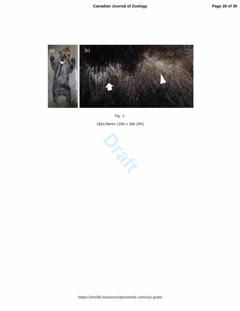
Abundant sebum secretion (Fig. 1) was observed on the back skin during the
transitional (n = 6/6) and breeding (n = 11/12) seasons. The secretion was observed at the
midline of the back skin from the first cervical vertebra to the second lumbar vertebra (Fig.
1). The secretion had a very strong sweet odor, and the color was brown or colorless. Oily
secretion was not evident beyond this specific area of back skin. During the pre- (n = 4)
and post-breeding (n = 12) seasons, no secretions from back skins of males were detected
from back skins of males. Throughout the experiment, no secretions were found on the
back skin of castrated males (n = 3, all seasons).
Seasonal changes in sebaceous skin glands and plasma testosterone concentrations
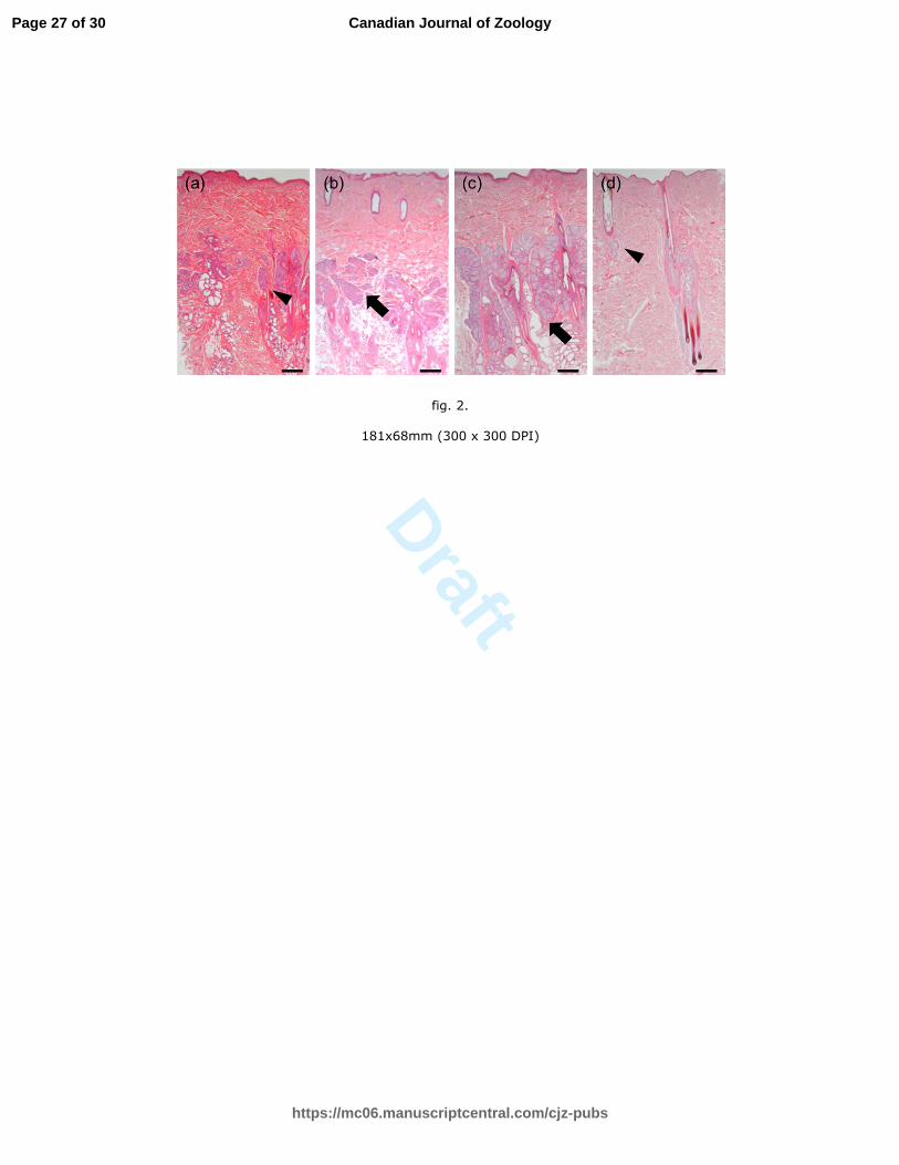
Figure 2 shows the histological findings of back skin samples from intact males
collected during the study period. Enlarged sebaceous glands were situated next to follicles
during the transitional (n = 6/6) and breeding (n = 11/12) seasons, with the exception of
one male sampled in June that had shriveled sebaceous glands that did not secrete sebum.
The sebaceous glands in back skins of intact males shriveled during the pre- (n = 4) and
post-breeding (n = 12) seasons. In castrated males, sebaceous glands did not become
enlarged throughout the experimental period (n = 3, all seasons). Changes in the index of
Page 10 of 30
https://mc06.manuscriptcentral.com/cjz-pubs
Canadian Journal of Zoology

Draft
11
sebaceous gland size in intact males during the pre-breeding (n = 4), transitional (n = 6),
breeding (n=12) and post-breeding (n=12) are shown in Fig. 3. Indices of sebaceous gland
size tended to be higher during the transitional and breeding seasons than during the
pre-breeding season (P = 0.05 and P = 0.07, respectively), and the indices were
significantly higher than that of the post-breeding season (P < 0.05 and P < 0.05,
respectively). The sebaceous gland indices were significantly higher in intact males than in
castrated males during the transitional and breeding seasons (transitional, P < 0.05; intact,
91.5 [45.4 - 148.8]; castrated, 3.0 [0.9 - 3.5] and breeding, P < 0.05; Intact, 100.2
[5.6-159.1]; castrated 9.7 [3.1 - 10.5]; intact, n = 12, castrated, n = 3). Indices of sebaceous
gland size in intact males were larger during June from back skin than the rump skin (n =
4; back, 71.1 [38.6 - 148.5]; rump 1.2 [0.4 - 2.4]; P < 0.05). The present study indicated
that the plasma testosterone concentrations of the intact male bears were seasonally
influenced (pre-breeding: n = 4; transitional: n = 6, breeding: n = 12; post-breeding: n = 12;
Fig. 3). Moreover, the plasma concentration of testosterone tended to be higher during the
transitional season than the pre-breeding season (P = 0.05), and significantly higher than
during the post-breeding season (P < 0.05). The plasma testosterone concentrations were
significantly higher during the breeding season than during the pre- and post-breeding
seasons (both P < 0.05). Plasma testosterone concentrations were significantly higher in
intact males than in castrated males during the transitional and breeding seasons
(transitional, P < 0.05; intact, 7.38 [2.20 - 33.85]; castrated, 0.08 [0.05 - 0.23]; breeding, P
< 0.05; intact, 7.39 [1.20 - 46.25], castrated 0.16 [0.13 - 0.17], respectively; intact: n = 12,
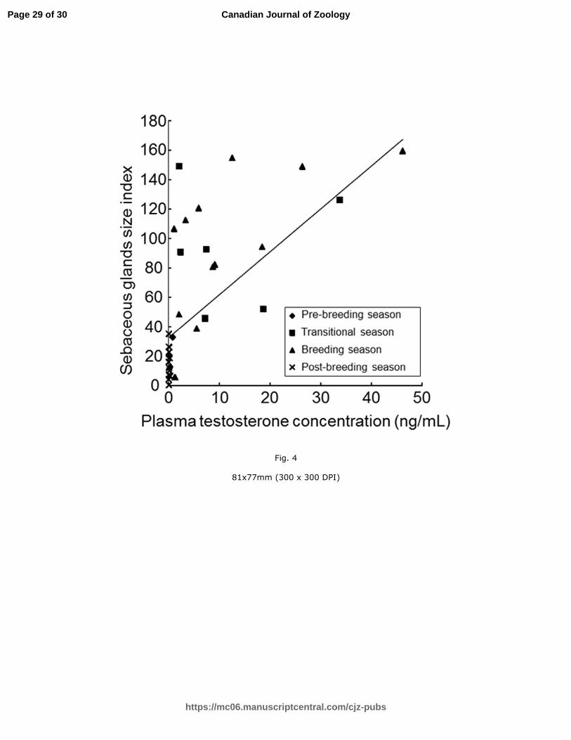
castrated: n = 3; Fig. 3). Significant positive correlations were observed between plasma
Page 11 of 30
https://mc06.manuscriptcentral.com/cjz-pubs
Canadian Journal of Zoology
Draft
12
testosterone concentrations and the index of sebaceous gland size in intact males (P < 0.05;
r = 0.78; pre-breeding: n = 4; transitional: n = 6, breeding: n = 12; post-breeding: n = 12;
Fig. 4).
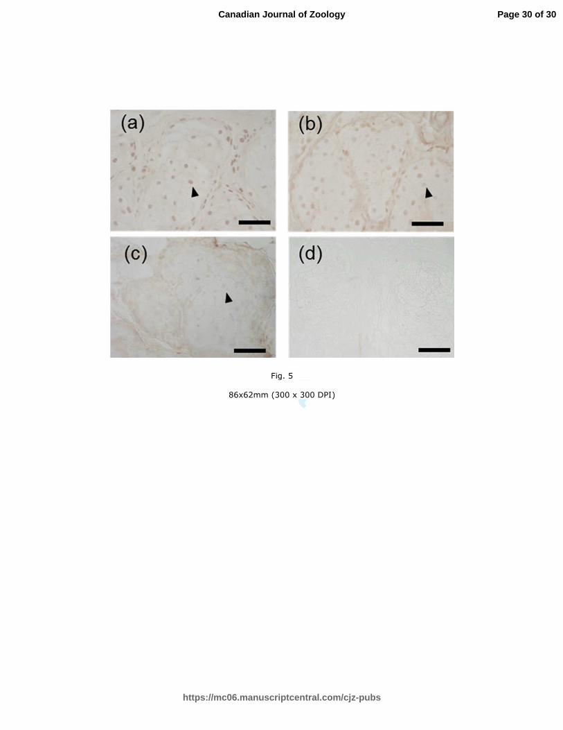
Immunohistochemistry of AR
Staining for AR in the nuclei of sebaceous gland cells was positive in the back skin
samples collected from all eight intact and three castrated bears in June (Fig. 5). Staining
for AR was also positive in the intact male with shriveled sebaceous glands in the back
skin. Furthermore, the sebaceous glands in the rump skin (n = 4/4) samples of four males
exhibited positive staining for AR.
Discussion
To our knowledge, this is the first study to show enlarged sebaceous glands and oily
skin secretions in the backs of male brown bears during the breeding season. These
findings supported our hypothesis that bipedal back rubbing is associated with chemical
communication in reproduction.
Glandular structures in mammalian skin are composed of sebaceous, apocrine and
eccrine glands. Secretions from skin glands are closely associated with olfactory
communication (Müller-Schwarze 2006). The structures of these glands might become
more complex structures in specific skin areas (Müller-Schwarze et al. 1977). The location
of mammalian scent glands is often linked to motor patterns of scent marking
(Stoeckelhuber et al. 2000; Rosell et al. 2011). In fact, pedal scent glands (prominent
apocrine and sebaceous glands) in brown bears might be associated with pedal marking
Page 12 of 30
https://mc06.manuscriptcentral.com/cjz-pubs
Canadian Journal of Zoology
Draft
13
behavior (Sergiel et al. 2017). Bipedal back rubbing is a core marking posture of bears.
The sebaceous glands in the withers of a male brown bear were enlarged compared with
those of breast in autumn (Sokolov 1982). We also found an oily secretion and enlarged
sebaceous glands in back skin during the breeding season. Thus, back sebaceous glands
seemed to be involved in back rubbing. We did not analyze the secretion and secretory
status of apocrine glands. Further study is needed to clarify the contribution of apocrine
glands to the oily secretion.
The finding that plasma testosterone concentrations were increased in all intact male
bears during transitional and breeding seasons is consistent with those of a previous study
(Tsubota and Kanagawa 1989). In rabbits Oryctolagus cuniculus (L., 1758), rats Rattus
norvegicus (Berkenhout, 1769), guinea pigs Cavia porcellus (L., 1758), and golden
hamsters, the enlargement of sebaceous glands is positively correlated with plasma
testosterone concentrations (Ebling 1977). Yet, paracloacal glands of marsupials are
thought to be a form of scent glands, and Helder-José and Freymüller (1995) suggested that
they are modified sebaceous glands. Bradley and Stoddart (1993) associated the
enlargement of these glands with high levels of testosterone in the breeding season in the
Australian marsupial Petaurus breviceps (Waterhouse, 1838). The neotropical marsupial
Metachirus nudicaudatus (É. Geoffroy Saint-Hilaire, 1803) has a pair of yellow and brown
paracloacal glands on both right and left sides of the cloaca. Their holocrine secretory
epithelium is modulated in a complex way by estrogens in females (Helder-José et al.
2014) and by testosterone in males (Helder-José et al. 2016). Our results corroborated the
influence of testosterone upon the sebaceous glands in the back skin of the male brown
Page 13 of 30
https://mc06.manuscriptcentral.com/cjz-pubs
Canadian Journal of Zoology
Draft
14
bears. Although sebaceous glands do not enlarge in castrated goats, testosterone
supplementation increased the size of these glands (Iwata et al. 2000; Wakabayashi et al.
2000). These results indicated that testosterone regulates the size of sebaceous glands. The
present study identified a significant positive correlation between plasma testosterone
concentrations and the size index of sebaceous gland in intact male brown bears, and
sebaceous glands in the back skin were AR positive. Sebaceous glands in the back skin of
three castrated bears shriveled during the transitional and breeding seasons, whereas those
in intact males were enlarged. Therefore, testosterone might induce the enlargement of
sebaceous glands in the back skin during the transitional and breeding seasons in this
species. The regulation of the size of sebaceous gland by testosterone would indicate that
back scent glands could convey reproductive information.
The sebaceous glands in the back skins of intact and castrated males stained positively
for AR, which also suggests that the size of sebaceous glands in back skin was regulated
by testosterone. Testosterone concentrations increased in intact males when the sebaceous
glands in back skin became enlarged, but remained at the basal level in castrated males,
which explains the shriveled sebaceous glands in back skin. Although sebaceous glands in
the rump skin of intact males also stained positively for AR, sebaceous glands in the rump
did not become enlarged under high plasma testosterone concentrations during the
breeding season. The reason why the oily secretion was absent and the sebaceous glands
did not become enlarged in rump skin during the breeding season remain unclear. The
differential expression of AR and enzymes that are associated with testosterone might
have contributed to the differences in the size of sebaceous glands between back and
Page 14 of 30
https://mc06.manuscriptcentral.com/cjz-pubs
Canadian Journal of Zoology
Draft
15
rump skin.
Enlarged sebaceous glands were not evident in one intact male during June, even
though the serum testosterone level did not differ from those of the other seven intact
males. Moreover, the sebaceous glands from this animal specimen stained positive for AR.
In Mongolian gerbils (Meriones unguiculatus; Milne-Edwards, 1867), social stress
decreased the size of the ventral gland independently of testosterone (Yamaguchi et al.
2005). This bear was housed with other bears that had enlarged sebaceous glands. Body
size is thought to be a determinant of male brown bear status (Zedrosser et al. 2007). We
postulate that this bear might have been under stress because it weighed less than the
others during June. Taken together, social stress might inhibit the enlargement of sebaceous
glands in brown bears.
In conclusion, the sebaceous glands in male bears became enlarged concurrently with
high plasma testosterone concentrations during the transitional and breeding seasons.
Moreover, sebaceous skin glands in the back have AR, so the size of the sebaceous glands
in the back skin of this species might be regulated by testosterone. Further investigations,
including analyses of secreted components and the effects of the secretion on the behavior
of male and female brown bears are required to elucidate the role of these secretions in this
species.
Acknowledgements
The authors thank the staff at Noboribetsu Bear Park (Noboribetsu, Japan), and Sahoro
Bear Mountain (Shintoku, Japan) for cooperation during this study.
Page 15 of 30
https://mc06.manuscriptcentral.com/cjz-pubs
Canadian Journal of Zoology
Draft
16
Ethical statement
This experiment was approved by the Animal Experiment Committee of the Obihiro
University of Agriculture and Veterinary Medicine, Japan (no. 28-218).
Page 16 of 30
https://mc06.manuscriptcentral.com/cjz-pubs
Canadian Journal of Zoology
Draft
17
References
Albers, A.C. 1992. Constraints on the design communication systems in terrestrial
vertebrates. Am. Nat. 139: S62-S89. doi:10.1086/285305.
Bradley, A.J., and Stoddart D.M. 1993. The dorsal paracloacal gland and its relationship
with seasonal changes in cutaneous scent gland morphology and plasma androgen in
the marsupial sugar glider (Petaurus breviceps; Marsupialia: Petauridae). J. Zool.
(Lond.) 229(2): 331–346. doi:10.1111/j.1469-7998.1993.tb02640.x.
Clapham, M., Nevin, O.T., Ramsey, A.D., and Rosell, F. 2012. A hypothetico-deductive
approach to assessing the social function of chemical signaling in a non-territorial
solitary carnivore. PLoS ONE, 7(4): e35404. doi:10.1371/journal.pone.0035404.
Clapham, M., Nevin, O.T., Ramsey, A.D., and Rosell, F. 2013. The function of strategic
tree selectivity in the chemical signaling of brown bears. Anim. Behav. 85: 1351–1357.
doi:10.1016/j.anbehav.2013.03.026.
Clapham, M., Nevin, O.T., Ramsey, A.D., and Rosell, F. 2014. Scent-marking investment
and motor patterns are affected by the age and sex of wild brown bears. Anim. Behav.
94: 107–116. doi:10.1016/j.anbehav.2014.05.017.
Dahle, B., and Swenson, J.E. 2003. Seasonal range size in relation to reproductive
strategies in brown bears Ursus arctos. J. Anim. Ecol. 72(4): 660–667.
doi:10.1046/j.1365-2656.2003.00737.x.
Ebling, F.J. 1977. Hormonal control of mammalian skin glands. In Chemical signals in
vertebrates. Edited by S.D. Müller and M.M. Mozell. Plenum Press, New York. pp.
17–33.
Page 17 of 30
https://mc06.manuscriptcentral.com/cjz-pubs
Canadian Journal of Zoology
Draft
18
Gorman, M.L., and Trowbridge, B.J. 1989. The role of odor in the social lives of
carnivores. In Carnivore behavior, ecology, and evolution. Edited by J.L. Gittleman.
Cornell University Press, New York. pp. 57–88.
Green, G.I., and Mattson, D.J. 2003. Tree rubbing by Yellowstone grizzly bears Ursus
arctos. Wildl. Biol. 9(1): 1–9.
Helder- José, H., and Freymüller, E. 1995. A morphological and ultrastructural study of the
paracloacal (scent) glands of the marsupial Metachirus nudicaudatus Geoffroy, 1803.
Acta. Anat. 153(1):31–38. doi:10.1159/000147712. PMID:8560957.
Helder-José, H., Mendes, E.G., Carneiro. N.M., Simões, M.J., and Freymüller, E. 2014.
Morphophysiology of the paracloacal (scent) glands in females of the marsupial
Metachirus nudicaudatus: action of estrogens. Zoomorphology, 133(2): 237–243.
doi:10.1007/s00435-014-0217-8.
Helder-José, H., Simões, M. J., and Freymüller, E. 2016. Testosterone modulation of the
male marsupial Metachirus nudicaudatus paracloacal (scent) glands. Zoomorphology,
135(3): 375–385, 2016. doi:10.1007/s00435-016-0314-y.
Hsu, S.M., Raine, L., and Fanger, H. 1981. Use of avidin-biotin-peroxidase complex
(ABC) in immunoperoxidase techniques: a comparison between ABC and unlabeled
antibody (PAP) procedures. J. Histochem. Cytochem. 29(4): 577–580.
PMID:6166661.
Ishikawa, A., Sakamoto, H., Katagiri, S., and Takahashi, Y. 2003. Changes in sexual
behavior and fecal steroid hormone concentrations during the breeding season in
Page 18 of 30
https://mc06.manuscriptcentral.com/cjz-pubs
Canadian Journal of Zoology
Draft
19
female Hokkaido brown bears (Ursus arctos yesoensis) under captive condition. J. Vet.
Med. Sci. 65(1): 99-102. doi:10.1292/jvms.65.99. PMID: 12576712.
Iwata, E., Wakabayashi, Y., Kakuma, Y., Kikusui, T., Takeuchi, Y., and Mori Y. 2000.
Testosterone-dependent primer pheromone production in the sebaceous gland of male
goat. Biol. Reprod. 62(3): 806–810. doi.10.1095/biolreprod62.3.806. PMID:10684827.
Jojola, S.M., Rosell, F., Warrington, I., Swenson, J.E., and Zedrossera, A. 2012. Subadult
brown bears (Ursus arctos) discriminate between unfamiliar adult male and female
anal gland secretion. Mamm. Biol. 77(5): 363–368.
doi:10.1016/j.mambio.2012.05.003.
Lu, L., Liu, S., Li, Q., Huang, S., Bao, L., Sheng, X., Han, Y., Watanabe, G., Taya, K., and
Weng, Q. 2014. Seasonal expression of androgen receptor in scented gland of muskrat
(Ondatra zibethicus). Gen. Comp. Endocrinol. 204: 1–7.
doi:10.1016/j.ygcen.2014.04.031. PMID:24818970.
Margaret, E.J., Mills, M.G.L., and Gorman, M.L. 1980. The scent marking behaviour of
the brown hyaena brunnea. Afr. Zool. 15(4): 240–248.
doi:10.1080/02541858.1980.11447718.
Miyamoto, A., Okuda, K., Schweigert, F.J., and Schams, D. 1992. Effects of basic
fibroblast growth factor, transforming growth factor-beta and nerve growth factor on
the secretory function of the bovine corpus luteum in vitro. J. Endocrinol. 135(1):
103–114. doi:10.1677/joe.0.1350103. PMID:1431675.
Page 19 of 30
https://mc06.manuscriptcentral.com/cjz-pubs
Canadian Journal of Zoology
Draft
20
Müller-Schwarze, D., Quay, W.B., and Brundin, A. 1977. The caudal gland in reindeer
(Rangifer tarandus L.): Its behavioral role, histology, and chemistry. J. Chem. Ecol.
3(5): 591–601. doi:10.1007/BF00989079.
Müller-Schwarze, D. 2006. Chemical ecology of vertebrates. Cambridge University Press,
Cambridge, U.K.
Rosell, F., Jojola, S.M., Ingdal, K., Lassen, B.A. Swenson, J.E., Arnemo, J.M., and
Zedrossera, A. 2011. Brown bears possess anal sacs and secretions may code for sex. J.
Zool. (Lond.) 283(2): 143–152. doi:10.1111/j.1469-7998.2010.00754.x.
Sato, Y., Kamiishi, C., Tokaji, T., Mori, M., Koizumi, S., Kobayashi, K., Itoh, T.,
Sonohara, W., Takada, B.M., and Urata, T. 2014. Selection of rub trees by brown
bears (Ursus arctos) in Hokkaido, Japan. Acta Theriol. 59(1): 129–137.
doi:10.1007/s13364-013-0143-z.
Sergiel, A., Naves, J., Kujawski, P., Maślak, R., Serwa,E., Ramos, D., Fernández-Gil, A.,
Revilla, E., Zwijacz-Kozica, T., Zięba, F., Painer, J., and Selva, N. 2017. Histological,
chemical and behavioural evidence of pedal communication in brown bears. Sci. Rep.
7:1052. doi:10.1038/s41598-017-01136-1. PMID: 28432322.
Sokolov, V.E. 1982. Mammal skin. University of California Press. Berkeley, C.A.
Støen, O.G., Bellemain, E., Sæbø, S., and Swenson, J.E. 2005. Kin-related spatial structure
in brown bears Ursus arctos. Behav. Ecol. Sociobiol. 59(2): 191–197.
doi:10.1007/s00265-005-0024-9.
Stoeckelhuber, M., Sliwa, A., and Welsch, U. 2000. Histo-physiology of the scent-marking
glands of the penile pad, anal pouch, and the forefoot in the aardwolf (Proteles
Page 20 of 30
https://mc06.manuscriptcentral.com/cjz-pubs
Canadian Journal of Zoology
Draft
21
cristatus). Anat. Rec. 259(3): 312–326.
doi:10.1002/1097-0185(20000701)259:3<312::AID-AR80>3.0.CO;2-X.
PMID:10861364.
Tsubota, T., Kanagawa, H., Takahashi, K., Yasue, K and Fukunaga, S. 1985. Observation
of sexual behavior under captive conditions in Hokkaido brown bears. Jpn. J. Anim.
Reprod. 31(4); 203–210. (in Japanese with English summary).
doi:10.1262/jrd1977.31.203.
Tsubota, T., and Kanagawa, H. 1989. Annual changes in serum testosterone levels and
spermatogenesis in the Hokkaido brown bear, Ursus arctos yesoensis. J. Mamm. Soc.
Jpn. 14(1): 11–17. doi:10.11238/jmammsocjapan1987.14.11.
Vandenbergh, J.G. 1973. Effects of gonadal hormones on the flank gland of the golden
hamster. Horm. Res. 4(1): 28–33. doi:10.1159/000178287. PMID:4748463.
Wakabayashi, Y., Iwata, E., Kikusui, T., Takeuchi, Y., and Mori, Y. 2000. Regional
differences of pheromone production in the sebaceous glands of castrated goats treated
with testosterone. J. Vet. Med. Sci. 62(10): 1067–1072. doi:10.1292/jvms.62.1067.
PMID:11073077.
White, A.M., Swaisgood, R.R., and Zhang, H. 2002. The highs and lows of chemical
communication in giant pandas (Ailuropoda melanoleuca): effect of scent deposition
height on signal discrimination. Behav. Ecol. Sociobiol. 51(6): 519–529
doi:10.1007/s00265-002-0473-3.
Page 21 of 30
https://mc06.manuscriptcentral.com/cjz-pubs
Canadian Journal of Zoology
Draft
22
Yamaguchi, H., Kikusui, T., Takeuchi, Y., Yoshimura, H., and Mori, Y. 2005. Social stress
decreases marking behavior independently of testosterone in Mongolian gerbils. Horm.
Behav. 47(5): 549–555, doi:10.1016/j.yhbeh.2004.12.009. PMID:15811356.
Zedrosser, A., Bellemain, E., Taberlet, P., and Swenson, J.E. 2007. Genetic estimates of
annual reproductive success in male brown bears: the effects of body size, age, internal
relatedness and population density. J. Anim. Ecol. 76(2): 368–375.
doi:10.1111/j.1365-2656.2006.01203.x.
Zhang, J.X., Zhang, Z.B., and Wang, Z.W. 2001. Scent, social status, and reproductive
condition in rat-like hamsters (Cricetulus triton). Physiol Behav. 74(4-5): 415–420.
doi:10.1016/S0031-9384(01)00506-6. PMID:11790399.
Page 22 of 30
https://mc06.manuscriptcentral.com/cjz-pubs
Canadian Journal of Zoology
Draft
23
Figure captions
Fig. 1. Dorsal view of adult male brown bear (a), and magnification of border region (b)
where the abundant sebum secretion was observed. Star indicates location of skin biopsy.
Area of skin with observed secretion is limited to the midline between the first cervical
vertebra and the second lumbar vertebra (dotted line). Border of abundant secretion is
marked with arrow (visible sebum covering skin and hair) and arrowhead (dry skin and
hair).
Fig. 2. Sebaceous glands in the back skin of a male brown bear and associated seasonal
changes in size. Enlarged sebaceous glands were observed during the transitional (b) and
breeding seasons (c). In pre- (a) and post- (d) breeding seasons, no enlarged sebaceous
glands were observed. Arrow: enlarged sebaceous glands. Arrowhead: shriveled sebaceous
glands. Scale bar = 1000 µm.
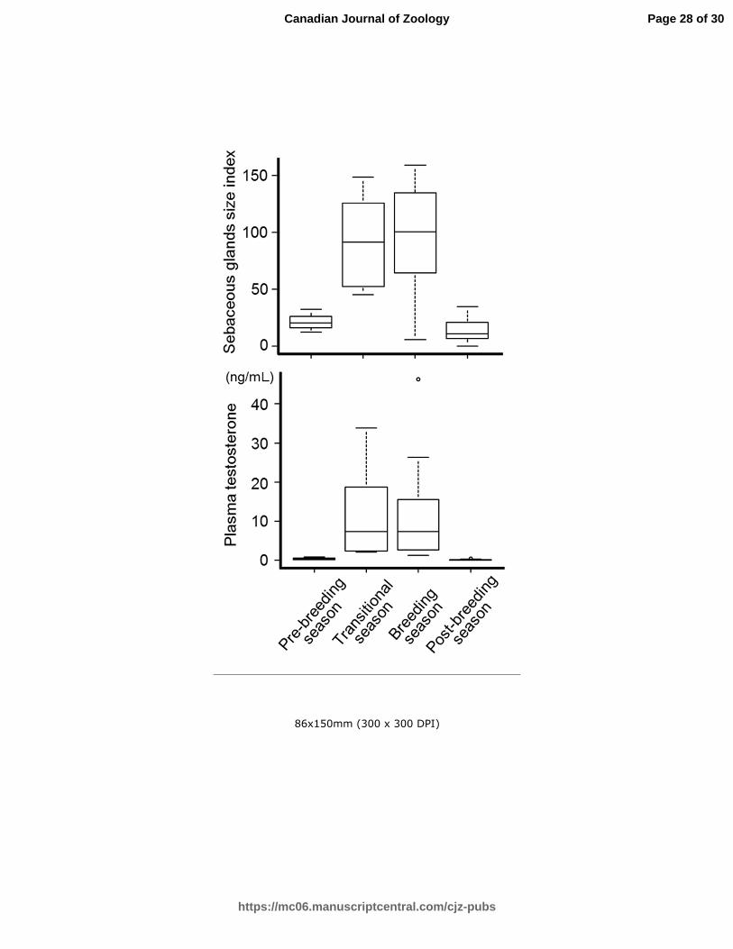
Fig. 3. Seasonal changes in indices of the sebaceous gland size in the back skin and plasma
testosterone concentrations of intact bears. Indices tended to be higher during the
transitional and breeding seasons than during the pre-breeding season (P = 0.05 and P =
0.07, respectively), and were significantly higher than during post breeding season (P <
0.05). Testosterone concentrations tended to be higher during the transitional, than during
the pre-breeding season (P = 0.05), and significantly higher than during the post-breeding
season (P < 0.05). Plasma testosterone concentrations during the breeding season were
significantly higher than during pre- and post-breeding seasons (P < 0.05). Pre-breeding, n
= 4; Transitional, n = 6; Breeding, n = 12; Post-breeding, n = 12.
Fig. 4. Scatter plot of positively correlated sebaceous gland size indices and plasma
testosterone concentrations in intact males (r = 0.78; P < 0.05).
Fig. 5. Immunohistochemical staining for the androgen receptor (AR) in the sebaceous
glands found in the back (a) and rump (b) skin samples from an intact male and in the back
Page 23 of 30
https://mc06.manuscriptcentral.com/cjz-pubs
Canadian Journal of Zoology
Draft
24
skin (c) sample of a castrated male, and negative control (d). Positive AR staining was
observed in the nuclei of sebaceous gland cells in the back skin. Scale bar = 50 µm. These
sections were photographed at a magnification ×400 (× 10 ocular lens and × 40 objective
lens).
Page 24 of 30
https://mc06.manuscriptcentral.com/cjz-pubs
Canadian Journal of Zoology
Draft
1
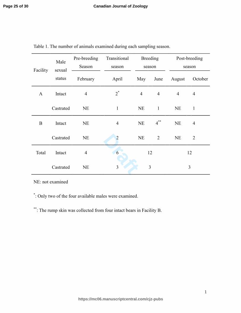
Table 1. The number of animals examined during each sampling season.
Facility
Male
sexual
status
Pre-breeding
Season
Transitional
season
Breeding
season
Post-breeding
season
February
April
May
June
August
October
A Intact 4
2*
4
4
4
4
Castrated NE
1
NE
1
NE
1
B Intact NE
4
NE
4**
NE
4
Castrated NE
2
NE
2
NE
2
Total Intact 4
6
12
12
Castrated NE
3
3
3
NE: not examined
*: Only two of the four available males were examined.
**: The rump skin was collected from four intact bears in Facility B.
Page 25 of 30
https://mc06.manuscriptcentral.com/cjz-pubs
Canadian Journal of Zoology
Draft
Fig. 1.
182x78mm (300 x 300 DPI)
Page 26 of 30
https://mc06.manuscriptcentral.com/cjz-pubs
Canadian Journal of Zoology
Draft
fig. 2.
181x68mm (300 x 300 DPI)
Page 27 of 30
https://mc06.manuscriptcentral.com/cjz-pubs
Canadian Journal of Zoology
Draft
86x150mm (300 x 300 DPI)
Page 28 of 30
https://mc06.manuscriptcentral.com/cjz-pubs
Canadian Journal of Zoology
Draft
Fig. 4
81x77mm (300 x 300 DPI)
Page 29 of 30
https://mc06.manuscriptcentral.com/cjz-pubs
Canadian Journal of Zoology
Draft
Fig. 5
86x62mm (300 x 300 DPI)
Page 30 of 30
https://mc06.manuscriptcentral.com/cjz-pubs
Canadian Journal of Zoology


