

sdw
Case Reports www.AJOG.org
Early prenatal diagnosis of tricuspid stenosis
Nizar Khatib, MD; Zeev Blumenfeld, MD; Moshe Bronshtein, MDfl
p(itwt
Tricuspid stenosis (TS) is a rare formof congenital heart disease; it is a cy-
anotic heart disease because of decreasedpulmonary flow. The valve dysfunctioncan result from primary or secondarycauses. Secondary TS is caused by rheu-matic heart disease, congenital abnor-malities, carcinoid disease, pacemakercatheters, and metabolic or enzymaticabnormalities such as Fabry’s or Whip-ple’s disease.1,2 In this case report, we de-cribe, for the first time, early prenataliagnosis of tricuspid stenosis at 15eeks’ gestational age.
CASE REPORTIn Israel, early vaginal sonography forthe detection of fetal structural malfor-mations is a common practice, and themajority of pregnant women undergothis scan between 14 and 16 weeks of ges-tation. A 28 year old healthy primigrav-ida presented at 15 weeks of a spontane-ous pregnancy for early transvaginalultrasound screening. The patient hasprophylactically taken folic acid and de-
From the Department of Obstetrics-Gynecology, Rambam Health Care Campus,The Rappaport Faculty of Medicine, Technion–Israel Institute of Technology (Drs Khatib andBlumenfeld), and Faculty of Social WelfareHealth Sciences, University of Haifa (DrBronshtein), Haifa, Israel.
Received June 29, 2012; accepted Aug. 20,2012.
The authors report no conflict of interest.
Reprints: Zeev Blumenfeld, MD, AssociateProfessor, Director, ReproductiveEndocrinology, Department of Obstetrics-Gynecology, RambamHealth Care Campus,Rappaport Research Institute, Technion-Faculty of Medicine, 8 Ha’Aliyah St., Haifa,Israel 31096. [email protected]; or [email protected].
0002-9378/free© 2012 Mosby, Inc. All rights reserved.http://dx.doi.org/10.1016/j.ajog.2012.08.030
VIDEOClick Supplementary Content underthe title of this article in the onlineTable of Contents
e6 American Journal of Obstetrics & Gynecology N
nied exposure to any teratogenic drugs.Her family history was unremarkable.
The ultrasound examination showed anormal left ventricle and a small rightventricle (Figure 1 and Video 1), normal
ow to the left ventricle, narrow main
FIGURE 1Stenotic tricuspid valve
LA, left atrium; LV, left ventricle; RV, right ventricle.
Khatib. Tricuspid stenosis–prenatal diagnosis. Am J Obstet G
Although the prenatal diagnosis of heart anlast 2 decades, the diagnosis of heart anomhas not been previously diagnosed in the eaearly detection of tricuspid stenosis at 15 watrium, small right ventricle, narrow pulmotricuspid valve. The diagnosis was confirmreport we describe, for the first time, earlyweeks’ gestational age.
Key words: cardiac malformations, fetal anstenosis
OVEMBER 2012
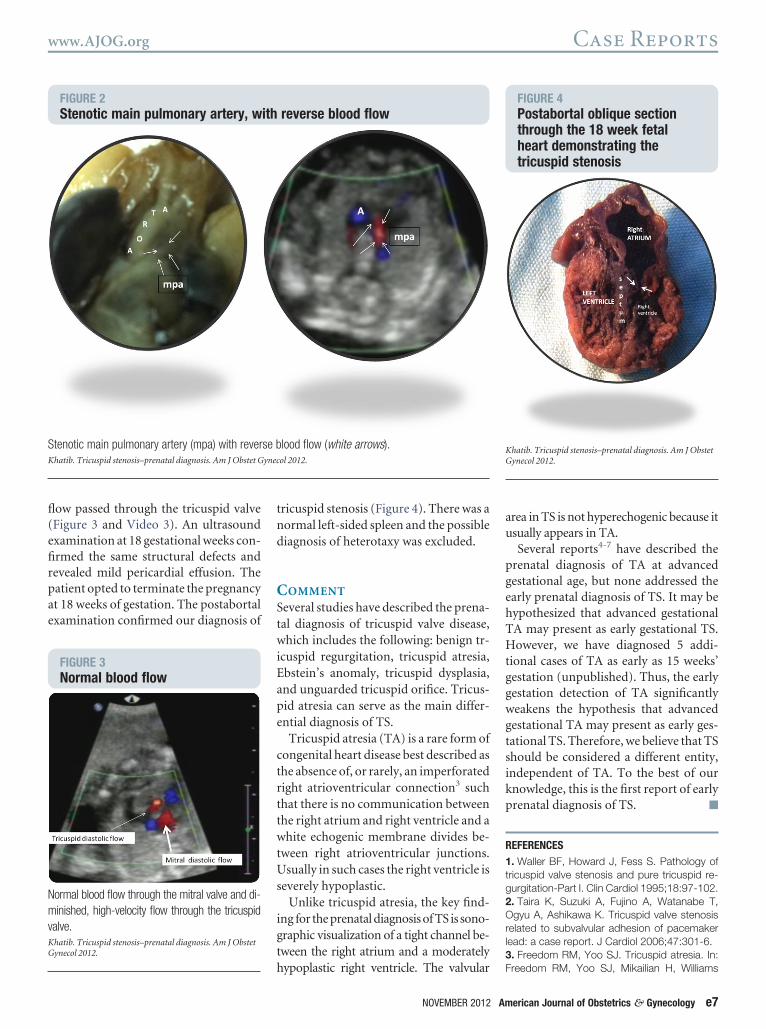
ulmonary artery with reversed flowFigure 2 and Video 2), and absence ofnferior vena cava with azygos continua-ion. The right atrium and right ventricleere connected by a narrow tunnel
hrough which severely decreased blood
ol 2012.
alies has improved dramatically during thees remains a challenge. Tricuspid stenosisecond trimester. The sonographic signs of
ks of gestation included normal sized righty artery, and diminished flow through theby postabortal examination. In this case
natal diagnosis of tricuspid stenosis at 15
alies, prenatal diagnosis, tricuspid
ynec
omali
rly seenaredpre
om
efirpae
pgehTHtggwgtsikp
www.AJOG.org Case Reports
flow passed through the tricuspid valve(Figure 3 and Video 3). An ultrasoundxamination at 18 gestational weeks con-rmed the same structural defects andevealed mild pericardial effusion. Theatient opted to terminate the pregnancyt 18 weeks of gestation. The postabortalxamination confirmed our diagnosis of
FIGURE 2Stenotic main pulmonary artery, w
Stenotic main pulmonary artery (mpa) with reverKhatib. Tricuspid stenosis–prenatal diagnosis. Am J Obstet G
FIGURE 3Normal blood flow
Normal blood flow through the mitral valve and di-minished, high-velocity flow through the tricuspidvalve.Khatib. Tricuspid stenosis–prenatal diagnosis. Am J ObstetGynecol 2012.
tricuspid stenosis (Figure 4). There was anormal left-sided spleen and the possiblediagnosis of heterotaxy was excluded.
COMMENT
Several studies have described the prena-tal diagnosis of tricuspid valve disease,which includes the following: benign tr-icuspid regurgitation, tricuspid atresia,Ebstein’s anomaly, tricuspid dysplasia,and unguarded tricuspid orifice. Tricus-pid atresia can serve as the main differ-ential diagnosis of TS.
Tricuspid atresia (TA) is a rare form ofcongenital heart disease best described asthe absence of, or rarely, an imperforatedright atrioventricular connection3 suchthat there is no communication betweenthe right atrium and right ventricle and awhite echogenic membrane divides be-tween right atrioventricular junctions.Usually in such cases the right ventricle isseverely hypoplastic.
Unlike tricuspid atresia, the key find-ing for the prenatal diagnosis of TS is sono-graphic visualization of a tight channel be-tween the right atrium and a moderately
reverse blood flow
lood flow (white arrows).ol 2012.
hypoplastic right ventricle. The valvular
NOVEMBER 2012 Am
area in TS is not hyperechogenic because itusually appears in TA.
Several reports4-7 have described therenatal diagnosis of TA at advancedestational age, but none addressed thearly prenatal diagnosis of TS. It may beypothesized that advanced gestationalA may present as early gestational TS.owever, we have diagnosed 5 addi-
ional cases of TA as early as 15 weeks’estation (unpublished). Thus, the earlyestation detection of TA significantlyeakens the hypothesis that advancedestational TA may present as early ges-ational TS. Therefore, we believe that TShould be considered a different entity,ndependent of TA. To the best of ournowledge, this is the first report of earlyrenatal diagnosis of TS. f
REFERENCES1. Waller BF, Howard J, Fess S. Pathology oftricuspid valve stenosis and pure tricuspid re-gurgitation-Part I. Clin Cardiol 1995;18:97-102.2. Taira K, Suzuki A, Fujino A, Watanabe T,Ogyu A, Ashikawa K. Tricuspid valve stenosisrelated to subvalvular adhesion of pacemakerlead: a case report. J Cardiol 2006;47:301-6.3. Freedom RM, Yoo SJ. Tricuspid atresia. In:
FIGURE 4Postabortal oblique sectionthrough the 18 week fetalheart demonstrating thetricuspid stenosis
Khatib. Tricuspid stenosis–prenatal diagnosis. Am J ObstetGynecol 2012.
ith
se bynec
Freedom RM, Yoo SJ, Mikailian H, Williams
erican Journal of Obstetrics & Gynecology e7

Case Reports www.AJOG.org
WG, eds. The natural and modified history ofcongenital heart disease. New York: BlackwellPublishing; 2004:381-6.4. Sharland G. Tricuspid valve abnormalities. In:Allan L, Hornberger LK, Sharland G, eds. Text-book of fetal cardiology. London (UK): Green-
wich Medical Media; 2000:133-47.e8 American Journal of Obstetrics & Gynecology N
5. Wald RM, Tham EB, McCrindle BW, et al.Outcome after prenatal diagnosis of tricuspidatresia: a multicenter experience. Am Heart J2007;153:772-8.6. Lato K, Gmbruch U, Geipel A, et al. Tricus-pid atresia with absent pulmonary valve and
intraventricular septum: intrauterine courseOVEMBER 2012
and outcome of an unusual congenital heartdefect. Ultrasound Obstet Gynecol 2010;35:243-5.7. Berg C, Lachman R, C. Kaiser C, et al. Pre-natal diagnosis of tricuspid atresia: intrauterinecourse and outcome. Ultrasound Obstet Gyne-
col 2010;35:183-90.


