
www.labmedonline.org 169eISSN 2093-6338
are associated with a 2–15.1 Mb deletion of the q11–q31 region,
with substantial phenotypic variation. However, only a small num-
ber of previously reported cases have been con�rmed by high-
resolution array comparative genomic hybridization (aCGH).
In the present study, we report the case of a patient with an in-
terstitial deletion of a maximum, 22.8 Mb on chromosome 4q. Clin-
ical and genetic characteristics were compared against previously
reported patients with deletion of the same region, con�rmed by
aCGH. Further, we discuss candidate genes, associated with this
syndrome, as potential markers, which could provide new insights
into the phenotypic spectrum and genotype-phenotype correlation.
CASE REPORT
The present case looks at an 18-year-old male patient with an
unremarkable family history. He is the third child of non-consan-
guineous, healthy, Korean parents and was born at a gestational
age of 40 weeks, at 2.8 kg, by vaginal delivery. Upon presentation,
INTRODUCTION
Chromosome 4q deletion syndrome is a rare disease caused by
interstitial or terminal deletion of the long arm of chromosome 4
[1-3]. These deletions are typically de novo, although some cases
have been found to result from an unbalanced product of paren-
tal reciprocal translocation [4]. The majority of 4q deletion cases
고해상도 염색체 마이크로어레이법으로 확인된 4번 염색체 장완 근위부 결실의 특징: 증례보고 및 문헌검토Characteristics of Interstitial Deletion in Chromosome 4q Confirmed by Array Comparative Genomic Hybridization: A Case Report and Literature Review
정우영1·이선주2·김혜란3·전경란4
Woo Yeong Chung, M.D.1, Sun Joo Lee, M.D.2, Hye Ran Kim, M.D.3, Kyung Ran Jun, M.D.4
인제대학교 의과대학 부산백병원 소아청소년과1, 영상의학과2, 진단검사의학과3, 인제대학교 의과대학 해운대백병원 진단검사의학과4
Departments of Pediatrics1, Radiology2, and Laboratory Medicine3, Inje University Busan Paik Hospital, Busan; Department of Laboratory Medicine4, Inje University Haeundae Paik Hospital, Busan, Korea
증례보고Lab Med OnlineVol. 10, No. 2: 169-174, April 2020https://doi.org/10.3343/lmo.2020.10.2.169
진단유전학
Corresponding author: Kyung Ran Jun, M.D., Ph.D.
https://orcid.org/0000-0001-8904-2327Department of Laboratory Medicine, Haeundae Paik Hospital, College of Medicine Inje University, 875 Haeun-daero, Haeundae-gu, Busan 48108, KoreaTel: +82-51-797-3191, Fax: +82-51-797-3194, E-mail: [email protected]
Received: May 10, 2019Revision received: July 1, 2019Accepted: July 1, 2019
This article is available from https://www.labmedonline.org 2020, Laboratory Medicine Online This is an Open Access article distributed under the terms of the Creative Commons
Attribution Non-Commercial License (https://creativecommons.org/licenses/by-nc/4.0/) which permits unrestricted non-commercial use, distribution, and reproduction in any medium, provided the original work is properly cited.
Chromosome 4q deletion syndrome is a rare disease caused by partial deletion of the long arm of chromosome 4. Phenotypic severity and expres-sivity vary among patients with chromosome 4q deletions, depending on the size and region of the deletion of the affected chromosome. Although there have been many reports of proximal 4q deletion cases, very few have been confirmed by high-resolution array comparative genomic hybrid-ization (aCGH). In the current study, we presented a new case of 4q proximal deletion, with detailed genetic and clinical characteristics, and com-pared these characteristics to those of six previous cases with available aCGH data. According to our review, several genes known to be associat-ed with specific phenotypes of 4q12q21.1 deletion cannot sufficiently explain the variable phenotypes observed among the cases. These pheno-types include mental retardation, microcephaly, ocular anomalies, dental anomaly, and piebaldism. Consequently, we recommend further detailed investigations into the genes associated with 4q12q21.1 deletion to assist in identifying genotype-phenotype associations more clearly.
Key Words: Chromosome 4q- syndrome, Comparative genomic hybridization, Genetic association studies
1 / 1CROSSMARK_logo_3_Test
2017-03-16https://crossmark-cdn.crossref.org/widget/v2.0/logos/CROSSMARK_Color_square.svg
정우영 외: Characteristics of 4q12-q21.1 Deletion
https://doi.org/10.3343/lmo.2020.10.2.169170 www.labmedonline.org
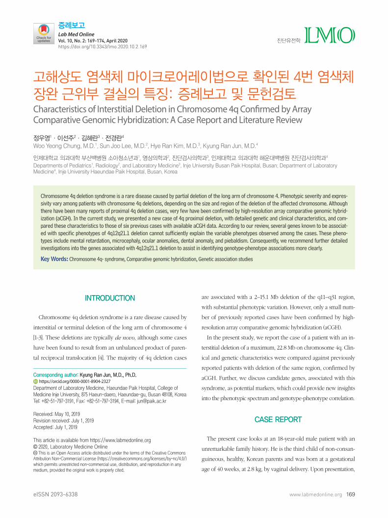
his height and weight were less than 1 percentile. The patient fur-
ther presented with microcephaly and clear dysmorphic facial
features, including cranial asymmetry, prominent forehead pro-
trusion, a bilateral epicanthal fold, hypertelorism, depressed nasal
root with a broad or beaked nose and low-set, simple ears with
preauricular dimples. No spots or piebaldism were observed. A
neurological examination indicated hypotonia, but the deep ten-
don re�ex was well preserved. Exophthalmos, ptosis, and strabis-
mus were observed in both eyes with a cataract also present in
the left eye. A dental examination revealed white plaque, due to
the generalized amelogenesis imperfecta, with normal maxillo-
mandibular growth, but bimaxillary protrusion was evident. Due
to substantially delayed bone maturation, bone age was delayed
(Fig. 1A). Abnormalities in the spine (Fig. 1B) and both feet (Fig.
1C) were also present. There were no abnormalities in the right
hip, according to both hip radiographs; however, sequelae of Legg-
Calvé-Perthes was observed in the left hip, with a short, broad left
femoral head and relative shortening of the left limb with coxa
magna deformity (Fig. 1D). This caused a discrepancy of 2 cm in
leg length.
The patient had a no history of seizure but did present with a
learning disability and speech disorder, along with severe devel-
Fig. 1. Skeletal radiographs of the proband. (A) Both AP hand radiographs showed no fusion of the phalangeal physis’ and the bone age was con-sidered to be in the order of 14 years, determined by the Greulich-Pyle method at the age of 17.3 years. (B) Whole spine, AP and lateral, radio-graphs showed minimal scoliotic curvature of the T-L spine, loss of normal lordotic curvature of the cervical spine, moderate spondylolytic spondy-lolisthesis at L5 on S1 (arrow), and a deformity of the anterosuperior corner of the S1 body (arrowhead). (C) AP radiographs of both feet showed deformity of the right first distal phalangeal bone (arrow). (D) Both hip AP radiographs showed sequelae of Legg-Calve-Perthes disease in the left hip, with a short, broad femoral head (arrow) and relative shortening of the left limb, in keeping with a coxa magna deformity.
A B
C D
정우영 외: Characteristics of 4q12- q21.1 Deletion
https://doi.org/10.3343/lmo.2020.10.2.169 www.labmedonline.org 171
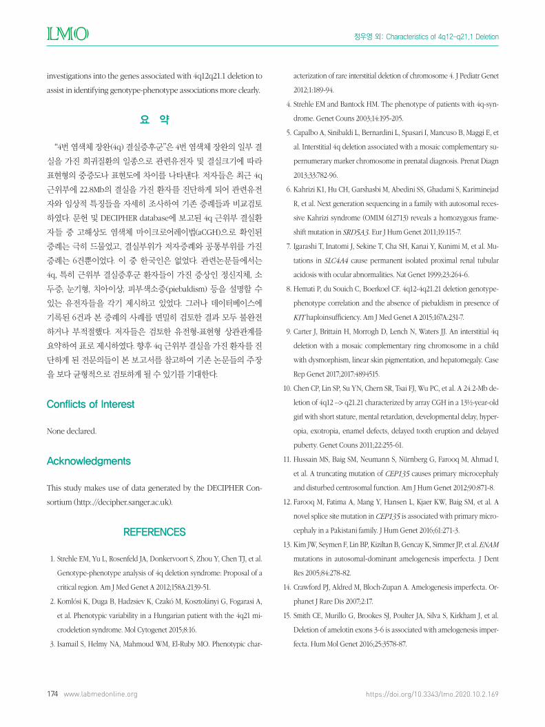
Fig. 2. GTL-banded partial karyotype and aCGH results of the proband: 46,XY,del(4)(q12q21.1).arr[GRCh38] 4q12q21.1(53575744_76379310)x1.
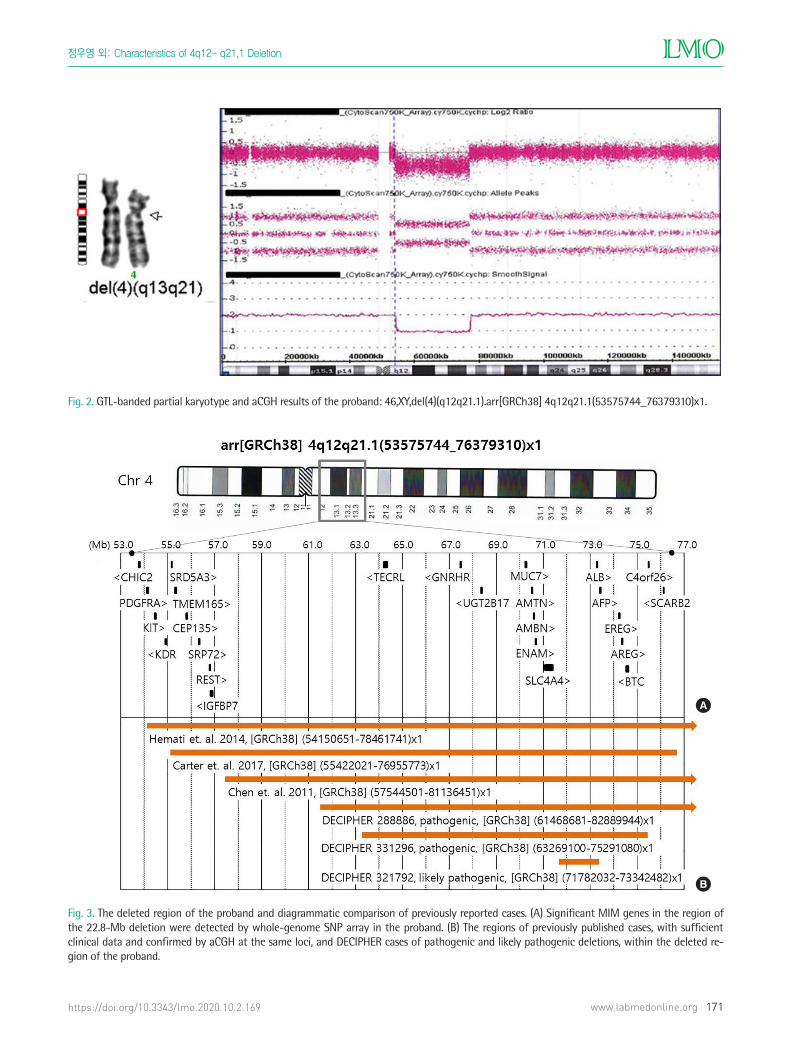
Fig. 3. The deleted region of the proband and diagrammatic comparison of previously reported cases. (A) Significant MIM genes in the region of the 22.8-Mb deletion were detected by whole-genome SNP array in the proband. (B) The regions of previously published cases, with sufficient clinical data and confirmed by aCGH at the same loci, and DECIPHER cases of pathogenic and likely pathogenic deletions, within the deleted re-gion of the proband.
A
B
정우영 외: Characteristics of 4q12-q21.1 Deletion
https://doi.org/10.3343/lmo.2020.10.2.169172 www.labmedonline.org
opmental delay. A psychiatric evaluation revealed signs of atten-
tion de�cit hyperactivity disorder (ADHD) and autism spectrum
disorder (ASD). An investigation of his pubertal status showed a
testes volume of 18/18 mL, pubic hair score of 4–5, genitalia score
of 5 with appropriate adult external genitalia overall. His serum
testosterone, luteinizing hormone and follicle stimulating hormone
levels were all elevated, at 3.95 (reference range: 1.7–8.6 mIU/mL),
3.37 (1.5–12.4 mIU/mL) and 8.57 (1.88–8.82 ng/mL), respectively.
No abnormal �ndings were observed in complete blood cell count,
urinalysis, biochemical tests, hearing tests, electroencephalogra-
phy, echocardiography, pelvic sonography, and the kidney-ureters-
bladder sonography survey.
Cytogenetic analysis and aCGH of the patient revealed an inter-
stitial, heterozygous deletion of chromosome 4q: 46,XY,del(4)
(q12q21.1) arr[GRCh38] 4q12q21.1(53575744_76379310)x1 (Fig. 2)
with the maximum length of the deleted region being 22.8 Mb.
His mother had a normal karyotype, but no chromosome analysis
was performed on his father. Neither parent showed any of the
phenotypes of the proband. Chromosome analysis of peripheral
blood lymphocytes was performed, according to standard tech-
niques, at a 550-band level. The aCGH was performed using the
Affymetrix Cytoscan 750K array. Copy number variants were com-
pared with public databases, including the DECIPHER database
(http://decipher.sanger.ac.uk) and USCS Genome Browser (http://
www.genome.ucsc.edu/cgi-bin/hgGateway). No pathogenic or
likely pathogenic variant was found in whole-exome sequencing
using the peripheral blood sample of the proband. All exon re-
gions of all human genes (~22,000) were captured using Agilent’s
SureSelect kit and the captured regions of the genome were se-
quenced with the Illumina sequencing platform.
The deleted region of the proband comprised 109 known Men-
delian Inheritance in Man (MIM) genes, including 25 genes asso-
ciated with disease phenotypes; however, few of these genes were
associated with speci�c phenotypes (Fig. 3A). The patient and his
parents provided written informed consent for the publication of
patient information.
DISCUSSION
Many patients with 4q proximal deletion of various sizes have
been reported to date, including 4q12q21.1. Capalbo et al. [5] sum-
marized the clinical characteristics of 31 patients with pure 4q
proximal deletions; however, detailed information on the deleted
region was not provided for these patients. Only three of these
cases had similar sized deletions (21.59–24.37 Mb) at the same loci
as our present case, and this was con�rmed by aCGH (Fig. 3B). In
addition, the DECIPHER database (version 9.23, released on May
23, 2018) showed three cases recorded as “pathogenic” or “likely
pathogenic”, which were smaller sized deletions of this region, as
pure copy number variations (DECIPHER number 288886, 321792,
and 331296) (Fig. 3B). Therefore, including the present case, there
are only seven cases of 4q proximal deletion con�rmed with aCGH
and detailed clinical information (Table 1). The common charac-
teristics of these cases include mental retardation, learning disabil-
ity, growth retardation, minor facial anomalies, hypotonia, skele-
tal abnormalities, and eye anomalies. Therefore, more in-depth
comparisons of these cases could prove useful in understanding
the genotype-phenotype relationship of the proximal 4q region.
As shown in Table 1, mental retardation and/or learning disabil-
ity was a common clinical �nding in all seven cases (100%). The
chromosomal region currently analyzed (4q) was found to be as-
sociated with two distinct syndromes linked to mental retardation
and growth retardation: Kahrizi syndrome (MIM#612713) and re-
nal tubular acidosis, proximal, with ocular abnormalities and men-
tal retardation (RTA-OA-MR, MIM#604278). Kahrizi syndrome,
which can be caused by a homozygous mutation of SRD5A3 (MIM*
611715) [6], is an autosomal recessive, neurodevelopmental disor-
der characterized by mental retardation, cataracts, coloboma, ky-
phosis, and coarse facial features. RTA-OA-MR is also an autoso-
mal recessive disease, and it is caused by a homozygous mutation
in SLC4A4 (MIM*603345) [7]. SRD5A3 and SLC4A4 are both re-
cessively inherited genes; however, our patient, and some other
patients with mental retardation and/or learning dif�culties [8, 9],
show heterozygous deletion of both genes. Moreover, some symp-
tomatic patients only harbor the SLC4A4 deletion [10], or no dele-
tion of either gene (DECIPHER 321791), suggesting that a more
complicated relationship exists between SRD5A3, SLC4A4 or
possibly other unidenti�ed genes in this region, in the process of
neurodevelopment.
The common craniofacial features of 4q deletions, which were
also observed in our case, are microcephaly and ocular defects
such as exophthalmos, astigmatism, strabismus, and cataracts. Mi-
crocephaly is known to be associated with CEP135 (MIM*611423),
and was identi�ed in patients with autosomal recessive primary
정우영 외: Characteristics of 4q12- q21.1 Deletion
https://doi.org/10.3343/lmo.2020.10.2.169 www.labmedonline.org 173
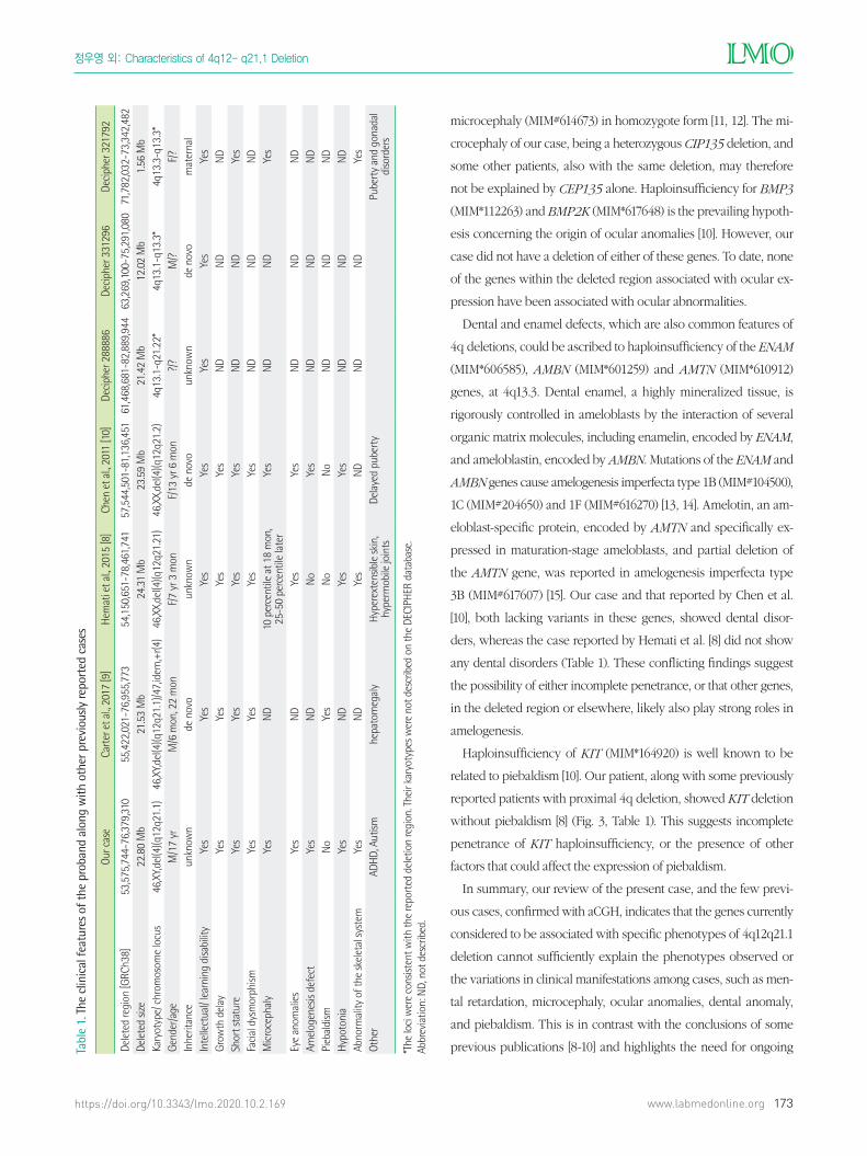
Tabl
e 1.
The
clin
ical
feat
ures
of t
he p
roba
nd a
long
with
oth
er p
revi
ously
repo
rted
cas
es
Our c
ase
Cart
er e
t al.,
2017
[9]
Hem
ati e
t al.,
2015
[8]
Chen
et a
l., 20
11 [1
0]De
ciph
er 2
8888
6De
ciph
er 3
3129
6De
ciph
er 3
2179
2
Dele
ted
regi
on [G
RCh3
8]53
,575
,744
-76,
379,
310
55,4
22,0
21-7
6,95
5,77
354
,150
,651
-78,
461,
741
57,5
44,5
01-8
1,13
6,45
161
,468
,681
-82,
889,
944
63,2
69,1
00-7
5,29
1,08
071
,782
,032
-73,
342,
482
Dele
ted
size
22.8
0 M
b21
.53
Mb
24.3
1 M
b23
.59
Mb
21.4
2 M
b12
.02
Mb
1.56
Mb
Kary
otyp
e/ c
hrom
osom
e lo
cus
46,X
Y,del
(4)(q
12q2
1.1)
46,X
Y,del
(4)(q
12q2
1.1)
/47,
idem
,+r(4
)46
,XX,
del(4
)(q12
q21.
21)
46,X
X,de
l(4)(q
12q2
1.2)
4q13
.1-q
21.2
2*4q
13.1
-q13
.3*
4q13
.3-q
13.3
*Ge
nder
/age
M/1
7 yr
M/6
mon
, 22
mon
F/7
yr 3
mon
F/13
yr 6
mon
?/?
M/?
F/?
Inhe
ritan
ceun
know
nde
nov
oun
know
nde
nov
oun
know
nde
nov
om
ater
nal
Inte
llect
ual/
lear
ning
disa
bilit
yYe
sYe
sYe
sYe
sYe
sYe
sYe
sGr
owth
del
ayYe
sYe
sYe
sYe
sN
DN
DN
DSh
ort s
tatu
reYe
sYe
sYe
sYe
sN
DN
DYe
sFa
cial
dys
mor
phism
Yes
Yes
Yes
Yes
ND
ND
ND
Mic
roce
phal
yYe
sN
D10
per
cent
ile a
t 18
mon
, 25
-50
perc
entil
e la
ter
Yes
ND
ND
Yes
Eye
anom
alie
sYe
sN
DYe
sYe
sN
DN
DN
DAm
elog
enes
is de
fect
Yes
ND
No
Yes
ND
ND
ND
Pieb
aldi
smN
oYe
sN
oN
oN
DN
DN
DHy
poto
nia
Yes
ND
Yes
Yes
ND
ND
ND
Abno
rmal
ity o
f the
skel
etal
syst
emYe
sN
DYe
sN
DN
DN
DYe
sOt
her
ADHD
, Aut
ismhe
pato
meg
aly
Hype
rext
ensib
le sk
in,
hype
rmob
ile jo
ints
Dela
yed
pube
rty
Pube
rty
and
gona
dal
diso
rder
s
*The
loci
wer
e co
nsist
ent w
ith th
e re
port
ed d
elet
ion
regi
on. T
heir
kary
otyp
es w
ere
not d
escr
ibed
on
the
DECI
PHER
dat
abas
e. Ab
brev
iatio
n: N
D, n
ot d
escr
ibed
.
microcephaly (MIM#614673) in homozygote form [11, 12]. The mi-
crocephaly of our case, being a heterozygous CIP135 deletion, and
some other patients, also with the same deletion, may therefore
not be explained by CEP135 alone. Haploinsuf�ciency for BMP3
(MIM*112263) and BMP2K (MIM*617648) is the prevailing hypoth-
esis concerning the origin of ocular anomalies [10]. However, our
case did not have a deletion of either of these genes. To date, none
of the genes within the deleted region associated with ocular ex-
pression have been associated with ocular abnormalities.
Dental and enamel defects, which are also common features of
4q deletions, could be ascribed to haploinsuf�ciency of the ENAM
(MIM*606585), AMBN (MIM*601259) and AMTN (MIM*610912)
genes, at 4q13.3. Dental enamel, a highly mineralized tissue, is
rigorously controlled in ameloblasts by the interaction of several
organic matrix molecules, including enamelin, encoded by ENAM,
and ameloblastin, encoded by AMBN. Mutations of the ENAM and
AMBN genes cause amelogenesis imperfecta type 1B (MIM#104500),
1C (MIM#204650) and 1F (MIM#616270) [13, 14]. Amelotin, an am-
eloblast-speci�c protein, encoded by AMTN and speci�cally ex-
pressed in maturation-stage ameloblasts, and partial deletion of
the AMTN gene, was reported in amelogenesis imperfecta type
3B (MIM#617607) [15]. Our case and that reported by Chen et al.
[10], both lacking variants in these genes, showed dental disor-
ders, whereas the case reported by Hemati et al. [8] did not show
any dental disorders (Table 1). These con�icting �ndings suggest
the possibility of either incomplete penetrance, or that other genes,
in the deleted region or elsewhere, likely also play strong roles in
amelogenesis.
Haploinsuf�ciency of KIT (MIM*164920) is well known to be
related to piebaldism [10]. Our patient, along with some previously
reported patients with proximal 4q deletion, showed KIT deletion
without piebaldism [8] (Fig. 3, Table 1). This suggests incomplete
penetrance of KIT haploinsuf�ciency, or the presence of other
factors that could affect the expression of piebaldism.
In summary, our review of the present case, and the few previ-
ous cases, con�rmed with aCGH, indicates that the genes currently
considered to be associated with speci�c phenotypes of 4q12q21.1
deletion cannot suf�ciently explain the phenotypes observed or
the variations in clinical manifestations among cases, such as men-
tal retardation, microcephaly, ocular anomalies, dental anomaly,
and piebaldism. This is in contrast with the conclusions of some
previous publications [8-10] and highlights the need for ongoing
정우영 외: Characteristics of 4q12-q21.1 Deletion
https://doi.org/10.3343/lmo.2020.10.2.169174 www.labmedonline.org
investigations into the genes associated with 4q12q21.1 deletion to
assist in identifying genotype-phenotype associations more clearly.
요 약
“4번 염색체 장완(4q) 결실증후군”은 4번 염색체 장완의 일부 결
실을 가진 희귀질환의 일종으로 관련유전자 및 결실크기에 따라
표현형의 중증도나 표현도에 차이를 나타낸다. 저자들은 최근 4q
근위부에 22.8Mb의 결실을 가진 환자를 진단하게 되어 관련유전
자와 임상적 특징들을 자세히 조사하여 기존 증례들과 비교검토
하였다. 문헌 및 DECIPHER database에 보고된 4q 근위부 결실환
자들 중 고해상도 염색체 마이크로어레이법(aCGH)으로 확인된
증례는 극히 드물었고, 결실부위가 저자증례와 공통부위를 가진
증례는 6건뿐이었다. 이 중 한국인은 없었다. 관련논문들에서는
4q, 특히 근위부 결실증후군 환자들이 가진 증상인 정신지체, 소
두증, 눈기형, 치아이상, 피부색소증(piebaldism) 등을 설명할 수
있는 유전자들을 각기 제시하고 있었다. 그러나 데이터베이스에
기록된 6건과 본 증례의 사례를 면밀히 검토한 결과 모두 불완전
하거나 부적절했다. 저자들은 검토한 유전형-표현형 상관관계를
요약하여 표로 제시하였다. 향후 4q 근위부 결실을 가진 환자를 진
단하게 된 전문의들이 본 보고서를 참고하여 기존 논문들의 주장
을 보다 균형적으로 검토하게 될 수 있기를 기대한다.
Conflicts of Interest
None declared.
Acknowledgments
This study makes use of data generated by the DECIPHER Con-
sortium (http:.//decipher.sanger.ac.uk).
REFERENCES
1. Strehle EM, Yu L, Rosenfeld JA, Donkervoort S, Zhou Y, Chen TJ, et al.
Genotype-phenotype analysis of 4q deletion syndrome: Proposal of a
critical region. Am J Med Genet A 2012;158A:2139-51.
2. Komlósi K, Duga B, Hadzsiev K, Czakó M, Kosztolányi G, Fogarasi A,
et al. Phenotypic variability in a Hungarian patient with the 4q21 mi-
crodeletion syndrome. Mol Cytogenet 2015;8:16.
3. Isamail S, Helmy NA, Mahmoud WM, El-Ruby MO. Phenotypic char-
acterization of rare interstitial deletion of chromosome 4. J Pediatr Genet
2012;1:189-94.
4. Strehle EM and Bantock HM. The phenotype of patients with 4q-syn-
drome. Genet Couns 2003;14:195-205.
5. Capalbo A, Sinibaldi L, Bernardini L, Spasari I, Mancuso B, Maggi E, et
al. Interstitial 4q deletion associated with a mosaic complementary su-
pernumerary marker chromosome in prenatal diagnosis. Prenat Diagn
2013;33:782-96.
6. Kahrizi K1, Hu CH, Garshasbi M, Abedini SS, Ghadami S, Kariminejad
R, et al. Next generation sequencing in a family with autosomal reces-
sive Kahrizi syndrome (OMIM 612713) reveals a homozygous frame-
shift mutation in SRD5A3. Eur J Hum Genet 2011;19:115-7.
7. Igarashi T, Inatomi J, Sekine T, Cha SH, Kanai Y, Kunimi M, et al. Mu-
tations in SLC4A4 cause permanent isolated proximal renal tubular
acidosis with ocular abnormalities. Nat Genet 1999;23:264-6.
8. Hemati P, du Souich C, Boerkoel CF. 4q12-4q21.21 deletion genotype-
phenotype correlation and the absence of piebaldism in presence of
KIT haploinsuf�ciency. Am J Med Genet A 2015;167A:231-7.
9. Carter J, Brittain H, Morrogh D, Lench N, Waters JJ. An interstitial 4q
deletion with a mosaic complementary ring chromosome in a child
with dysmorphism, linear skin pigmentation, and hepatomegaly. Case
Rep Genet 2017;2017:4894515.
10. Chen CP, Lin SP, Su YN, Chern SR, Tsai FJ, Wu PC, et al. A 24.2-Mb de-
letion of 4q12 --> q21.21 characterized by array CGH in a 13½-year-old
girl with short stature, mental retardation, developmental delay, hyper-
opia, exotropia, enamel defects, delayed tooth eruption and delayed
puberty. Genet Couns 2011;22:255-61.
11. Hussain MS, Baig SM, Neumann S, Nürnberg G, Farooq M, Ahmad I,
et al. A truncating mutation of CEP135 causes primary microcephaly
and disturbed centrosomal function. Am J Hum Genet 2012;90:871-8.
12. Farooq M, Fatima A, Mang Y, Hansen L, Kjaer KW, Baig SM, et al. A
novel splice site mutation in CEP135 is associated with primary micro-
cephaly in a Pakistani family. J Hum Genet 2016;61:271-3.
13. Kim JW, Seymen F, Lin BP, Kiziltan B, Gencay K, Simmer JP, et al. ENAM
mutations in autosomal-dominant amelogenesis imperfecta. J Dent
Res 2005;84:278-82.
14. Crawford PJ, Aldred M, Bloch-Zupan A. Amelogenesis imperfecta. Or-
phanet J Rare Dis 2007;2:17.
15. Smith CE, Murillo G, Brookes SJ, Poulter JA, Silva S, Kirkham J, et al.
Deletion of amelotin exons 3-6 is associated with amelogenesis imper-
fecta. Hum Mol Genet 2016;25:3578-87.


