

* Corresponding author. E-mail: [email protected]. Telephone: 1-718-997-3271.
Elucidating Potential Energy Surfaces for Singlet O2 Reactions with Protonated, Deprotonated
and Di-deprotonated Cystine Using A Combination of Approximately Spin Projected Density
Functional Theory and Guided-Ion-Beam Mass Spectrometry
Wenchao Lu,ab I-Hsien (Midas) Tsai,c Yan Sun,ab Wenjing Zhou,a and Jianbo Liu*ab
a Department of Chemistry and Biochemistry, Queens College of the City University of New York,
65-30 Kissena Blvd., Queens, NY 11367, USA
b Ph.D. Program in Chemistry, The Graduate Center of the City University of New York,
365 5th Ave., New York, NY 10016, USA
c Department of Natural Sciences, LaGuardia Community College,
31-10 Thomson Ave., Long Island City, NY 11101, USA

2
Abstract: The reactivity of cystine towards electronically excited singlet O2 (a1g) has been long
debated, despite the fact that most organic disulfides are susceptible to oxidation by singlet O2. We
report a combined experimental and computational study on reactions of singlet O2 with gas-phase cystine
at different ionization and hydration states, aimed to determine reaction outcomes, mechanisms and
potential energy surfaces. Ion-molecule collisions of protonated and di-deprotonated cystine ions with
singlet O2, in both the absence and the presence of a water ligand, were measured over a center-of-mass
collision energy (Ecol) range from 0.1 to 1.0 eV, using a guided-ion-beam scattering tandem mass
spectrometer. No oxidation was observed for these reactant ions except collision-induced dissociation at
high energies. Guided by density functional theory (DFT) calculated potential energy surfaces, reaction
coordinates were established to unravel the origin of the non-reactivity of cystine ions towards singlet O2.
To account for mixed open- and closed-shell characters, singlet O2 and critical structures along reaction
coordinates were evaluated using broken-symmetry, open-shell DFT with spin contamination errors
removed by an approximate spin projection method. It was found that collision of protonated cystine
with singlet O2 follows a repulsive potential surface, and possesses no chemically significant interaction;
and collision-induced dissociation of protonated cystine is dominated by loss of water and CO.
Collision of di-deprotonated cystine with singlet O2, on the other hand, forms a short-lived
electrostatically bonded precursor complex at low Ecol. The latter may evolve to a covalently bonded
persulfoxide, but the conversion is blocked by an activation barrier lying 0.39 eV above reactants. At
high Ecol, C-S bond cleavage dominates the collision-induced dissociation of di-deprotonated cystine,
leading to charge-separated fragmentation. Cross section for the ensuing fragment ion
H2NCH(CO2-)CH2SS was measured as a function of Ecol, and the mechanism for charge-separated
fragmentation was discussed. It was also found that the reaction of deprotonated cystine with singlet O2
follows a similar mechanism as that of di-deprotonated cystine, but with an even higher activation barrier
(0.72 eV).

3
1. Introduction
The lowest electronically excited singlet O2[a1g] can be produced in biological systems by energy
transfer to ground-state O2 from protein-bound or other chromophores on exposure to UV-Vis light (i.e.
photosensitized oxidation).1 1O2 can also be generated by a range of endogenous enzymatic and chemical
reactions.2 Oxidation of biomolecules by 1O2 leads to protein misfunctionality, mutagenic alterations to
DNA, lipid peroxidation, and membrane degradation.2-4 These oxidatively generated damages are
involved in aging, mutation, carcinogenesis and cellular death, as well as in photodynamic therapy.5
Reactions of 1O2 are highly selective and specific to certain residues in biological systems. Of the
20 standard amino acids that make up proteins, tryptophan (Trp), histidine (His), tyrosine (Tyr),
methionine (Met) and cysteine (Cys) are the five most susceptible to 1O2 attack.2 Oxidation of the thiol
group of Cys may create a disulfide linkage with another Cys,6 producing dimeric compound cystine
(CySSCy). Disulfide bonds of cystine residues are responsible for the tertiary structures of
polypeptides,7 and play a central role in stabilizing the correct, biologically active conformations of
proteins against denaturation. Controlled oxidation of Cys and reduction of cystine act as a redox switch
to control the structures and functions of a number of key proteins.8 In addition to biological milieu,
cystine is one of the amino acids abundant in tropospheric particles and depositions.9-10 Questions have
arisen as to whether cystine is vulnerable to further photodynamic oxidation in cells, and to 1O2-mediated
photochemical transformations in the atmosphere.11
Studies on photobleaching of wool keratin implied possible reactions involving cystine with 1O2.12
However, many aspects of cystine oxidation (mechanism, products, kinetics, etc.) have not been well
understood. It was suggested that cystine may react with 1O2 in a manner similar to dialkyl sulfides and
dialkyl disulfides,13-14 i.e., forms a zwitterion persulfoxide CySS+(OO-)Cy, and the latter reacts with a
second cystine to give rise to two molecules of CySS(=O)Cy.15-16 However, the photosensitized
oxidation experiments of cystine led to disputing results.16-17 Murray and Jindal investigated methylene
blue-sensitized photooxidation of cystine model compounds, 3,3-dithiobis(dipropionic acid), 3,3-dithiobis

4
(dipropionic acid diethylamide), and cystamine.18 Subsequently, Fliss and Viswanatha studied
2,3-butanedione-sensitized photooxidation of cystine.19 All these compounds were reported to be
oxidized by 1O2. In contradistinction to these observations, Weil reported that both cystine and
S-S-glutathione were sluggish to photooxidation20 (although in a previous report Weil et al. reported that
cystine was susceptible to methylene blue-sensitized photooxidation21). The non-reactivity of cystine
towards 1O2 was also observed by Iori et al. using other sensitizers.22 It is not clear what might have
been misinterpreted in the past studies. However, Type I (radical-mediated) and Type II (1O2-mediated)
mechanisms may co-exist in photooxidation and simultaneously contributed to reactions. Solution
experimental results were also coupled to many other factors, e.g., pH, solvent composition, type of
sensitizer, etc. All these apparently complicated the interpretation of photooxidation outcomes.
We have recently investigated the 1O2 oxidation of protonated/deprotonated Cys23-25 and Met26-27 in
the gas phase, in both the absence and the presence of water ligands. Experiments were carried out on a
guided-ion-beam mass spectrometer, where reactions were separated from bulk solution environments.
1O2 was produced through the reaction of H2O2 and Cl2 in basic solution28-29 without forming radical
byproducts. Gas-phase experiments avoided the complexities arising from conventional solution-phase
photooxidation experiments, and eliminated the competition of 1O2 oxidation with radical-mediated
reactions. They have allowed us to distinguish intrinsic reactivities of amino acids towards 1O2.
In the present work, we have extended gas-phase experiment to protonated/deprotonated cystine,
aimed at directly measuring the reactivity of cystine with 1O2. Experimental results were interpreted in
light of density function theory (DFT) calculations. To this end, B3LYP and BHandHLYP hybrid
functionals were employed to characterize intermediates, products and activation barriers. Owing to the
mixing of open- and closed-shell characters of 1O2 and reaction intermediates,30 closed-shell DFT
methods led to large errors in energy calculations; on the other hand, broken-symmetry, open-shell DFT
brought about spin contamination from high-spin states. To correct for spin contamination errors,31 an
approximate spin-projection method32-33 was extended to the broken-symmetry, open shell DFT
calculations for cystine with 1O2.
5
2. Experimental and computational details
2.1. Gas-phase 1O2 experiment
Reactions of cystine ions with 1O2 were measured on a home-made guided-ion-beam tandem mass
spectrometer.34 The apparatus consists of an electrospray ionization (ESI) ion source, a radio frequency
(rf) hexapole ion guide, a quadrupole mass filter, an rf octopole ion guide surrounded by a scattering cell,
a second quadrupole mass filter, and a pulse-counting electron multiplier detector.
A sample solution for protonated [CySSCy + H]+ was prepared by adding 48 mg Cys (99%, Alfa
Aesar) to 50 mL 0.1 M HCl, leaving it overnight for complete dissolving and then diluting to 0.004 mM
with ethanol/water (3:1 vol. ratio). Solution for di-deprotonated [CySSCy – 2H]2- was prepared in
ethanol/water (3:1) containing 0.2 mM cystine and 1.0 mM NaOH. According to cystine pK values (pK1
1.50, pK2 2.05, and pK3 8.03, and pK4 8.80,),35 di-protonated [CySSCy + 2H]2+ dominates at pH < 1.5,
whereas deprotonated [CySSCy – H]- forms within a pH range of 7.5 9.3. Unfortunately, acidic
solution of pH < 1.5 suppressed electrospray of cystine ions, whereas aqueous of pH 7 – 9 could not
dissolve cystine. Therefore, neither of [CySSCy + 2H]2+ and [CySSCy – H]- could be examined
experimentally.
The solution of [CySSCy + H]+ or [CySSCy – 2H]2- was sprayed into the ambient atmosphere
through an electrospray needle at a rate of 0.06 mL/h. The ESI needle was held at 2.3 and -2.35 kV
relative to ground for producing positively and negatively charged species, respectively. Charged
droplets entered the source chamber of the mass spectrometer through the sampling aperture of a
pressure-reducing desolvation capillary, which was heated to 140 C and biased at 115 V for positive ions
and -80 V for negative ones. The distance between the tip of the ESI needle and the sampling orifice of
the capillary was 10 mm. Liquid aerosols underwent desolvation as they passed through the heated
capillary, and were converted to gas-phase ions in the source chamber. Under mild heating conditions,
not all of the solvent was evaporated, resulting in the formation of both dehydrated and monohydrated
ions. Ions were transported into the hexapole ion guide at a pressure of 24 mTorr, undergoing

6
collisional focusing and cooled to ~310 K. Ions subsequently passed into a conventional quadrupole for
selection of specific reactant ions. Reactant ions were collected into the octopole ion guide, which
trapped ions in the radial direction. In addition to rf voltages, DC bias voltage was applied to the
octopole ion guide with variable amplitude to determine initial kinetic energy distributions of reactant
ions using retarding potential analysis.36 The DC bias voltage also allowed control of the kinetic energy
of reactant ions in the laboratory frame (ELab), thereby setting the collision energy (Ecol) between reactant
ions and neutral gas molecules in the center-of-mass frame, i.e., Ecol = ELab × mneutral/(mion + mneutral), where
mneutral and mion are the masses of neutral and ionic reactants, respectively. The octopole passes through
the scattering cell containing neutral reactant gas. The cell pressure was measured by a Baratron
capacitance manometer (MKS 690 head and 670 signal conditioner). Product ions resulting from
ion-molecule collisions and unreacted primary ions drifted to the end of the octopole, and were mass
analyzed by the second quadrupole, and counted by the electron multiplier. Intensities of the reactant
ion beam were 5 104 counts/s for [CySSCy + H]+, 1.4 105 counts/s for [CySSCy 2H]2- and 2.4 104
counts/s for [CySSCy 2H]2-∙H2O. Initial kinetic energy of the ion beam was 0.8 eV for [CySSCy +
H]+, 1.0 eV for [CySSCy 2H]2- and 0.8 eV for [CySSCy 2H]2-H2O, with an energy spread of 0.6 eV
that corresponded to an Ecol resolution better than 0.1 eV.
1O2 was generated by the reaction of H2O2 + Cl2 + 2KOH 1O2/3O2 + 2KCl + 2H2O.26, 37 10.5 mL
of 8 M KOH was added to 20 mL of 35 wt% aqueous H2O2 in a sparger held at -16 ºC. The resulting
mixture was degassed. 4.4 sccm of Cl2 (~ 99.5%, Sigma Aldrich) was mixed with 96 sccm of He and
bubbled through the H2O2/KOH slush. All of the Cl2 reacted with H2O2 to produce a mixture of 1O2,
3O2, and water.26, 37 Gas products passed through a -70 ºC cold trap to remove water vapor. Only 1O2,
3O2 and He remained in the gas mixture. Before leaking into the scattering cell of the mass spectrometer
for ion-molecule collisions, the concentration of 1O2 was determined by measuring 1O2 emission (a1g
X3Σ )38 at 1270 nm in an optical emission cell.26 The scattering cell pressure was set at 0.25 mTorr
containing 5% of 1O2/3O2 and 95% of He. Under these conditions, cystine ions underwent at most a

7
single collision with O2. Ions also collided with He, but heavy ion-light neutral combination made these
collisions insignificant at experimental Ecol. The 1O2 generator also produced 3O2. To determine if
reactions are 1O2 specific, control experiments were performed under the same conditions using pure 3O2.
2.2. Electronic structure calculations and the approximate spin projection method
Structures of reactants, intermediates, transition states (TSs) and products were optimized at the B3LYP
level of theory paired with 6-31+G(d,p) and 6-311++G(d,p) basis sets using Gaussian 09.39 Tautomer/
rotamer search was conducted for cystine ions, and the most stable conformations were used as starting
structures in construction of reaction potential energy surfaces (PESs). B3LYP calculated vibrational
frequencies and zero-point energies (ZPEs) were scaled by a factor of 0.952 and 0.981,40 respectively.
All TSs were verified as first-order saddle points, and the vibrational mode associated with an imaginary
frequency corresponds to the anticipated reaction pathway. Aside from the local criterion, intrinsic
reaction coordinate (IRC) calculations were carried out to identify reactant and product minima connected
through the identified TSs. PES scans were carried out using B3LYP and RI-MP2 with the same basis
set 6-31+G(d,p). RI-MP2 calculations were completed using ORCA 4.0.0.2.41 Charge populations
were analyzed using NBO 6.0.42 RRKM43 unimolecular rates were calculated with the program of Zhu
and Hase,44 using direct state count algorithm.
For the reasons discussed above, closed-shell DFT and MP2 methods led to large errors in the
calculations of 1O2 excitation energy, and MP2 overestimated the diatomic bond length by ~ 0.1 Å. On
the other hand, broken-symmetry, open-shell calculations ran into spin contamination from 3O2 that
adversely affects the quality of calculations, too. In our previous studies,23, 25 the DFT energy of 1O2 was
obtained by adding the experimental excitation energy of 0.98 eV38 to the DFT energy of 3O2. But the
spin contamination problem exists not only for reactant but also in the reaction intermediate region,45
where the contamination from triplet states shifts the energies of stationary points and TSs. Therefore,
for intermediates that were invalidated by large contributions from species other than Hartree-Fock
configurations, we had utilized the multi-reference CASMP2//CASSCF method.46-47 However,
application of CASSCF to the present system of sixteen heavy atoms is impractical in terms of
8
computational cost. Yamaguchi and co-workers developed an approximate spin projection method to
remove spin contamination errors from broken-symmetry singlet states.32-33 The approach was accessed
at various DFT levels (including B3LYP and BHandHLYP) using the cycloaddition of 1O2 to ethylene as
a test, which goes through a diradical intermediate. More recently, Schlegel and co-workers applied the
same method to the 1O2 oxidation of guanine,48 and obtained satisfied results. Inspired by these works,
we have adopted spin-projected broken-symmetry, open-shell DFT calculations in the present study.
3. Results and discussion
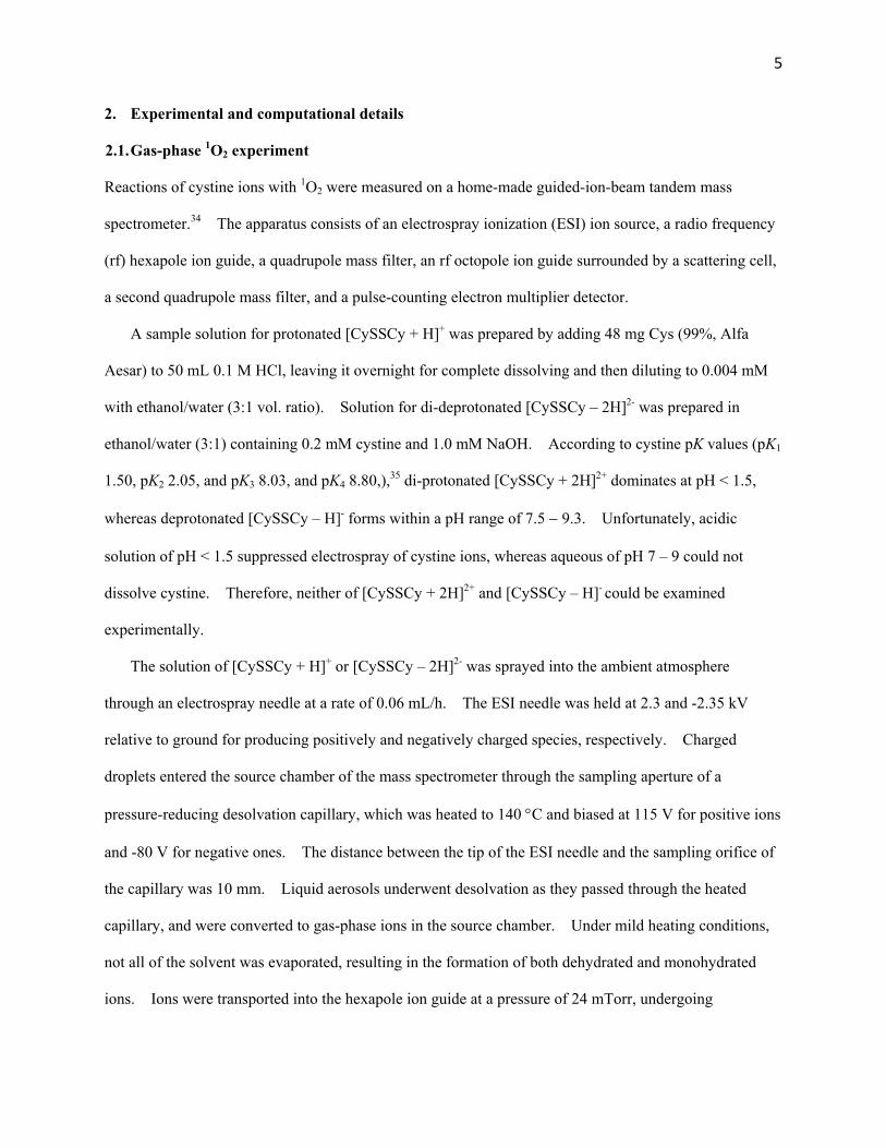
3.1. Structures of protonated, deprotonated and di-deprotonated cystine
Cystine ions have numerous conformations. To find the global minima of their conformational
landscape, a grid search method was applied. We rotated each of the torsion angles along molecules
through 360 at 60 increments to generate trial staggered conformations for each ionization state.
Every conformation so generated was subjected to optimization at B3LYP/6-31+G(d) to derive the
associated local minimum conformation. Many of the initial conformations optimized to the same local
minima. All local minimum conformations were re-optimized at B3LYP/6-311++G(d,p). For each
ionization state, all of the conformers with relative energy within 0.2 eV are provided in the Supporting
Information together with their Cartesian coordinates and energies. Each conformer has a number suffix
to indicate the order of stability within its category. We have compared relative energies of conformers
at a pressure of one atmosphere vs. mTorr, and found no obvious changes. Major conformers for each
series are summarized in Fig. 1.
For [CySSCy + H]+, a total of fourteen stable conformers/rotamers were located within 0.2 eV.
They are gathered in Fig. S1, of which the two most significant, [CySSCy + H]+_1 and 2, are depicted in
the top row of Fig. 1. The two conformers represent 54 and 34% of protonated ions at 298 K,
respectively. In both conformers, two amino groups interact to stabilize the proton. Seven low-energy
conformers were identified for [CySSCy – H]- (Fig. S2). The first three lowest-energy conformers lie
within 0.1 eV with a population of 69, 15 and 14%, respectively. Both protonated and deprotonated
cystines are featured by cyclic structures via proton sharing and charge complexation between two Cys

9
residues. A total of twenty three [CySSCy – 2H]2- conformers lie within 0.2 eV, as summarized in Fig.
S3. In contrast to cyclization of singly charged structures, [CySSCy – 2H]2- is constrained to linear
structures, in order to maximize the distance and thus minimize Coulomb repulsion between the two
negative charges. The two lowest-energy conformers, accounting for 44 and 33% of the population,
have the two carboxylate termini separated from each other more than 9 Å.
Our conformation search not only reproduced the protonated cystine conformers that were reported at
the B3LYP/6-31+G(d,p) and RI-MP2 levels of theory49, but found many new structures and identified
new global minima. On the basis of calculated populations, [CySSCy + H]+_1, [CySSCy – H]- _1 and
[CySSCy – 2H]2- _1 in Fig. 1 represent dominating reactant ions in gas-phase experiments, and were used
as starting structures in PESs. It is certainly possible that interconversion between different conformers
may occur during ion-molecule collisions. However, it seems less likely that different conformers
would significantly alter reaction coordinates, as confirmed by direct dynamics trajectory simulations of
protonated/deprotonated Cys with 1O2.23-25
3.2. Products from ion-molecule collisions
[CySSCy + H]+ + 1O2. We started by first examining the reaction of [CySSCy + H]+ (m/z 241) over the
center-of-mass Ecol range of 0.1 1.0 eV. Product ions were observed at m/z 120, 152, 154, 195 and 223.
A representative product ion mass spectrum and formation scheme are illustrated in Fig. S4 in the
Supporting Information. Product ions of m/z 120 are due to bond cleavage at the disulfide link.50-52
Its structure could be assigned to H2NCH(CO2H)CH2S+ (i.e. [Cys – H]+, see Fig. S4),51 and the S-S bond
rupture was preceded by an intramolecular proton transfer from the ammonium group to the disulfide
bond. While m/z 120 is minor in collision-induced dissociation (CID), the same fragment is dominant in
electron-induced dissociation (EID) of [CySSCy + H]+.52 The m/z 152 can be attributed to cyclic
[-+H2NCH(CO2H)CH2SS-],50-54 produced by C-S cleavage via a Grob-like fragmentation process.51 The
m/z 154 might be assigned as a protonated thiocysteine H3N+CH(CO2H)CH2SSH. 51-53 Product ions of
m/z 195 and 223 correspond to concomitant loss of neutral (H2O + CO) and elimination of a single water
from [CySSCy + H]+, respectively.51-53
10
All of these products were observed upon collisions of [CySSCy + H]+ with ground-state O2, too; and
therefore could be excluded from 1O2-specific reactions. Intensities of most product ions increased at
high Ecol, except for m/z 120 that had only a small amount and diminished at high energies. The latter
does not resemble typical CID in which high collision energy would enhance fragmentation.
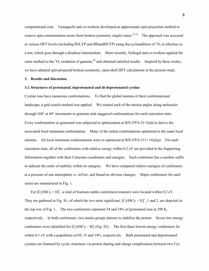
[CySSCy – 2H]2- + 1O2 and disulfide bond cleavage. For [CySSCy – 2H]2- (m/z 119) + 1O2, only four
CID product ions were detected, as shown in Fig. 2a along with fragmentation scheme. The first three
correspond to m/z 73 (H2NCHCO2-), 110 (elimination of a water from [CySSCy – 2H]2- ) and 101
(elimination of two water molecules from reactant ion), all of which have minor intensities. The major
product is an anionic radical H2NCH(CO2-)CH2SS at m/z 151. m/z 151 could also be assigned to
H2NCH(CO2-)CH2SO2 a product that may form by breaking the S-S bond of an O2 adduct
[CySS(OO)Cy – 2H]2-. To identify the structure of m/z 151, an isotopic labeling experiment was carried
out using [Cy32S34SCy – 2H]2- (m/z 120) as the reactant ion. If H2NCH(CO2-)CH2SO2 were produced,
the product would have contained a mixture of m/z 151(containing a 32S) and 153 (containing a 34S) with a
1:1 intensity ratio. However, only a single ion peak was observed at m/z =153, consistent with the
structure of H2NCH(CO2-)CH2
32S34S∙.
The observation that CID of [CySSCy – 2H]2- is dominated by C-S cleavage is consistent with the
previous report that the absence of a mobile proton facilitates disulfide bond cleavage.51 Identical
product ions were obtained in the collisions of [CySSCy – 2H]2- with pure 3O2 and Ar. We have
measured the cross sections for the product ion H2NCH(CO2-)CH2SS∙ in collisions of [CySSCy – 2H]2-
with O2 and Ar. Cross sections were determined from the ratios of reactant and product ion intensities at
each Ecol, the pressure of collision gas in the scattering cell, and the effective cell length. Results are
plotted in Fig. 2b over the Ecol range from 0.1 to 5 eV. The two collision systems present similar
“apparent” thresholds for production of H2NCH(CO2-)CH2SS∙. At high Ecol, Ar produced slightly larger
cross sections.
Based on PES scans and stationary point calculations at B3LYP/6-311++G(d,p), the C-S cleavage of

11
[CySSCy – 2H]2- may occur via four possible pathways:
[CySSCy – 2H]2 H2NCH(CO2)CH2SS∙ (m/z 151) + CH2CH(NH2)CO2
(m/z 87) H = 0.40 eV (1)
H2NCH(CO2)CH2SS∙ (m/z 151) + [CH2CHNH2]∙̅ (m/z 43) + CO2 H = 0.84 eV (2)
H2NCH(CO2)CH2SS∙ (m/z 151) + CH2CHNH2
+ CO2 (m/z 44) H = 0.57 eV (3)
[H2NCH(CO2)CH2SS]2 (m/z 75.5) + CH2CHNH2 + CO2 H = 1.15 eV (4)
Reaction (1) produces two radical anions. Despite being the least endothermic, this reaction bears
the highest activation barrier (1.84 eV), and thus is energetically less favorable than reactions (2) (4)
that are to be discussed blow. The height of the reaction (4) barrier is partly attributed to the
intramolecular repulsive Coulomb barrier (RCB) against charge-separated fragmentation. Here the RCB
is created as a result of the superposition of short-range binding of excess charges in the dianion and
long-range Coulomb repulsion between two negatively charged fragments.55-56 The open-shell
dissociation PES for reaction (1) is shown in Fig. 2c, obtained by relaxed PES scan along the dissociating
rC-S bond using uB3LYP/6-31+G(d,p).
Reactions (2) (4) are governed by a single transition state TS(C-S) of 1.29 eV as shown in Fig. 2d.
TS(C-S) is located at the early stage of the reactions, and its imaginary frequency corresponds to the
motions of dissociating both C-S and CH-COO- bonds simultaneously, as indicated by green-colored
displacement vectors. A movie of TS(C-S) along the reaction path is available in the Supporting
Information. The structures of the reactant and product minima connected through TS(C-S) were
verified by IRC trajectory calculation. Once crossing TS(C-S), the system forms a product-like complex.
The latter may produce three sets of fragments via reactions (2) – (4) that differ only in which fragments
carry charges. Their energetics are indicated by bold lines in the figure. Reaction (4) could be ruled
out since no dianionic product [H2NCH(CO2)CH2SS]2- was detected in the experiment. The absence of
this reaction is not surprising because this dissociation channel has largest endothermicity among
reactions (2) (4), so the system switches to low-energy electronic state(s) that may lead to reactions (2)
and/or (3). The likelihood for reactions (2) and (3) depends on the extent of charge delocalization and
12
the nature of how charge states start to unmix as products separate. In theory, product branching
between (2) and (3) could be determined by the intensities of the complementary ions [CH2CHNH2]∙ ̅ and
CO2- that were produced concurrently with H2NCH(CO2
-)CH2SS in reactions (2) and (3). Unfortunately,
neither of the two complementary ions was present in product ion mass spectra a not unusual
phenomenon in CID of multiply charged ions.57-58
Natural population analysis indicates that the charges of the H2NCH(CO2)CH2SS, CH2CHNH2 and
CO2 moieties are -1.68, -0.06, and -0.26 at TS(C-S), respectively; and the CO2 moiety has a O-C-O
bend angle of 154 which is closer to CO2- anion (138 ) than to neutral CO2 (180 ). The question is at
what point and how the charge separation among fragments gets decided. Fig. 2d tracks the charges
upon individual moieties along the IRC trajectory. The CO2 moiety starts to lose negative charge as
soon as the trajectory approaches the activation barrier, and becomes neutral after the trajectory crosses
over TS(C-S). This may imply that reaction (2) is favored by the system. Charges between
H2NCH(CO2)CH2SS and CH2CHNH2, on the other hand, mix together during the trajectory. Both open-
and closed-shell DFT calculations converged to the same product-like complex. Within this complex,
charge populations are -1.9 on H2NCH(CO2)CH2SS, -0.1 on H2NCHCH2, and none on CO2. It seems
that RCB is sufficiently large to bind excess electrons within a single H2NCH(CO2)CH2SS moiety. We
speculate that charge separation between H2NCH(CO2)CH2SS and CH2CHNH2 did not occur until the
final stage when products start to separate beyond equilibrium distance.
A similar late-stage charge separation scenario was reported for H2CO+ + C2H4 HCO+ + C2H5 and
HCO + C2H5+.59 In this reaction, two product channels (a pair of charge states for [HCO + C2H5]
+) share
a common transition state which leads to the formation of a product-like complex. Actual atom transfer
takes place in the geometry where reactants are interacting intimately, and charge separation occurs at the
last step when products separate. Such late-stage charge separation was verified by the competition
between the two product channels and their dependence on Ecol and H2CO+ vibrational modes.
The branching between reactions (2) and (3) must depend on how the energies and coupling strength
13
of these charge states vary with separation. It would be ideal to follow charge separation and
dissociation of the product-like complex in Fig. 2d, and locate the barrier on the PES that leads to two
separate valleys. Note that the reaction of [CySSCy – 2H]2- takes place on a single potential surface up to
the product-like complex, then undergoes a transition from one electronic state to another. This
electronic state transition is evidenced by the formation of charge-separated products rather than dianionic
[H2NCH(CO2)CH2SS]2-. However, the DFT method used for PES calculations is not applicable to this
non-adiabatic inter-system crossing or surface hopping, particularly when the transition between the two
charge states is abrupt. In addition, near the crossing seam a single reference wavefunction may be
inadequate for calculations. For these reasons, we did not attempt to calculate the branching between
reactions (2) and (3).
We have also calculated the symmetric S-S bond rupture process of [CySSCy 2H]2- 2[Cys – H]∙ ̅
(anionic radical, m/z 119). The reaction has H of only 0.2 eV calculated at B3LYP/6-311++G(d,p), but
bears a dissociation barrier of 2.5 eV and thus is not expected to be accessible during low-energy CID.
We could not rule out the possibility of S-S cleavage at high Ecol. However, product ion [Cys – H]∙ ̅ has
the same m/z as the reactant and thus could not be distinguished by mass spectrometry.
The adiabatic electron detachment energy for [CySSCy 2H]2- was calculated to be 1.2 eV at
B3LYP/6-31+G(d,p). The unfavorable electron detachment is related to the fact the resulting anionic
radical tends to eliminate two CO2 moieties. Charge transfer between [CySSCy 2H]2- and 1O2 is, on
the other hand, exothermic by 0.26 eV. Nevertheless, neither electron detachment nor charge transfer
was observed in the experiment. The inability to detect these reactions may be again due to the
existence of RCB. It follows that energy substantially higher than the electron binding energy is
required to remove an electron from [CySSCy 2H]2-.
Note that none of the aforementioned reactions are 1O2-specific. We had thought that the failure to
detect oxidation products of cystine ions might be because the peroxides formed in the reactions were
unstable and did not have sufficient lifetime, and in the meantime their conversions to end products were

14
blocked by high-energy barriers. Consequently, intermediates could only decay back to reactants via
physical quenching of 1O2. Such scenario indeed happened in the 1O2 reactions with gas-phase Cys23-25
and Met,27 where the decay of transient hydroperoxides CySOOH and MetOOH caused fragmentation of
amino acids. For those systems, we devised reaction routes by using hydrated Cys and Met as the
targets for collisions with 1O2. The point was to stabilize hydroperoxides via water evaporation cooling
of gas-phase hydrates. This strategy was proved to be successful in capturing CySOOH and MetOOH.
Following the same idea, we examined the collisions of 1O2 with monohydrated [CySSCy – 2H]2-H2O.
Contrary to our expectation, no oxidation product was detected for hydrated reactant ions, either.
3.3. Reaction PESs
Repulsive PES for [CySSCy + H]+ + 1O2. To address the origin of the non-reactivity of [CySSCy +
H]+ with 1O2, a 31 19 grid reaction PES was generated at B3LYP/6-31+G(d,p). The contour map is
visualized in Fig. 3a along rSO (the distance between the reacting S and O atoms) and rSS (the disulfide
bond length). During the PES scan, rSO and rSS varied from 4.5 to 1.5 Å and 1.9 to 2.8 Å at an interval
of 0.1 and 0.05 Å, respectively. All the other bond lengths and bond angles were optimized at each
point of the PES. On this PES, there is a strip of flat and shallow potential well leading from the
reactants to a precursor complex located at rSO = 3.0 Å and rSS = 2.1 Å with a binding energy of only
0.04 eV. The amount of binding energy roughly matches the charge-induced-dipole potential at an
ion-molecule center-of-mass distance of 4 Å.60 The fact that the potential becomes repulsive at rSO <
2.5 Å implies that either 1O2 is well shielded from the positive charge of the protonated amino group, or
the ion-induced dipole term is weaker than the repulsion between reactants.
We noticed that DFT was reported to be problematic in describing dimethyl persulfoxide
(CH3)2SOO.45 The stable B3LYP structure for (CH3)2SOO was characterized by a long rSO distance of
2.36 Å. On the other hand, MP2 was able to locate a covalently bound (CH3)2SOO with rSO of 1.59
1.65 Å.45, 61 In our PES study for [CySSCy H]- + 1O2 and [CySSCy 2H]2- + 1O2 (vide infra), DFT
was able to locate both weakly bound precursor complexes (rSO = 2.4 Å) and covalently bonded cystine
15
persulfoxides (rSO = 1.7 Å) as well as the transition states connecting the two structures. To further
ascertain that the short-range potential surface was not artificially raised by DFT, another PES scan was
completed along rSO at the RI-MP2 level of theory. The RI-MP2 method is almost equivalent to the
exact MP2 in calculating 1O2 reaction PESs,62 but with a higher computational efficiency. The similarities
and differences between DFT and RI-MP2 PESs are illustrated by comparing the two relaxed PESs in Fig.
3b, which were calculated at the same collision orientations and using the same basis set. As shown in
the figure, the RI-MP2 curve has slightly deeper well at large reactant separation. Both curves become
repulsive starting at rSO = 2.5 Å, and the RI-MP2 potential rises more quickly at short range.
During ion-molecule collisions, as the distance between reactants decreases, the kinetic energy which
was initially wholly translational was being converted to centrifugal energy.63 We have estimated the
orbital angular momentum L for the collision complex at Ecol = 0.1 eV, using L= μ ·vrel · (σcollision /π)1/2,
where μ and vrel are the reduced mass and relative velocity of the collision partners, respectively, and
σcollision is the ion-induced dipole capture cross section. 60 Resulting centrifugal barrier for the calculated L
reaches ~0.1 eV at rSO of 3 4 Å. Therefore, total potential, as the sum of ion-molecule potential and
centrifugal barrier, becomes consistently repulsive as 1O2 approaches [CySSCy + H]+.
[CySSCy – H]- and [CySSCy – 2H]2- may form persulfoxides with 1O2. We have mapped out
reaction PESs for both [CySSCy – H]- and [CySSCy – 2H]2- at B3LYP/6-31+G(d,p) in Fig. 4, so that we
may look at the effects of an excess negative charge on the reaction. To differentiate similar structures
between singly- and di-deprotonated ones, we included the number of missing protons and negative
charge in their acronyms. The two PESs share common features, so are described together. Both
[CySSCy – H]- and [CySSCy – 2H]2- may form precursor complexes with 1O2 at the early stage of
collisions. Their binding energies are -0.37 and -0.46 eV, respectively. Critical bond lengths are rSO =
2.49 and 2.38 Å, rOO =1.26 and 1.27 Å, and rSS = 2.08 and 2.10 Å for the singly- and di-deprotonated
precursors. In contrast to the protonated precursor that bounces off the repulsive potential surface, both
singly- and di-deprotonated precursors may evolve to high-energy but stable covalently bound
persulfoxides. A minimum energy pathway along intrinsic reaction coordinate is projected onto each of

16
the two PESs. IRC trajectories feature the so-called late-barriers located at [TS H]- and [TS 2H]2-
(the movies of [TS H]- and [TS 2H]2- along reaction pathways are available in the Supporting
Information). The transition states resemble their products in geometries except the changes of three
bond lengths, i.e., rSO = 1.76 vs. 1.64 Å, rSS = 2.17 vs. 2.21 Å, rOO = 1.37 vs. 1.43 Å for [TS H]- and
[CySS(OO)Cy H]-; and rSO = 1.85 vs. 1.68 Å, rSS = 2.14 vs. 2.15 Å and rOO = 1.35 vs. 1.40 Å for [TS
2H]2- and [CySS(OO)Cy 2H]2- , respectively.
The only difference between the PESs for [CySSCy – H]- and [CySSCy – 2H]2- concerns the long-
range potential. [CySSCy – 2H]2- remains attractive to 1O2 at large separations, and has a binding
energy of 0.2 eV with 1O2 at rSO = 4.5 Å; whereas the attractive potential between [CySSCy – H]- and
1O2 decreases quickly at large SO. This difference might be attributed to two facts. First, long-range
ion-induced-dipole potential (= -q2/2r4, where denotes the molecule polarizability, q the ion charge,
and r the ion-molecule separation) increases with ion charges. Secondly, at relatively large reactant
separation 1O2 could be locked into orientations close to the -NH2 and/or -CO2- groups of [CySSCy –
2H]2-. Electrostatic binding energy at such orientations reaches up to 0.7 eV. Formation of these
electrostatic complexes becomes most probable when 1O2 attacks the back side of the disulfide bond and
the two termini of [CySSCy – 2H]2-. Nevertheless, trapping in these collision complexes disfavors the
attack of 1O2 on the disulfide bond.
Reaction coordinates refined by an approximate spin projection method. Guided by the PESs in
Figs. 3 and 4, we are able to establish reaction coordinates for all three systems. 1O2 reactions present
multi-configurational characters and thus lead to poorly calculated stationary point energies and barrier
heights using restricted DFT. Calculating energies for the present systems using multi-reference
solutions, such as CASMP2,47 was not feasible due to large system sizes. Here approximately
spin-projected open-shell, broken-symmetry B3LYP calculations were used to refine local minima and
TSs. The spin contamination error was approximately removed following Yamaguchis methodology
described by eq. (5),

17
⟨ ⟩
⟨ ⟩ ⟨ ⟩
⟨ ⟩
⟨ ⟩ ⟨ ⟩ (5)
where E refers to energies with the superscripts AP, BS and HS represent the approximately spin-
projected state, the broken-symmetry unrestricted singlet state, and high-spin state, respectively. Only
triplet was considered in HS since the <S2> after annihilation was only ~ 0.03. As a test, the
spin-projected 1O2 excitation energy was calculated to be 0.89 eV at B3LYP/6-31+G(d,p) and 1.05 eV at
BHandHLYP/6-31+G(d,p), which are within 0.09 eV of the experimental value. It is therefore
reasonable to expect that this method would improve the description of reaction PESs. We found that, in
addition to 1O2, precursor complexes exhibit significant spin contaminations with triplet states, thus their
energies were much improved by the spin projection method. Open-shell, broken-symmetry calculations
of TSs and disulfide peroxides, on the other hand, all converged to closed shells. Natural population
charge analysis indicates [CySS+0.85(OO)-0.86 – H]- and [CySS+0.87(OO)-0.80 – 2H]2- at TSs, and
[CySS+0.85(OO)-0.86 – H]- and [CySS+0.87(OO)-0.97 – 2H]2- at peroxides. The zwitterionic characteristics of
TSs and end products are consistent with what was found for persulfoxides in reactions of 1O2 with
sulfides.13-14, 18, 61 It may be concluded that spin projection is required mostly for the early stage of the
reactions when structures contain loosely bound 1O2. We would not expect spin contamination to cause
significant issues at the late stage of the reactions when wavefunctions become stable in restricted DFT.
Both spin-projected, open-shell energies (indicated by bold lines) and restricted singlet state energies
(dashed lines) are plotted in Fig. 5, where reactants are located at zero energy. To verify that the spin-
projected results are not functional dependent, we calculated precursors for these systems using
BHandHLYP/6-31+G(d,p) another popular functional set for describing amino acid energies with
accuracy better than MP2.64 The latter was able to reproduce B3LYP results.
The mechanistic importance of the low-lying precursors in Fig. 5 depends on their lifetimes, so we
used RRKM theory to model their unimolecular kinetics. No barrier was expected for decay of the
precursor to reactants in excess of dissociation asymptote, thus an orbiting TS65 was assumed.
Rotational quantum number K was treated as active in evaluating rate constant k(E, J), and all (2J +1)K

18
levels were counted as shown by eq. (6):
J
JKr
J
JKr
KJEEN
KJEEEG
h
dJEk
)],([
)],([),(
0 (6)
where d is the reaction path degeneracy, G is the sum of accessible states from 0 to E - E0 - Er† at the
orbiting TS, N is the energized reactants density of states, E is the system energy, E0 is the activation
energy, and Er and Er† are the rotational energies for the reactant and the orbiting TS, respectively.
Precursors and reactants were described by B3LYP calculated frequencies, moments of inertia, and
spin-projected energies. At the Ecol regime below 0.2 eV where a complex-mediated mechanism might
be important, the dissociation rate constant is 109 - 1011 s-1 for the precursors at different ionization states.
This corresponds to a lifetime (< ns) that was insignificant during mass spectrometric measurements (on
the time scale ~ 102 s). As a result, kinetic analysis predicted no chemical reactivity for cystine ions.
4. Conclusions
We present guided-ion-beam scattering mass spectrometric and theoretical study on the collisions of 1O2
with protonated, deprotonated and di-deprotonated cystine. Ion-molecule scattering measurements
confirmed no oxidation of cystine ions by 1O2, despite the fact that alkyl disulfides are oxidizable. A
series of DFT potential surfaces, with the aid of approximately spin-projected open-shell, broken-
symmetry calculations, have provided rationalization for experimental results. One remarkable finding
is that the collisions of pronated cystine with 1O2 follow a repulsive potential surface starting from the
initial approaching of reactants. Formation of short-lived, weak complexes are possible in the collisions
of singly- and di-deprotonated cystine with 1O2, but their conversions to stable persulfoxides (akin to the
key intermediates proposed for oxidation of alkyl sulfides) are hampered by high activation barriers. We
reported previously that 1O2 oxidation of Cys23-25 and Met26-27 is mediated by hydroperoxide intermediates
CySOOH and MetOOH. CySOOH and MetOOH are an analogue of (but yet more stable than) the
S-hydroperoxysulfonium ylide in 1O2 oxidation of organic sulfides.6, 45, 61 The non-reactivitiy of cystine
may be traced back to the lack of a mobile proton that would otherwise stabilize peroxide intermediates

19
via formation of hydropersulfoxides.
Disulfide bond cleavage (both C-S and S-S cleavages) of disulfide-containing peptides may open
access to the previously covered backbone regions and help identify peptide primary structures. Various
approaches were reported for inducing disulfide bond cleavage such as UV photodissociation,49, 66
negative ion dissociation67-68, EID,52 and radical-induced dissociation.69-70 The present experiment
probed CID of cystine ions, including di-deprotonated cystine for the first time. In contrast to minor
disulfide bond cleavage in the CID of protonated cystine, C-S cleavage dominates the CID of
di-deprotonated cystine and produces unique H2NCH(CO2-)CH2SS anionic radical fragment.
Supporting Information Available: Tautomer/rotamers of [CySSCy + H]+, [CySSCy H]- and
[CySSCy 2H]2-, CID mass spectrum and fragmentation scheme for [CySSCy + H]+, structures in Figs.
2d 4, and movies of TS(C-S), [TS – H]- and [TS – 2H]2-.
Acknowledgements
This work was supported by the National Science Foundation (Grants No. CHE 0954507 and 1464171).
WL and YS acknowledge CUNY Doctoral Student Research Grants.
References (1) Ogilby, P. R. Singlet Oxygen: There Is Indeed Something New under the Sun. Chem. Soc. Rev. 2010, 39, 3181-3209. (2) Davies, M. J. Reactive Species Formed on Proteins Exposed to Singlet Oxygen. Photochem. Photobiol. Sci. 2004, 3, 17-25. (3) Cadet, J.; Ravanat, J.-L.; Martinez, G. R.; Medeiros, M. H. G.; Mascio, P. D. Singlet Oxygen Oxidation of Isolated and Cellular DNA: Product Formation and Mechanistic Insights. Photochem. Photobiol. 2006, 82, 1219-1225. (4) Marchetti, B.; Karsili, T. N. V. An Exploration of the Reactivity of Singlet Oxygen with Biomolecular Constituents. Chem. Commun. 2016, 52, 10996-10999. (5) Palumbo, G. Photodynamic Therapy and Cancer: A Brief Sightseeing Tour. Expert Opin. Drug Delivery 2007, 4, 131-148. (6) Clennan, E. L. Persulfoxide: Key Intermediate in Reactions of Singlet Oxygen with Sulfides. Acc. Chem. Res. 2001, 34, 875-884. (7) Thornton, J. M. Disulfide Bridges in Globular Proteins. J. Mol. Biol. 1981, 151, 261-287. (8) Hogg, P. J. Disulfide Bonds as Switches for Protein Function. Trends Biochem. Sci. 2003, 28, 210-214. (9) Mace, K. A.; Duce, R. A.; Tindale, N. W. Organic Nitrogen in Rain and Aerosol at Cape Grim, Tasmania, Australia. J. Geophys. Res. 2003, 108, 4338. (10) Mader, B. T.; Yu, J. Z.; Xu, J. H.; Li, Q. F.; Hu, W. S.; Flagan, R. C.; Seinfeld, J. H. Molecular Composition of the Water-Soluble Fraction of Atmospheric Carbonaceous Aerosols Collected During ACE-Asia. J. Geophys. Res. 2004, 109, D06206.

20
(11) Anastasio, C.; McGregor, K. G. Photodestruction of Dissolved Organic Nitrogen Species in Fogwaters. Aerosol Sci. Technol. 2000, 32, 106-119. (12) Millington, K. R.; Church, J. S. The Photodegradation of Wool Keratin II. Proposed Mechanisms Involving Cystine. J. Photochem. Photobiol. B 1997, 39, 204-212. (13) Foote, C. S.; Peters, J. W. Chemistry of Singlet Oxygen. XIV. Reactive Intermediate in Sulfide Photooxidation. J. Am. Chem. Soc. 1971, 93, 3795-3796. (14) Murray, R. W.; Jindal, S. L. Photosensitized Oxidation of Dialkyl Disulfides. J. Org. Chem. 1972, 37, 3516-3520. (15) Creed, D. The Photophysics and Photochemistry of the near-UV Absorbing Amino Acids. III. Cystine and Its Simple Derivatives. Photochem. Photobiol. 1984, 39, 577-583. (16) Ando, W.; Takata, T., Photooxidation of Sulfur Compounds. In Singlet O2, Frimer, A. A., Ed. CRC Press: Boca Raton, 1985; Vol. 3: Reaction Modes and Products Part 2, pp 1-117. (17) Straight, R. C.; Spikes, J. D., Photosensitized Oxidation of Biomolecules. In Singlet O2, Frimer, A. A., Ed. CRC Press: Boca Raton, Florida, 1985; Vol. 4: Polymers and Biomolecules, pp 91-143. (18) Murray, R. W.; Jindal, S. L. Photosensitized Oxidation of Disulfides Related to Cystine. Photochem. Photobiol. 1972, 16, 147-151.
(19) Fliss, H.; Viswanatha, T. 2,3-Butanedione as a Photosensitizing Agent: Application to -Amino Acids and -Chymotrypsin. Can. J. Biochem. 1979, 57, 1267-1272. (20) Weil, L. On the Mechanism of the Photo-Oxidation of Amino Acids Sensitized by Methylene Blue. Arch. Biochem. Biophys. 1965, 110, 57-68. (21) Weil, L.; Gordon, W. G.; Buchert, A. R. Photooxidation of Amino Acids in the Presence of Methylene Blue. Arch. Biochem. 1951, 33, 90-109. (22) Benassi, C. A.; Scoffone, E.; Galiazzo, G.; Iori, G. Proflavine-Sensitized Photooxidation of Tryptophan and Related Peptides. Photochem. Photobiol. 1967, 6, 857-866. (23) Liu, F.; Fang, Y.; Chen, Y.; Liu, J. Dissociative Excitation Energy Transfer in the Reactions of Protonated Cysteine and Tryptophan with Electronically Excited Singlet Molecular Oxygen (a1g). J. Phys. Chem. B 2011, 115, 9898-9909. (24) Fang, Y.; Liu, F.; Emre, R.; Liu, J. Guided-Ion-Beam Scattering and Direct Dynamcis Trajectroy Study on the Reaction of Deprotonated Cysteine with Singlet Molecualar Oxygen. J. Phys. Chem. B 2013, 117, 2878-2887. (25) Liu, F.; Emre, R.; Lu, W.; Liu, J. Oxidation of Gas-Phase Hydrated Protonated/Deprotonated Cysteine: How Many Water Ligands Are Sufficient to Approach Solution-Phase Photooxidation Chemistry? Phys. Chem. Chem. Phys. 2013, 15, 20496-20509. (26) Fang, Y.; Liu, F.; Bennett, A.; Ara, S.; Liu, J. Experimental and Trajectory Study on Reaction of Protonated Methionine with Electronically Excited Singlet Molecular Oxygen (a1g): Reaction Dynamics and Collision Energy Effects. J. Phys. Chem. B 2011, 115, 2671-2682. (27) Liu, F.; Liu, J. Oxidation Dynamics of Methionine with Singlet Oxygen: Effects of Methionine Ionization and Microsolvation. J. Phys. Chem. B 2015, 119, 8001-8012. (28) Seliger, H. H. A Photoelectric Method for the Measurement of Spectra of Light Sources of Rapidly Varying Intensities. Anal. Biochem. 1960, 1, 60-65. (29) Khan, A.; Kasha, M. Red Chemiluminescence of Oxygen in Aqueous Solution. J. Chem. Phys. 1963, 39, 2105-2106.
(30) Maranzana, A.; Ghigo, G.; Tonachini, G. Diradical and Peroxirane Pathways in the [2 + 2] Cycloaddition Reactions of 1g Dioxygen with Ethene, Methyl Vinyl Ether, and Butadiene: A Density Functional and Multireference Perturbation Theory Study. J. Am. Chem. Soc. 2000, 122, 1414-1423. (31) Szabo, A.; Ostlund, N. S., Modern Quantum Chemistry: Introduction to Advanced Electronic Structure Theory Dover Publications: New York, NY, 1996; p 480. (32) Saito, T.; Nishihara, S.; Kataoka, Y.; Nakanishi, Y.; Matsui, T.; Kitagawa, Y.; Kawakami, T.; Okumura, M.; Yamaguchi, K. Transition State Optimization Based on Approximate Spin-Projection (AP) Method. Chem. Phys. Lett. 2009, 483, 168-171. (33) Saito, T.; Nishihara, S.; Kataoka, Y.; Nakanishi, Y.; Kitagawa, Y.; Kawakami, T.; Yamanaka, S.; Okumura, M.; Yamaguchi, K. Reinvestigation of the Reaction of Ethylene and Singlet Oxygen by the Approximate Spin

21
Projection Method. Comparison with Multireference Coupled-Cluster Calculations. J. Phys. Chem. A 2010, 114, 7967-7974. (34) Fang, Y.; Liu, J. Reaction of Protonated Tyrosine with Electronically Excited Singlet Molecular Oxygen (a1g): An Experimental and Trajectory Study. J. Phys. Chem. A 2009, 113, 11250-11261. (35) Lide, D. R., CRC Handbook of Chemistry and Phycsics. CRC Press: Boca Raton, 2007-2008. (36) Ervin, K. M.; Armentrout, P. B. Translational Energy Dependence of Ar+ + XY → ArX+ + Y (XY = H2,D2,HD) from Thermal to 30 eV C.M. J. Chem. Phys. 1985, 83, 166-189.
(37) Midey, A.; Dotan, I.; Viggiano, A. A. Temperature Dependences for the Reactions of O- and O2- with O2(a
1g) from 200 to 700 K. J. Phys. Chem. A 2008, 112, 3040-3045. (38) Lafferty, W. J.; Solodov, A. M.; Lugez, C. L.; Fraser, G. T. Rotational Line Strengths and Self-Pressure-Broadening Coefficients for the 1.27 Mm, a1g-X
3g-, =0-0 Band of O2. Appl. Opt. 1998, 37,
2264-2270. (39) Frisch, M. J.; Trucks, G. W.; Schlegel, H. B.; Scuseria, G. E.; Robb, M. A.; Cheeseman, J. R.; Scalmani, G.; Barone, V.; Mennucci, B.; Petersson, G. A.; Nakatsuji, H.; Caricato, M.; Li, X.; Hratchian, H. P.; Izmaylov, A. F.; Bloino, J.; Zheng, G.; Sonnenberg, J. L.; Hada, M.; Ehara, M.; Toyota, K.; Fukuda, R.; Hasegawa, J.; Ishida, M.; Nakajima, T.; Honda, Y.; Kitao, O.; Nakai, H.; Vreven, T.; J. A. Montgomery, J.; Peralta, J. E.; Ogliaro, F.; Bearpark, M.; Heyd, J. J.; Brothers, E.; Kudin, K. N.; Staroverov, V. N.; Keith, T.; Kobayashi, R.; Normand, J.; Raghavachari, K.; Rendell, A.; Burant, J. C.; Iyengar, S. S.; Tomasi, J.; Cossi, M.; Rega, N.; Millam, J. M.; Klene, M.; Knox, J. E.; Cross, J. B.; Bakken, V.; Adamo, C.; Jaramillo, J.; Gomperts, R.; Stratmann, R. E.; Yazyev, O.; Austin, A. J.; Cammi, R.; Pomelli, C.; Ochterski, J. W.; Martin, R. L.; Morokuma, K.; Zakrzewski, V. G.; Voth, G. A.; Salvador, P.; Dannenberg, J. J.; Dapprich, S.; Daniels, A. D.; Farkas, O.; Foresman, J. B.; Ortiz, J. V.; Cioslowski, J.; Fox, D. J. Gaussian 09, Revision D.01, Gaussian, Inc: Wallingford, CT, 2013. (40) Alecu, I. M.; Zheng, J.; Zhao, Y.; Truhlar, D. G. Computational Thermochemistry: Scale Factor Databases and Scale Factors for Vibrational Requencies Obtained from Electronic Model Chemistries. J. Chem. Theory Comput. 2010, 6, 2872-2887. (41) Neese, F. The Orca Program System. Wiley Interdiscip. Rev.: Comput. Mol. Sci. 2012, 2, 73-78. (42) Glendening, E. D.; Badenhoop, J. K.; Reed, A. E.; Carpenter, J. E.; Bohmann, J. A.; Morales, C. M.; Landis, C. R.; Weinhold, F. NBO 6.0, Theoretical Chemistry Institute, University of Wiscosin: Madison, WI, 2013. (43) Marcus, R. A. Unimolecular Dissociations and Free-Radical Recombination Reactions. J. Chem. Phys. 1952, 20, 359-364. (44) Zhu, L.; Hase, W. L. A General RRKM Program(QCPE 644), Quantum Chemistry Program Exchange, Chemistry Department, University of Indiana: Bloomington, 1993. (45) McKee, M. L. A Theoretical Study of Unimolecular Reactions of Dimethyl Persulfoxide. J. Am. Chem. Soc. 1998, 120, 3963-3969. (46) Lu, W.; Liu, J. Capturing Transient Endoperoxide in the Singlet Oxygen Oxidation of Guanine. Chem. Eur. J. 2016, 22, 3127-3138. (47) Lu, W.; Teng, H.; Liu, J. How Protonation and Deprotonation of 9-Methylguanine Alter Its Singlet O2 Addition Path: About the Initial Stage of Guanine Nucleoside Oxidation. Phys. Chem. Chem. Phys. 2016, 18, 15223-15234. (48) Thapa, B.; Munk, B. H.; Burrows, C. J.; Schlegel, H. B. Computational Study of Oxidation of Guanine by Singlet Oxygen (1g) and Formation of Guanine:Lysine Cross-Links. Chem. Eur. J. 2017, 23, 5804-5813. (49) Soorkia, S.; Dehon, C.; Kumar, S. S.; Pedrazzani, M.; Frantzen, E.; Lucas, B.; Barat, M.; Fayeton, J. A.; Jouvet, C. UV Photofragmentation Dynamics of Protonated Cystine: Disulfide Bond Rupture. J. Phys. Chem. Lett. 2014, 5, 1110-1116.
(50) Burrows, E. P. Dimethyl Ether Chemical Ionization Mass Spectrometry of -Amino Acids. J. Mass Spectrom. 1998, 33, 221-228. (51) Lioe, H.; O'Hair, R. A. J. A Novel Salt Bridge Mechanism Highlights the Need for Nonmobile Proton Conditions to Promote Disulfide Bond Cleavage in Protonated Peptides under Low-Energy Collisional Activation. J. Am. Soc. Mass Spectrom. 2007, 18, 1109-1123. (52) Lioe, H.; O’Hair, R. A. J. Comparison of Collision-Induced Dissociation and Electron-Induced Dissociation of Singly Protonated Aromatic Amino Acids, Cystine and Related Simple Peptides Using a Hybrid Linear Ion Trap–FT-ICR Mass Spectrometer. Anal. Bioanal. Chem. 2007, 389, 1429-1437.

22
(53) Rubino, F. M.; Verduci, C.; Giampiccolo, R.; Pulvirenti, S.; Brambilla, G.; Colombi, A. Characterization of the Disulfides of Bio-Thiols by Electrospray Ionization and Triple-Quadrupole Tandem Mass Spectrometry. J. Mass Spectrom. 2004, 39, 1408-1416. (54) Ghumman, C. A. A.; Moutinho, A. M. C.; Santos, A.; Tolstogouzov, A.; Teodoro, O. M. N. D. TOF-SIMS Study of Cystine and Cholesterol Stones. J. Mass Spectrom. 2012, 47, 547-551. (55) Wang, L.-S.; Ding, C.-F.; Wang, X.-B.; Nicholas, J. B. Probing the Potential Barriers and Intramolecular Electrostatic Interactions in Free Doubly Charged Anions. Phys. Rev. Lett. 1998, 81, 2667-2670. (56) Dreuw, A.; Cederbaum, L. S. Multiply Charged Anions in the Gas Phase. Chem. Rev. 2002, 102, 181-200. (57) Smith, R. D.; Loo, J. A.; Barinaga, C. J.; Edmonds, C. G.; Udseth, H. R. Collisional Activation and Collision-Activated Dissociation of Large Multiply Charged Polypeptides and Proteins Produced by Electrospray Ionization. J. Am. Soc. Mass Spectrom. 1990, 1, 53-65. (58) McLuckey, S. A.; Glish, G. L.; Berkel, G. J. V. Charge Determination of Product Ions Formed from Collision- Induced Dissociation of Multiply Protonated Molecules Via Ion/Molecule Reactions. Anal. Chem. 1991, 63, 1971-1978. (59) Liu, J.; van Devener, B.; Anderson, S. L. Vibrational Mode and Collision Energy Effects on Reaction of H2CO+ with C2D4. J. Chem. Phys. 2004, 121, 11746-11759. (60) Troe, J. Statistical Adiabatic Channel Model of Ion-Neutral Dipole Capture Rate Constants. Chem. Phys. Lett. 1985, 122, 425-430. (61) Jensen, F.; Greer, A.; Clennan, E. L. Reaction of Organic Sulfides with Singlet Oxygen. A Revised Mechanism. J. Am. Chem. Soc. 1998, 120, 4439-4449. (62) Lu, W.; Liu, J. Deprotonated Guanine·Cytosine and 9-Methylguanine·Cytosine Base Pairs and Their "Non-Statistical" Kinetics: A Combined Guided-Ion Beam and Computational Study. Phys. Chem. Chem. Phys. 2016, 18, 32222-32237. (63) Levine, R. D.; Bernstein, R. B., Molecular Reaction Dynamics and Chemical Reactivity. Oxford University Press: New York, 1987. (64) Yu, W.; Liang, L.; Lin, Z.; Ling, S.; Haranczyk, M.; Gutowski, M. Comparison of Some Representative Density Functional Theory and Wave Function Theory Methods for the Studies of Amino Acids. J. Comput. Chem. 2009, 30, 590-600. (65) Rodgers, M. T.; Ervin, K. M.; Armentrout, P. B. Statistical Modeling of Collision-Induced Dissociation Thresholds. J. Chem. Phys. 1997, 106, 4499-4508. (66) Fung, Y. M. E.; Kjeldsen, F.; Silivra, O. A.; Chan, T. W. D.; Zubarev, R. A. Facile Disulfide Bond Cleavage in Gaseous Peptide and Protein Cations by Ultraviolet Photodissociation at 157 nm. Angew. Chem. Int. Ed. 2005, 44, 6399-6403. (67) Chrisman, P. A.; McLuckey, S. A. Dissociations of Disulfide-Linked Gaseous Polypeptide/Protein Anions: Ion Chemistry with Implications for Protein Identification and Characterization. J. Proteome Res. 2002, 1, 549-557. (68) Zhang, M.; Kaltashov, I. A. Mapping of Protein Disulfide Bonds Using Negative Ion Fragmentation with a Broadband Precursor Selection. Anal. Chem. 2006, 78, 4820-4829. (69) Xia, Y.; Cooks, R. G. Plasma Induced Oxidative Cleavage of Disulfide Bonds in Polypeptides During Nanoelectrospray Ionization. Anal. Chem. 2010, 82, 2856-2864. (70) Stinson, C. A.; Xia, Y. Radical Induced Disulfide Bond Cleavage within Peptides Via Ultraviolet Irradiation of an Electrospray Plume. Analyst 2013, 138, 2840-2846.

23
Figure Captions
Fig. 1 Low-lying tautomers/conformers of [CySSCy + H]+, [CySSCy – H]- and [CySSCy – 2H]2-.
Relative energies (eV, with respect to global minima) and thermal populations (in parentheses)
were evaluated at B3LYP/6-311++G(d,p) with 298 K thermal corrections.
Fig. 2 (a) Product mass spectrum for [CySSCy – 2H]2- + 1O2 obtained at Ecol = 1.0 eV, and
fragmentation scheme; (b) cross sections for product ion m/z 151 in collisions of [CySSCy –
2H]2- with oxygen and Ar; (c) open-shell C-S bond dissociation PES for [CySSCy – 2H]2- and
product charge distributions, calculated at uB3LYP/6-31+G(d,p); and (d) closed-shell IRC
trajectory for C-S bond cleavage of [CySSCy – 2H]2-, and changes of product charge partitions,
calculated at B3LYP/6-311++G(d,p). Vibrational mode corresponding to TS(C-S) imaginary
frequency is indicated by displacement vectors. A movie of TS(C-S) along reaction pathway
is available in the Supporting Information.
Fig. 3 (a) 2D PES for [CySSCy + H]+ + 1O2. Numbers in contour map are potential energies
calculated at B3LYP/6-31+G(d,p); and (b) Comparison of B3LYP vs. RI-MP2 calculated PESs
for [CySSCy + H]+ + 1O2.
Fig. 4 2D PESs for (a) [CySSCy – H]- + 1O2 and (b) [CySSCy – 2H]2- + 1O2. Numbers in the contour
maps are potential energies calculated at B3LYP/6-31+G(d,p). Dotted lines represent IRC
trajectories. Movies of [TS – H]- and [TS – 2H]2- along reaction pathways are available in the
Supporting Information.
Fig. 5 Reaction coordinates for 1O2 with [CySSCy + H]+, [CySSCy – H]- and [CySSCy – 2H]2-,
calculated at B3LYP/6-31+G(d,p). Solid lines indicate approximately spin-projected,
open-shell energies, and dotted lines indicate close-shell energies. For protonated cystine,
only precursor exists.

24
Fig. 1

25
Fig. 2
rCS (Å)2 3 4 5
PE
0
1
2
(charge)
-2
-1
0
Collision Energy (eV)0 1 2 3 4 5
0
2
4
6
m/z
15
1)
(Å2 )
151
fragment ions (x1000)
m/z 119
[CySSCy - 2H]2-
m/z80 100 120 140
a)
b)
[CySSCy - 2H]2- + Ar
[CySSCy - 2H]2- + O2
11010173
Intrinsic Reaction Coordinate
(charge)
-2
-1
0
PE
TS(C-S)
product-like complex
[CySSCy - 2H]2-
d)
0.0 eV
1.29 eV
H2NCH(CO2)CH2SS
CO2
CH2CHNH2
c)
H2NCH(CO2)CH2SS
H2NCH(CO2)CH2
0.0 eV
1.84 eV
rCS
[CySSCy - 2H]2-
1.15 eV, [H2NCH(CO2)CH2SS]2- + CH2CHNH2 + CO2
0.84 eV, [H2NCH(CO2)CH2SS.]- + [CH2CHNH2].- + CO2
0.57 eV, [H2NCH(CO2)CH2SS.]- + CH2CHNH2 + CO2-
Dissociation channels
H2NS
SNH2
COO
COO
H2N COO
(m/z 119)
(m/z 73)
HNS
SNH2
OC
COO
H2O+
HNS
SNH
CO
OC
2H2O+
H2NS
S
COO
NH2+
(m/z 110)
(m/z 101)
(m/z 151) (m/z 43)
CO2+

26
Fig. 3

27
Fig. 4

28
Fig. 5
reactants-0.02
TS
0.39
cystine persulfoxide
(-0.05)
(-0.38)
-0.02
(-0.05) -0.13
(-0.37)
precursor
(-0.40)
(-0.07)
0.38
0.72
(-0.46)
precursor
/ protonated
/ deprotonated
/ di-deprotonated
0.70

29
TOC


