

Article
Foldability of a Natural De
Novo Evolved ProteinGraphical Abstract
Highlights
d The young, functional de novo protein Bsc4 has a
rudimentary ability to fold
d Bsc4 forms compact oligomers with high b sheet content and
a hydrophobic core
d Bsc4 lacks a specific quaternary state and binds dyes
suggestive of amyloid oligomers
d Young de novo proteins can have some structural order and
native-like properties
Bungard et al., 2017, Structure 25, 1687–1696November 7, 2017 ª 2017 Elsevier Ltd.https://doi.org/10.1016/j.str.2017.09.006
Authors
Dixie Bungard, Jacob S. Copple,
Jing Yan, ..., Joanna Masel,
Vicki H. Wysocki, Matthew H.J. Cordes
In Brief
Recent studies have shown that new
protein-coding genes can arise ‘‘de novo’’
from noncoding DNA. The properties of
the brand new proteins encoded by these
genes remain poorly understood. Here,
Bungard et al. show that a very young de
novo protein from yeast folds to a partially
ordered three-dimensional structure.

Structure
Article
Foldability of a NaturalDe Novo Evolved ProteinDixie Bungard,1 Jacob S. Copple,1 Jing Yan,3 Jimmy J. Chhun,1 Vlad K. Kumirov,1 Scott G. Foy,2 Joanna Masel,2
Vicki H. Wysocki,3 and Matthew H.J. Cordes1,4,*1Department of Chemistry and Biochemistry, University of Arizona, Tucson, AZ 85721-0088, USA2Department of Ecology and Evolutionary Biology, University of Arizona, Tucson, AZ 85721-0088, USA3Department of Chemistry and Biochemistry, Ohio State University Columbus, Columbus, OH 43210-1173, USA4Lead Contact
*Correspondence: [email protected]
https://doi.org/10.1016/j.str.2017.09.006
SUMMARY
The de novo evolution of protein-coding genes fromnoncoding DNA is emerging as a source of molecularinnovation in biology. Studies of random sequencelibraries, however, suggest that young de novo pro-teins will not fold into compact, specific structurestypical of native globular proteins. Here we showthat Bsc4, a functional, natural de novo protein en-coded by a gene that evolved recently from noncod-ing DNA in the yeast S. cerevisiae, folds to a partiallyspecific three-dimensional structure. Bsc4 formssoluble, compact oligomers with high b sheet con-tent and a hydrophobic core, and undergoes co-operative, reversible denaturation. Bsc4 lacks aspecific quaternary state, however, existing insteadas a continuous distribution of oligomer sizes,and binds dyes indicative of amyloid oligomers ormolten globules. The combination of native-like andnon-native-like properties suggests a rudimentaryfold that could potentially act as a functional interme-diate in the emergence of new folded proteinsde novo.
INTRODUCTION
Protein folding is difficult and poses a potential roadblock to
evolving protein structures from scratch. In classic textbook
views, natural proteins such as myoglobin fold cooperatively
into specific, stable, soluble, globular structures; these elegant,
intricate native states then serve as scaffolds for biological
functions such as oxygen binding. Such native structures are,
however, rare among amino acid sequences. Soluble proteins
with significant secondary structure content have been re-
covered from unevolved random amino acid sequence libraries,
but they do not have specific, well-defined tertiary structures
(Chiarabelli et al., 2006; Davidson et al., 1995; Doi et al., 1998).
Evenwhen such libraries are biased toward compositions or pat-
terns found in natural proteins, the structures recovered tend to
have ‘‘rudimentary’’ or ‘‘molten globule’’ characteristics lacking
clearly specific tertiary structure (Graziano et al., 2008; Labean
Stru
et al., 2011; Matsuura et al., 2002). Only in a single well-known
case, in which random sequence libraries were subjected to
extensive in vitro functional evolution, have clearly native-like
structures been recovered (Keefe and Szostak, 2001; Lo Surdo
et al., 2004; Mansy et al., 2007). Even among ancient, highly
evolved sequences with specific native states, chaperones are
often necessary to avoid pitfalls such as aggregation. The diffi-
culty of folding is one justification for the common perception
that evolution conservatively ‘‘tinkers’’ with an ancient repertoire,
for example by duplicating and modifying existing protein-cod-
ing genes, instead of inventing entirely new proteins (Jacob,
1977; Zuckerkandl, 1975).
Not all proteins, however, require specific folding to function,
and the relaxation of classic assumptions about structure/
function relationships could make more radical mechanisms
for molecular innovation far more plausible. Intrinsically dis-
ordered proteins (IDPs) that cannot fold independently are a
sizable minority of proteins and serve a variety of biological func-
tions (Dyson and Wright, 2005; Meszaros et al., 2007; Schles-
singer et al., 2011; Tompa and Kovacs, 2010). IDPs vary widely
in the level of disorder, from random coils to ‘‘pre-molten glob-
ules’’ to molten globule states, which are able to fold compactly
and have high levels of secondary structure but lack specific
tertiary structures (Dunker and Obradovic, 2001; Habchi et al.,
2014; Uversky, 2002). Molten globule states may be functional
(DeGrado, 1993; Pervushin et al., 2007; Vamvaca et al., 2004),
and even themost IDPs can function through short linear binding
motifs (Davey et al., 2012). A functional protein might therefore
evolve de novo even if it cannot fold specifically. A protein
born as an IDP might later evolve more native-like properties
(Zhu et al., 2016) or continue indefinitely as a partially or
completely disordered protein.
Until the last decade, there was no clear evidence that whole
functional protein-coding genes could evolve de novo, e.g.,
from previously noncoding DNA; any suggestion that evolution
does anything other than ‘‘tinkering’’ with existing scaffolds
was speculative. Since 2006 (Levine et al., 2006), however,
numerous studies in a variety of organisms have suggested
that some genes trace their origins to the appearance and
expression of a new open reading frame in noncoding DNA
(Andersson et al., 2015; McLysaght and Guerzoni, 2015;
Schlotterer, 2015; Tautz and Domazet-Loso, 2011). Such cases
provide opportunities to test whether very young proteins
emerge as IDPs and, if so, whether they nonetheless have
cture 25, 1687–1696, November 7, 2017 ª 2017 Elsevier Ltd. 1687

Figure 1. Alignment of Unique Bsc4 Sequences from S. cerevisiae Strains
Perfectly conserved residues are shown in color. Bsc4 sequences and strain names were obtained from Blastp searches using the S288C sequence, and aligned
using ClustalX. The alignment is annotated with JRONN and IUPRED disorder prediction for S288C.
some level of order, for example a molten globule state, that
might constitute a nascent folded structure.
Despite plenty of genetic evidence for de novo proteins,
however, there has been almost no reported experimental char-
acterization of their structures (Schmitz and Bornberg-Bauer,
2017). Recently born de novo proteins have been predicted by
sequence analysis to have high levels of intrinsic disorder on
average (Wilson et al., 2017), but no systematic experimental
study of their ability to fold has been done. The antifreeze protein
AFGP from Antarctic notothenioid fishes contains a tripeptide
repeat that evolveddenovo fromasplice junction in a trypsinogen
gene (Chen et al., 1997), and the unusual structure of the repeat
region, matching its unusual function as an antifreeze protein,
has been probed by numerous methods (Urbanczyk et al.,
2017). Some structural data also exist for the special category
of de novo proteins encoded by viral ‘‘overprinted’’ genes, which
evolved from an alternative reading frame of an existing coding
gene, rather than from noncoding DNA. Overprinting proteins
are generally predicted to have high intrinsic disorder (Kovacs
et al., 2010; Rancurel et al., 2009), but least two are known to
fold into specific, compact, novel structures (Meier et al., 2006;
Pavesi et al., 2013; Shukla and Hilgenfeld, 2015; Vargason et al.,
2003). It is not clear how old these proteins are, and they may
retain little signature of their de novo origins. Their structures do,
however, point to the existence of pathways for evolving native-
like folds de novo. No experimental structure of any de novo pro-
tein (young or old), verified to have evolved from a new open
reading frame in noncoding DNA, has been reported.
As a step toward structural characterization of young de novo
proteins, we present a case study of the yeast protein Bsc4.
A serious issue with case studies of individual newborn genes
is the difficulty in proving, in the absence of evolutionary conser-
vation, that they are both protein coding and functional, in addi-
tion to proving that they arose from noncoding sequences
(McLysaght and Hurst, 2016). The yeast gene BSC4 is an excep-
tionally well-supported case of an entire functional protein-
coding gene that recently evolved de novo from an ancestral
noncoding sequence (Cai et al., 2008). The nameBSC4 (‘‘bypass
of stop codon’’) derives from belonging to a set of Saccharo-
myces cerevisiae genes with 9%–25% stop codon bypass
efficiency (Namy et al., 2003). BSC4 is conserved in all strains
of S. cerevisiae, but no homologous open reading frame is
present in other fungal species, and the hypothetical Bsc4 pro-
tein sequence is not similar to any other known protein
sequence. A thorough analysis of synteny and phylogeny among
1688 Structure 25, 1687–1696, November 7, 2017
numerous fungal species demonstrated that BSC4 is homolo-
gous to, and evolved recently from, a region of noncoding DNA
in the intergenic region between LYP1 and ALP1 (Cai et al.,
2008).BSC4 is nonessential but has two synthetic lethal partners
(RPN4 and DUN1) (Pan et al., 2006). Its sequence is >90%
conserved across known S. cerevisiae strains (Figure 1) and
shows a low dN/dS ratio, indicating purifying selection. RT-PCR
and mass spectrometry (MS) data demonstrate expression of
BSC4 at the RNA and protein level, respectively, under normal
culture conditions (Cai et al., 2008). Heightened expression of
BSC4 is observed in stationary phase (Aragon et al., 2008; Gasch
et al., 2000), and both synthetic lethal partners function in DNA
damage repair pathways, suggesting that BSC4 plays a role in
DNA damage repair during stationary phase (Cai et al., 2008).
Bsc4 is a functional, whole de novo protein-coding gene and,
given its presence in only a single yeast species, a notably young
one that can provide a window into de novo gene origin.
We predict that the Bsc4 protein has at least some folded
structure despite the de novo origin and youth of the BSC4
gene. The Bsc4 protein from S. cerevisiae reference strain
S288C has 131 amino acid residues, easily long enough to
form a domain. Its sequence is rich in positively charged resi-
dues, which disfavors folding, but also rich in hydrophobic
residues, which favors folding (Uversky et al., 2000). Based on
aweighting of these two factors, the program FoldIndex predicts
that Bsc4will fold (Prilusky et al., 2005). IUPRED (Dosztanyi et al.,
2005) and JRONN (Troshin et al., 2011; Yang et al., 2005) also
predict relatively low disorder except near the termini (Figure 1).
RESULTS
Recombinant Overexpression, Purification, andRefolding of Two Bsc4 VariantsAs noted above, extant Bsc4 sequences are highly conserved
(Figure 1). The most significant variation is the presence or
absence of a ten-residue hydrophobic C-terminal tail, IVII(YC)
VVRFH, which is predicted by TANGO (Fernandez-Escamilla
et al., 2004) and AGGRESCAN (Conchillo-Sole et al., 2007) to
be an aggregation hotspot. For our experiments we selected
Bsc4 sequences from strains EC1118 and S288C, which are
identical except that S288C has the C-terminal tail, whereas
EC1118 does not. The BSC4 gene is expressed, at least at the
transcript level, in both strains (Rossouw et al., 2009).
We overexpressed Bsc4 EC1118 and S288C in Escherichia
coli from synthetic, codon-optimized BSC4 genes in T7-based
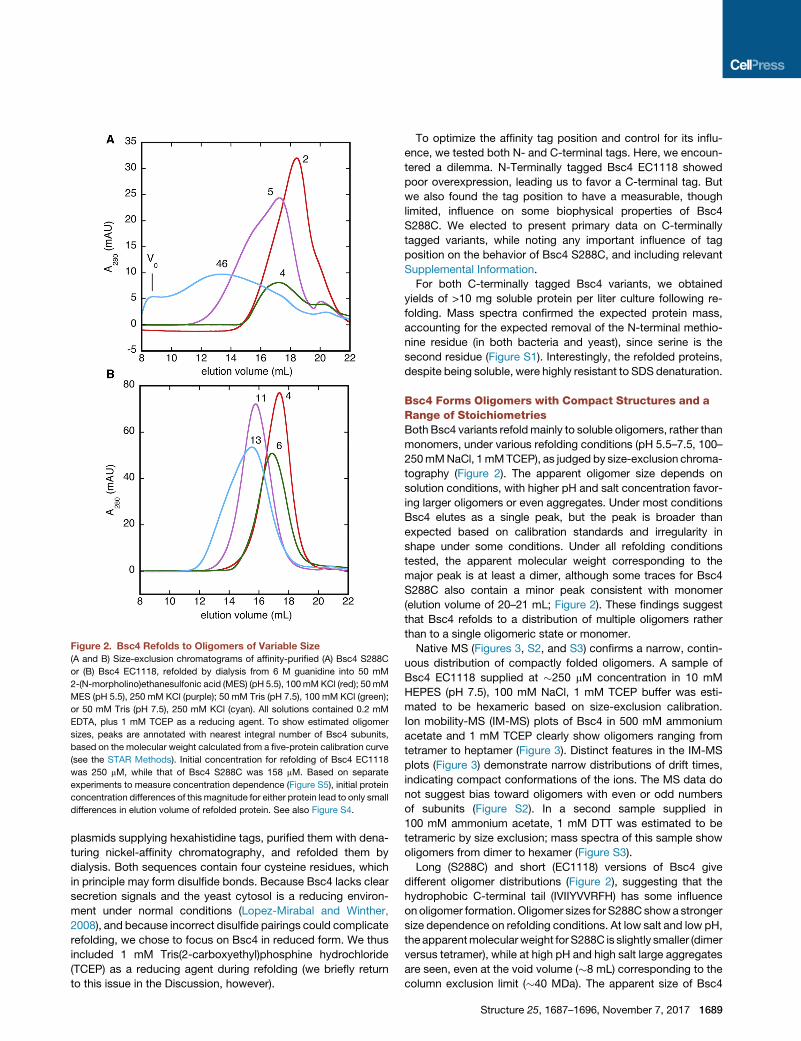
Figure 2. Bsc4 Refolds to Oligomers of Variable Size
(A and B) Size-exclusion chromatograms of affinity-purified (A) Bsc4 S288C
or (B) Bsc4 EC1118, refolded by dialysis from 6 M guanidine into 50 mM
2-(N-morpholino)ethanesulfonic acid (MES) (pH 5.5), 100mMKCl (red); 50mM
MES (pH 5.5), 250 mM KCl (purple); 50 mM Tris (pH 7.5), 100 mM KCl (green);
or 50 mM Tris (pH 7.5), 250 mM KCl (cyan). All solutions contained 0.2 mM
EDTA, plus 1 mM TCEP as a reducing agent. To show estimated oligomer
sizes, peaks are annotated with nearest integral number of Bsc4 subunits,
based on the molecular weight calculated from a five-protein calibration curve
(see the STAR Methods). Initial concentration for refolding of Bsc4 EC1118
was 250 mM, while that of Bsc4 S288C was 158 mM. Based on separate
experiments to measure concentration dependence (Figure S5), initial protein
concentration differences of this magnitude for either protein lead to only small
differences in elution volume of refolded protein. See also Figure S4.
plasmids supplying hexahistidine tags, purified them with dena-
turing nickel-affinity chromatography, and refolded them by
dialysis. Both sequences contain four cysteine residues, which
in principle may form disulfide bonds. Because Bsc4 lacks clear
secretion signals and the yeast cytosol is a reducing environ-
ment under normal conditions (Lopez-Mirabal and Winther,
2008), and because incorrect disulfide pairings could complicate
refolding, we chose to focus on Bsc4 in reduced form. We thus
included 1 mM Tris(2-carboxyethyl)phosphine hydrochloride
(TCEP) as a reducing agent during refolding (we briefly return
to this issue in the Discussion, however).
To optimize the affinity tag position and control for its influ-
ence, we tested both N- and C-terminal tags. Here, we encoun-
tered a dilemma. N-Terminally tagged Bsc4 EC1118 showed
poor overexpression, leading us to favor a C-terminal tag. But
we also found the tag position to have a measurable, though
limited, influence on some biophysical properties of Bsc4
S288C. We elected to present primary data on C-terminally
tagged variants, while noting any important influence of tag
position on the behavior of Bsc4 S288C, and including relevant
Supplemental Information.
For both C-terminally tagged Bsc4 variants, we obtained
yields of >10 mg soluble protein per liter culture following re-
folding. Mass spectra confirmed the expected protein mass,
accounting for the expected removal of the N-terminal methio-
nine residue (in both bacteria and yeast), since serine is the
second residue (Figure S1). Interestingly, the refolded proteins,
despite being soluble, were highly resistant to SDS denaturation.
Bsc4 Forms Oligomers with Compact Structures and aRange of StoichiometriesBoth Bsc4 variants refoldmainly to soluble oligomers, rather than
monomers, under various refolding conditions (pH 5.5–7.5, 100–
250mMNaCl, 1mMTCEP), as judged by size-exclusion chroma-
tography (Figure 2). The apparent oligomer size depends on
solution conditions, with higher pH and salt concentration favor-
ing larger oligomers or even aggregates. Under most conditions
Bsc4 elutes as a single peak, but the peak is broader than
expected based on calibration standards and irregularity in
shape under some conditions. Under all refolding conditions
tested, the apparent molecular weight corresponding to the
major peak is at least a dimer, although some traces for Bsc4
S288C also contain a minor peak consistent with monomer
(elution volume of 20–21 mL; Figure 2). These findings suggest
that Bsc4 refolds to a distribution of multiple oligomers rather
than to a single oligomeric state or monomer.
Native MS (Figures 3, S2, and S3) confirms a narrow, contin-
uous distribution of compactly folded oligomers. A sample of
Bsc4 EC1118 supplied at �250 mM concentration in 10 mM
HEPES (pH 7.5), 100 mM NaCl, 1 mM TCEP buffer was esti-
mated to be hexameric based on size-exclusion calibration.
Ion mobility-MS (IM-MS) plots of Bsc4 in 500 mM ammonium
acetate and 1 mM TCEP clearly show oligomers ranging from
tetramer to heptamer (Figure 3). Distinct features in the IM-MS
plots (Figure 3) demonstrate narrow distributions of drift times,
indicating compact conformations of the ions. The MS data do
not suggest bias toward oligomers with even or odd numbers
of subunits (Figure S2). In a second sample supplied in
100 mM ammonium acetate, 1 mM DTT was estimated to be
tetrameric by size exclusion; mass spectra of this sample show
oligomers from dimer to hexamer (Figure S3).
Long (S288C) and short (EC1118) versions of Bsc4 give
different oligomer distributions (Figure 2), suggesting that the
hydrophobic C-terminal tail (IVIIYVVRFH) has some influence
onoligomer formation. Oligomer sizes for S288C showa stronger
size dependence on refolding conditions. At low salt and low pH,
the apparentmolecularweight for S288C is slightly smaller (dimer
versus tetramer), while at high pH and high salt large aggregates
are seen, even at the void volume (�8 mL) corresponding to the
column exclusion limit (�40 MDa). The apparent size of Bsc4
Structure 25, 1687–1696, November 7, 2017 1689

Figure 4. Bsc4 Has b Sheet Secondary Structure
Far UV circular dichroism spectra of Bsc4 oligomers (S288C, solid line;
EC1118, dashed line) from 195 to 240 nm at 20�C, at 100 mM protein con-
centration in a 0.1 mm pathlength cell, in 50 mM MES (pH 5.5), 100 mM KCl,
1 mM TCEP.
Figure 3. Ion Mobility-Mass Spectrometry Plots of the Mass
Spectrum of Bsc4 Oligomers
The spectrum of Bsc4 EC1118 (266 mM) in 500 mM ammonium acetate and
1 mM TCEP indicates a continuous distribution of oligomers from tetramer to
octamer. The relative abundance of the species is shown in linear scale (color
bar inset top left). Distinct spots in the ionmobility-mass spectrometry (IM-MS)
plot demonstrate narrow distributions of drift times, indicating relatively
compact conformations of the ions. Similar distribution can be observed with
Bsc4 concentration diluted to 18 mM. The spectrum of each of the species is
extracted and shown in Figure S2. The stoichiometry of each of the peaks was
confirmed by collision-induced dissociation and surface-induced dissociation.
See also Figures S1–S3.
S288C aggregates at high pH also increases over time upon stor-
age of refolded protein at 4�C. These observations agreewith the
prediction that the C-terminal tail is aggregation prone (see
above). We also note, however, that this tendency is less pro-
nounced in N-terminally tagged S288C (Figure S4).
The oligomerization of Bsc4 is not simply aggregation resulting
fromhigh protein concentration (�250mM)during refolding. Bsc4
refolds predominantly to oligomers even at more modest con-
centrations (�50 mM) under solution conditions where large olig-
omers are least favored (low salt/low pH) (Figure S5). The elution
volume of the peakmaximumshows only a small increase across
a 4-fold dilution of Bsc4 during refolding. The narrow, continuous
distribution of oligomers seen by MS would, in fact, be expected
to give rise to such dependence, since higher protein concentra-
tion should gradually shift the distribution upwards.
Bsc4 Oligomers Have b Sheet Secondary Structure anda Hydrophobic CoreFar UV circular dichroism (CD) spectra under conditions that
favor smaller oligomers show the presence of b sheet secondary
structure (Figure 4). The combination of mean residue ellipticity
values at 200 nm (near +2,000 deg cm2 dmol–1) and 222 nm
(near �7,000 deg cm2 dmol–1) is also directly inconsistent with
a highly unfolded structure, such as a random coil or ‘‘pre-molten
globule’’ (Uversky, 2002). Analysis of secondary structure con-
tent using the program K2D3 (Louis-Jeune et al., 2012) gives
�30% b strand, �10% a helix content for both variants. The
CD results also agree with our observation (see above) that
Bsc4 is SDS-resistant. SDS-resistance correlates with a combi-
nation of oligomerization and high b strand content (Manning and
Colon, 2004).
Tryptophan fluorescence spectra show strong evidence for
burial of the single tryptophan residue (Trp 47). Spectra obtained
1690 Structure 25, 1687–1696, November 7, 2017
in native buffers (Figure 5) exhibit maximum fluorescence near
328 nm, consistent with tryptophan burial. Spectra obtained in
6 M guanidine (Figure 5) show maxima near 351 nm, indicating
that guanidine denaturation exposes the tryptophan to solvent.
These data suggest that tertiary and/or quaternary interactions
between side chains form a hydrophobic interior in refolded
Bsc4, in agreement with the compact structure inferred from
mass spectra.
Near UV CD spectra of Bsc4 (Figure 5) are somewhat weak in
intensity and show less fine structure relative to those of many
native proteins (Kelly et al., 2005), suggesting that the hydro-
phobic core of Bsc4 could have a partially ‘‘molten’’ character
(Price et al., 2005; Ptitsyn, 1995). As one point of comparison,
the C 33 domain of immunoglobulin E, a molten globule of similar
subunit size (110 versus 120–130 residues) as the two Bsc4
variants, and with about the same number of Trp/Tyr residues
(4 versus 4–5), has a near UVCD spectrum that is similar in shape
and intensity (in mean residue ellipticity), although with less fine
structure (Price et al., 2005). Precise structural interpretation of
near UV CD spectra is not possible, however, so the structure
of Bsc4 cannot be conclusively classified as either molten or
native-like on this basis.
The folded structure of Bsc4 EC1118 does not appear to
include the regions near the N- and C termini. An heteronuclear
single quantum coherence spectrum of a 13C/15N/2H-labelled
sample of Bsc4 EC1118 (Figure S6), refolded under conditions
that favor small oligomers, shows at least 50 resolvable amide
proton resonances. We were able to assign the strongest reso-
nances to regions near the N terminus (3–14) and C terminus
(95–98 and 105–121). TALOS analysis of chemical shifts (Fig-
ure S6) shows low S2 values (<0.7) for these residues, indicative
of highly dynamic character, which is also consistent with the low
spectral dispersion and high intensity of the amide peaks. In
addition, peptides released during limited trypsinolysis corre-
spond primarily to these regions of sequence, while other

Figure 5. Tryptophan Fluorescence and Near UV Circular Dichroism
of Bsc4
(A) Tryptophan fluorescence emission spectrum of Bsc4 S288C (S288C, solid
line; EC1118, dashed line) at 50 mM in 50mMMES (pH 5.5), 100mMKCl, 1mM
TCEP with (red) or without (blue) 6 M guanidine.
(B) Near UV circular dichroism spectra of Bsc4 oligomers (S288C reduced,
solid line; EC1118, dashed line) from 310 to 260 nm at 20�C, at 100 mMprotein
concentration in a 1 cm pathlength cell, in 50 mM MES (pH 5.5), 100 mM
KCl, 1 mM TCEP. The tryptophan fluorescence spectra show a maximum
near 328 nm for folded Bsc4 and 351 nm for guanidine denatured Bsc4.
Figure 6. Cycled Reversible Thermal Denaturation of Small Oligo-
mers of Bsc4
(A) Bsc4 S288C, (B) Bsc4 EC1118 in 50 MES (pH 5.5), 100 mM KCl, 1 mM
TCEP, at 50 mM protein concentration in a 1 mm pathlength cell from 20�C to
98�C (293 K–371 K), monitored by circular dichroism at 222 nm. Filled and
unfilled red circles represent the forward and reverse phases, respectively, of a
first melt, while filled and unfilled blue squares represent forward and reverse
melts of a second melt (remelt) of the same sample. Solid and dashed lines
represent fits of forward and reverse denaturation curves (see the STAR
Methods for details of fitting). For Bsc4 S288C, upper baselines are particularly
poorly defined, and the data could not be fit to any unique solution. The fits
shown for S288C are therefore for illustrative purposes only. See also Figures
S7–S9.
regions appear to be protected (Figure S6). These findings sup-port the predictions by JRONN and IUPRED that the termini are
the least-ordered regions (Figure 1). The rest of the sequence
(residues 15–95, approximately) likely contains a folded domain,
and the apparent resistance to proteolysis suggests that it may
have a high level of structural order. The lack of clearly assign-
able resonances, and the lack of wide chemical shift dispersion,
may reflect the absence of a unique quaternary and/or tertiary
structure for the folded regions; alternatively, or additionally, it
may reflect line broadening due to the high molecular weight of
the oligomers (a pentamer of Bsc4 EC1118 has Mr �75 kDa,
for example). We return to this subject in the Discussion.
Bsc4 Oligomers Undergo Cooperative, ReversibleThermal, and Chemical DenaturationBsc4 oligomers are highly resistant to thermal denaturation, but
both S288C and EC1118 can be melted at least partially under
low salt/low pH conditions (Figure 6). Bsc4 S288C is more resis-
tant to thermal denaturation than Bsc4 EC1118 and does not un-
fold completely, even at 98�C, suggesting that the hydrophobic
C-terminal tail contributes stabilizing interactions (this enhanced
stability is less pronounced for N-terminally affinity-tagged
Structure 25, 1687–1696, November 7, 2017 1691

Figure 7. Cooperative Guanidine Denaturation of Small Oligomers
of Bsc4
(A) Bsc4 S288C, (B) Bsc4 EC1118 in 50 MES (pH 5.5), 100 mM KCl, 1 mM
TCEP, at 60 mM protein concentration in a 0.5 mM pathlength cell at 20�C,monitored by circular dichroism from 250 to 215 nm.Guanidine concentrations
range from 0 M (purple) to 6 M (maroon) in 0.75 M increments. Note the
transition in spectral shape toward a random coil-like spectrumwith increasing
guanidine concentration. Insets show fitting of the ellipticity at 222 nm to a
standard two-state chemical denaturation model. See also Figure S7.
S288C; however; see Figure S7). For both variants, the unfolding
transition is reversible, and the protein can be melted and
refolded at least twice with a similar apparent denaturation
midpoint. Consistent with an oligomeric folded state, the dena-
turation midpoint is concentration dependent, showing a 5�Cincrease for Bsc4 EC1118 over the concentration range 25–
100 mM (Figure S8).
Curiously, both variants, especially S288C, show a gain in di-
chroism signal after the first recooling cycle (Figure 6). Difference
spectra from before and after thermal denaturation show
maximum signal gain near 215 nm, consistent with gain of b
strand secondary structure (Figure S9). Comparisons of size-
exclusion traces before and after melting show that the oligomer
distribution shifts to larger size following the melt (Figure S9).
1692 Structure 25, 1687–1696, November 7, 2017
Thus, the gain in dichroism signal is probably attributable to
renaturation of the protein to larger oligomers with enriched
b sheet content.
The observation of different elution volumes in the same
sample, before and after a thermal melt, suggests that equilibra-
tion of oligomers may be slow at room temperature. To investi-
gate this idea further, we injected samples of refolded Bsc4
EC1118 and isolated different size-exclusion fractions repre-
senting approximately the low and high elution volume halves
of the major peak. Reinjection of these fractions within a few
hours led to different peak elution volumes; incubation of the
samples at 35�C, however, led to apparent equilibration to a
common elution volume over one to several days, depending
upon conditions. Thus, different oligomers of Bsc4 do equilibrate
slowly at ambient temperature.
Bsc4 oligomers undergo cooperative chemical denaturation
by guanidine (Figure 7). Bsc4 S288C has a slightly higher
denaturation midpoint than Bsc4 EC1118 (3.2 versus 2.8 M),
consistent with its greater resistance to thermal denaturation
(this is also observed with N-terminally affinity-tagged Bsc4
S288C; see Figure S7). Free energies of unfolding at zero dena-
turant, derived from the data fitting, are +5.4 and +3.6 kcal/mol
for Bsc4 S288C and EC1118, respectively. Fitted guanidine
m values are 1.7 and 1.3 kcal/mol, respectively, lower than ex-
pected for a typical globular protein of this molecular weight
(specifically, a guanidine m value of 1.5 corresponds to
2,600 A2 of surface area burial, which is typical of a �40-residue
protein rather than a 120-residue protein) (Myers et al., 1995).
One possible contributor to a low m value is that the folded re-
gion does not include the termini.
Bsc4 Oligomers Bind Dyes Indicative of Amyloids orMolten GlobulesThe formation of compact b sheet-rich oligomers without a
specific quaternary state (or specific tertiary interactions,
perhaps) led us to wonder whether the folded state of Bsc4
can be compared with oligomeric intermediates in the formation
of amyloid fibrils, or perhaps to molten globule intermediates in
protein folding. We tested the binding of both Bsc4 variants to
Congo red, Thioflavin T, and ANS, all of which have been re-
ported to bind oligomeric amyloid intermediates (Fandrich,
2012). Changes in ANS fluorescence upon binding have also
classically been used as a measure of molten globule character
(Ptitsyn, 1995). Bsc4 oligomers of both variants bind all three
dyes (Figure 8), showing a shift in the absorbance maximum of
Congo red, with the largest difference near 550 nm; enhance-
ment of fluorescence at 480 nm for Thioflavin T; and a large
enhancement of fluorescence plus a blue shift of the maximum
from �535 to �480 nm for ANS. The dye-binding behavior of
Bsc4 suggests that its structure may indeed resemble amyloid
oligomers or molten globules.
DISCUSSION
We have demonstrated for the first time that a young, naturally
functional protein, encoded by a gene that evolved recently de
novo from noncoding DNA, folds to a structure with some
properties found in native globular proteins. These properties
include compactness, stable secondary structure, side-chain

Figure 8. Dye Binding by Bsc4 Oligomers
Binding of Bsc4 S288C (solid blue) and Bsc4 EC1118 (dashed blue) to (A)
20 mM Congo red, (B) 5 mM Thioflavin T, and (C) 50 mM ANS. Black lines in (B
and C) show dye signal alone, with maximum fluorescence normalized to 1,
while for Congo red the data are plotted as absorbance difference spectra.
Protein concentrations are 6, 10, and 5 mM in (A, B, and C), respectively.
burial, cooperative denaturation, and some resistance to prote-
olysis. The structure of Bsc4 is not entirely native-like, however,
lacking a specific quaternary state. In addition, we found no
conclusive evidence for specific tertiary interactions, and the
behavior of Bsc4 in dye-binding experiments is similar to that
of amyloid oligomers or molten globules. In sum, Bsc4 is neither
an IDP (at least not a highly unfolded one), nor does it appear
likely to be a uniquely folded globular protein. Some observa-
tions support a molten globule state, but protease resistance
suggests a higher level of order, so its placement in common
classifications of structural order, such as the Uversky quartet
model, remains in some doubt (Uversky, 2002). Bsc4 might be
conservatively described as having a ‘‘rudimentary fold’’ (Labean
et al., 2011), and it also bears some comparison with a folding or
misfolding intermediate. In any case, a nascent structure with
such an unusual combination of properties seems reasonable
for the ‘‘birth’’ of folding in a de novo evolved protein. Whether
such proteins can later evolve more specific, native-like struc-
tures remains speculative.
If some de novo proteins can fold, even partially, then in prin-
ciple they could be a source of structural innovation, namely new
protein domain folds or novel modes of oligomerization. For very
young, nascent proteins such as Bsc4, it may prove difficult to
characterize structure at a resolution sufficient to assess struc-
tural novelty. Bsc4 has steadfastly resisted our attempts at crys-
tallization thus far, and does not appear to be a good candidate
for structure determination by nuclear magnetic resonance
(NMR). The apparent lack of dispersed amide resonances in
NMR spectra despite deuteration (Figure S6) may reflect dy-
namic conformational averaging within a ‘‘molten’’ structure.
Alternatively, it could reflect static conformational heterogeneity,
either among subunits within individual, low-symmetry oligo-
meric states, or in different high-symmetry oligomeric states
(or some combination thereof). For dispersed NMR peaks in
folded regions, static conformational heterogeneity could split
peaks and thereby weaken their intensity to the point where
they cannot be observed. If a single oligomeric state of Bsc4
could be isolated under some set of conditions, there might be
more hope for high-resolution structural studies.
Bsc4 forms smaller oligomers at lower pH and salt concentra-
tion (Figure 2), a behavior that we speculate may be due to elec-
trostatic repulsions. Bsc4 S288C and EC1118 are very positively
charged proteins near neutral pH, with predicted pI values of
11.2–11.3 owing to 25–26 Lys/Arg residues compared with 4
Glu/Asp residues. Bsc4 EC1118 also contains three His resi-
dues, while Bsc4 S288C has four owing to an additional His
residue at the C terminus. Lowering the pH from 7.5 to 5.5 is ex-
pected to lead to protonation of the histidines and an increase in
the already large net positive charge, potentially increasing elec-
trostatic repulsions between subunits in oligomers. Lowering the
salt concentration from 250 to 100 mM would be expected to
exacerbate such repulsions. Bsc4 may respond by forming
smaller oligomers, which would have lower overall positive
charge and potentially less internal electrostatic repulsion.
The structural properties of the Bsc4 variants are not likely to
be artifacts of simple covalent modifications such as affinity
tagging, N-terminal processing, or cysteine oxidation state. As
we have noted, the N-terminal methionine is processed in
E. coli, but this is also expected to occur in native yeast. The
affinity tag does have some effect on oligomer distribution, but
the qualitative properties of Bsc4 S288C were largely indepen-
dent of tag location. We further note that removal of the N-termi-
nal tag for Bsc4 S288C by thrombin cleavage does not strongly
affect the oligomerization behavior or secondary structure (Fig-
ure S10). As to cysteine oxidation, we focused on reduced
Structure 25, 1687–1696, November 7, 2017 1693

Bsc4 based on the lack of secretion signals in the sequence and
the reducing nature of the yeast cytosol under normal conditions
(Lopez-Mirabal and Winther, 2008). However, to test whether
disulfide bonding could impart a more specific structure to the
protein, we also tried refolding Bsc4 under nonreducing condi-
tions (see the STAR Methods). We did observe limited loss of
free cysteine, but these samples showed no changes in near
UV CD or in size-exclusion chromatography that would indicate
a change to a more specific tertiary or quaternary structure.
Is it nonetheless possible that Bsc4 might fold to a different
(and perhapsmore native-like) structure under some conditions?
The formation of oligomers under a range of solution conditions
and protein concentrations, and the reversibility of thermal dena-
turation, strongly suggests that the b sheet-rich oligomers repre-
sent the most stable structure for Bsc4, at least at micromolar
concentrations. Typical Bsc4 concentrations under normal con-
ditions in yeast are unknown, however, and could be lower. It is
possible that, while Bsc4 is prone to form oligomers, the native
functional form is monomeric, and it is unclear whether such a
monomer would fold or be intrinsically disordered. Even if the
native form of Bsc4 differs from the structures studied here,
however, the results still demonstrate for the first time the rudi-
mentary foldability of a natural de novo evolved sequence.
Some de novo proteins may be born with rudimentary folds
that resemble molten globules or amyloid-like states, but such
folds need to support function and be nontoxic. Molten globule
states can be functional (DeGrado, 1993; Pervushin et al.,
2007; Vamvaca et al., 2004), but their potential for cytotoxicity
has not been studied systematically. Amyloid cross-b structures
are generic, stable folding patterns for polypeptides (Dobson,
2003), and have been proposed as early peptide structures on
ancient Earth (Greenwald and Riek, 2012; Maury, 2009). Some
amyloid fibrils can support function (Fowler et al., 2007), and
some functional natural proteins such as the small heat-shock
proteins behave like amyloid oligomers (Breydo and Uversky,
2015), exchanging between multiple b sheet-rich oligomeric
states (Delbecq and Klevit, 2013; Haslbeck and Vierling, 2015).
Amyloid oligomers can be highly cytotoxic, but toxicity varies
considerably with oligomer size and structural features (Breydo
and Uversky, 2015), so this danger may be avoidable.
STAR+METHODS
Detailed methods are provided in the online version of this paper
and include the following:
d KEY RESOURCES TABLE
d CONTACT FOR REAGENT AND RESOURCE SHARING
d EXPERIMENTAL MODEL AND SUBJECT DETAILS
d METHOD DETAILS
169
B Cloning
B Overexpression, Purification and Refolding
B Size Exclusion Chromatography
B Circular Dichroism Spectroscopy
B Mass Spectrometry of Oligomers
B Tryptophan Fluorescence and Dye Binding
B NMR Spectroscopy
B Limited Proteolysis
d QUANTIFICATION AND STATISTICAL ANALYSIS
4 Structure 25, 1687–1696, November 7, 2017
SUPPLEMENTAL INFORMATION
Supplemental Information includes ten figures and can be found with this
article online at https://doi.org/10.1016/j.str.2017.09.006.
AUTHOR CONTRIBUTIONS
D.B., J.S.C., J.Y., J.C., V.K.K., and M.H.J.C. conducted the experiments.
S.G.F. and M.H.J.C. conducted database studies. M.H.J.C., V.H.W., and
J.M. designed the experiments. M.H.J.C. and J.M. wrote the paper.
ACKNOWLEDGMENTS
This work was supported by NIH grant GM104040 (R01 to M.H.J.C. and J.M.),
GM113658 (to V.H.W.), and John Templeton Foundation grant 39667 (to J.M.).
Mass spectrometry data for limited proteolysis experiments were acquired by
the Arizona Proteomics Consortium supported by NIEHS grant ES06694 to
SWEHSC, NIH/NCI grant CA023074 to the UA Cancer Center, and by the
Bio5 Institute of the University of Arizona. The Thermo Fisher LTQ Orbitrap Ve-
los mass spectrometer was provided by grant 1S10 RR028868-01 from
NIH/NCRR.
Received: March 25, 2017
Revised: July 22, 2017
Accepted: September 15, 2017
Published: October 12, 2017
SUPPORTING CITATIONS
The following references appear in the Supplemental Information: Marty
et al. (2015).
REFERENCES
Andersson, D.I., Jerlstrom-Hultqvist, J., and Nasvall, J. (2015). Evolution of
new functions de novo and from preexisting genes. Cold Spring Harb.
Perspect. Biol. 7, https://doi.org/10.1101/cshperspect.a017996.
Aragon, A.D., Rodriguez, A.L., Meirelles, O., Roy, S., Davidson, G.S., Tapia,
P.H., Allen, C., Joe, R., Benn, D., and Werner-Washburne, M. (2008).
Characterization of differentiated quiescent and nonquiescent cells in yeast
stationary-phase cultures. Mol. Biol. Cell 19, 1271–1280.
Becktel, W.J., and Schellman, J.A. (1987). Protein stability curves.
Biopolymers 26, 1859–1877.
Breydo, L., and Uversky, V.N. (2015). Structural, morphological, and functional
diversity of amyloid oligomers. FEBS Lett. 589, 2640–2648.
Cai, J., Zhao, R., Jiang, H., and Wang, W. (2008). De novo origination of a new
protein-coding gene in Saccharomyces cerevisiae. Genetics 179, 487–496.
Chen, L., DeVries, A.L., and Cheng, C.H. (1997). Evolution of antifreeze glyco-
protein gene from a trypsinogen gene in Antarctic notothenioid fish. Proc. Natl.
Acad. Sci. USA 94, 3811–3816.
Chiarabelli, C., Vrijbloed, J.W., De Lucrezia, D., Thomas, R.M., Stano, P.,
Polticelli, F., Ottone, T., Papa, E., and Luisi, P.L. (2006). Investigation of de
novo totally random biosequences, Part II: on the folding frequency in a totally
random library of de novo proteins obtained by phage display. Chem.
Biodivers. 3, 840–859.
Conchillo-Sole, O., de Groot, N.S., Aviles, F.X., Vendrell, J., Daura, X., and
Ventura, S. (2007). AGGRESCAN: a server for the prediction and evaluation
of ‘‘hot spots’’ of aggregation in polypeptides. BMC Bioinformatics 8, 65.
Davey, N.E., Van Roey, K., Weatheritt, R.J., Toedt, G., Uyar, B., Altenberg, B.,
Budd, A., Diella, F., Dinkel, H., andGibson, T.J. (2012). Attributes of short linear
motifs. Mol. Biosyst. 8, 268–281.
Davidson, A.R., Lumb, K.J., and Sauer, R.T. (1995). Cooperatively folded pro-
teins in random sequence libraries. Nat. Struct. Biol. 2, 856–864.
DeGrado, W.F. (1993). Peptide engineering. Catalytic molten globules. Nature
365, 488–489.
Delaglio, F., Grzesiek, S., Vuister, G.W., Zhu, G., Pfeifer, J., and Bax, A. (1995).
NMRPipe: a multidimensional spectral processing system based on UNIX
pipes. J. Biomol. NMR 6, 277–293.
Delbecq, S.P., and Klevit, R.E. (2013). One size does not fit all: the oligomeric
states of alphaB crystallin. FEBS Lett. 587, 1073–1080.
Dobson, C.M. (2003). Protein folding and misfolding. Nature 426, 884–890.
Doi, N., Yomo, T., Itaya, M., and Yanagawa, H. (1998). Characterization of
random-sequence proteins displayed on the surface of Escherichia coli
RNase HI. FEBS Lett. 427, 51–54.
Dosztanyi, Z., Csizmok, V., Tompa, P., and Simon, I. (2005). IUPred: web
server for the prediction of intrinsically unstructured regions of proteins based
on estimated energy content. Bioinformatics 21, 3433–3434.
Dunker, A.K., andObradovic, Z. (2001). The protein trinity – linking function and
disorder. Nat. Biotechnol. 19, 805–806.
Dyson, H.J., and Wright, P.E. (2005). Intrinsically unstructured proteins and
their functions. Nat. Rev. Mol. Cell Biol. 6, 197–208.
Fandrich, M. (2012). Oligomeric intermediates in amyloid formation: structure
determination and mechanisms of toxicity. J. Mol. Biol. 421, 427–440.
Fernandez-Escamilla, A.M., Rousseau, F., Schymkowitz, J., and Serrano, L.
(2004). Prediction of sequence-dependent and mutational effects on the
aggregation of peptides and proteins. Nat. Biotechnol. 22, 1302–1306.
Fowler, D.M., Koulov, A.V., Balch, W.E., and Kelly, J.W. (2007). Functional am-
yloid – from bacteria to humans. Trends Biochem. Sci. 32, 217–224.
Gasch, A.P., Spellman, P.T., Kao, C.M., Carmel-Harel, O., Eisen, M.B., Storz,
G., Botstein, D., and Brown, P.O. (2000). Genomic expression programs in
the response of yeast cells to environmental changes. Mol. Biol. Cell 11,
4241–4257.
Graziano, J.J., Liu, W., Perera, R., Geierstanger, B.H., Lesley, S.A., and
Schultz, P.G. (2008). Selecting folded proteins from a library of secondary
structural elements. J. Am. Chem. Soc. 130, 176–185.
Greenwald, J., and Riek, R. (2012). On the possible amyloid origin of protein
folds. J. Mol. Biol. 421, 417–426.
Habchi, J., Tompa, P., Longhi, S., and Uversky, V.N. (2014). Introducing pro-
tein intrinsic disorder. Chem. Rev. 114, 6561–6588.
Haslbeck, M., and Vierling, E. (2015). A first line of stress defense: small heat
shock proteins and their function in protein homeostasis. J. Mol. Biol. 427,
1537–1548.
Jacob, F. (1977). Evolution and tinkering. Science 196, 1161–1166.
Keefe, A.D., and Szostak, J.W. (2001). Functional proteins from a random-
sequence library. Nature 410, 715–718.
Kelly, S.M., Jess, T.J., and Price, N.C. (2005). How to study proteins by circular
dichroism. Biochim. Biophys. Acta 1751, 119–139.
Kovacs, E., Tompa, P., Liliom, K., and Kalmar, L. (2010). Dual coding in
alternative reading frames correlates with intrinsic protein disorder. Proc.
Natl. Acad. Sci. USA 107, 5429–5434.
Labean, T.H., Butt, T.R., Kauffman, S.A., and Schultes, E.A. (2011). Protein
folding absent selection. Genes (Basel) 2, 608–626.
LeFevre, K.R., and Cordes, M.H. (2003). Retroevolution of lambda Cro toward
a stable monomer. Proc. Natl. Acad. Sci. USA 100, 2345–2350.
Levine, M.T., Jones, C.D., Kern, A.D., Lindfors, H.A., and Begun, D.J. (2006).
Novel genes derived from noncoding DNA in Drosophila melanogaster are
frequently X-linked and exhibit testis-biased expression. Proc. Natl. Acad.
Sci. USA 103, 9935–9939.
Lo Surdo, P., Walsh, M.A., and Sollazzo, M. (2004). A novel ADP- and zinc-
binding fold from function-directed in vitro evolution. Nat. Struct. Mol. Biol.
11, 382–383.
Lopez-Mirabal, H.R., andWinther, J.R. (2008). Redox characteristics of the eu-
karyotic cytosol. Biochim. Biophys. Acta 1783, 629–640.
Louis-Jeune, C., Andrade-Navarro, M.A., and Perez-Iratxeta, C. (2012).
Prediction of protein secondary structure from circular dichroism using theo-
retically derived spectra. Proteins 80, 374–381.
Manning, M., and Colon, W. (2004). Structural basis of protein kinetic stability:
resistance to sodium dodecyl sulfate suggests a central role for rigidity and a
bias toward beta-sheet structure. Biochemistry 43, 11248–11254.
Mansy, S.S., Zhang, J., Kummerle, R., Nilsson, M., Chou, J.J., Szostak, J.W.,
and Chaput, J.C. (2007). Structure and evolutionary analysis of a non-biolog-
ical ATP-binding protein. J. Mol. Biol. 371, 501–513.
Marty, M.T., Baldwin, A.J., Marklund, E.G., Hochberg, G.K., Benesch, J.L.,
and Robinson, C.V. (2015). Bayesian deconvolution of mass and ion mobility
spectra: from binary interactions to polydisperse ensembles. Anal. Chem.
87, 4370–4376.
Matsuura, T., Ernst, A., and Pluckthun, A. (2002). Construction and character-
ization of protein libraries composed of secondary structure modules. Protein
Sci. 11, 2631–2643.
Maury, C.P. (2009). Self-propagating beta-sheet polypeptide structures as
prebiotic informational molecular entities: the amyloid world. Orig. Life Evol.
Biosph. 39, 141–150.
McLysaght, A., and Guerzoni, D. (2015). New genes from non-coding
sequence: the role of de novo protein-coding genes in eukaryotic evolutionary
innovation. Philos. Trans. R. Soc. Lond. B Biol. Sci. 370, 20140332.
McLysaght, A., and Hurst, L.D. (2016). Open questions in the study of de novo
genes: what, how and why. Nat. Rev. Genet. 17, 567–578.
Meier, C., Aricescu, A.R., Assenberg, R., Aplin, R.T., Gilbert, R.J., Grimes,
J.M., and Stuart, D.I. (2006). The crystal structure of ORF-9b, a lipid binding
protein from the SARS coronavirus. Structure 14, 1157–1165.
Meszaros, B., Tompa, P., Simon, I., and Dosztanyi, Z. (2007). Molecular prin-
ciples of the interactions of disordered proteins. J. Mol. Biol. 372, 549–561.
Myers, J.K., Pace, C.N., and Scholtz, J.M. (1995). Denaturant m values and
heat capacity changes: relation to changes in accessible surface areas of pro-
tein unfolding. Protein Sci. 4, 2138–2148.
Namy, O., Duchateau-Nguyen, G., Hatin, I., Hermann-Le Denmat, S., Termier,
M., and Rousset, J.P. (2003). Identification of stop codon readthrough genes in
Saccharomyces cerevisiae. Nucleic Acids Res. 31, 2289–2296.
Nesvizhskii, A.I., Keller, A., Kolker, E., and Aebersold, R. (2003). A statistical
model for identifying proteins by tandem mass spectrometry. Anal. Chem.
75, 4646–4658.
Pan, X., Ye, P., Yuan, D.S., Wang, X., Bader, J.S., and Boeke, J.D. (2006). A
DNA integrity network in the yeast Saccharomyces cerevisiae. Cell 124,
1069–1081.
Pavesi, A., Magiorkinis, G., and Karlin, D.G. (2013). Viral proteins originated de
novo by overprinting can be identified by codon usage: application to the
‘‘gene nursery’’ of Deltaretroviruses. PLoS Comput. Biol. 9, e1003162.
Pervushin, K., Vamvaca, K., Vogeli, B., and Hilvert, D. (2007). Structure and dy-
namics of a molten globular enzyme. Nat. Struct. Mol. Biol. 14, 1202–1206.
Price, N.E., Price, N.C., Kelly, S.M., andMcDonnell, J.M. (2005). The key role of
protein flexibility in modulating IgE interactions. J. Biol. Chem. 280,
2324–2330.
Prilusky, J., Felder, C.E., Zeev-Ben-Mordehai, T., Rydberg, E.H., Man, O.,
Beckmann, J.S., Silman, I., and Sussman, J.L. (2005). FoldIndex: a simple
tool to predict whether a given protein sequence is intrinsically unfolded.
Bioinformatics 21, 3435–3438.
Ptitsyn, O.B. (1995). Molten globule and protein folding. Adv. Protein Chem.
47, 83–229.
Rancurel, C., Khosravi, M., Dunker, A.K., Romero, P.R., and Karlin, D. (2009).
Overlapping genes produce proteins with unusual sequence properties and
offer insight into de novo protein creation. J. Virol. 83, 10719–10736.
Rossouw, D., Olivares-Hernandes, R., Nielsen, J., and Bauer, F.F. (2009).
Comparative transcriptomic approach to investigate differences in wine yeast
physiology and metabolism during fermentation. Appl. Environ. Microbiol. 75,
6600–6612.
Schlessinger, A., Schaefer, C., Vicedo, E., Schmidberger, M., Punta, M., and
Rost, B. (2011). Protein disorder – a breakthrough invention of evolution?
Curr. Opin. Struct. Biol. 21, 412–418.
Structure 25, 1687–1696, November 7, 2017 1695

Schlotterer, C. (2015). Genes from scratch – the evolutionary fate of de novo
genes. Trends Genet. 31, 215–219.
Schmitz, J.F., and Bornberg-Bauer, E. (2017). Fact or fiction: updates on how
protein-coding genesmight emerge de novo frompreviously non-coding DNA.
F1000Res. 6, 57.
Shen, Y., and Bax, A. (2015). Protein structural information derived from NMR
chemical shift with the neural network program TALOS-N. Methods Mol. Biol.
1260, 17–32.
Shukla, A., and Hilgenfeld, R. (2015). Acquisition of new protein domains by
coronaviruses: analysis of overlapping genes coding for proteins N and 9b in
SARS coronavirus. Virus Genes 50, 29–38.
Tautz, D., and Domazet-Loso, T. (2011). The evolutionary origin of orphan
genes. Nat. Rev. Genet. 12, 692–702.
Tompa, P., and Kovacs, D. (2010). Intrinsically disordered chaperones in
plants and animals. Biochem. Cell Biol. 88, 167–174.
Troshin, P.V., Procter, J.B., and Barton, G.J. (2011). Java bioinformatics
analysis web services for multiple sequence alignment – JABAWS: MSA.
Bioinformatics 27, 2001–2002.
Urbanczyk, M., Gora, J., Latajka, R., and Sewald, N. (2017). Antifreeze glyco-
peptides: from structure and activity studies to current approaches in chemical
synthesis. Amino Acids 49, 209–222.
Uversky, V.N. (2002). Natively unfolded proteins: a point where biology waits
for physics. Protein Sci. 11, 739–756.
1696 Structure 25, 1687–1696, November 7, 2017
Uversky, V.N., Gillespie, J.R., and Fink, A.L. (2000). Why are ‘‘natively
unfolded’’ proteins unstructured under physiologic conditions? Proteins 41,
415–427.
Vamvaca, K., Vogeli, B., Kast, P., Pervushin, K., and Hilvert, D. (2004). An
enzymatic molten globule: efficient coupling of folding and catalysis. Proc.
Natl. Acad. Sci. USA 101, 12860–12864.
Vargason, J.M., Szittya, G., Burgyan, J., and Hall, T.M. (2003). Size selective
recognition of siRNA by an RNA silencing suppressor. Cell 115, 799–811.
Wilson, B.A., Foy, S.G., Neme, R., and Masel, J. (2017). Young genes are
highly disordered as predicted by the preadaptation hypothesis of de novo
gene birth. Nat. Ecol. Evol. 1, 0146–146.
Yang, Z.R., Thomson, R., McNeil, P., and Esnouf, R.M. (2005). RONN: the
bio-basis function neural network technique applied to the detection of
natively disordered regions in proteins. Bioinformatics 21, 3369–3376.
Zhou, M., Dagan, S., and Wysocki, V.H. (2012). Protein subunits released by
surface collisions of noncovalent complexes: nativelike compact structures
revealed by ion mobility mass spectrometry. Angew. Chem. Int. Ed. 51,
4336–4339.
Zhu, H., Sepulveda, E., Hartmann, M.D., Kogenaru, M., Ursinus, A., Sulz, E.,
Albrecht, R., Coles, M., Martin, J., and Lupas, A.N. (2016). Origin of a folded
repeat protein from an intrinsically disordered ancestor. Elife 5, https://doi.
org/10.7554/eLife.16761.
Zuckerkandl, E. (1975). The appearance of new structures and functions in
proteins during evolution. J. Mol. Evol. 7, 1–57.
STAR+METHODS
KEY RESOURCES TABLE
REAGENT or RESOURCE SOURCE IDENTIFIER
Bacterial and Virus Strains
Escherichia coli BL21(lDE3) Stratagene Cat#200131
Chemicals, Peptides, and Recombinant Proteins
Bsc4 from S.cerevisiae EC1118 (recombinant protein) this paper; see Recombinant DNA CAY82343 (natural sequence)
Bsc4 from S.cerevisiae S288C (recombinant protein) this paper; see Recombinant DNA DAA10291 (natural sequence)
Critical Commercial Assays
QuikChange mutagenesis kit Stratagene Cat#200158
Mag-Trypsin magnetic beads Clontech Cat#635646
Experimental Models: Organisms/Strains
Escherichia coli BL21(lDE3) Stratagene Cat#200131
S.cerevisiae EC1118 not used directly; source organism
for gene/protein sequences
S.cerevisiae S288C not used directly; source organism
for gene/protein sequences
Recombinant DNA
Bsc4 EC1118 in pET21b vector BioBasic/Novagen pMC500 (plasmid)
Bsc4 S288C in pET21b vector BioBasic/Novagen pJC401 (plasmid)
Bsc4 EC1118 in pET15b vector BioBasic/Novagen pDB102 (plasmid)
Bsc4 S288C in pET15b vector BioBasic/Novagen pDB104 (plasmid)
Software and Algorithms
NMRPipe https://spin.niddk.nih.gov/bax/
software/NMRPipe/
Sparky 3 https://www.cgl.ucsf.edu/home/sparky/ RRID: SCR_014228
TALOS-N https://spin.niddk.nih.gov/bax/
software/TALOS-N/
Sequest Thermo Fisher
Scaffold4 Proteome Software
CONTACT FOR REAGENT AND RESOURCE SHARING
Further information and requests for resources and reagents should be directed to and will be fulfilled by the Lead Contact, Matthew
Cordes ([email protected]).
EXPERIMENTAL MODEL AND SUBJECT DETAILS
Bsc4 proteins from Saccharomyces cerevisiae were not obtained from a yeast source, but were overexpressed in Escherichia coli
strain BL21(lDE3) using synthetic genes constructed andmodified based on sequences in the NCBI nonredundant protein database
(Genbank accession CAY82343 in the case of S. cerevisiae strain EC1118, and DAA10291 in the case of strain S288C).
METHOD DETAILS
CloningA synthetic, codon-optimized gene encoding the Bsc4 sequence from Saccharomyces cerevisiae strain S288C was supplied by
BioBasic (Markham, Ontario, Canada) in a pUC57 cloning vector. The synthetic gene was flanked by NdeI and XhoI restriction sites,
which were then used to subclone the gene into a pET-21b expression vector (Novagen), which supplied an in-frame 3’ sequence
encoding a C-terminal LEHHHHHH affinity tag. An equivalently tagged expression plasmid encoding Bsc4 from strain EC1118
Structure 25, 1687–1696.e1–e4, November 7, 2017 e1

was subsequently obtained by deletion of 30 bases encoding the last 10 amino-acid residues of the natural S288C sequence
(IVIIYVVRFH) using QuikChange mutagenesis (Stratagene). Expression plasmids encoding N-terminally tagged Bsc4 S288C and
EC1118 were obtained starting from these constructs in the following way: first, a stop codon was introduced prior to the
XhoI site by QuikChangemutagenesis to remove the sequence encoding the C-terminal tag; second, the resulting tagless constructs
were digested with NdeI and XhoI; third, the NdeI-XhoI fragment was ligated into a pET-15b backbone produced by digestion with
NdeI and XhoI. The pET-15b vector (Novagen) contains a sequence, upstream of the NdeI cloning site, that supplies an N-terminal
MGSSHHHHHHSSGLVPRGSH affinity tag.
Overexpression, Purification and RefoldingBsc4 variants were overexpressed in Escherichia coli strain BL21(lDE3) and purified by denaturing Ni-NTA affinity chromatography
essentially as described (LeFevre and Cordes, 2003). To prevent disulfide bond formation, 15 mM b-mercaptoethanol was used in
the lysis and wash buffers and 3-5 mM b-mercaptoethanol in the elution buffer. Proteins were then typically dialyzed into one of
several refolding buffers of varying pH and salt concentration: 50 mM MES (pH 5.5), 100 mM KCl; 50 mM MES (pH 5.5), 250 mM
KCl; 50 mM Tris (pH 7.5), 100 mM KCl; or 50 mM Tris (pH 7.5), 250 mM KCl. Each refolding buffer also contained 0.2 mM EDTA,
and 1 mM TCEP to maintain cysteines in a reduced state. To test whether disulfide bonds could form and affect structure, however,
we also conducted refolding experiments without 1 mM TCEP, as well as parallel 20-fold dilution refolding experiments into buffers
containing either 1mMTCEPormixtures of oxidized (0.4mM) and reduced (2mM) glutathione, In both cases, standard Ellman’s tests
showed loss of 1-2 (out of 4) free cysteines in samples folded under nonreducing conditions. Such samples showed only minor
differences in size exclusion elution volumes or near ultraviolet circular dichroism spectra, suggesting that any disulfide formation
does not greatly perturb tertiary or quaternary structure of Bsc4.
Concentrations of refolded purified protein were obtained from A280 values using an estimated extinction coefficient of
9530 M-1 cm-1 for Bsc4 EC1118 and 10810 M-1 cm-1 for Bsc4 S288C, based on the number of tryptophan and tyrosine residues
in each sequence.
Size Exclusion ChromatographySize exclusion chromatography was carried out on an AKTA FPLC instrument (General Electric) with a Superose 6 10/300GL column,
using an injection volume of 0.5 mL and a flow rate of 0.5 mL/min. This column has a void volume of approximately 8 mL, a total
column volume of approximately 24 mL, and an exclusion limit of�40 MDa. The column was calibrated with 5 standards: ovalbumin
(Mr 43000), bovine serum albumin (Mr 67000), aldolase (Mr 158000), ferritin (Mr 440000), and thyroglobulin (Mr 669000). The
calibration was found to be insensitive to variations in pH and salt across the ranges used in our experiments. Estimated Mr values
and numbers of subunits for Bsc4 oligomers were obtained from observed elution volumes based on a calibration curve obtained at
pH 7.5 and 250 mM salt.
Circular Dichroism SpectroscopyCircular dichroism spectra, thermal denaturation curves, and guanidine denaturation profiles were obtained on an OLIS DSM-20 CD
spectropolarimeter, using 50 MES (pH 5.5), 100 mM KCl, 1 mM TCEP, 0.2 mM EDTA as refolding buffer to generate small Bsc4 olig-
omers. Far ultraviolet wavelength scans were obtained at 20 �C at a protein concentration of 100 mM in a 0.1mmpathlength cell, from
240 to 195 nm in 1 nm stepswith an integration time of 30 s andwith signal averaging from 5 scans. Near ultraviolet wavelength scans
were obtained at 20 �C at a protein concentration of 100 mM in a 1 cm pathlength cell, from 310 to 260 nm in 1 nm steps with an
integration time of 15 s and with signal averaging from 5 scans. For guanidine denaturation profiles, scans were obtained at 20 �Cat a protein concentration of 60 mM in a 0.5 mm pathlength cell, from 250 to 215 nm in 1 nm steps with an integration time of 5 s
and with signal averaging from 5 scans. Guanidine concentrations ranging from 0 M to 6 M in 0.75 M increments were obtained
by 5x dilution of 300 mM protein stocks into 50 MES (pH 5.5), 100 mM KCl, 1 mM TCEP with guanidine concentrations ranging
from 0M to 7.5Mguanidine. All spectra abovewere corrected for buffer baseline signals. Thermal denaturation curves were obtained
at 50 mM protein concentration in a 1 mm pathlength cell from 20-98 �C (293-371 K), monitored by circular dichroism at 222 nm.
Mass Spectrometry of OligomersOne sample of Bsc4 EC1118 (�250 mM) was supplied directly in 100 mM ammonium acetate, 1 mMDTT; a second sample was sup-
plied in 10mMHEPES (pH 7.5), 100mMNaCl, 1mMTCEP, and exchangedwith an Amicon Ultra 0.5mL volume centrifugal filter (with
a molecular weight cutoff of 3000 Da) into 500 mM ammonium acetate (pH 7.5) plus 1 mM TCEP prior to analysis. Due to a very slow
rate of equilibration between oligomers (see Results), changes in the buffer prior to mass spectrometric analysis are not expected to
alter the oligomer distribution significantly, making it possible to compare size distributions from mass spectra with those from size
exclusion chromatography on the source samples. The mass spectrometry experiments were performed on a modified Waters
Synapt G2S HDMS mass spectrometer (Wilmslow, U.K.) with an surface induced dissociation (SID) device installed between the
truncated trap travelling wave ion guide (TWIG) and the ion mobility cell (Zhou et al., 2012). The samples were sprayed with a nano-
electrospray source at a voltage of 1.0 kV. The sampling cone and source offset were set to 20 V. The gas flow rates for Trap, helium
cell, and ion mobility cell were set to 10 mL/min, 120 mL/min, and 60 mL/min, respectively. The ion mobility wave velocity and wave
height were 350 m/s and 16.0 V. The stoichiometry of the oligomers was confirmed by collision induced dissociation (CID) and SID,
which are activation methods providing noncovalent products (Zhou et al., 2012). The CID experiments were conducted in the trap
e2 Structure 25, 1687–1696.e1–e4, November 7, 2017

TWIG by accelerating the ions before entering the trap TWIG. The SID experiments were performed in the SID device by steering the
ions to collide with the surface by changing the voltage on the front bottom deflector, and the voltages on the other electrodes were
tuned to maximize the transmission of the product ions.
Tryptophan Fluorescence and Dye BindingProteins for these experiments were refolded in 50 mM MES (pH 5.5), 100 mM KCl, 1 mM TCEP, 0.2 mM EDTA to generate small
oligomers. Tryptophan fluorescence spectra were obtained on an ISS PC1 photon counting spectrofluorimeter in L-format with
excitation monochromator set at 280 nm and emission wavelengths scanned from 300 to 450 nm. Proteins were scanned at
50 mM in 50 MES (pH 5.5), 100 mM KCl, 1 mM TCEP, 0.2 mM EDTA. For Congo Red binding, a stock solution of Congo Red was
made by dissolving the dye to a concentration of 7 mg/mL (10 mM) in stock buffer (10 mM sodium phosphate [pH 7], 100mM sodium
chloride). 2 mL of the stock solutionwere then diluted into 1mL of stock buffer in a plastic cuvette with a 1mL pathlength, for aworking
concentration of 20 mM Congo Red. Bsc4 was then titrated into the Congo Red sample. Binding was monitored by changes in the
absorbance spectrum from 400 to 700 nm, measured using a Cary 50 UV-visible spectrophotometer. The spectra were corrected for
wavelength-dependent scattering prior to subtraction to obtain difference spectra. Changes in Congo Red absorbance appeared to
saturate at �5 mM protein concentration, suggesting that binding is quite strong and that there are multiple Congo Red binding sites
per Bsc4 subunit. For thioflavin T binding, a 250 mMstock solution of thioflavin T was prepared by dissolving 8 mg of the dye in 10 mL
phosphate buffer (10mMsodiumphosphate [pH 7], 150mMsodium chloride). The stockwas then diluted to 5 mM in phosphate buffer
in a microcuvette with a 1 cm excitation pathlength. Bsc4 was then titrated into the thioflavin T sample. Binding was monitored using
an ISS PC1 photon counting spectrofluorometer in L-format with excitation monochromator set at 440 nm and emission wavelengths
scanned from 460 to 560 nm. Thioflavin T showed an approximately linear increase in fluorescence as a function of protein concen-
tration from 10-40 mM. For ANS binding, ANSwas diluted from a 1.1mM stock solution in 50MES (pH 5.5), 100mMKCl, 1 mMTCEP,
0.2 mM EDTA to a concentration of 50 mM in the same buffer with or without 5-50 mM Bsc4 EC1118 or S288C. Changes in fluores-
cence were monitored using an ISS PC1 photon counting spectrofluorometer in L-format with excitation monochromator set at
380 nm and emission wavelengths scanned from 400 to 600 nm. Fluorescence of ANS at 480 nm increased (but not linearly) with
protein concentration from 5-50 mM.
NMR Spectroscopy13C/15N/2H-labelled Bsc4 EC1118 was produced by overexpression in M9 minimal media containing 0.8 mg/mL 15NH4Cl as sole
nitrogen source, 2.5 mg/mL 13C6-glucose as sole carbon source, and 100% 2H2O as a source of deuterium. To condition cells to2H-labelled media, a 40 mL starter culture was initially grown in M9 media in 100% 1H2O up to an OD600 of 0.1, then switched to
50% 2H2O until OD600�0.2, then switched to 100% 2H2O until OD600�0.4. At this point the starter culture was transferred to a larger
flask, and the cells kept at an OD600 of 0.1-0.4 by gradual addition of deuterated media up to a volume of 950 mL. When this culture
reached OD600�0.5, it was induced by addition of 100 mg/mL IPTG, and growth continued for 5 h. Following affinity purification,
the protein was refolded by dialysis into 50 MES (pH 5.5), 50 mM KCl, 1 mM TCEP. The refolded protein sample was split and con-
centrated to 200 mM, 430 mM, 770 mM, and 1 mM. All four samples had highly similar HSQC spectra lacking detectable dispersed
resonances (temperature range 15-45 �C). Triple-resonance spectra for assignment of disordered regions were acquired at 15 �Con the 1 mM sample on a Varian Inova-600 spectrometer equipped with a triple-resonance cryogenic probe. The relaxation delay
was set to 1.5 s. 1H-15N-HSQC, HNCO and HNCACB spectra were acquired using 8 scans, utilizing the TROSY pulse sequence
to enhance sensitivity. NMR data were processed using NMRPipe (Delaglio et al., 1995)and resonances were assigned manually
using SPARKY (T. Goddard and D.G. Kneller, SPARKY 3, University of California, San Francisco). Backbone chemical shifts
(CA, CB, CO, N, HN) were analyzed by TALOS-N (Shen and Bax, 2015) to predict backbone order parameters (S2).
Limited ProteolysisMag-trypsin magnetic beads (Clontech Laboratories; 0.3 mL of a 5% suspension) were equilibrated in 10 mM HEPES (pH 7.5),
100 mM NaCl, 1 mM TCEP. Nickel-affinity and size-exclusion purified Bsc4 EC1118 (at a concentration of 4 mg/mL in the same
buffer) wasmixedwith an equal volume of equilibratedMag-Trypsin beads and incubated for 10min. To end the reaction, the reaction
mixture was placed on a magnetic separator and the protein solution pipetted away from the beads. Prior to analysis, solutions were
flash frozen in liquid nitrogen and stored at -80 �C. Tandem mass spectrometric analysis (MS/MS) was conducted using a Thermo
Fisher LTQ Orbitrap Velos mass spectrometer at the Arizona Proteomics Core Facility, and tandem mass spectra were extracted.
Charge state deconvolution and deisotoping were not performed. All MS/MS samples were analyzed using Sequest (XCorr Only)
(Thermo Fisher Scientific, San Jose, CA, USA; version 1.3.0.339). Sequest (XCorr Only) was set up to search a database including
E. coli proteins, common contaminants, and the tagged Bsc4 EC1118 sequence (5191 entries), assuming the digestion enzyme
trypsin. Sequest (XCorr Only) was searched with a fragment ion mass tolerance of 0.80 Da and a parent ion tolerance of
10.0 PPM. Oxidation of methionine and carbamidomethyl of cysteine were specified in Sequest (XCorr Only) as variable modifica-
tions. Scaffold (version Scaffold_4.7.5, Proteome Software Inc., Portland, OR) was used to validate MS/MS based peptide and
protein identifications.
Structure 25, 1687–1696.e1–e4, November 7, 2017 e3

QUANTIFICATION AND STATISTICAL ANALYSIS
For chemical denaturation monitored by circular dichroism, unfolding free energies (DGu, 20�C) and denaturant m values were
obtained by nonlinear least squares fitting of the ellipticity at 222 nm as a function of guanidine concentration, to a model in which
DGu was assumed to vary linearly with guanidine concentration and the slopes and intercepts of folded and unfolded baselines were
allowed to vary.
For thermal denaturation monitored by circular dichroism, temperature-dependent dichroism data were fitted to the following
relationship (Becktel and Schellman, 1987):
DGu = DHuð1� T=TmÞ+ DCp½T � Tm � T � lnðT=TmÞ�Baseline slopes and intercepts for folded and unfolded states were allowed to vary in some fits, though the upper (unfolded)
baseline was very poorly defined in some cases and had to be restrained based on values observed in cases where the baseline
was well defined. The heat capacity of unfolding (DCp) was fixed at 1400 cal mol-1 K-1 based on an estimate of 100 residues of folded
sequence (subtracting disordered N and C termini based on NMR data) and 14 cal mol-1 K-1 per residue (Myers et al., 1995). Because
of the poor definition of the upper baselines for S288C, the thermal denaturation fits were used for illustrative purposes only (see Fig-
ure 6), to highlight the cooperativity of the denaturation rather than to extract reliable Tm values.
In MS/MS analysis of limited proteolysis solutions using Sequest and Scaffold (see above), tagged Bsc4 EC1118 was identified
with 100% probability based on identification of 22 exclusive unique peptides. Protein probabilities were assigned by the Protein
Prophet algorithm (Nesvizhskii et al., 2003). Peptide identifications were accepted if they could be established at greater than
10.0% probability to achieve an FDR less than 0.1% by the Scaffold Local FDR algorithm (with the exception of 15 identifications
of the N-Type equation here.terminal peptide SIVLR, all identifications included in Figure S6B, met a higher standard of 26.0%
probability of 0.1% FDR).
e4 Structure 25, 1687–1696.e1–e4, November 7, 2017


