
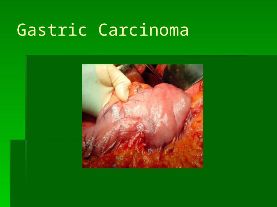
Gastric Carcinoma
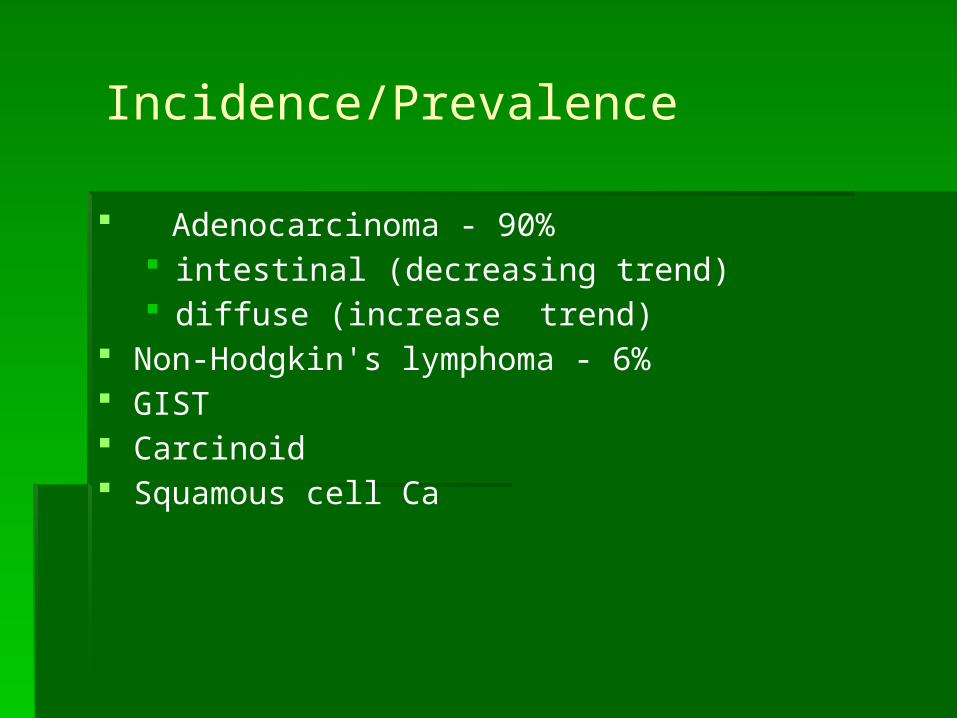
Incidence/Prevalence
Adenocarcinoma - 90% intestinal (decreasing trend) diffuse (increase trend)
Non-Hodgkin's lymphoma - 6% GIST Carcinoid Squamous cell Ca

Incidence/Prevalence
3rd most common GI malignancy (after colorectal and pancreatic)
Second causes of death from cancer
lung (17.8 %), gastric (10.4 %), and liver (8.8 %)
The worldwide incidence of gastric cancer has declined rapidly over the recent few decades
Part of the decline may be due to the recognition of certain risk factors such as H. pylori and other dietary and environmental risks
Despite the decline, the absolute number of new cases per year is increasing
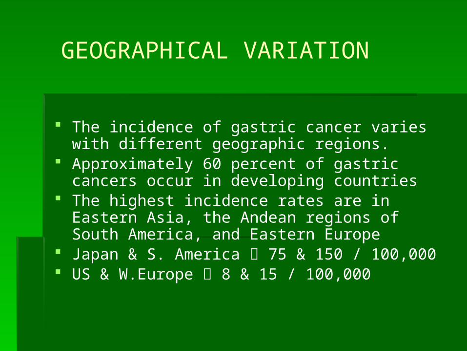
GEOGRAPHICAL VARIATION
The incidence of gastric cancer varies with different geographic regions.
Approximately 60 percent of gastric cancers occur in developing countries
The highest incidence rates are in Eastern Asia, the Andean regions of South America, and Eastern Europe
Japan & S. America 75 & 150 / 100,000 US & W.Europe 8 & 15 / 100,000

Incidence/Prevalence
Early Gastric Carcinoma
Japan 40 %
United States 6 -10 %

Incidence/Prevalence
Slowly developing
Usually discovered in advanced stages
Men>Women
Occurs between the ages of 50-70

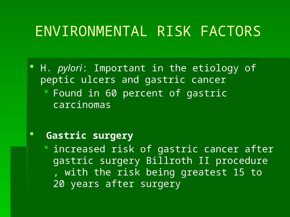
ENVIRONMENTAL RISK FACTORS
Emigrants from high-incidence to low-incidence countries often experience a decreased risk of developing gastric carcinoma.
Diet Foods such as pickled vegetables, salted fish and meat, smoked
foods and salt
Salt — High salt intake damages stomach mucosa and increases the susceptibility to carcinogenesis in rodents
Dietary nitrates (bacteria in stomach breaks down nitrites to compounds that are carcinogenic in animals)

ENVIRONMENTAL RISK FACTORS
• People who smoke cigarettes or use alcohol are 1.5 times more likely
Socioeconomic status
-The risk of distal gastric cancer is increased by approximately twofold in populations with low socioeconomic status
- By contrast, proximal gastric cancers have been associated with higher socioeconomic class
ENVIRONMENTAL RISK FACTORS
H. pylori: Important in the etiology of peptic ulcers and gastric cancer Found in 60 percent of gastric carcinomas
Gastric surgery increased risk of gastric cancer after gastric surgery
Billroth II procedure , with the risk being greatest 15 to 20 years after surgery
RISK FACTORS
Gastric polyps Gastric ulcer Genetic factors include:
First degree relatives Type A blood
Pernicious anemia

Anatomy of Stomach
Cardia
Body
Antrum
Pylorus

Stomach-normal histology
Parietal cells - in body produce HCl Chief cells - in body - pepsinogen Mucous cells - all over - mucus G cells-in antrum - gastrin

Anatomy of the stomach

Location
37% in the proximal third of the stomach 30% in the distal stomach 20% in the midsection Remaining 13% in the entire stomach
Gastric Carcinoma Lauren Classification
There are two main histologic variants of gastric adenocarcinoma.
The most frequent is the "intestinal type", so called because of its morphologic similarity to adenocarcinomas arising in the intestinal tract.
The less common diffuse type gastric cancers

Gastric Carcinoma Lauren Classification Intestinal
patients greater than 50, male>female arises from metaplastic glands in chronic gastritis;
associated with H. pylori incidence decreasing in USA
Diffuse (signet ring cell, linitis plastica) younger patients, no gender preference not associate with H. pylori incidence increasing

Intestinal type
One model for the "intestinal type" of gastric cancer describes a progression from chronic gastritis to chronic atrophic gastritis, to intestinal metaplasia, dysplasia, and eventually to adenocarcinoma

Morphologic types of Carcinoma Stomach
Fungating
Ulcerating
Diffuse

Ulcerated gastric adenocarcinoma

Thickened “linitis plastica” type adenocarcinoma infiltrating gastric wall
Physical Assessment
Early gastric cancer Abdominal discomfort initially relieved with antacids Feeling of fullness Epigastric, back, or retrosternal pain NOTE: most people will show no clinical
manifestations

Symptoms of Gastric Disorders
Heartburn Epigastric pain Dyspepsia (upset stomach) Vomiting Hematemesis
Frequently “coffee-ground” emesis Melena

Physical Assessment
Advanced stage:
Nausea/vomiting Obstructive symptoms Iron deficiency/anemia Palpable epigastric mass Enlarged lymph nodes Weakness/fatigue Progressive weight loss

DIAGNOSIS
Esophagogastroduodenoscopy
- Polypoid mass
- Ulcer crater
- Thickened fibrotic gastric wall
Preoperative evaluation
Abdominopelvic CT scan
Endoscopic ultrasonography
Chest CT For patients with a proximal gastric cancer
PET scan Sensitivity of PET scans for the detection of peritoneal
carcinomatosis is only about 50 percent.
Staging laparoscopy Between 20 and 30 % of patients who have disease that is beyond T1
stage on EUS will be found to have peritoneal metastases despite having a negative CT scan

Gastric carcinoma
Tumor Node Metastasis
T1 invades lamina propria or submucosa
N0 no mets in LN M0 no distant mets
T2 invades muscularis propria or subserosa
N1 mets in perigastric LN 1-6 LN
M1 Distant mets
T3 penetrates serosa
N2 mets in perigastric LN7 – 15 LN
T4 invades adjacent organs
N3 mts to > 15 LN

STAGE GROUPING
Stage 0 Tis N0 M0
Stage 1A T1 N0 M0
Stage IB T1 N1 M0
T2a/b N0 M0
Stage II T1 N2 M0
T2a/b N1 M0
T3 N0 M0
Stage IIIA T2a/b N2 M0
T3 N1 M0
T4 N0 M0
Stage IIIB T3 N2 M0
Stage IV T4 N1–3 M0
T1–3 N3 M0
Any T Any N M1From AJCC Cancer Staging Manual, 6th ed. New York, Springer-Verlag, 2001.

Spread of Gastric Ca
Spreads through stomach into the gastric wall to the Lymph nodes Pancreas Transverse colon
Omentum
Through portal vein into Liver
Through systemic circulation into lungs, and bone Peritoneum Ovaries Pelvic cul-de-sac Distant Lymph nodes

Spread of Gastric Ca

Physical signs – advanced or mts Palpable abdominal mass Palpable supraclavicular (Virchow’s) LN Palpable periumbilical (Sister Mary Joseph’s) LN Peritoneal mets palpable by rectal exam (Blumer’s
shelf) Palpable ovarian mass (Krukenberg’s tumor) Hepatomegaly
Clinical Presentation
Surgical Treatment
Absence of distant mts
Patient with distant mts but with complicated tumor
Line of resection at least 6 cm from the tumor mass to decrease recurrence at anastomosis

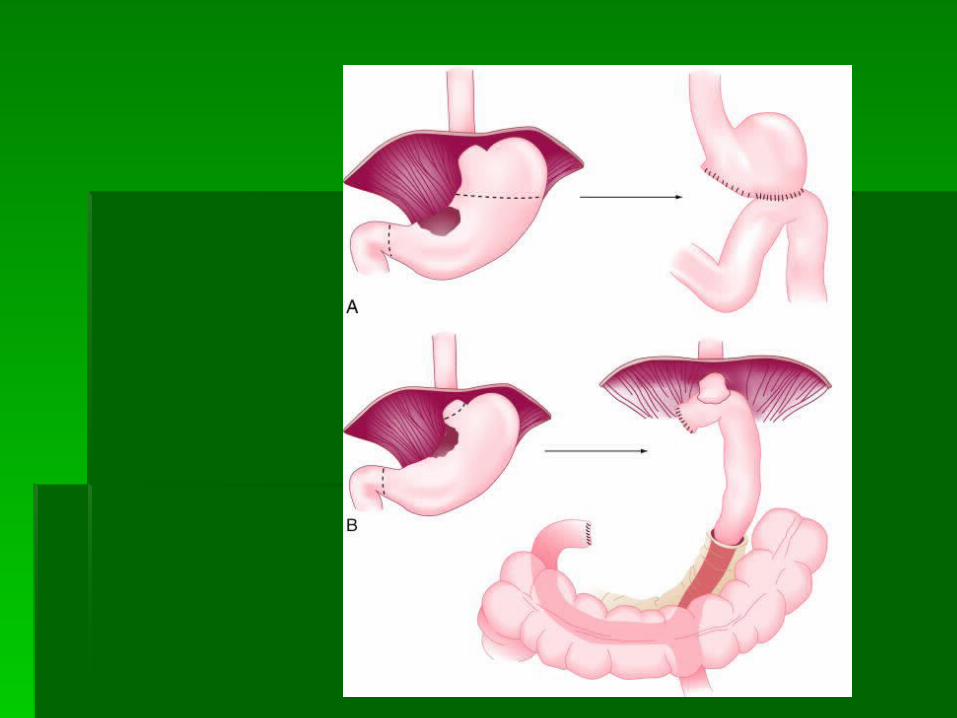
Carcinoma of Stomach
Surgical options
Total gastrectomyProximal tumoursMid-body tumours
Subtotal gastrectomy Distal tumours
Omentectomy

Distal Tumors
Account for ~ 35 % of all gastric cancers
No 5-year survival difference b/n subtotal vs total gastrectomy
Subtotal appropriate if negative margins Recurrence vs nonrecurrence depends on margin of
3.5 cm vs 6.5 cm margins 4 – 6 cm 10% involvement margins 2 cm 30 %

Proximal Tumors
Cardia / proximal ~ 35-50% of gastric adenocarcinomas
Proximal More advanced at presentation Curative resection is rare Total gastrectomy
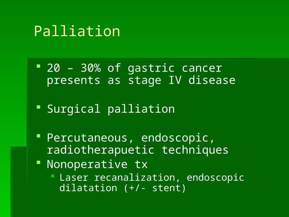
Palliation
20 – 30% of gastric cancer presents as stage IV disease
Surgical palliation
Percutaneous, endoscopic, radiotherapuetic techniques
Nonoperative tx Laser recanalization, endoscopic dilatation (+/-
stent)

Carcinoma of Stomach
Surgical treatment Overall 5 year survival rate 10 – 21% in western
series Japanese series 50%
Adjuvant therapy (postoperative) Neoadjuvant therapy (preoperative)
Response rates vary from 21 –31% clinical response rate to complete response rate of 0-15%
Adjuvant Therapy
Southwest Cancer Oncology Group trial
5-FU, Leucovorin w/ chemorad for R0
3 yr survival 41% Chem/Rad 3 yr survival 50%
28% benefit in survival
Gastric Carcinoma - Natural History
2/3 of patients have locally advanced or metastatic disease at diagnosis
50% recurrence following curative surgery
Adjuvant Chemo + R/T improves survival

Recurrence
After gastrectomy quite high 40 – 80 % Most occur w/in first 3 years Locoregional failure 38 – 45%
Anastomosis, gastric bed and regional nodes
Peritoneal dissemination – 54%
Annual endoscopy for subtotal gastrectomy
Gastric Carcinoma
Prognosis invasion is most important factor
early: limited to mucosa and submucosa; 90-95% survival at 5 years
late: beyond submucosa; less than 10 - 30% survival at 5 years
Five-year survival 95 % for patients with superficial T1 tumors and
negative lymph nodes (stage IA disease) 7 - 8 % for patients with N3 nodes or any distant
metastases LN Dissection


