
GASTROINTESTINAL PHYSIOLOGYEbaa M Alzayadneh, DDS, PhD
Tortora
1
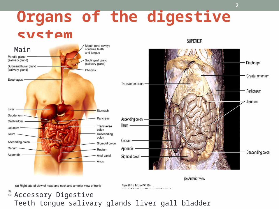
Organs of the digestive system
2
Main
Accessory DigestiveTeeth tongue salivary glands liver gall bladder pancreas
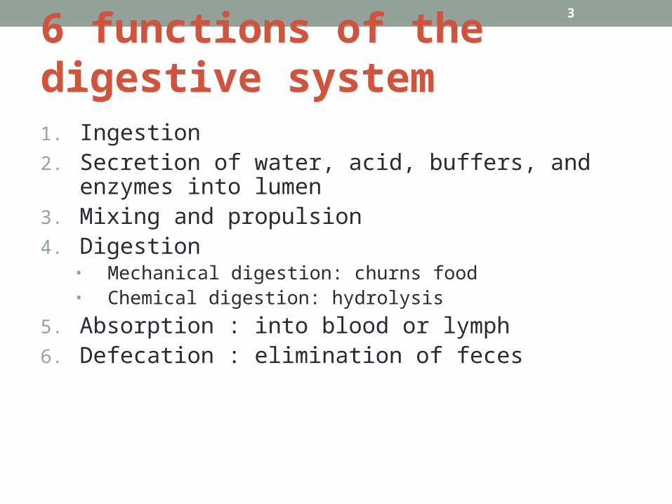
6 functions of the digestive system1. Ingestion2. Secretion of water, acid, buffers, and enzymes into
lumen3. Mixing and propulsion4. Digestion
• Mechanical digestion: churns food• Chemical digestion: hydrolysis
5. Absorption : into blood or lymph6. Defecation : elimination of feces
3

Layers of GI tract
Copyright 2009, John Wiley & Sons, Inc. 4

Layers of GI tractfrom lower esophagus to anal canal• MucosaInner lining of mucous membrane; epithelium and
enteroendocrine cells, lamina propria of connective tissue, thin layer of smooth muscle cells (muscularis mucosa).
• SubmucosaConnetive tissue binds mucosa to muscularis, blood vessels,
lymphatic and neurons.• MuscularisSkeletal in mouth, esophegus and external anal sphincterRest of GI; Smooth muscle cells: inner circular and outer
longtudinal. Involuntary contractions mix and propel. In between the two muscularis is the myenteric plexus.
• SerosaVisceral peritonium except esophagus
Copyright 2009, John Wiley & Sons, Inc. 5

Organization of the enteric nervous system
6
Autonomic NervousSystemParasympathetic; Vagus and sacral spinal (increase activity)Sympathetic;Thoracic and lumbar spinal (decrease activity)
• Enteric nervous system “Brain of the gut”
1. Myenteric(plexus of Auerobach)2. Submucosal (plexus of Meissner3. sensory neurons, motor neurons
and interneurons4. Myenteric: GI motility 5. motor neurons of submucosal
plexus control secretions from mucosal epithelial glands.
6. sensory neurons of submucosal epithelial. Some are chemoreceptors or stretch receptors
7. Interneurons interconnect the 2 plexuses
8. ENS can be independent but still subject to regulation of ANS
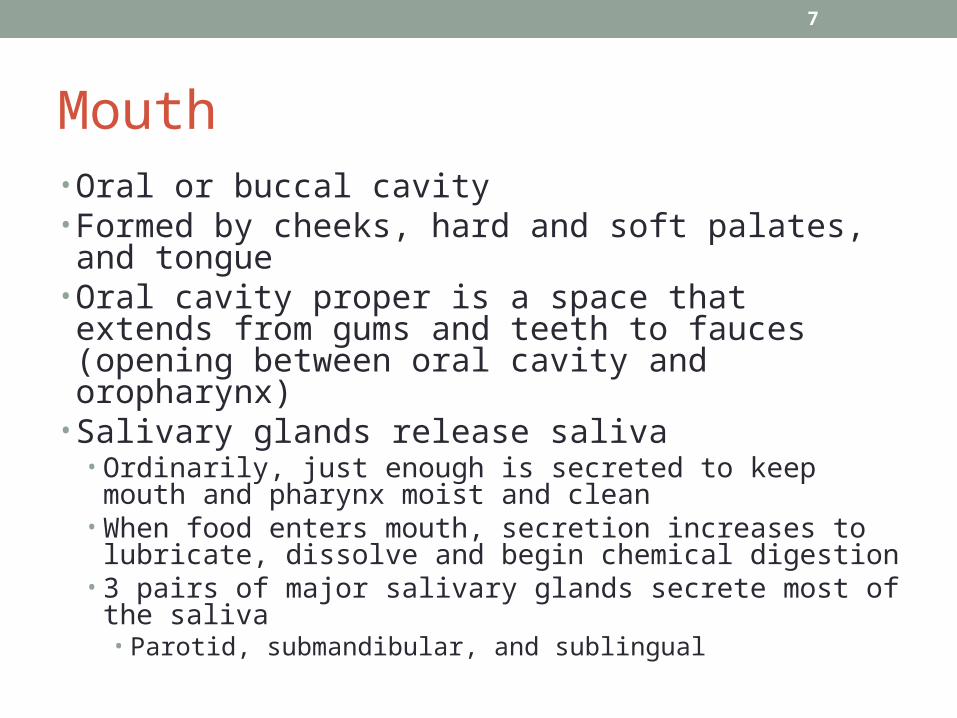
Mouth• Oral or buccal cavity• Formed by cheeks, hard and soft palates, and tongue• Oral cavity proper is a space that extends from gums and teeth to fauces (opening between oral cavity and oropharynx)
• Salivary glands release saliva• Ordinarily, just enough is secreted to keep mouth and
pharynx moist and clean• When food enters mouth, secretion increases to lubricate,
dissolve and begin chemical digestion• 3 pairs of major salivary glands secrete most of the saliva
• Parotid, submandibular, and sublingual
7
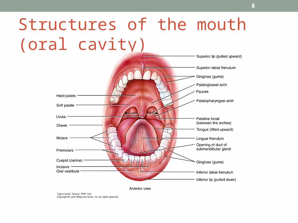
Structures of the mouth (oral cavity)
8
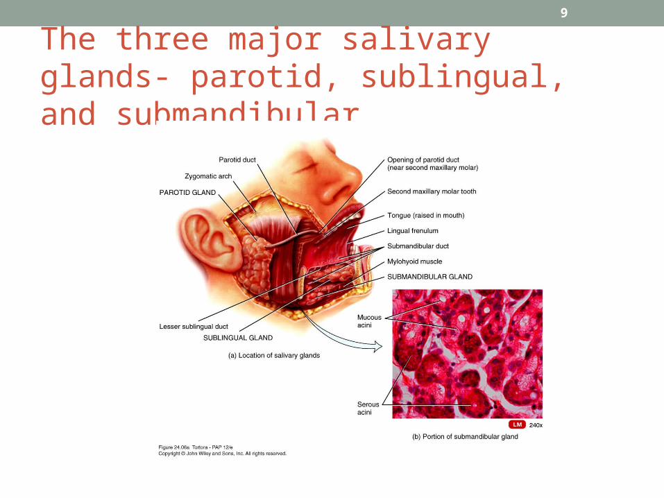
The three major salivary glands- parotid, sublingual, and submandibular
9
Saliva• Saliva
• Mostly water 99.5%• 0.5% solutes – ions, dissolved gases, urea, uric acid,
mucus, immunoglobulin A, lysozyme, and salivary amylase (acts on starch)
• Not all salivary glands produce the same saliva• Salivation
• Controlled by autonomic nervous system• Parasympathetic stimulation promotes secretion of
moderate amount of saliva• Sympathetic stimulation decreases salivation
10
Digestion in the mouth• Mechanical digestion in the mouth
• Chewing or mastication• Food manipulated by tongue, ground by teeth, and mixed
with saliva• Forms bolus
• Chemical digestion in the mouth• Salivary amylase secreted by salivary glands acts on
starches• Only monosaccharides can be absorbed• Continues to act until inactivated by stomach acid
• Lingual lipase secreted by lingual glands of tongue acts on triglycerides• Becomes activated in acidic environment of stomach
11
Pharynx• Passes from mouth into pharynx• 3 parts
• Nasopharynx• Functions only in respiration
• Oropharynx• Digestive and respiratory functions
• Laryngopharynx• Digestive and respiratory functions
12
Esophagus• Secretes mucous, transports food – no enzymes
produced, no absorption• Mucosa – protection against wear and tear• Submucosa• Muscularis divided in thirds
• Superior 1/3 skeletal muscle• Middle 1/3 skeletal and smooth muscle• Inferior 1/3 smooth muscle• 2 sphincters – upper esophageal sphincter (UES) regulates
movement into esophagus, lower esophageal sphincter (LES) regulates movement into stomach
• Adventitia – no serosa – attaches to surroundings
13
Deglutition• Act of swallowing• Facilitated by secretions of saliva and mucus• Involves mouth, pharynx, and esophagus• 3 stages
• Voluntary – bolus passed to oropharynx• Pharyngeal – involuntary passage through pharynx into
esophagus(the bolus stimulates receptors in the oropharynx to deglut. Center in medulla oblongata and lower pons-soft palate, uvula and epiglottis close off. And upper esophageal sphincter relaxes.
• Esophageal – involuntary passage through esophagus to stomach• Peristalsis pushes bolus forward
14
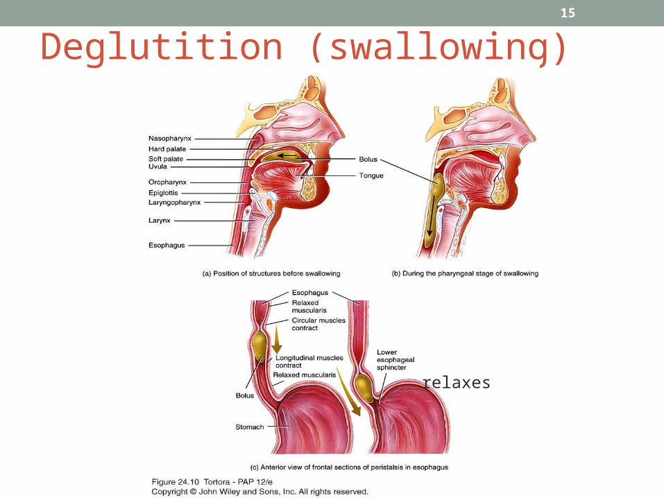
Deglutition (swallowing)15
relaxes

External and internal anatomy of the stomach
16
FUNCTIONS1.Mix to form chyme2.Reservoir :most distensible part of the GI3. gastric juice: HCl, pepsin, Intrensic factor, gastric lipase.4. Gastrin

Histology of the stomach17

Mechanical and Chemical Digestion• Mechanical digestion
• Mixing waves – gentle, rippling peristaltic movements – creates chyme
• Chemical digestion• Digestion by salivary amylase continues until inactivated by
acidic gastric juice• Acidic gastric juice activates lingual lipase
• Digest triglycerides into fatty acids and diglycerides• Parietal cells secrete H+ and Cl- separately but net effect is
HCl• Kills many microbes, denatures proteins and stimulates bile and
pancreatic juice• Parietal cells secrete intrinsic factor necessary for vitamin
B12 absorption.
18

Chemical Digestion• Chemical digestion
• Pepsin secreted by chief cells digest proteins• Secreted as pepsinogen ( become active at very acidic)
Why it doesn’t digest the stomach or the chief cells?
PH and mucus!
• Gastric lipase splits triglycerides into fatty acids and monoglycerides
• Small amount of nutrient absorption• Some water, ions, short chain fatty acids, certain drugs (aspirin)
and alcohol
• Gastrin hormone by G cells: stimulates both parietal and chief cells (HCl and Pepsinogen respectively) , contracts LES and relaxes pyloric s. .
19

Pancreas• Lies posterior to greater curvature of stomach• Pancreatic juice secreted into pancreatic duct and accessory duct and to small intestine (duedenum)• Pancreatic duct joins common bile duct and enters
duodenum at hepatopancreatic ampulla• Histology
• 99% of cells are acini• Exocrine• Secrete pancreatic juice – mixture of fluid and digestive enzymes
• 1% of cells are pancreatic islets (islets of Langerhans)• Endocrine• Secrete hormones glucagon, insulin, somatostatin, and
pancreatic polypeptide
20
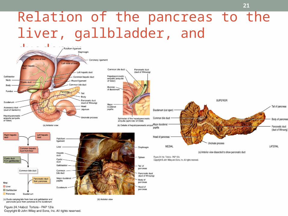
Relation of the pancreas to the liver, gallbladder, and duodenum
21
Pancreatic juice• 1200-1500ml daily• Mostly water, ALKALINE
• Sodium bicarbonate – buffers acidic stomach chyme• Enzymes
• Pancreatic amylase ( FOR starch)• Proteolytic enzymes – trypsin (secreted as trypsinogen),
chymotrypsin (chymotrypsinogen), carboxypeptidase (procarboxypeptidase), elastase (proelastase)
• Pancreatic lipase: The principal lipase in adults• Ribonuclease and deoxyribonuclease (for nucleic acids)• Trypsinogen activated by brush border enzyme
enterokinase. Trypsin activates Chymotrypsinogen, procarboxypeptidase and proelastase.
• Trypsin inhibitor
22

Histology of the Liver
23
Gallbladder
• Contraction of smooth muscle fibers eject contents of gall bladder into cystic duct
• Functions to store and concentrate bile produced by the liver until it is needed in the small intestine
• Jaundice: yellowish pigmentation (bilirubin)
24
Role and composition of bile
• Hepatocytes secrete 800-1000mL of bile daily• Mostly water, bile salts, cholesterol, lecithin, bile
pigments and several ions• Partially excretory product/ partially digestive secretion• Bilirubin – principal bile pigment
• Derived from the heme of recycled RBCs• Breakdown product stercobilin gives feces brown
color• Bile salts play a key role in emulsification
• absorption of lipids
25
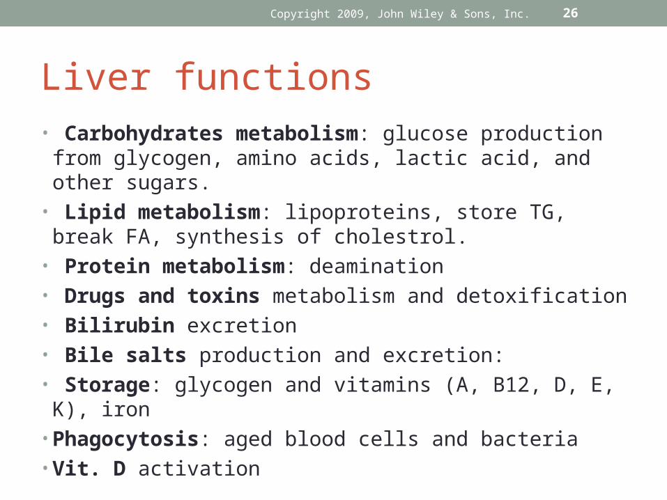
Liver functions• Carbohydrates metabolism: glucose production from
glycogen, amino acids, lactic acid, and other sugars.• Lipid metabolism: lipoproteins, store TG, break FA,
synthesis of cholestrol.• Protein metabolism: deamination• Drugs and toxins metabolism and detoxification• Bilirubin excretion• Bile salts production and excretion:• Storage: glycogen and vitamins (A, B12, D, E, K), iron• Phagocytosis: aged blood cells and bacteria• Vit. D activation
Copyright 2009, John Wiley & Sons, Inc. 26
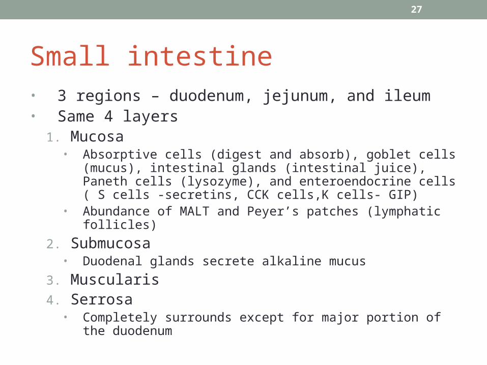
Small intestine• 3 regions – duodenum, jejunum, and ileum• Same 4 layers
1. Mucosa• Absorptive cells (digest and absorb), goblet cells (mucus),
intestinal glands (intestinal juice), Paneth cells (lysozyme), and enteroendocrine cells ( S cells -secretins, CCK cells,K cells- GIP)
• Abundance of MALT and Peyer’s patches (lymphatic follicles)
2. Submucosa• Duodenal glands secrete alkaline mucus
3. Muscularis4. Serrosa
• Completely surrounds except for major portion of the duodenum
27
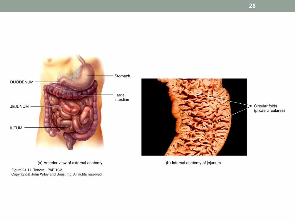
ANATOMY OF THE SMALL INTESTINE
28

Special structural features increase surface area for digestion and absorption
• Circular folds
Permanent ridges of mucosa and submucosa
Cause chyme to spiral• Villi
Fingerlike projections of mucosa
Contains arteriole, venule, blood capillary, and lacteal• Microvilli
Projects of apical plasma membrane of absorptive cells
Brush border has brush border enzymes
29
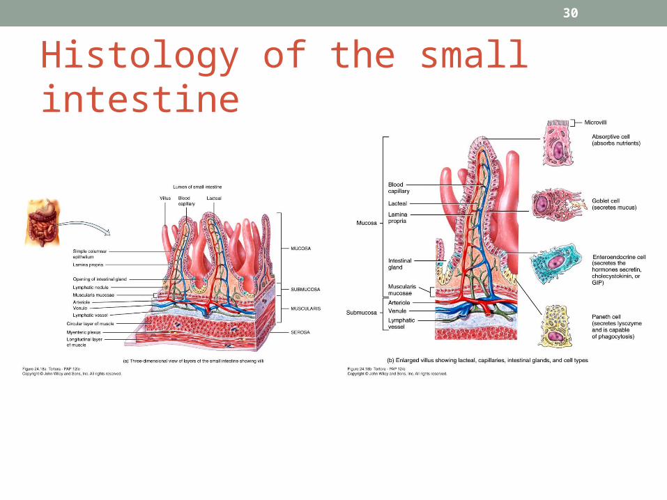
Histology of the small intestine
30
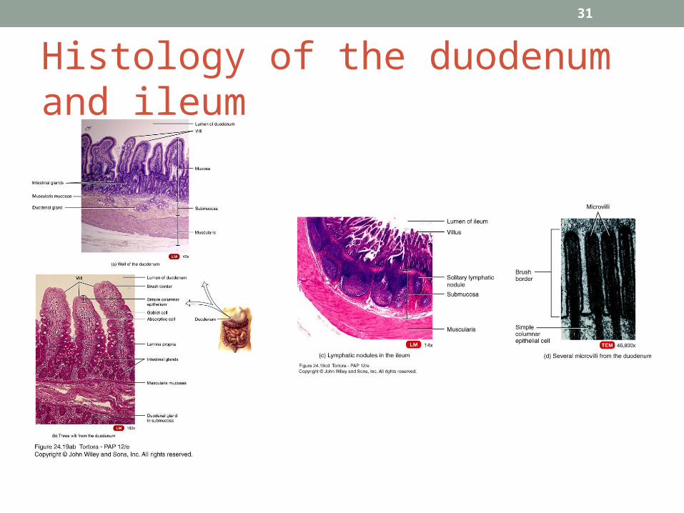
Histology of the duodenum and ileum
31
Intestinal juice and brush-border enzymes• Intestinal juice
• 1-2L daily• Contains water and mucus, slightly alkaline• Provide liquid medium aiding absorption
• Brush border enzymes• Inserted into plasma membrane of absorptive cells• Some enzymatic digestion occurs at surface rather than just
in lumen• α-dextrinase, maltase, sucrase, lactase, aminopetidase,
dipeptidase, nucleosidases and phosphatases
32

Mechanical Digestion• Governed by myenteric plexus• Segmentations
• Localized, mixing contractions to bring chyme to mucosa
• Does not move chyme along the Intestine.• Migrating motility complexes (MMC)
• A type of peristalsis• Pushes food forward to slowly migrate chyme down
the small intestine
33
Chemical digestion
• Carbohydrates• Pancreatic amylase• α-dextrinase ( to glucose), sucrase (to glucose and
fructose), lactase (to glucose and galactose), maltase (to glucose) in brush border
• Ends up with monosaccharides which can be absorbed
• Proteins• Trypsin, chymotrypsin, carboxypeptidase, and
elastase from pancreas (from proteins to peptides)• Aminopeptidase and dipeptidase in brush border (to
amino acids)
34
Lipids and Nucleic Acids• Lipids
• Pancreatic lipase most important in triglyceride digestion (to fatty acids and monoglycerides)
• Emulsification by bile salts increases surface area• Amphipathic – (hydrophobic and hydrophilic )
• Nucleic acids• Ribonuclease and deoxyribonuclease in pancreatic
juice• Nucleosidases and phosphatases in brush border ( to
pentose, phosphates and nitrogenous base)
35
Absorption:
• Monosaccharides• All dietary carbohydrates digested are absorbed• Only indigestible cellulose and fibers left in feces• Absorbed by facilitated diffusion or active transport
into blood• Amino acids, dipetides and tripeptides
• Most absorbed as amino acids via active transport into blood
36
Lipids• All dietary lipids absorbed by simple diffusion• Short-chain fatty acids go into blood for transport • Long-chain fatty acids and monoglycerides via:
• Large and hydrophobic• Bile salts form micelles to ferry them to absorptive
cell surface• Reform into triglycerides and coated by proteins
forming chylomicrons• Leave cell by exocytosis• Enter lacteals to eventually enter blood with protein
coat of chylomicron keeping them suspended and separate
37

ABSORPTION 38
Absorption
• Electrolytes• From GI secretions or food• Sodium ions (Na+) reclaimed by active transport• Other ions also absorbed by active transport
• Vitamins• Fat-soluble vitamins A, D, E, and K absorbed by simple diffusion
and transported with lipids in micelles• Most water-soluble vitamins also absorbed by simple diffusion
• Water • 9.3L comes from ingestion (2.3L) and GI secretions (7.0L)• Most absorbed in small intestine, some in large intestine• Only 100ml excreted in feces• All water absorption by osmosis
39
Large intestine• Overall function to complete absorption, produce certain vitamins, and form and expel feces
• 4 major regions – cecum, colon, rectum, and anal canal
• Ileocecal sphincter between small and large intestine• Colon divided into ascending, transverse, descending and sigmoid
• Opening of anal canal (anus) guarded by internal anal sphincter of smooth muscle and external anal sphincter of skeletal muscle
40
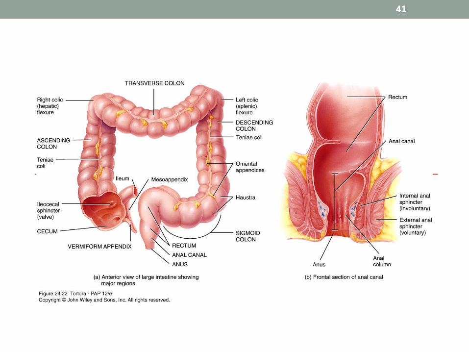
ANATOMY OF THE LARGE INTESTINE
41

Digestion of the Large Intestine• Mechanical digestion
• Haustral churning• Peristalsis• Mass peristalsis – drives contents of colon toward rectum
• Chemical digestion• Final stage of digestion through bacterial action
• Ferment carbohydrates, produce some B vitamins and vitamin K
• Mucus but no enzymes secreted
• Remaining water absorbed along with ions and some vitamins
42
Phases of digestion• Cephalic phase
• Smell, sight, thought or initial taste of food activates neural centers – prepares mouth and stomach for food to be eaten
• Gastric phase• Neural and hormonal mechanisms promote gastric secretion ( by
gastrin) and motility
• Intestinal phase• Begins when food enter small intestine• Inhibitory that slows exit of chyme from stomach• Stimulates flow of bile and pancreatic juice
43
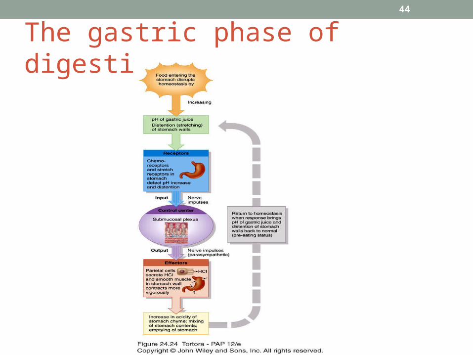
The gastric phase of digestion
44


