
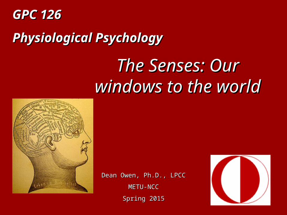
GPC 126GPC 126
Physiological PsychologyPhysiological Psychology
Dean Owen, Ph.D., LPCCDean Owen, Ph.D., LPCC
METU-NCCMETU-NCC
Spring 2015Spring 2015
The Senses: Our The Senses: Our windows to the windows to the
worldworld

Lecture 7This presentation has been created to assist in the mastery of the material contained in Chapter 6-7of the text
Foundations of Physiological PsychologyFoundations of Physiological Psychologyby
Neil R. Carlson
All of the material contained in the presentation is drawn from the text.
Plan for the day
3. Review the anatomy and physiology of the sensory organs
2. Review the fundamentals of sensation and sensory activation
4. Review major categories of sensory disturbance and disease
1. Selection of presentation dates

Presentation DateTeam 28 April 5 May 12 May
Bobcats X
Canaries X
Cheetahs X
Eagles X
Evil X
Indians X
Panthers X
Reindeer X
Sphinx X
Squirrels X
Tulips X
Venus X

But first…….But first…….
http://www.birdcheck.co.uk/whackthepenguin.htm
Where are you at this moment??
How do you know that??
What information are you using to make that decision???
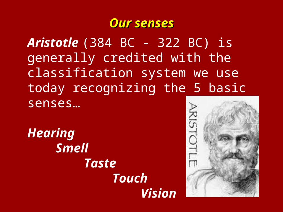
Our sensesOur senses
Aristotle (384 BC - 322 BC) is generally credited with the classification system we use today recognizing the 5 basic senses…
HearingSmell
TasteTouch
Vision
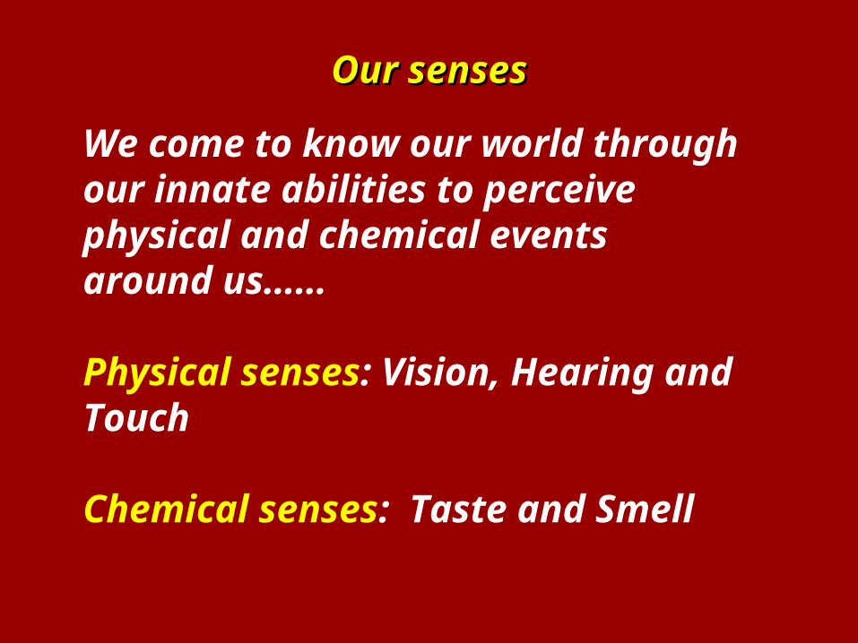
Our sensesOur senses
We come to know our world through our innate abilities to perceive physical and chemical events around us……
Physical senses: Vision, Hearing and Touch
Chemical senses: Taste and Smell
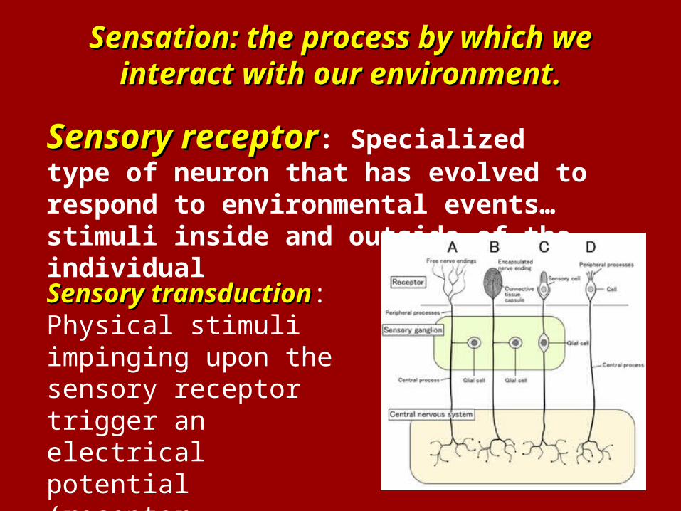
Sensation: the process by which we Sensation: the process by which we interact with our environment.interact with our environment.
Sensory receptorSensory receptor: Specialized type of neuron that has evolved to respond to environmental events…stimuli inside and outside of the individual
Sensory transductionSensory transduction: Physical stimuli impinging upon the sensory receptor trigger an electrical potential (receptor potential)
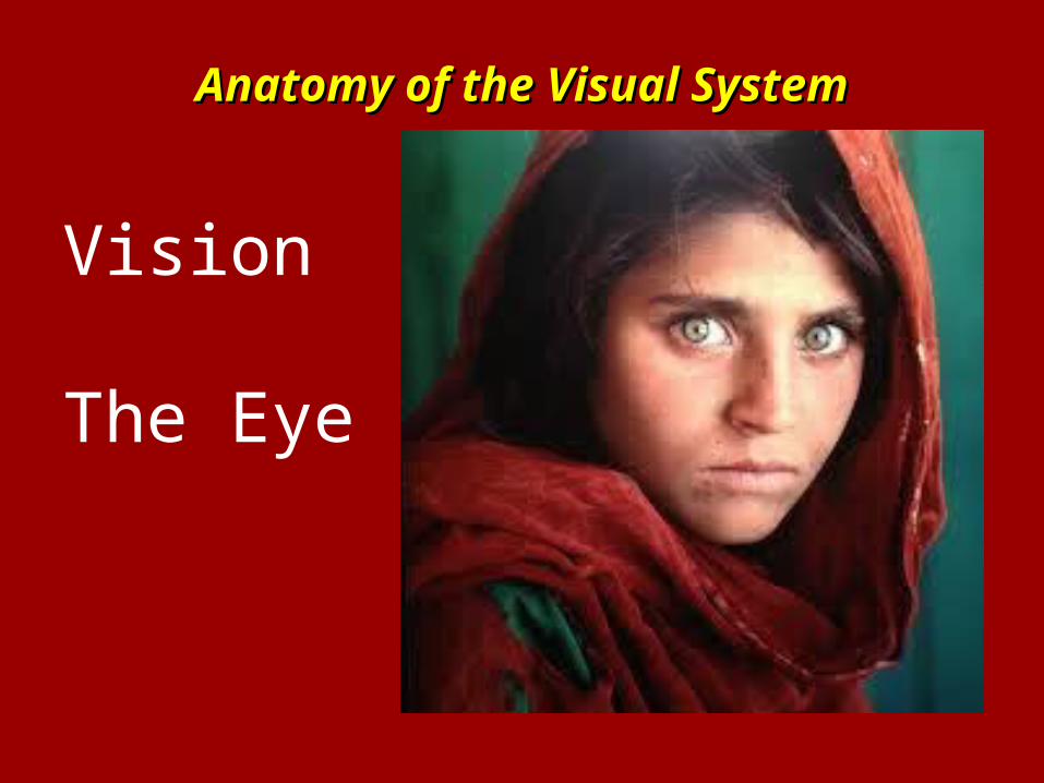
Anatomy of the Visual SystemAnatomy of the Visual System
Vision
The Eye
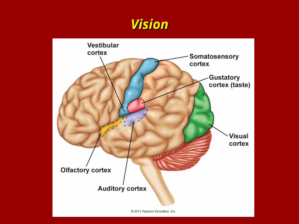
VisionVision
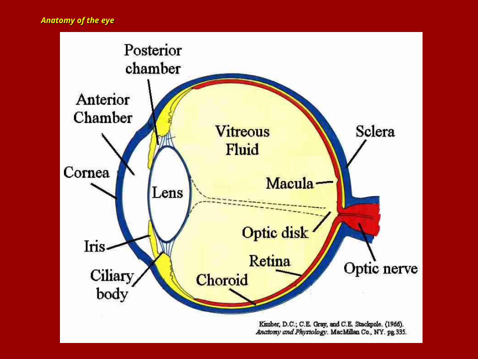
Anatomy of the eyeAnatomy of the eye
Anatomy of the eyeAnatomy of the eye
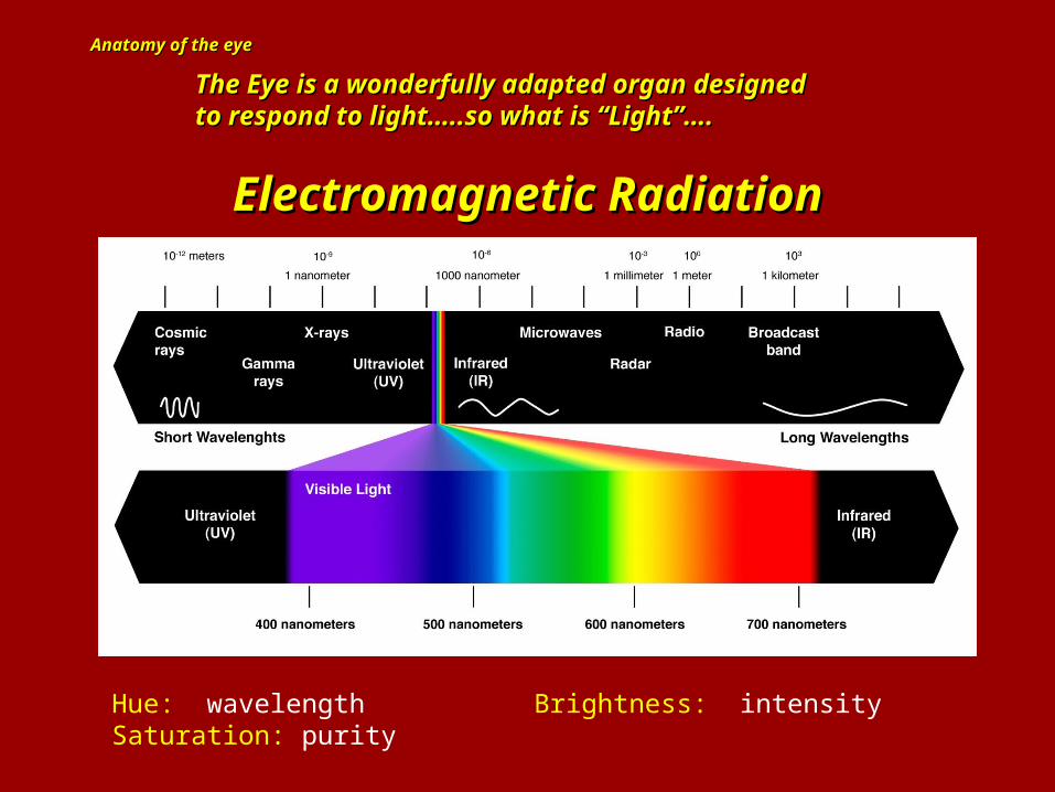
Hue: wavelength Brightness: intensity Saturation: purity
The Eye is a wonderfully adapted organ designed The Eye is a wonderfully adapted organ designed to respond to light…..so what is “Light”….to respond to light…..so what is “Light”….
Electromagnetic RadiationElectromagnetic Radiation
Anatomy of the eyeAnatomy of the eye
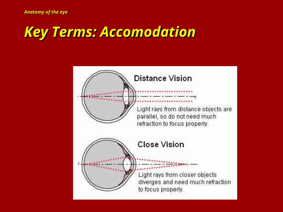
Key Terms: AccomodationKey Terms: Accomodation
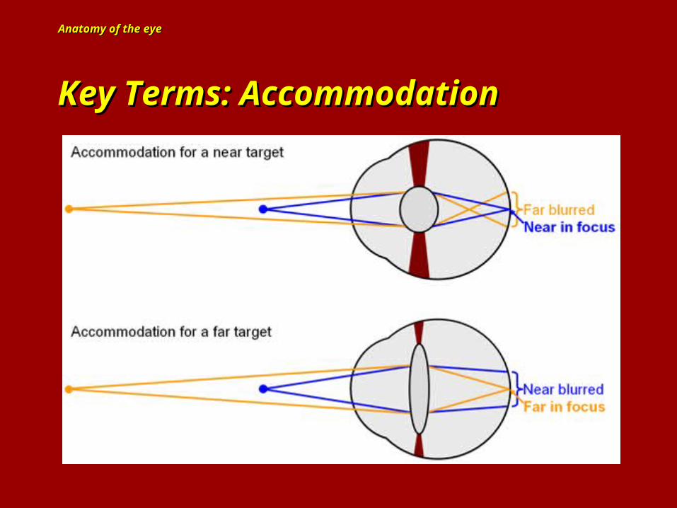
Anatomy of the eyeAnatomy of the eye
Key Terms: AccommodationKey Terms: Accommodation
Anatomy of the eyeAnatomy of the eye
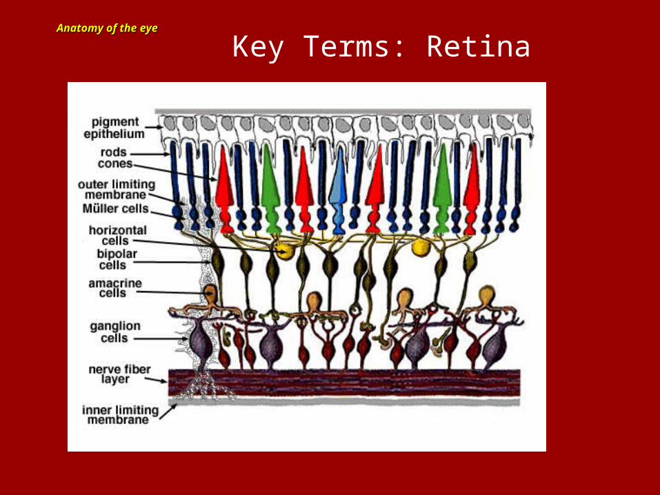
Key Terms: Retina
Anatomy of the eyeAnatomy of the eye
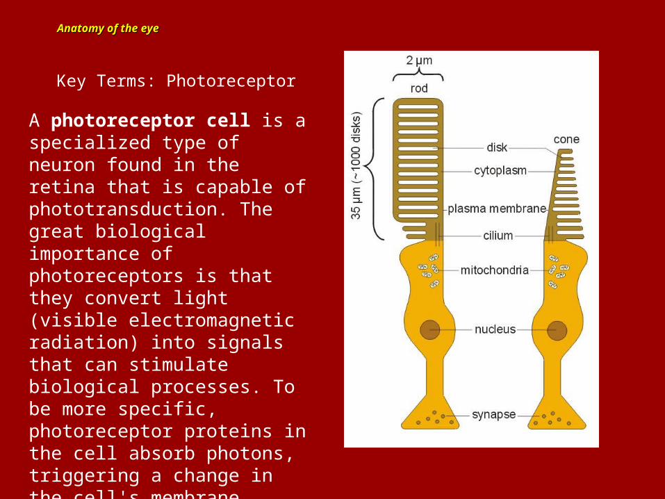
Key Terms: Photoreceptor
A photoreceptor cell is a specialized type of neuron found in the retina that is capable of phototransduction. The great biological importance of photoreceptors is that they convert light (visible electromagnetic radiation) into signals that can stimulate biological processes. To be more specific, photoreceptor proteins in the cell absorb photons, triggering a change in the cell's membrane potential.
Anatomy of the eyeAnatomy of the eye
Key Terms: Photoreceptor
The two classic photoreceptor cells are rods and
cones, each of which makes a contribution to sight. The rods are more sensitive to photon stimulation and are distributed more evenly across the retinal field. The cones are sensitive to a broad spectrum of light frequencies and are concentrated in high numbers in the fovea.
A third class of photoreceptor cells was discovered during the 1990s:] the photosensitive ganglion cells. These cells do not contribute to sight directly, but are thought to support circadian rhythms and pupillary reflex.
Anatomy of the eyeAnatomy of the eyeKey Terms: Rods and Cones
Rods are extremely sensitive, and can be triggered by as few as 6 photons. At very low light levels, visual experience is based solely on the rod signal. This explains why colors cannot be seen at low light levels: only one type of photoreceptor cell is active.
Dark adaptation: The process of enhanced visual sensitivity to light in low light environments….may take up to 30 minutes to achieve maximum light sensitivity…….
Anatomy of the eyeAnatomy of the eye
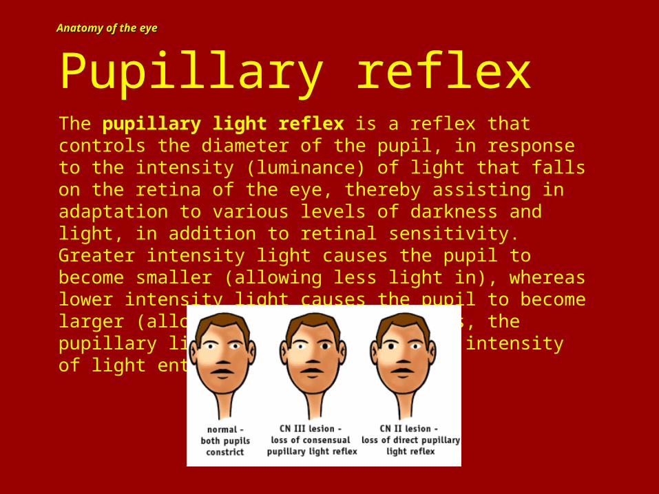
Pupillary reflexThe pupillary light reflex is a reflex that controls the diameter of the pupil, in response to the intensity (luminance) of light that falls on the retina of the eye, thereby assisting in adaptation to various levels of darkness and light, in addition to retinal sensitivity. Greater intensity light causes the pupil to become smaller (allowing less light in), whereas lower intensity light causes the pupil to become larger (allowing more light in). Thus, the pupillary light reflex regulates the intensity of light entering the eye.
Anatomy of the eyeAnatomy of the eyeKey Terms: Rods and Cones
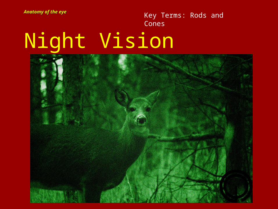
Night Vision
Anatomy of the eyeAnatomy of the eye Key Terms: Rods and Cones
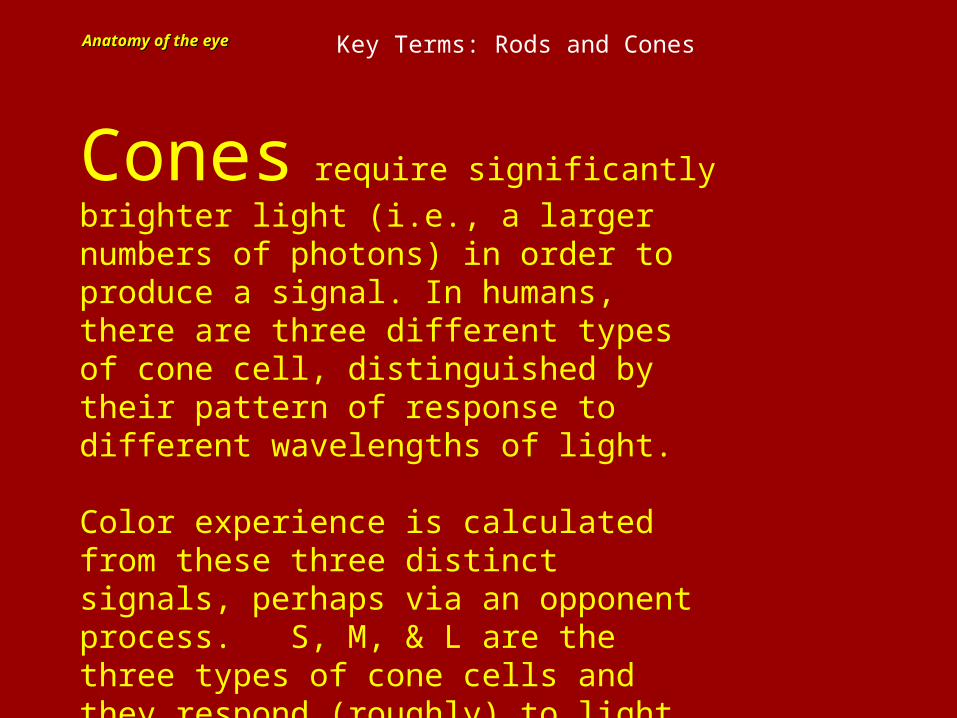
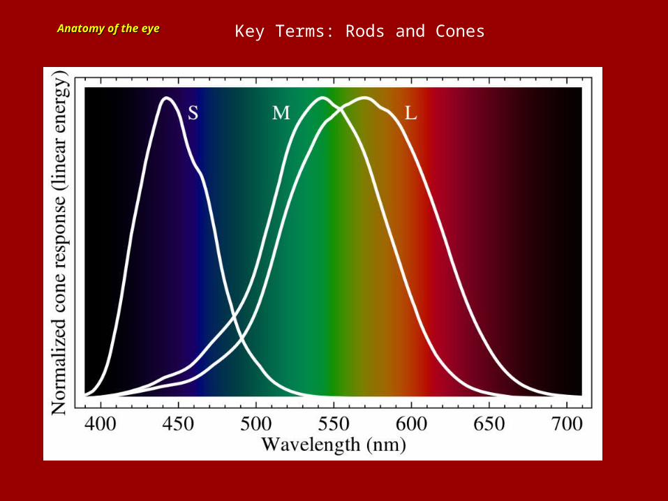
Cones require significantly brighter
light (i.e., a larger numbers of photons) in order to produce a signal. In humans, there are three different types of cone cell, distinguished by their pattern of response to different wavelengths of light.
Color experience is calculated from these three distinct signals, perhaps via an opponent process. S, M, & L are the three types of cone cells and they respond (roughly) to light of short, medium, and long wavelengths.
Anatomy of the eyeAnatomy of the eye Key Terms: Rods and Cones
Anatomy of the eyeAnatomy of the eye Key Terms: Rods and Cones
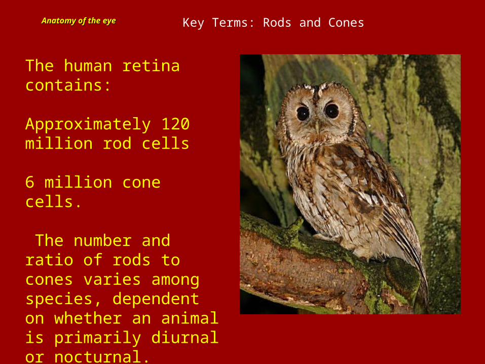
The human retina contains:
Approximately 120 million rod cells
6 million cone cells.
The number and ratio of rods to cones varies among species, dependent on whether an animal is primarily diurnal or nocturnal.
Anatomy of the eyeAnatomy of the eye
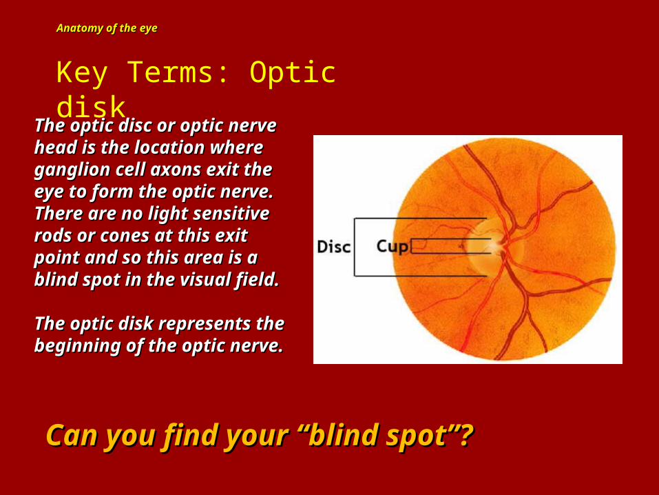
Key Terms: Optic disk
The optic disc or optic nerve The optic disc or optic nerve head is the location where head is the location where ganglion cell axons exit the ganglion cell axons exit the eye to form the optic nerve. eye to form the optic nerve. There are no light sensitive There are no light sensitive rods or cones at this exit rods or cones at this exit point and so this area is a point and so this area is a blind spot in the visual field.blind spot in the visual field.
The optic disk represents the The optic disk represents the beginning of the optic nerve.beginning of the optic nerve.
Can you find your “blind spot”?Can you find your “blind spot”?
Anatomy of the eyeAnatomy of the eye
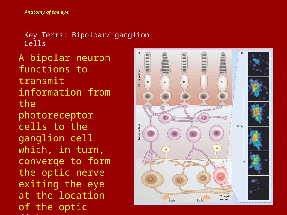
Key Terms: Bipoloar/ ganglion Cells
A bipolar neuron functions to transmit information from the photoreceptor cells to the ganglion cell which, in turn, converge to form the optic nerve exiting the eye at the location of the optic disk.

Anatomy of the eyeAnatomy of the eye
http://www.youtube.com/watch?v=ajnsDVsP0Uk
Anatomy of the eyeAnatomy of the eye
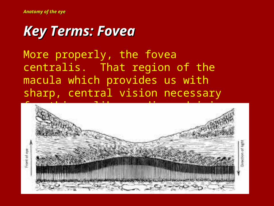
Key Terms: FoveaKey Terms: Fovea
More properly, the fovea centralis. That region of the macula which provides us with sharp, central vision necessary for things like reading, driving, sewing….etc.
Anatomy of the eyeAnatomy of the eye
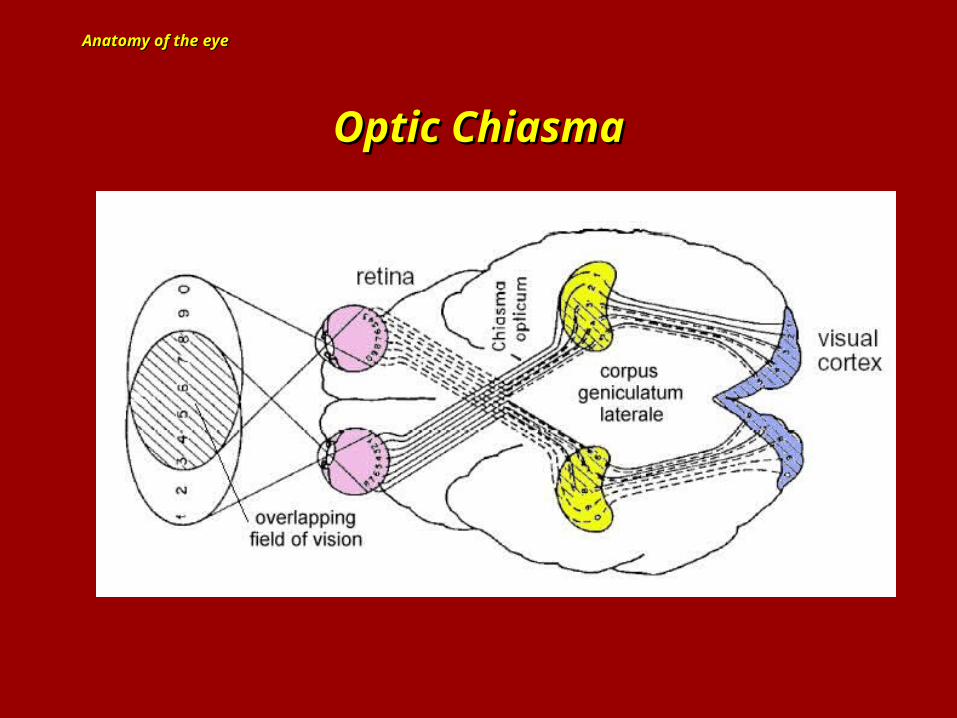
Optic ChiasmaOptic Chiasma
Anatomy of the eyeAnatomy of the eye
Blurred vision (refractive errors)
Nearsightedness (called myopia) is when you can see clearly up close but blurry in the distance.
Farsightedness (called hyperopia) is when you can see clearly in the distance but blurry up close.
Presbyopia is age related and caused by loss of elasticity of the lens…in ability to focus for older people (after age 40). One in every three people 40 years or older in the U.S. will need glasses to read smaller print.
Astigmatism is another condition that causes blurred vision, but it is because of the shape of the cornea.
Anatomy of the eyeAnatomy of the eye
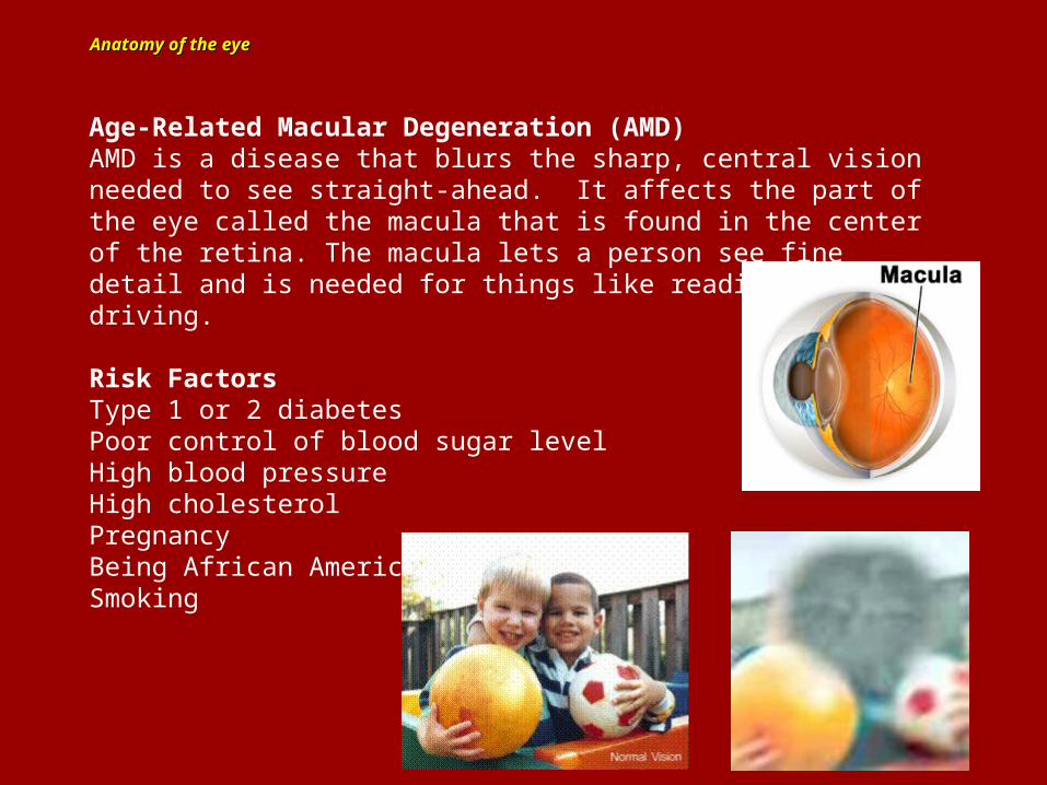
Age-Related Macular Degeneration (AMD)AMD is a disease that blurs the sharp, central vision needed to see straight-ahead. It affects the part of the eye called the macula that is found in the center of the retina. The macula lets a person see fine detail and is needed for things like reading and driving.
Risk FactorsType 1 or 2 diabetesPoor control of blood sugar levelHigh blood pressureHigh cholesterolPregnancyBeing African American or HispanicSmoking
Anatomy of the eyeAnatomy of the eye
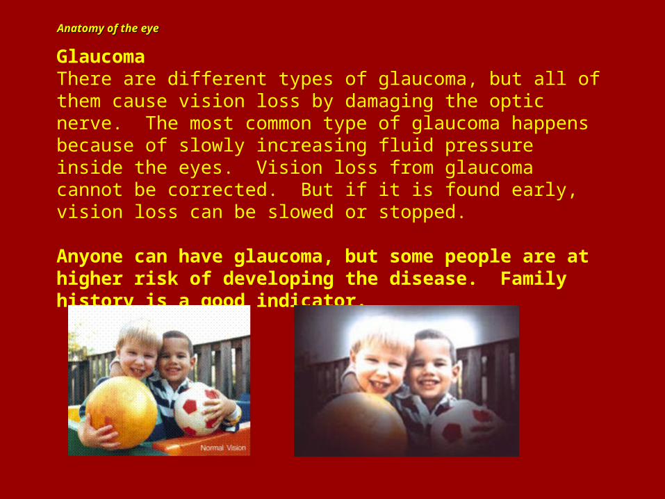
GlaucomaThere are different types of glaucoma, but all of them cause vision loss by damaging the optic nerve. The most common type of glaucoma happens because of slowly increasing fluid pressure inside the eyes. Vision loss from glaucoma cannot be corrected. But if it is found early, vision loss can be slowed or stopped.
Anyone can have glaucoma, but some people are at higher risk of developing the disease. Family history is a good indicator.
Anatomy of the eyeAnatomy of the eye
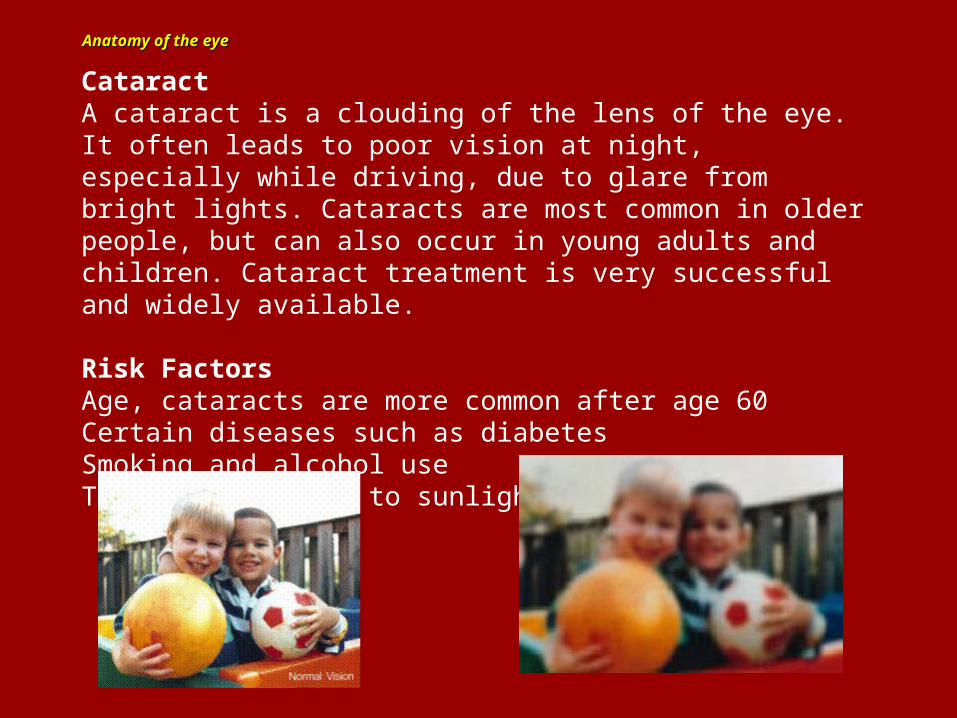
CataractA cataract is a clouding of the lens of the eye. It often leads to poor vision at night, especially while driving, due to glare from bright lights. Cataracts are most common in older people, but can also occur in young adults and children. Cataract treatment is very successful and widely available.
Risk FactorsAge, cataracts are more common after age 60Certain diseases such as diabetesSmoking and alcohol useToo much exposure to sunlight
Anatomy of the eyeAnatomy of the eye
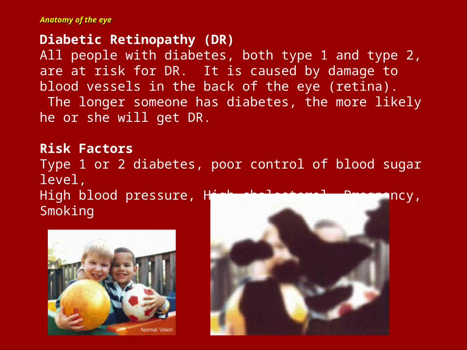
Diabetic Retinopathy (DR)All people with diabetes, both type 1 and type 2, are at risk for DR. It is caused by damage to blood vessels in the back of the eye (retina). The longer someone has diabetes, the more likely he or she will get DR.
Risk FactorsType 1 or 2 diabetes, poor control of blood sugar level,High blood pressure, High cholesterol, Pregnancy, Smoking

A Little Test for You….A Little Test for You….
FINISHED FILES ARE THE RESULT OF YEARS OF SCIENTIFIC STUDY COMBINED WITHTHE EXPERIENCE OF YEARS.....
Read the following text and count the number of “F”s you find
How many did you find? 66
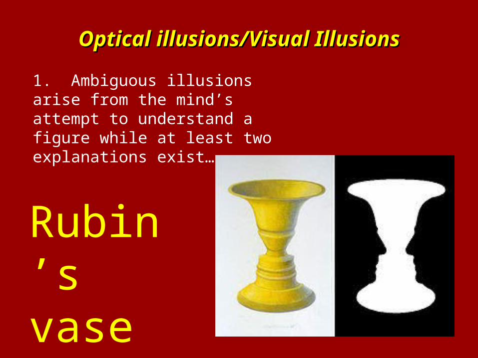
Optical illusions/Visual IllusionsOptical illusions/Visual Illusions
1. Ambiguous illusions arise from the mind’s attempt to understand a figure while at least two explanations exist…..
Rubin’s vase
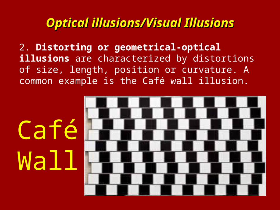
Optical illusions/Visual IllusionsOptical illusions/Visual Illusions
2. Distorting or geometrical-optical illusions are characterized by distortions of size, length, position or curvature. A common example is the Café wall illusion.
Café Wall
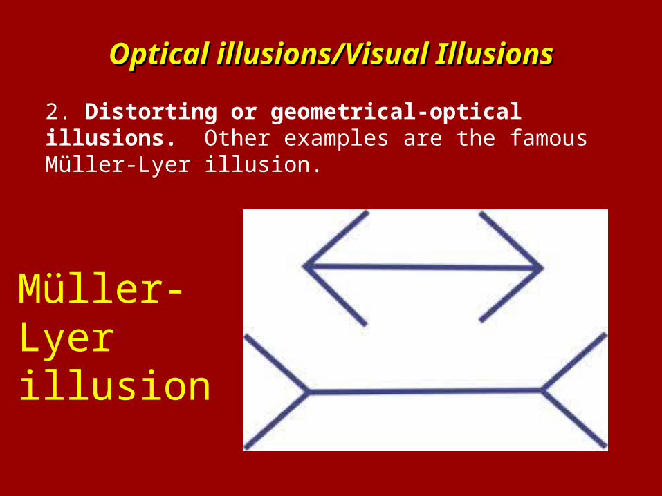
Optical illusions/Visual IllusionsOptical illusions/Visual Illusions
2. Distorting or geometrical-optical illusions. Other examples are the famous Müller-Lyer illusion.
Müller-Lyer illusion
Optical illusions/Visual IllusionsOptical illusions/Visual Illusions
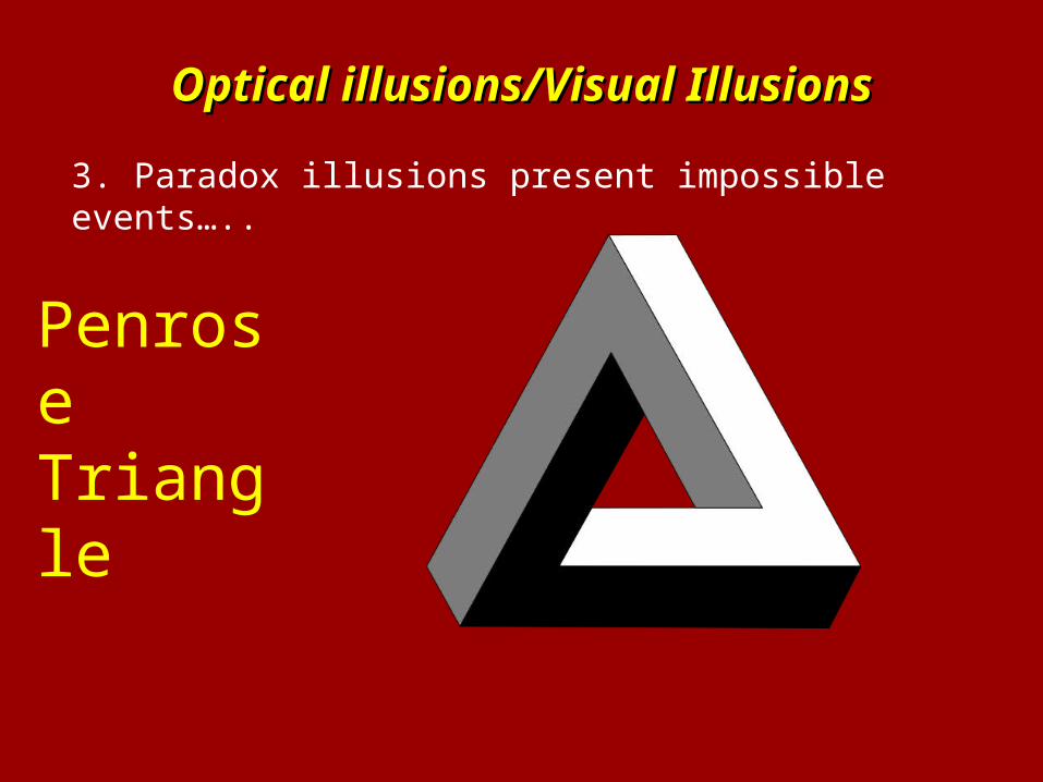
3. Paradox illusions present impossible events…..
Penrose Triangle
Optical illusions/Visual IllusionsOptical illusions/Visual Illusions
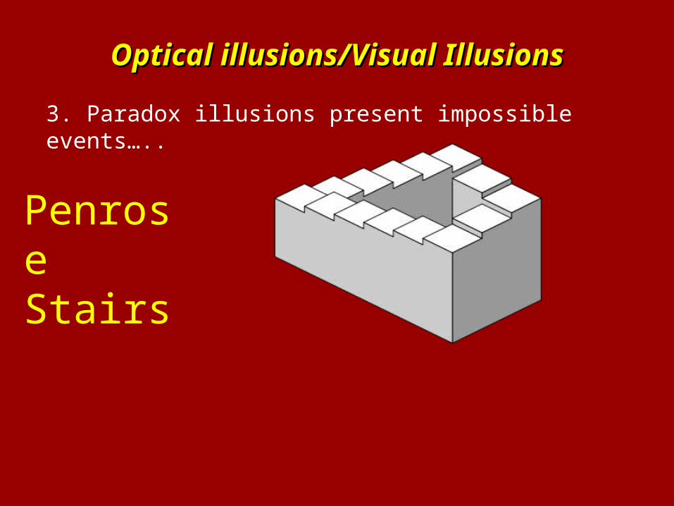
3. Paradox illusions present impossible events…..
Penrose Stairs
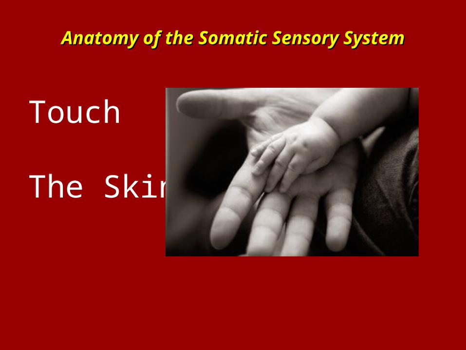
Anatomy of the Somatic Sensory SystemAnatomy of the Somatic Sensory System
Touch
The Skin
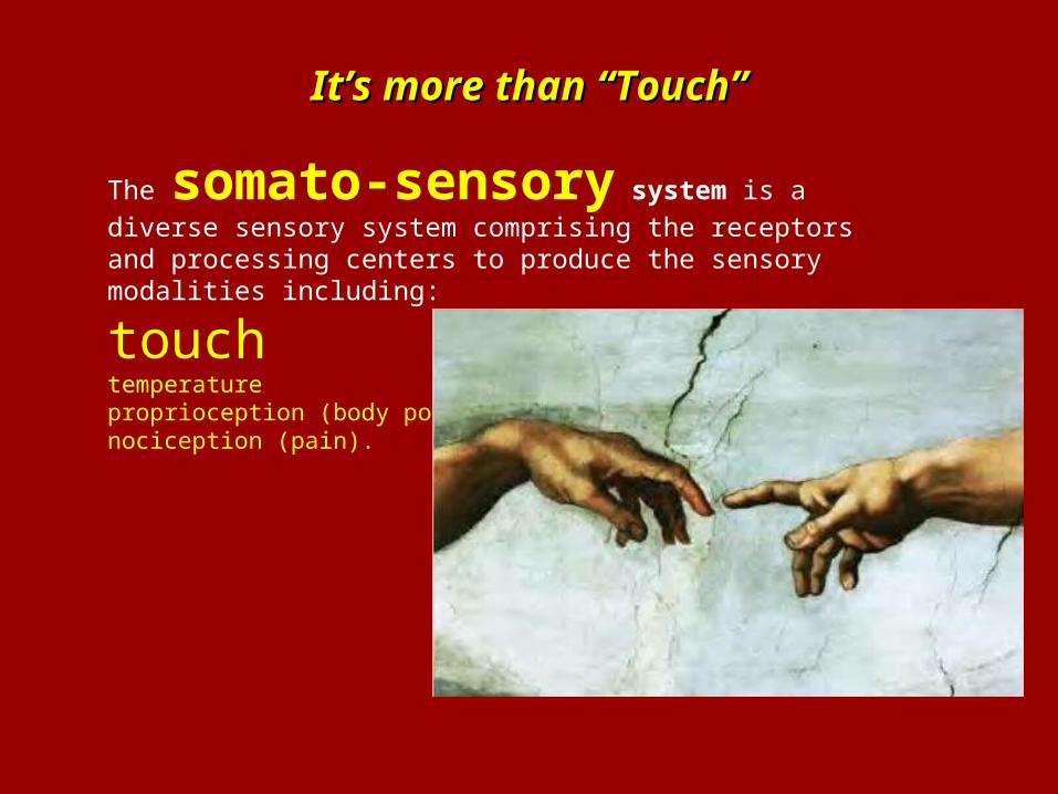
It’s more than “Touch”It’s more than “Touch”
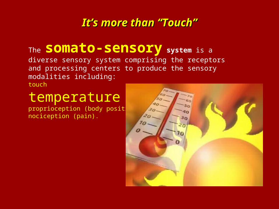
The somato-sensory system is a diverse
sensory system comprising the receptors and processing centers to produce the sensory modalities including:
touchtemperatureproprioception (body position) nociception (pain).
It’s more than “Touch”It’s more than “Touch”
The somato-sensory system is a diverse
sensory system comprising the receptors and processing centers to produce the sensory modalities including:touch
temperatureproprioception (body position) nociception (pain).
It’s more than “Touch”It’s more than “Touch”
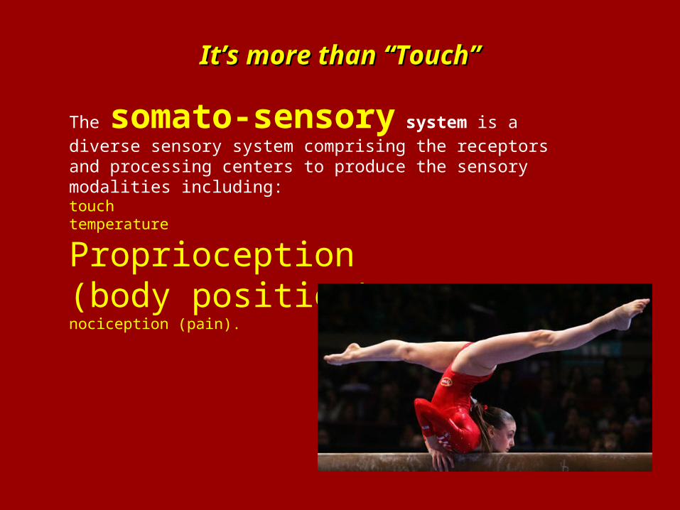
The somato-sensory system is a diverse
sensory system comprising the receptors and processing centers to produce the sensory modalities including:touchtemperature
Proprioception(body position) nociception (pain).
It’s more than “Touch”It’s more than “Touch”
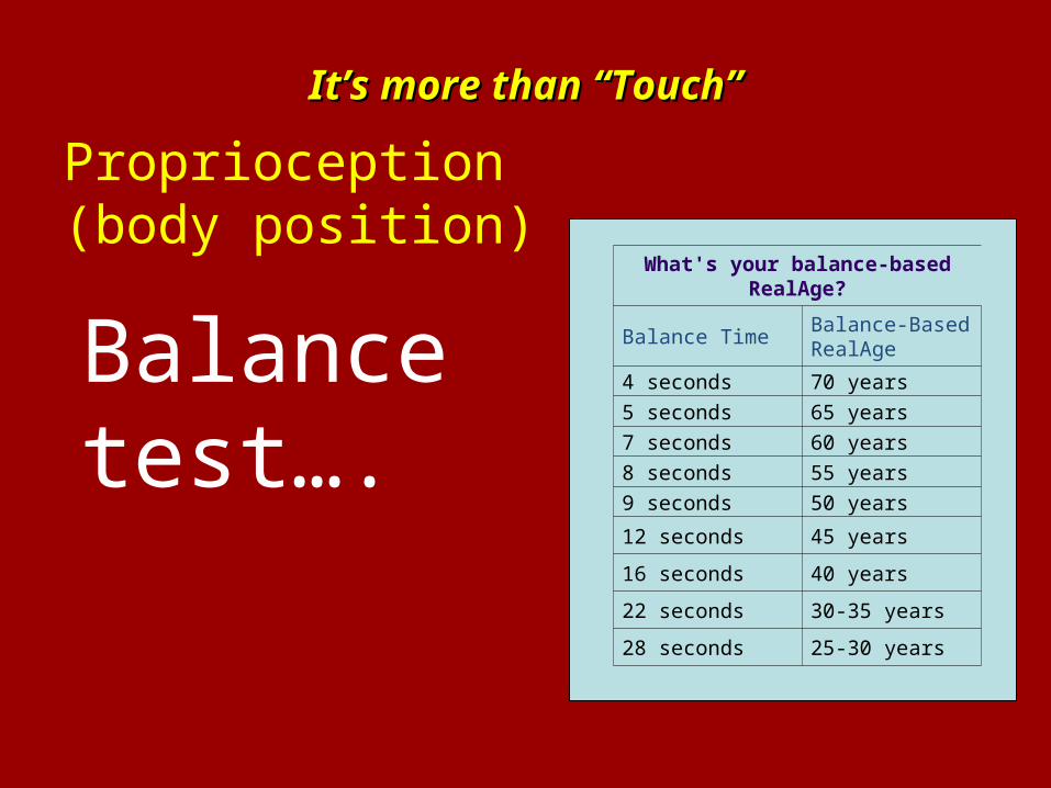
Proprioception(body position)
What's your balance-basedRealAge?
Balance TimeBalance-BasedRealAge
4 seconds 70 years
5 seconds 65 years
7 seconds 60 years
8 seconds 55 years
9 seconds 50 years
12 seconds 45 years
16 seconds 40 years
22 seconds 30-35 years
28 seconds 25-30 years
Balance test….
It’s more than “Touch”It’s more than “Touch”
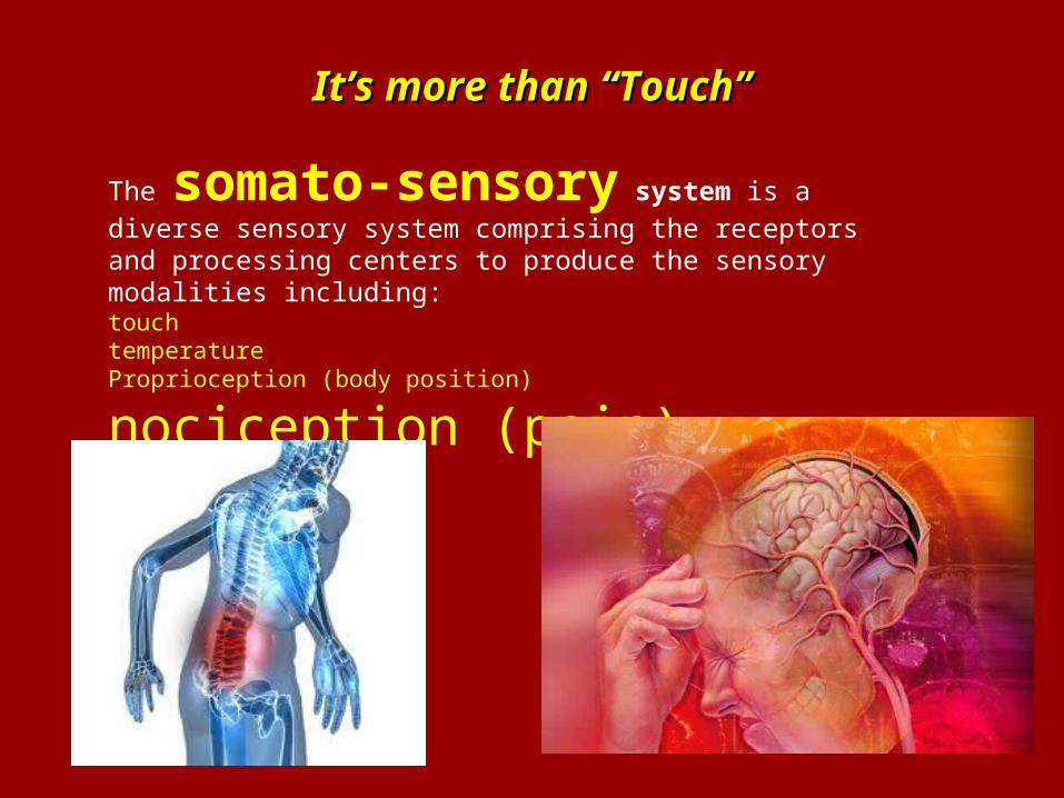
The somato-sensory system is a diverse
sensory system comprising the receptors and processing centers to produce the sensory modalities including:touchtemperatureProprioception (body position)
nociception (pain).
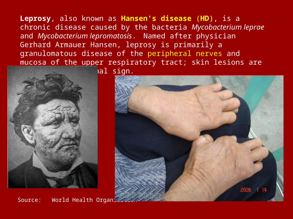
Leprosy, also known as Hansen's disease (HD), is a chronic disease caused by the bacteria Mycobacterium leprae and Mycobacterium lepromatosis. Named after physician Gerhard Armauer Hansen, leprosy is primarily a granulomatous disease of the peripheral nerves and mucosa of the upper respiratory tract; skin lesions are the primary external sign.
Source: World Health Organization
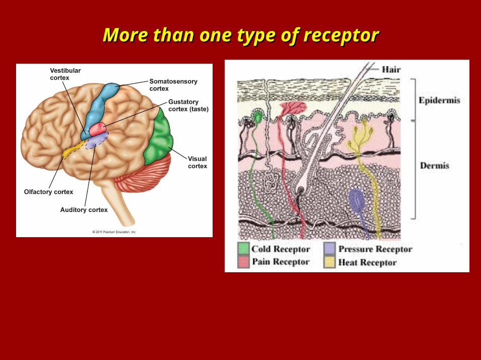
More than one type of receptorMore than one type of receptor
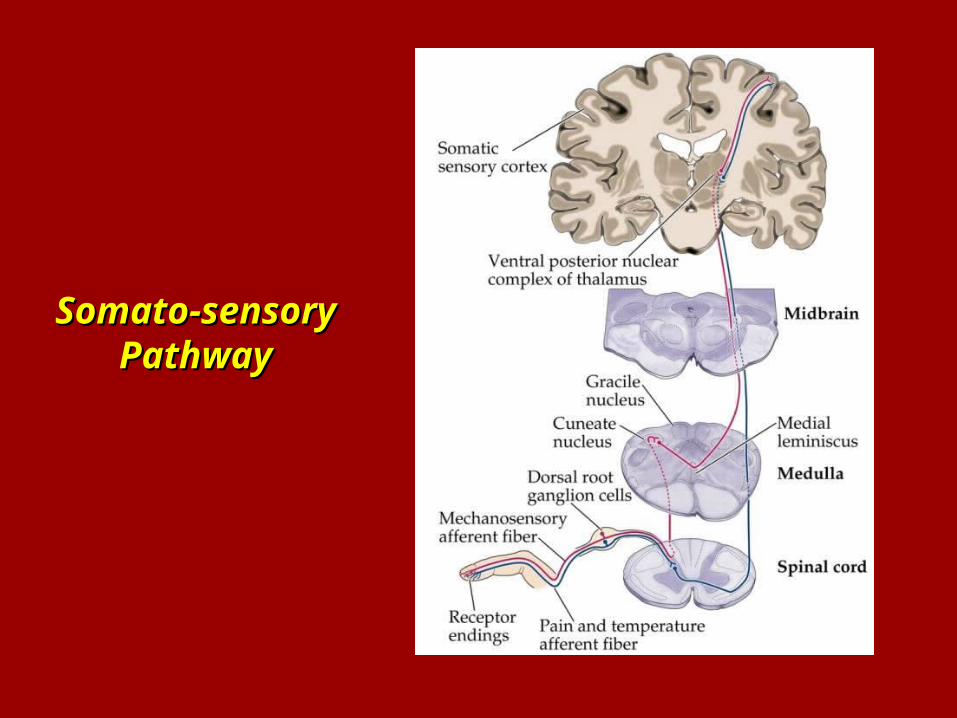
Somato-sensory Somato-sensory PathwayPathway
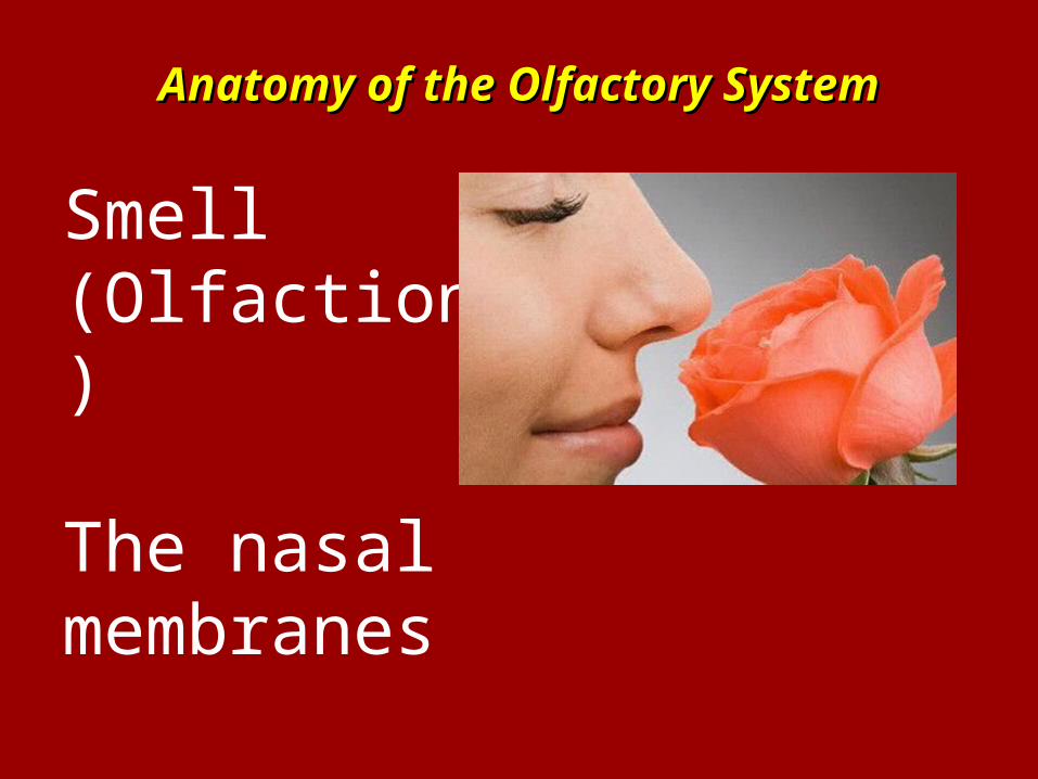
Anatomy of the Olfactory SystemAnatomy of the Olfactory System
Smell(Olfaction)
The nasal membranes
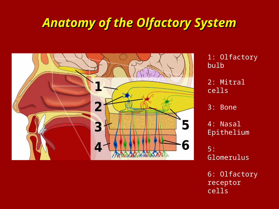
Anatomy of the Olfactory SystemAnatomy of the Olfactory System
1: Olfactory bulb
2: Mitral cells
3: Bone
4: Nasal Epithelium
5: Glomerulus
6: Olfactory receptor cells
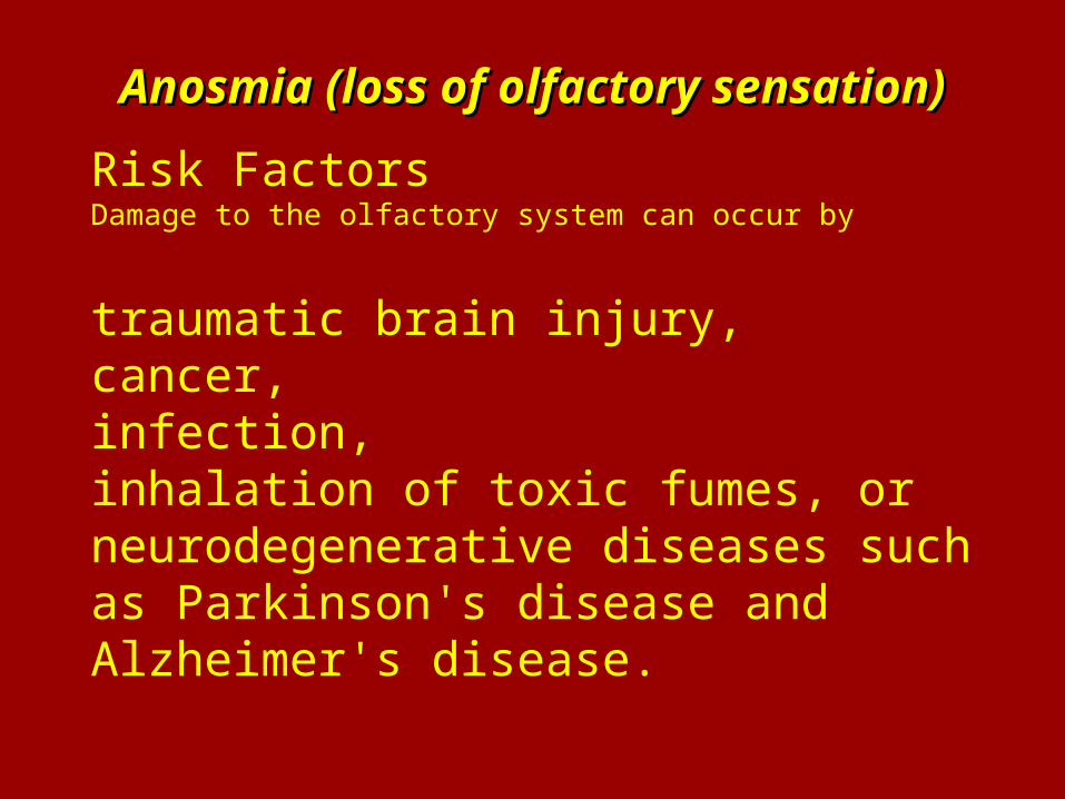
Anosmia (loss of olfactory sensation)Anosmia (loss of olfactory sensation)
Risk FactorsDamage to the olfactory system can occur by
traumatic brain injury, cancer, infection, inhalation of toxic fumes, or neurodegenerative diseases such as Parkinson's disease and Alzheimer's disease.
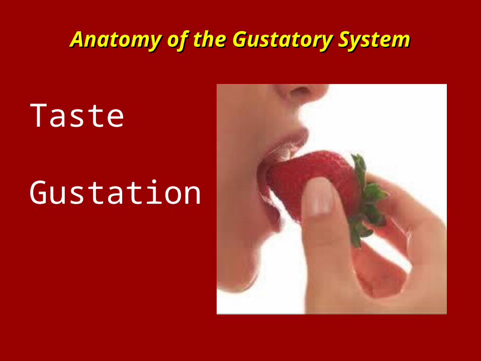
Anatomy of the Gustatory SystemAnatomy of the Gustatory System
Taste
Gustation
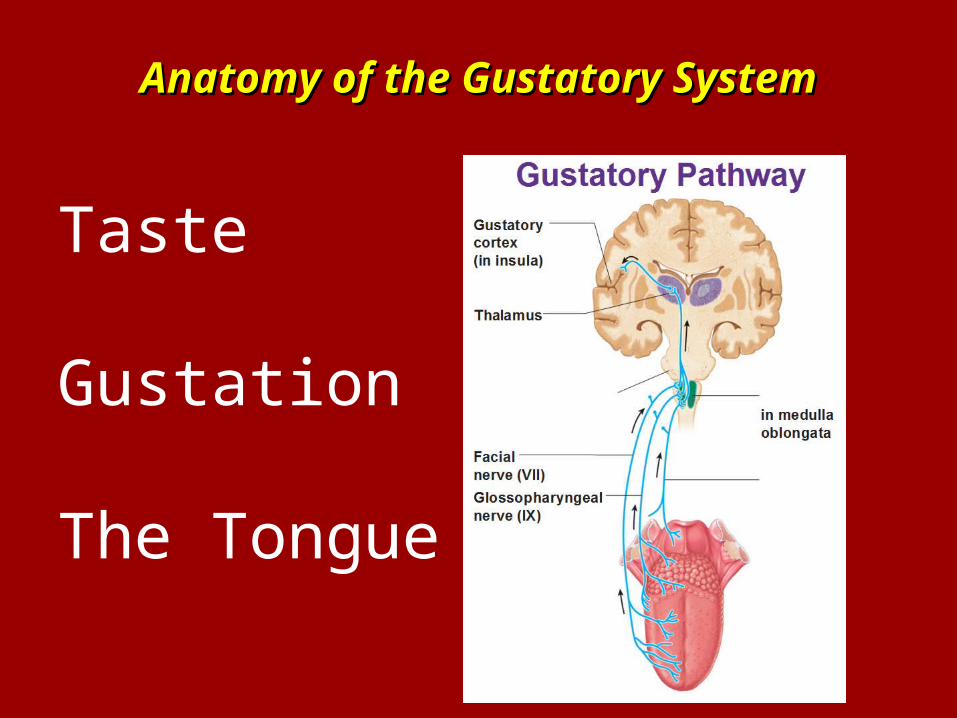
Anatomy of the Gustatory SystemAnatomy of the Gustatory System
Taste
Gustation
The Tongue
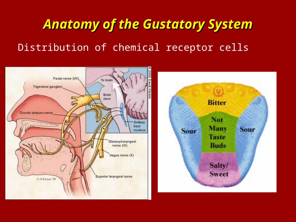
Anatomy of the Gustatory SystemAnatomy of the Gustatory System
Distribution of chemical receptor cells
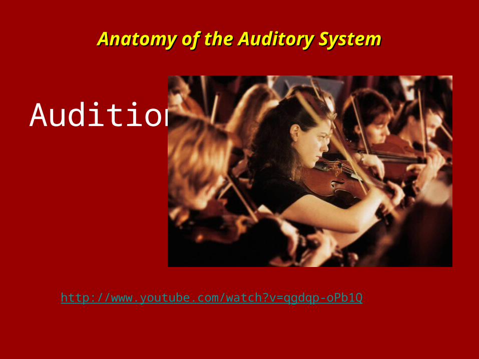
Anatomy of the Auditory SystemAnatomy of the Auditory System
Audition
http://www.youtube.com/watch?v=qgdqp-oPb1Q
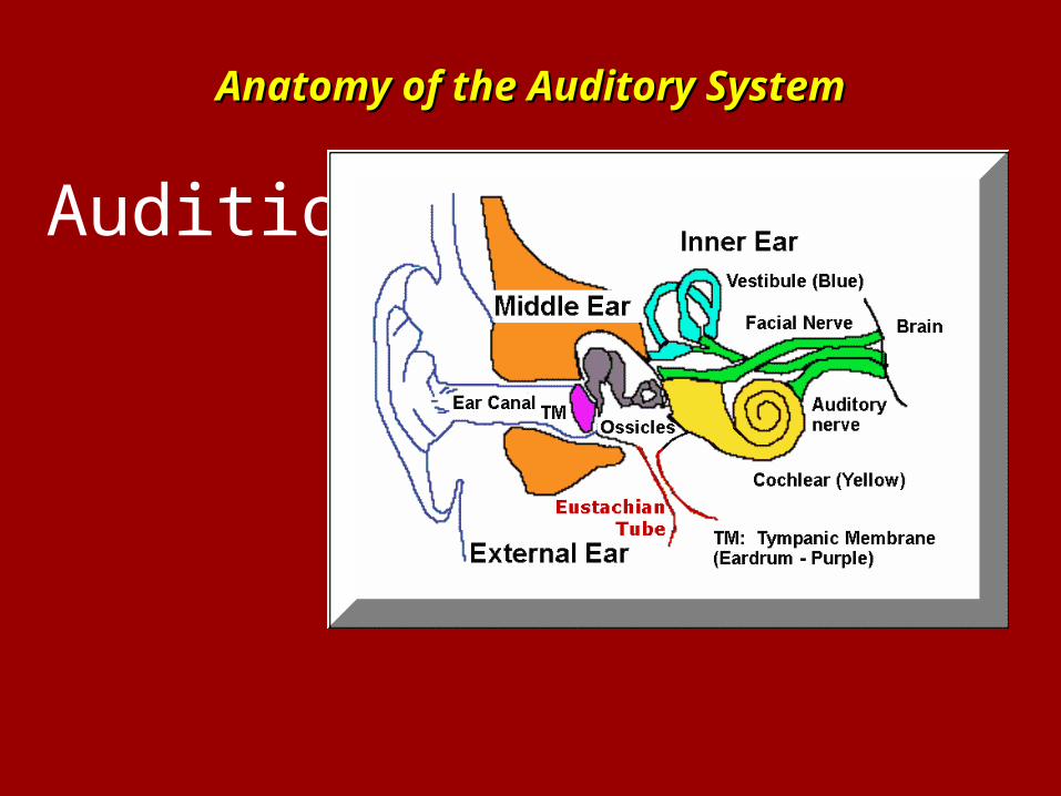
Anatomy of the Auditory SystemAnatomy of the Auditory System
Audition
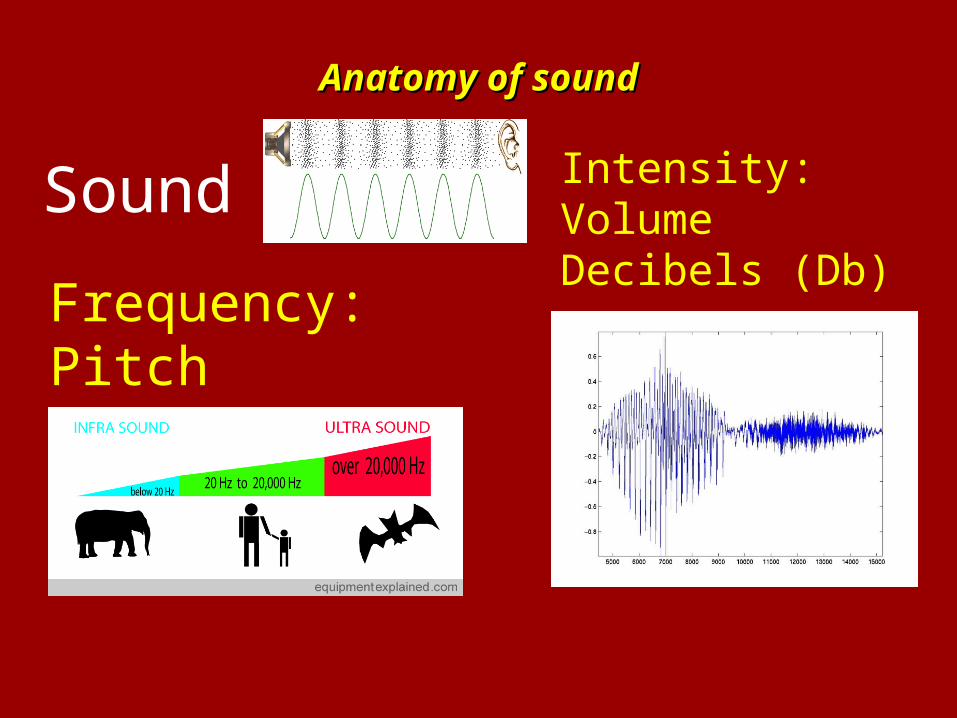
Anatomy of soundAnatomy of sound
Sound
Frequency: PitchHertz (Hz)
Intensity: Volume Decibels (Db)
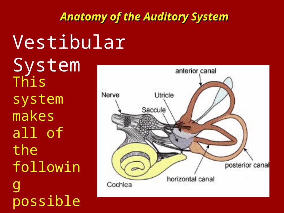
Anatomy of the Auditory SystemAnatomy of the Auditory System
Vestibular System
This system makes all of the following possible…..
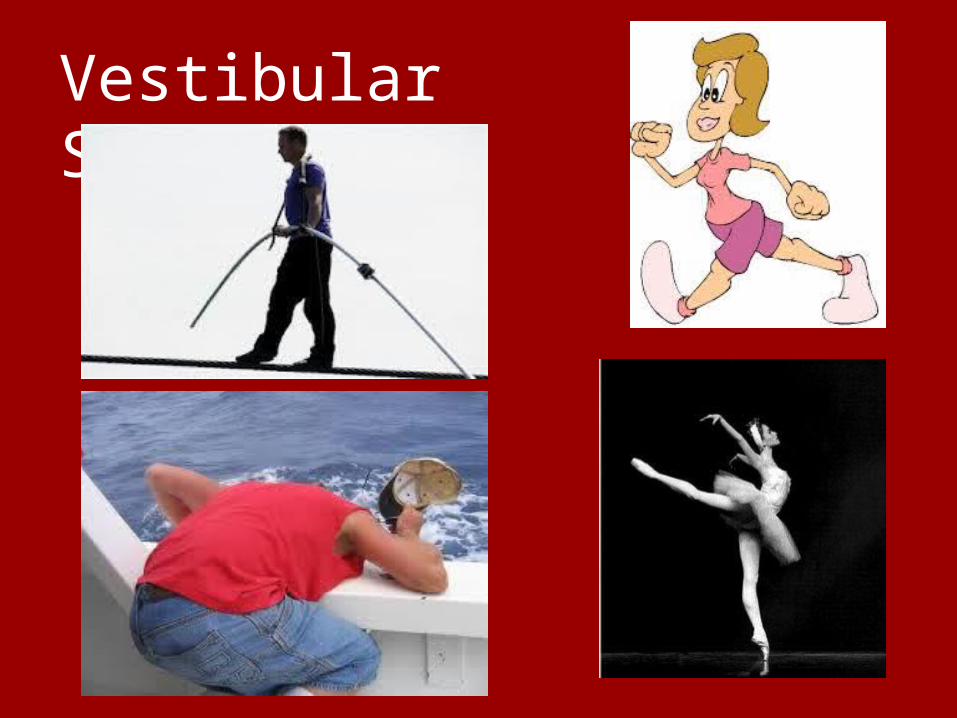
Vestibular System
Vestibular System
Controls Balance and Spatial Orientation
This system is the primary source of information about movement and a sense of balance.
The system senses rotations and linear accelerations and sends information to the
eyes and to the muscles that control balance.
Vestibular System
Kinesthesia: Perception of the body’s own movement.
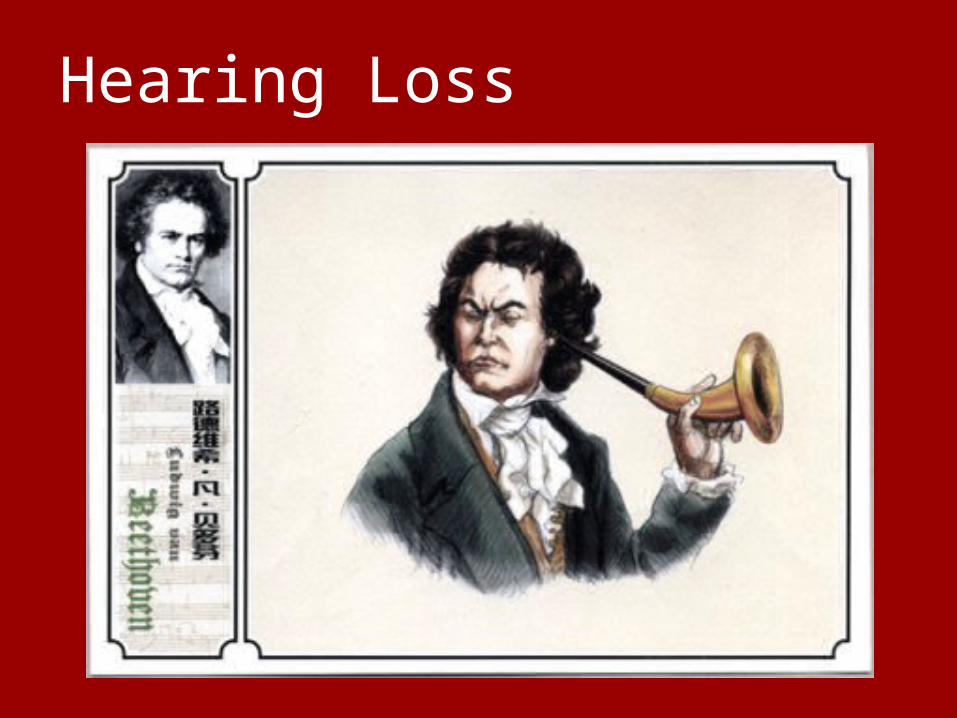
Hearing Loss
Hearing Loss
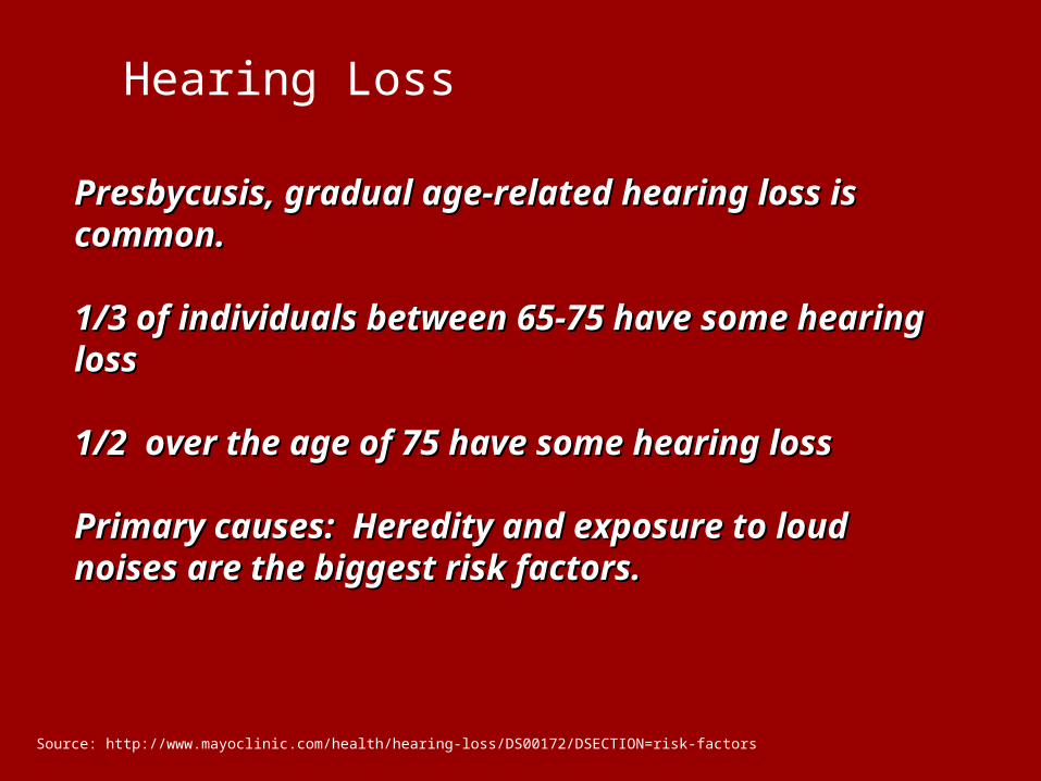
Presbycusis, gradual age-related hearing loss is Presbycusis, gradual age-related hearing loss is common.common.
1/3 of individuals between 65-75 have some hearing 1/3 of individuals between 65-75 have some hearing lossloss
1/2 over the age of 75 have some hearing loss1/2 over the age of 75 have some hearing loss
Primary causes: Heredity and exposure to loud Primary causes: Heredity and exposure to loud noises are the biggest risk factors.noises are the biggest risk factors.
Source: http://www.mayoclinic.com/health/hearing-loss/DS00172/DSECTION=risk-factors
Anatomy of the Auditory SystemAnatomy of the Auditory System
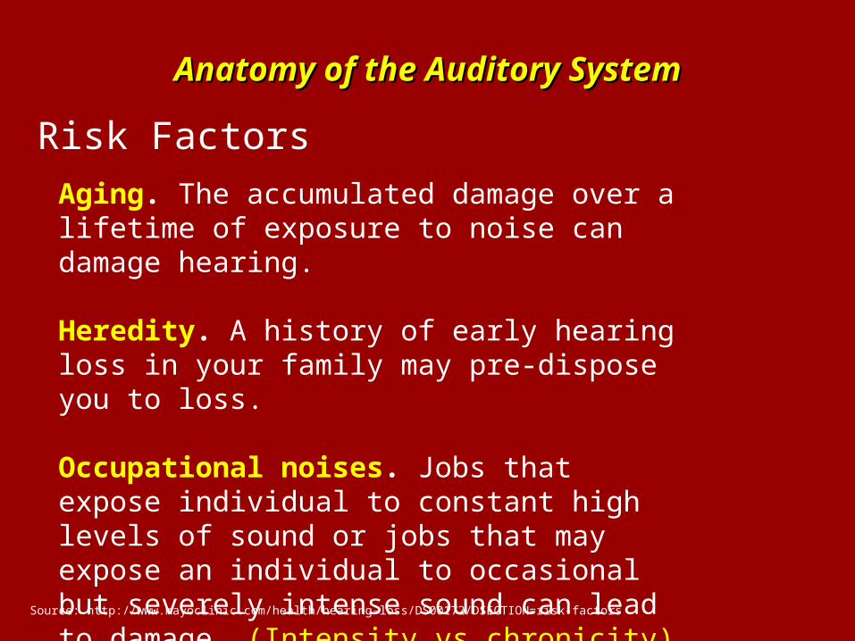
Risk Factors
Aging. The accumulated damage over a lifetime of exposure to noise can damage hearing.
Heredity. A history of early hearing loss in your family may pre-dispose you to loss.
Occupational noises. Jobs that expose individual to constant high levels of sound or jobs that may expose an individual to occasional but severely intense sound can lead to damage. (Intensity vs chronicity)……
Source: http://www.mayoclinic.com/health/hearing-loss/DS00172/DSECTION=risk-factors
Anatomy of the Auditory SystemAnatomy of the Auditory System
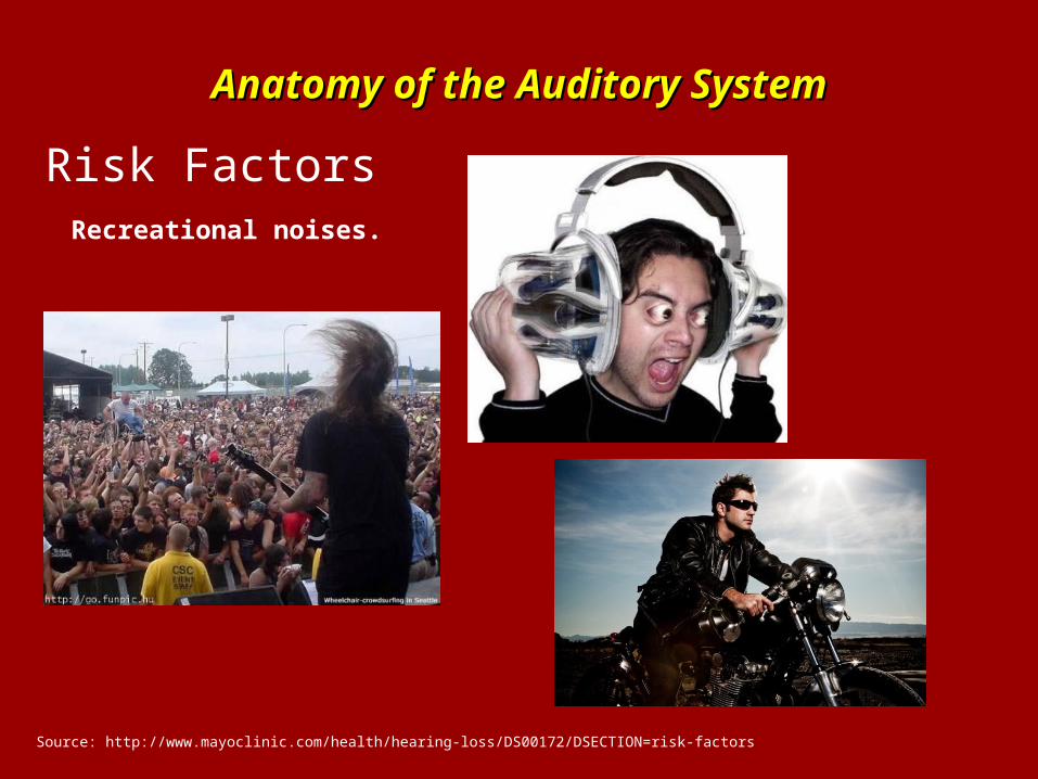
Risk FactorsRecreational noises.
Source: http://www.mayoclinic.com/health/hearing-loss/DS00172/DSECTION=risk-factors
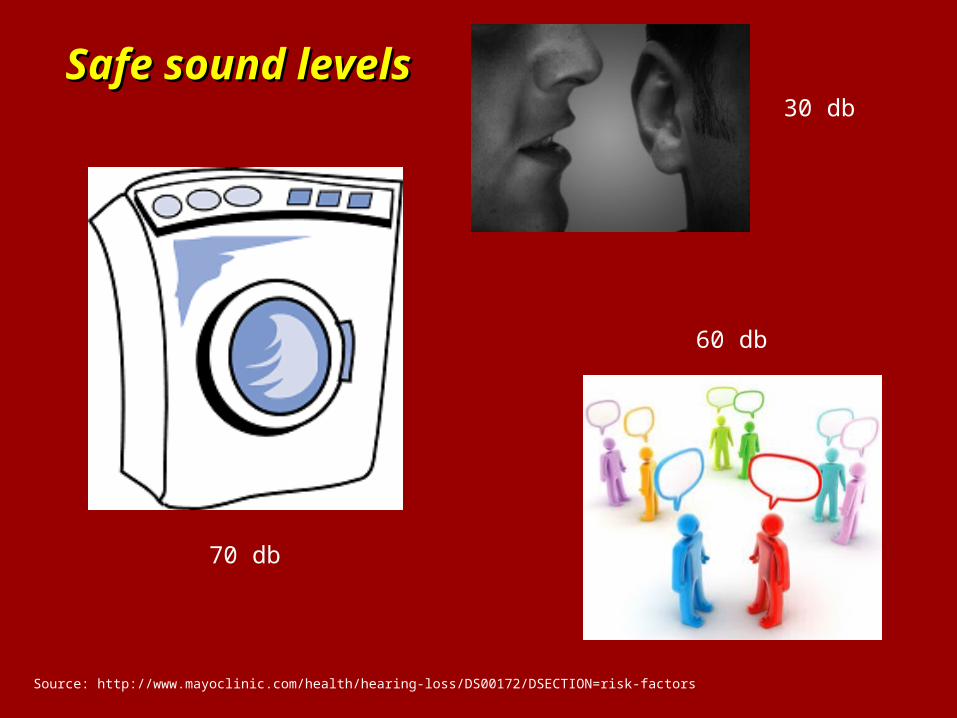
Safe sound levelsSafe sound levels
Source: http://www.mayoclinic.com/health/hearing-loss/DS00172/DSECTION=risk-factors
30 db
60 db
70 db
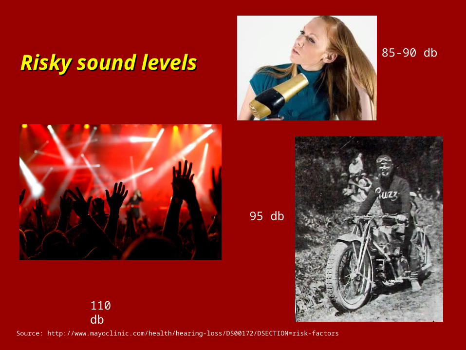
Risky sound levelsRisky sound levels
Source: http://www.mayoclinic.com/health/hearing-loss/DS00172/DSECTION=risk-factors
85-90 db
95 db
110 db
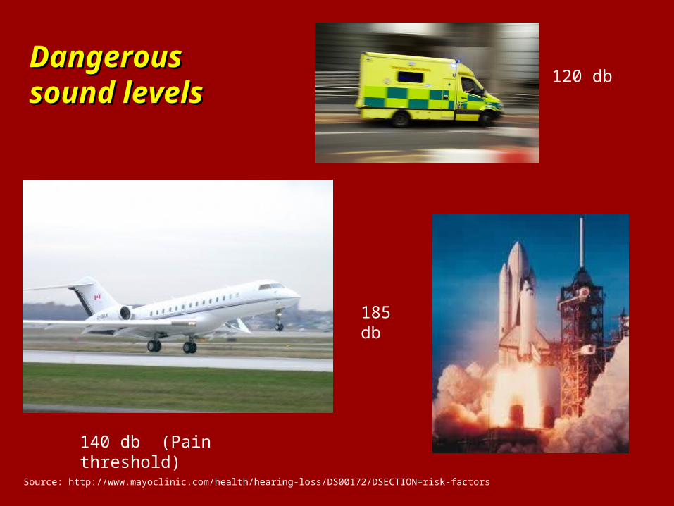
DangerousDangeroussound levelssound levels
Source: http://www.mayoclinic.com/health/hearing-loss/DS00172/DSECTION=risk-factors
120 db
185 db
140 db (Pain threshold)
Anatomy of the Auditory SystemAnatomy of the Auditory System
Risk FactorsSome medications. Drugs, such as the antibiotic gentamicin and certain chemotherapy drugs, can damage the inner ear. Temporary effects on your hearing — ringing in the ear (tinnitus) or hearing loss — can occur if you take very high doses of aspirin, other pain relievers, antimalarial drugs or loop diuretics.
Some illnesses. Diseases or illnesses that result in high fever, such as meningitis, may damage the cochlea.
Source: http://www.mayoclinic.com/health/hearing-loss/DS00172/DSECTION=risk-factors
Causes of hearing loss
Source: World Health Organization
Genetic causes
Syndromic: Deafness is the result of an illness and hearing loss is only one symptom, among many others, of the disease. (30%)
Nonsyndromic: Deafness is the only result of this genetic defect. (70%)
Causes of hearing loss
Source: World Health Organization
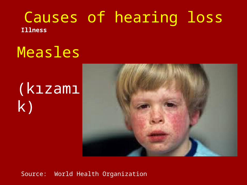
Illness
Measles (kızamık)
Causes of hearing loss
Source: World Health Organization
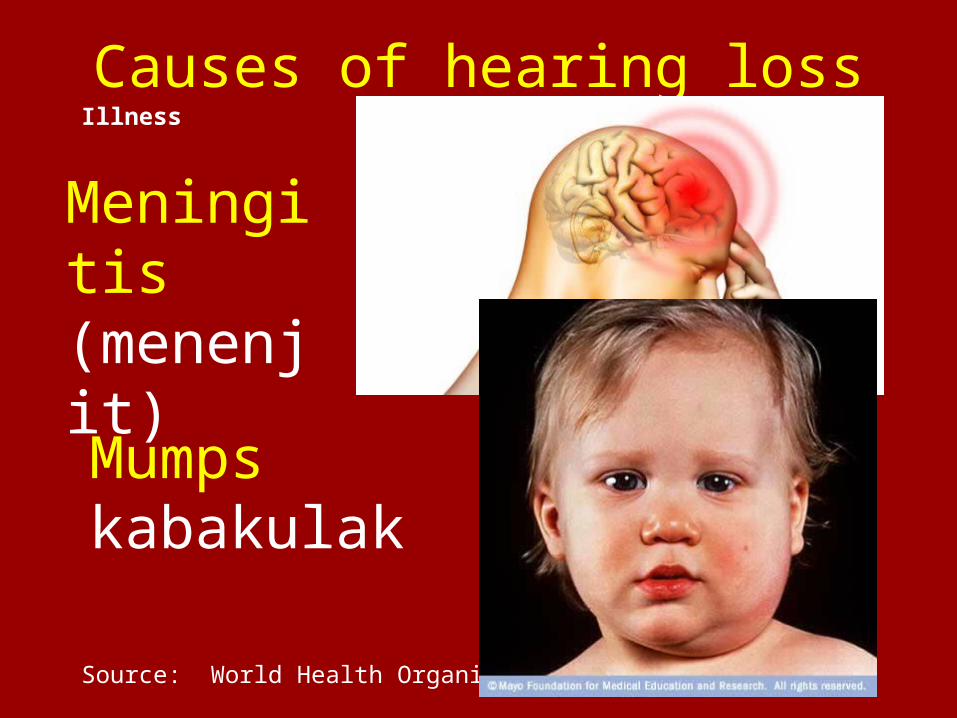
Illness
Meningitis (menenjit)
Mumps kabakulak
Causes of hearing loss
Source: World Health Organization
Illness
PresbycusisLoss of blood flow to ear..
Chlamydia (STD)Chlamydia
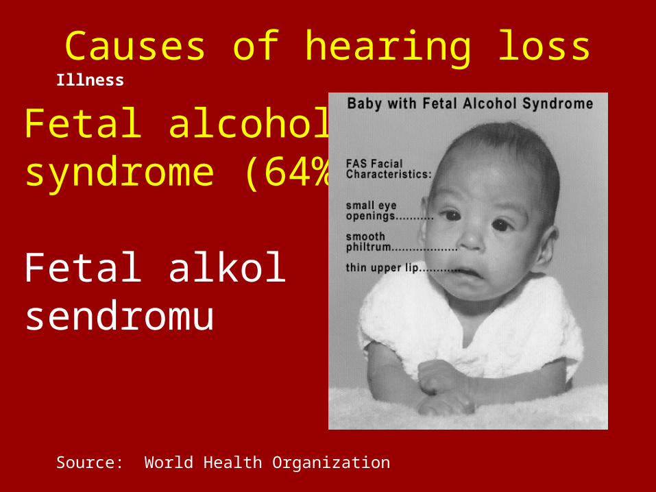
Causes of hearing loss
Source: World Health Organization
Illness
Fetal alcohol syndrome (64%)
Fetal alkol sendromu
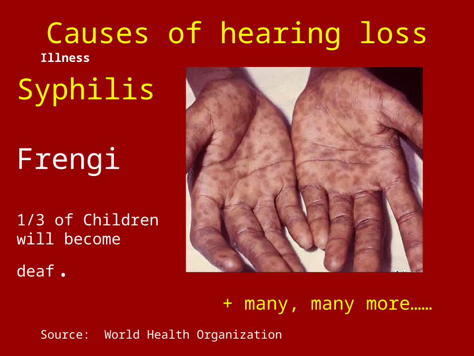
Causes of hearing loss
Source: World Health Organization
Illness
Syphilis
Frengi
1/3 of Children will
become deaf.
+ many, many more……
Causes of hearing loss
Source: World Health Organization
Neurological Disorders
Multiple Sclerosis
Strokes
Brain tumors
Causes of hearing loss
Source: World Health Organization
Medication
This includes some diuretics, aspirin, non steroidal anti-inflammatory drugs (NSAIDs),Antibiotics, antimalarials
Causes of hearing loss
Source: World Health Organization
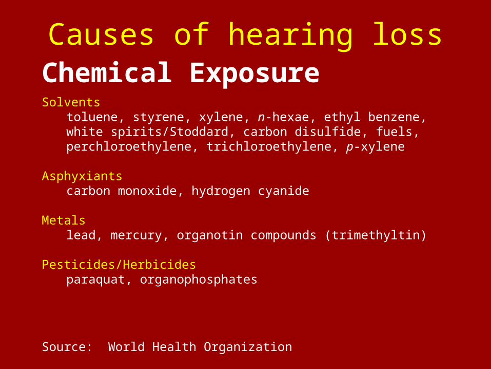
Chemical ExposureSolvents
toluene, styrene, xylene, n-hexae, ethyl benzene, white spirits/Stoddard, carbon disulfide, fuels, perchloroethylene, trichloroethylene, p-xylene
Asphyxiants carbon monoxide, hydrogen cyanide
Metals lead, mercury, organotin compounds (trimethyltin)
Pesticides/Herbicides paraquat, organophosphates
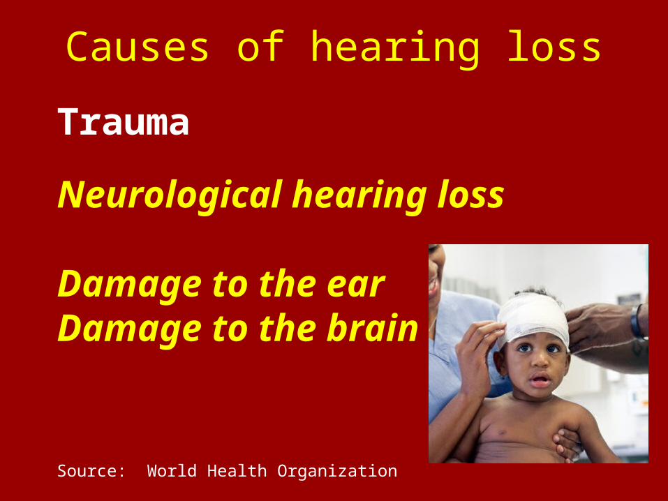
Causes of hearing loss
Source: World Health Organization
Trauma
Neurological hearing loss
Damage to the earDamage to the brain

Homework Assignment for next weekHomework Assignment for next weekDue 14 AprilDue 14 April
Complete Sleep Log
Return for class on 14 April and bring sleep log…..

and finally, some more housekeeping
Please help me return the classroom to it Please help me return the classroom to it original condition…..original condition…..
1. Take your rubbish with you……
2. Place the student desks in their original order.
Thank you…., Gracias, Merci, Danke, teşekkür ederim, ありがとう , Asante, gratias ago vos,Dank u, Takk skal du ha, спасибо ……
Harika!!
Too many drugs??


