
Hanyang University Guri HospitalJae Ung Lee MD.

Male /83
Chest pain 6hrs agoContinuousDizzinessShortness of breath
Vital sign
150/90mmHg86/min
CAD risk factors
HypertensionEx‐smokerOld age

ECG
ST segment elevation with pathologic Q waves in lead
III, aVF

Chest X‐ray
Mild cardiomegalyNo pulmonary edema

Coronary angiography
mLAD (80%)Grade 1 collaterals dRCA(100%) TIMI 0

Primary PCI
Predilatation(2.0x20mm, Lacrosse)
6 atm

OCT
OCT image wire(Image‐wire TM , LightLab)
0.4cc/sec
Occlusion balloon catheter(Helios TM Goodman)
Removal of GW

OCT
Intimal tearSoft plaque rupture
Thrombus


Fibrous plaque
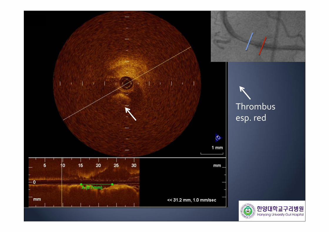
Thrombusesp. red

Intimal tear
Soft plaque

Thrombusesp. white
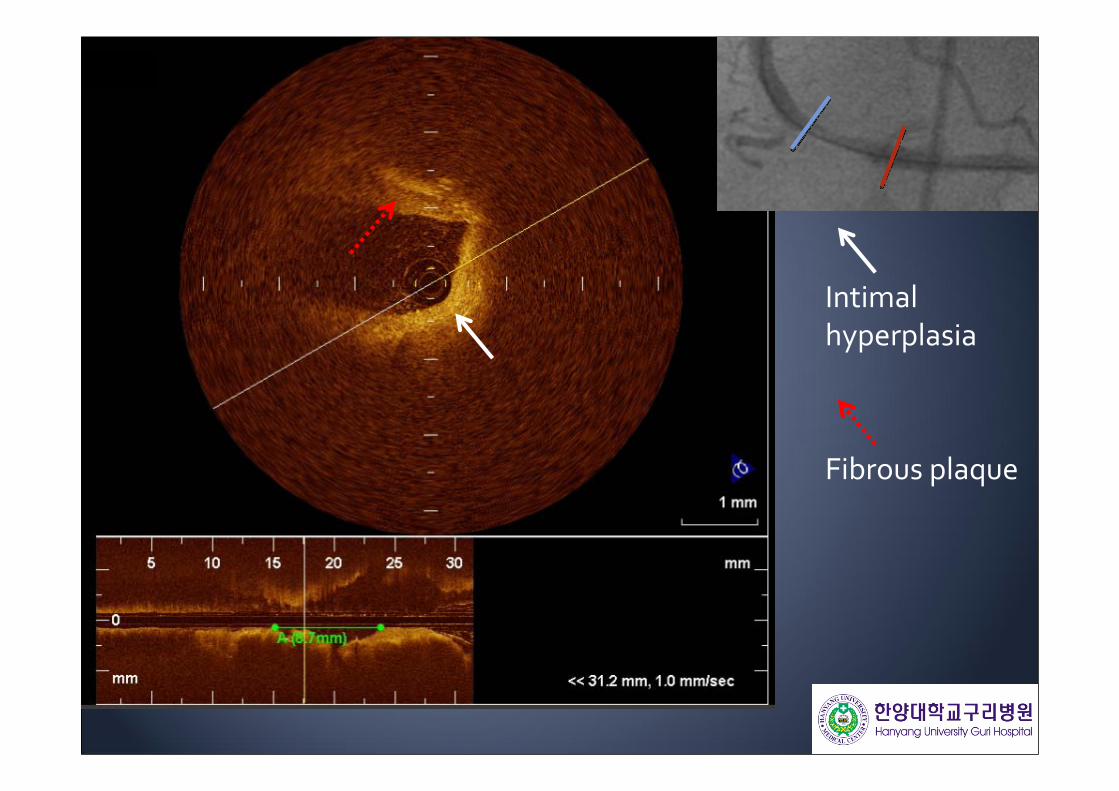
Intimal hyperplasia
Fibrous plaque

Acute marginalbranch
Soft plaque
Calcification


Stent Implantation
Endeavor RX, 3.0x24mm14atm

OCT
Occlusion catheterRemoval of GW
Insertion of OCT wire

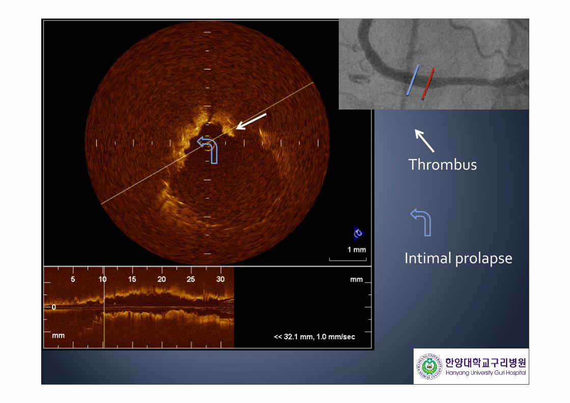
OCTIntimal prolapse
Thrombus fragments directly attached to strutsComplete stent apposition

Strut of stent
Thrombus
Intimal prolapse

Intimal prolapse

Strut of stentover the AM branch
Ostium of AM branch was narrowed
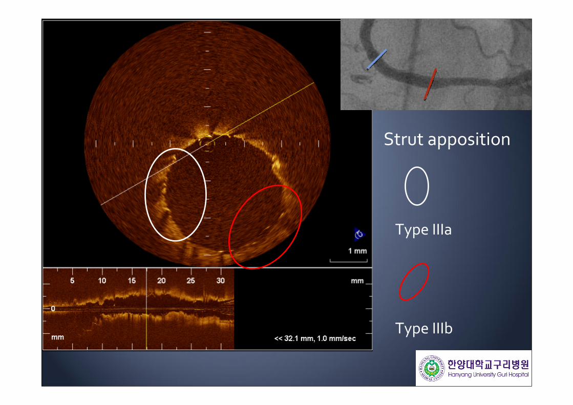
Type IIIa
Type IIIb
Strut apposition

Classification of strut apposition by OCT
Type I
Type II
Type IIIa
Type IIIb
Type IV
totally embedded strut
Embedded subintimally without disruption of lumen contour
completely embedded with disruption of lumen contour
Partially embedded with extension of strut into lumen
complete strut malapposition
Giulio Guagliumi et al. Catheterization and Cardiovascular Interventions 72:237–247 (2008)

Proximal border of stent

Intima
TearErosion
plaque
Lipid‐richFibrouscalcified
Thrombus
WhiteRed
Stent
Intimal prolapseThrombus
Stent apposition
Stent
OCT findings in STEMI
Optimal sizeLength

Conclusion
OCT can be the powerful tool to evaluate the pathophysiology of STEMI

Thank you for your attention !


