

Headache Clinics UK
Using a GoniometerLynn Wilkes
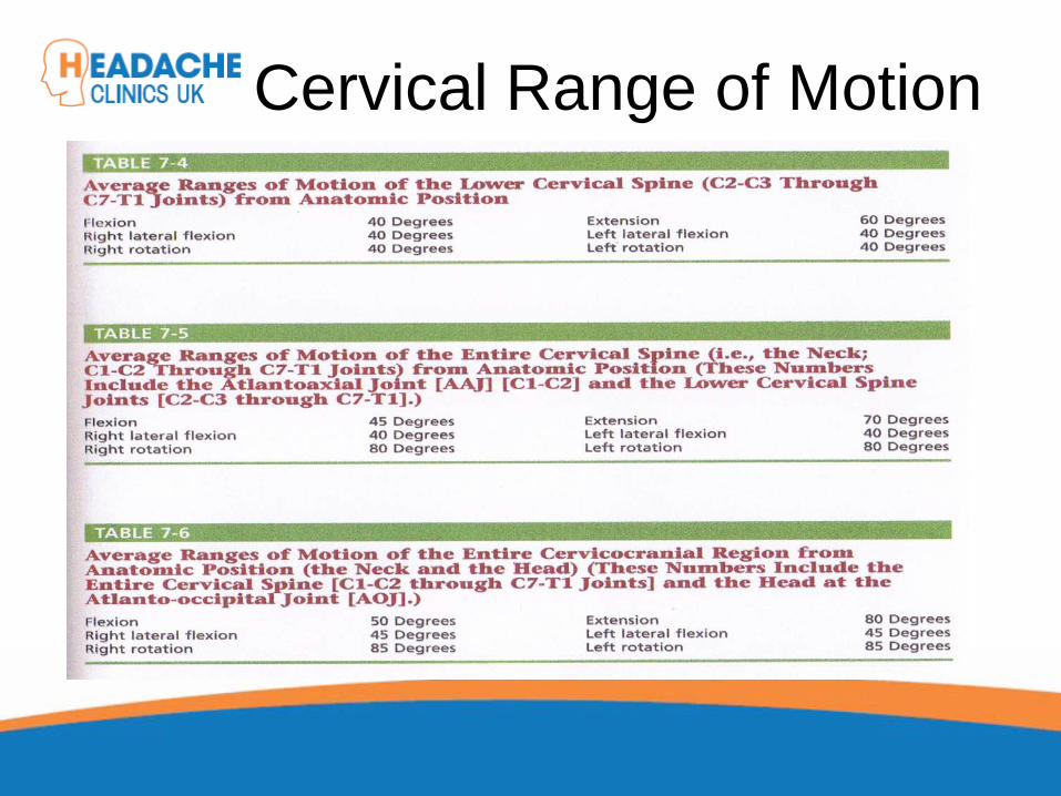
Cervical Range of Motion
Cervical Range of Motion Reliability of Goniometer
A range of studies have confirmed the goniometer, including the basic plastic one, is a valid and reliable measurement instrument for cervical spine range of motion

Cervical Range of Motion
Flexion and extension (A and B) the goniometer axis was positioned at the level of the seventh cervical vertebra,
the fixed arm was kept parallel to the floor and,
at the end of the movement, the moving arm was aligned with the earlobe

Cervical Range of Motion Rotation (C): the goniometer axis was positioned at the centre of the head,
the fixed arm was positioned at the centreof the head, at the sagittal suture,
at the end of the movement, the moving arm was aligned with the nose;

Cervical Range of Motion
Lateral flexion (D): the goniometer axis was placed on the spinous process of the seventh cervical vertebra, the fixed arm was placed parallel to the floor and the moving arm was aligned with the midline of the cervical spine;

Headache Clinics UK
Trigger Point Therapy For Headaches
Trigger Points
Travell defined a TrP as “a hyperirritable spot in skeletalmuscle that is associated with a hypersensitive palpablenodule in a taut band. The spot is tender when pressed andcan give rise to characteristic referred pain, motor dysfunction,and autonomic phenomena.”.
Trigger PointsResearch
Travell Trigger Points—Molecular and Osteopathic PerspectivesJohn M. McPartland, DO, MS
Myofascial trigger points and sensitization: an updated painmodel for tension-type headacheC Fernández-de-las-Peñas1,2, ML Cuadrado2,3,
CHIROPRACTIC MANAGEMENT OF MYOFASCIAL TRIGGER POINTS AND MYOFASCIAL PAIN SYNDROME: A SYSTEMATIC REVIEW OF THE LITERATUREHoward Vernon, DC, PhD,a and Michael Schneider, DCb
Myofascial trigger points in the suboccipital muscles in episodictension - type headacheCesar Fernandez-de-las-Penasa,
Contribution of Myofascial Trigger Points to Migraine SymptomsMaria Adele Giamberardino, Emmanuele Tafuri,

Key Muscles in Headaches

Key Muscles in Headaches
Trapezius Sub OccipitalSternocleidomastoid Splenius capitisFrontalis Splenius cervicusMedial Ptyergoid Rectus capitis posterior minor
Lateral Ptyergoid Rectus capitis posterior major
Masseter Semispinalis capitisTemporalis Semi spinalis cervicis

Referral PatternsTRAPEZIUS
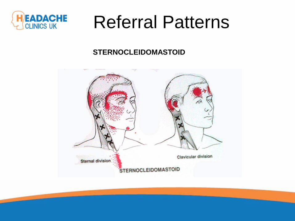
Referral PatternsSTERNOCLEIDOMASTOID

Referral Patterns
Temporalis
Diagastric
Occipital Frontalis
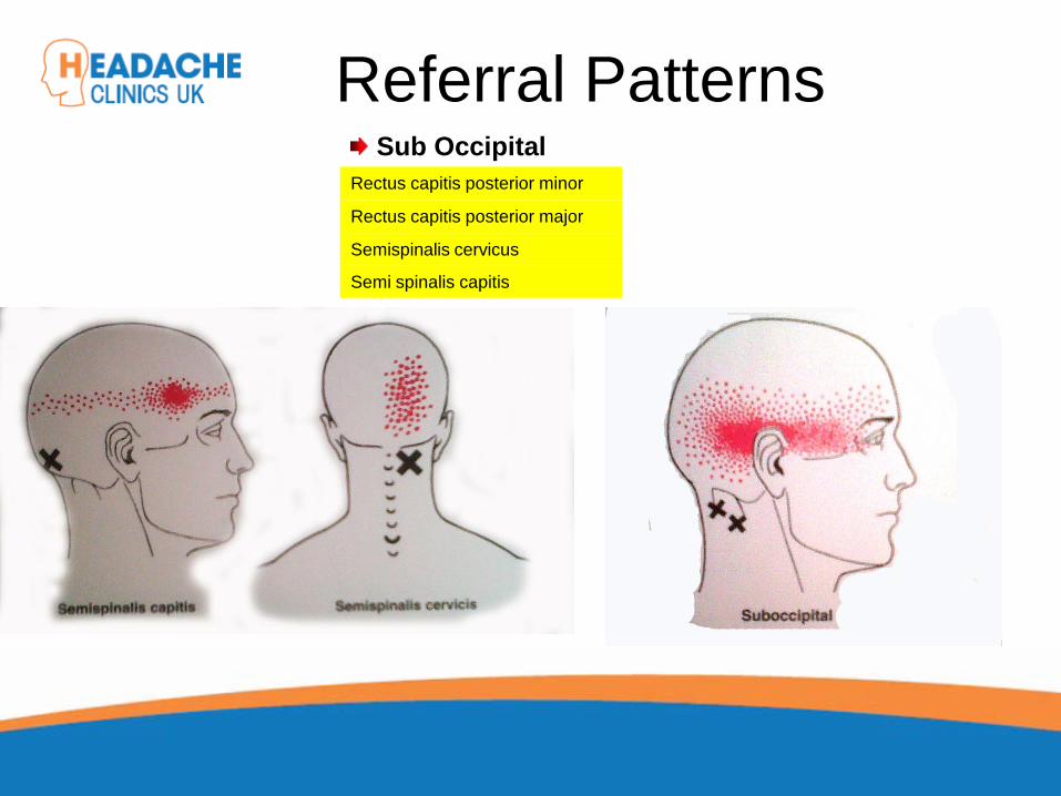
Referral PatternsSub Occipital
Rectus capitis posterior minor
Rectus capitis posterior major
Semispinalis cervicus
Semi spinalis capitis

Referral PatternsSub Occipital
Splenius capitis
Splenius cervicus
TMJ Research Leads to Discovery
In addition to a new muscle of mastication, he also discovered a connective tissue bridge that attaches the rectus capitus posterior minor muscle to the dura at the atlanto – occipital junction. (it was present in all 10 cadavers)
Hack GD. Anatomic relation between the rectus capitus posterior minor muscle and the dura matter. Spine 1995: 20:2484-2486

The Neuromuscular Connection
• Dr. Hack stated: – Maryland Scientists speculate that the newly
described muscle – dura connection may transmit forces from the neck muscles to the pain sensitive dura.
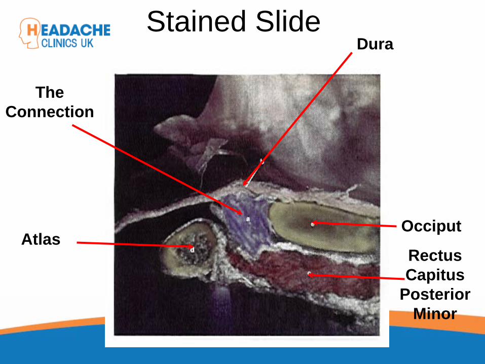
Stained SlideDura
Occiput
RectusCapitus
PosteriorMinor
TheConnection
Atlas
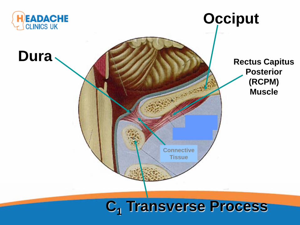
Dura
Occiput
C1 Transverse Process
ConnectiveTissue
Rectus Capitus Posterior (RCPM) Muscle
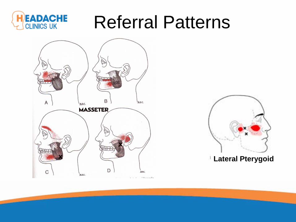
Referral Patterns
Lateral Pterygoid
Trigger Point TherapyPractical
Mary Sanderson


