

Hemorrhage & Shock
Review of Hemorrhage Location Anatomical Type & Timing Coagulation Fibrinolysis Assessment Management
Review of Hemorrhage
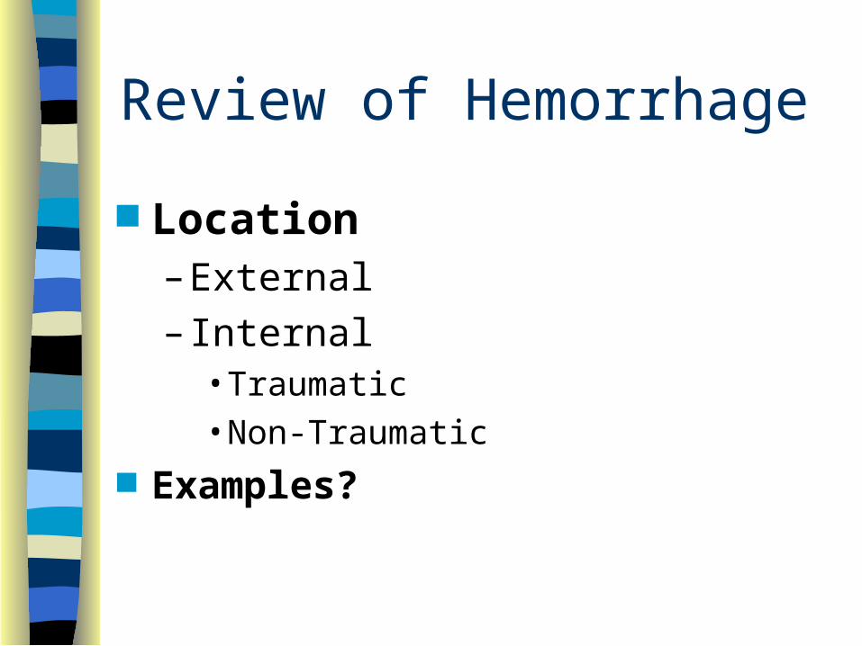
Location– External
– Internal• Traumatic• Non-Traumatic
Examples?

Review of Hemorrhage
Anatomical Type– Arterial
– Venous
– Capillary Timing
– Acute
– Chronic

Severity of Hemorrhage
Comparison of Adult vs Child
Hematocrit
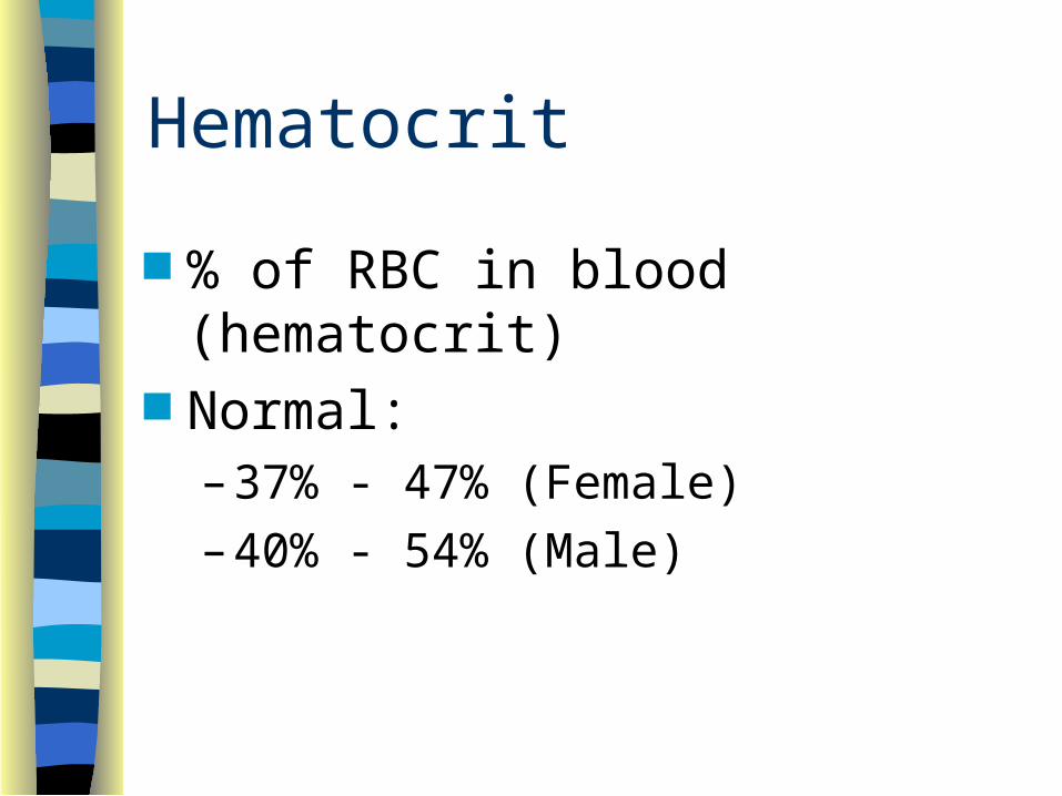
% of RBC in blood (hematocrit) Normal:
– 37% - 47% (Female)
– 40% - 54% (Male)
Thrombocytes
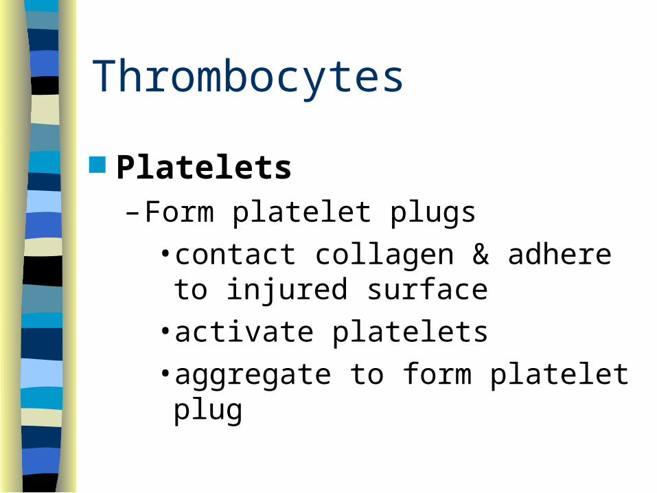
Platelets– Form platelet plugs
• contact collagen & adhere to injured surface
• activate platelets
• aggregate to form platelet plug

Coagulation
Formation of blood clots– Prothrombin activator– Prothrombin Thrombin– Fibrinogen Fibrin
• entrap platelets, blood cells & plasma
– Clot retraction

Fibrinolysis
Breaking up the clot– tissue plasminogen activator (tPA)
– plasminogen plasmin
Assessing Hemorrhage Clues
– Bright red blood from wound, mouth, rectum or other orifice
– Hematemesis• Coffee ground appearance of vomitus
– Hematochezia• Melena
– Orthostatic hypotension• Dizziness or syncope on sitting or standing
– Signs and symptoms of hypovolemic shock

Management of Hemorrhage Airway and Ventilatory Support Circulatory Support
– From nose or ears after head trauma = loose drsg– Control bleeding
• direct pressure, elevation, pressure points• tourniquet• packing of large wounds• splinting• PASG• transport to appropriate facility
Shock“A rude unhinging of the machinery of life”
“A brief pause in the act of dying”
Shock
Inadequate peripheral perfusion leading to failure of
tissue oxygenation may lead to anaerobic metabolism

Shock
Homeostasis– cellular state of balance
– perfusion of cells with oxygen is one of its cornerstones
Shock
Adequate Cellular Oxygenation– Red Cell Oxygenation
– Red Cell Delivery To Tissues
Fick Principle

Fick Principle
Air’s gotta go in and out.Blood’s gotta go round and round.Any variation of the above is not a good thing!
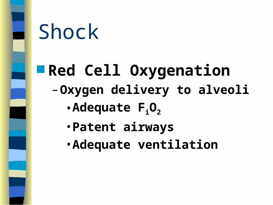
Shock
Red Cell Oxygenation– Oxygen delivery to alveoli
• Adequate FiO2
• Patent airways
• Adequate ventilation

Shock Red Cell Oxygenation
– Oxygen exchange with blood• Adequate oxygen diffusion into blood• Adequate RBC flow past alveoli• Adequate RBC mass/Hgb levels
• Adequate RBC capacity to bind O2
–pH–Temperature
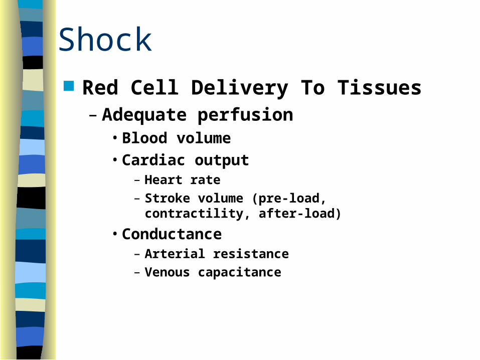
Shock Red Cell Delivery To Tissues
– Adequate perfusion• Blood volume• Cardiac output
– Heart rate– Stroke volume (pre-load, contractility,
after-load)
• Conductance– Arterial resistance– Venous capacitance
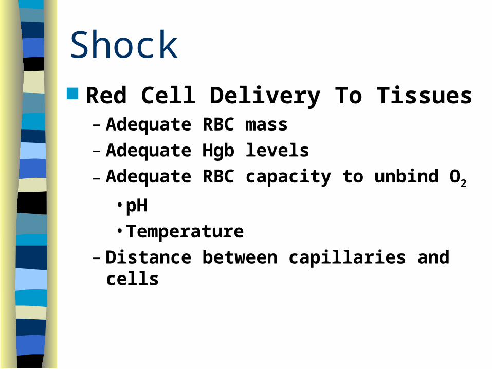
Shock Red Cell Delivery To Tissues
– Adequate RBC mass– Adequate Hgb levels
– Adequate RBC capacity to unbind O2
• pH• Temperature
– Distance between capillaries and cells
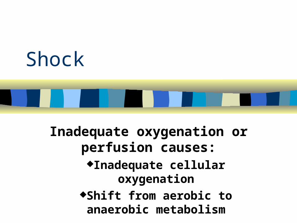
Shock
Inadequate oxygenation or perfusion causes:
Inadequate cellular oxygenationShift from aerobic to anaerobic
metabolism

AEROBIC METABOLISM
6 O2
GLUCOSE
METABOLISM
6 CO2
6 H2O
36 ATP
HEAT (417 kcal)
Glycolysis: Inefficient source of energy production; 2 ATP for every glucose; produces pyruvic acid
Oxidative phosphorylation: Each pyruvic acid is converted into 34 ATP

ANAEROBIC METABOLISM
GLUCOSE METABOLISM
2 LACTIC ACID
2 ATP
HEAT (32 kcal)
Glycolysis: Inefficient source of energy production; 2 ATP for every glucose; produces pyruvic acid

Anaerobic Metabolism Occurs without oxygen
– oxydative phosphorylation can’t occur without oxygen
– glycolysis can occur without oxygen– cellular death leads to tissue and organ
death– can occur even after return of perfusion
organ or organism death

InadequateCellularOxygenDelivery
AnaerobicMetabolism
InadequateEnergyProduction
MetabolicFailure
LacticAcidProduction
MetabolicAcidosisCELL
DEATH
Ultimate Effects of Anaerobic Metabolism
Maintaining perfusion requires: Volume Pump Vessels Failure of one or more of these
causes shock
Shock Hypovolemic Shock = Low Volume
–Trauma–Non-traumatic blood loss
VaginalGIGU
–Burns–Diarrhea
–Vomiting–Diuresis–Sweating–Third space losses
PancreatitisPeritonitisBowel obstruction
Shock
Cardiogenic Shock = Pump Failure
–Acute M I–CHF–Bradyarrhythmias–Tachyarrhythmias
–Mechanical obstruction (“distributive shock”)
Cardiac tamponadeTension pneumothoraxPulmonary embolism

Shock
Vasogenic Shock = Low Resistance
– Spinal cord trauma• neurogenic shock
– Depressant drug toxicity– Simple fainting
Shock Mixed Shock
– Septic Shock• Overwhelming infection• Inflammatory response occurs• Blood vessels
–Dilate (loss of resistance)–Leak (loss of volume)
Shock Mixed Shock
– Septic Shock• Fever
– Increased O2 demand
– Increased anaerobic metabolism• Bacterial toxins
– Impaired tissue metabolism

Shock
Mixed Shock– Anaphylactic Shock
• Severe allergic reaction• Histamine is released• Blood vessels
–Dilate (loss of resistance)–Leak (loss of volume)

Shock
Mixed Shock– Anaphylactic Shock
• Histamine release• Extravascular smooth muscle spasm
– Laryngospasm– Bronchospasm

Shock
Progressive syndrome Three phases
– Compensated
– Decompensated
– Irreversible

Shock
Signs and symptoms due to:– Hypoperfusion
– Compensatory responses

Compensated Shock
Baroreceptors detect fall in BP– Usually 60-80 mm Hg (adult)
Sympathetic nervous system activates– What are the primary SNS
Neurotransmitters & their effects?
Compensated Shock
Cardiac effects• Increased force of contractions
• Increased rate
• Increased cardiac output

Compensated Shock
Peripheral effects• Arteriolar constriction
• Pre-/post-capillary sphincter contraction
• Increased peripheral resistance
• Shunting of blood to core organs
Compensated Shock Decreased renal blood flow
– Renin released from kidney arteriole– Renin & Angiotensinogen combine– Converts to Angiotensin I– Angiotensin I converts to Angiotensin II
• Peripheral vasoconstriction• Increased aldosterone release (adrenal
cortex)– promotes reabsorption of sodium & water

Compensated Shock Decreased blood flow to
hypothalamus Release of antidiuretic hormone
(ADH or Arginine Vasopressin) from posterior pituitary– Retention of salt, water– Peripheral vasoconstriction

Compensated Shock Insulin
secretion caused by epinephrine– contributes to hyperglycemia
Glucagon release caused by epinephrine– promotes liver glycogenolysis & gluconeogenesis
ACTH– stimulates adrenal cortex release of cortisol glucose production

Compensated Shock Peripheral capillaries contain
minimal blood Stagnation Aerobic metabolism changes to
anaerobic Extracellular potassium shifts
begin
Compensated Shock Presentation
– Restlessness, anxiety • Earliest sign of shock
– Tachycardia • ?Bradycardia in cardiogenic,
neurogenic

Compensated Shock Presentation
– Normal BP, narrow pulse pressure – Falling BP = late sign of shock– Mild orthostatic hypotension (15 to
25 mm Hg)– “Possible” delay in capillary refill

Compensated Shock Presentation
– Pale, cool skin
• Cardiogenic• Hypovolemic
– Flushed skin
• Anaphylactic• Septic• Neurogenic
Compensated Shock Presentation
– Slight tachypnea– Respiratory compensation for
metabolic acidosis
Compensated Shock Presentation
– Nausea, vomiting
– Thirst
– Decreased body temperature
– Feels cold
– Weakness
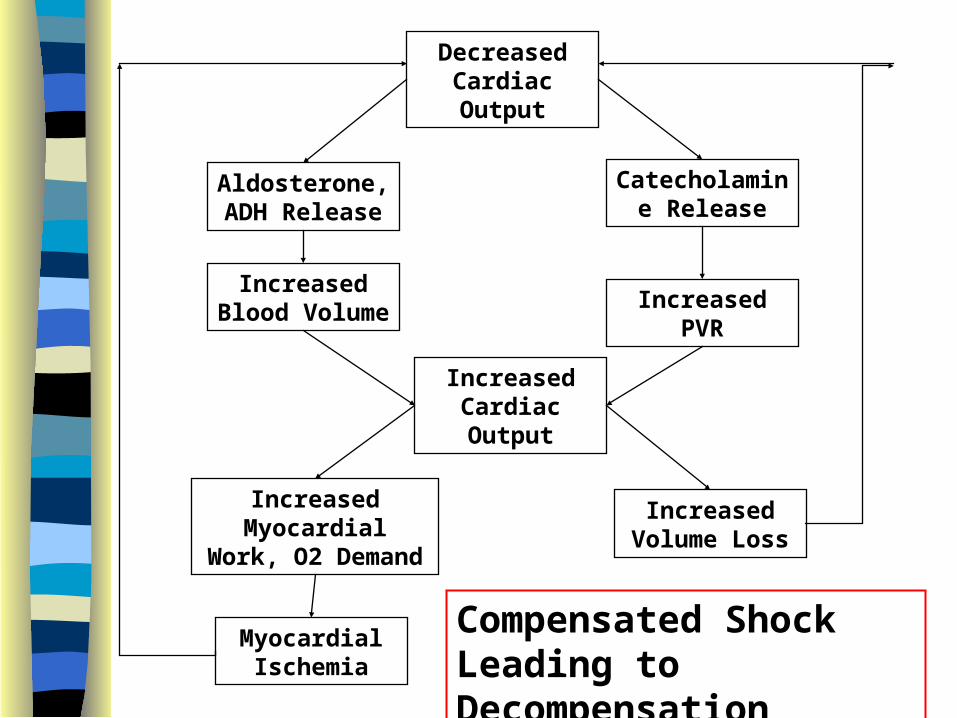
Decreased Cardiac Output
Aldosterone, ADH Release
Catecholamine Release
Increased Blood Volume Increased PVR
Increased Cardiac Output
Increased Myocardial Work,
O2 Demand
Increased Volume Loss
Myocardial Ischemia
Compensated Shock Leading to Decompensation

Decompensated Shock
Presentation– Cardiac Effects
• Decreased RBC oxygenation
• Decreased coronary blood flow
• Myocardial ischemia
• Decreased force of contraction
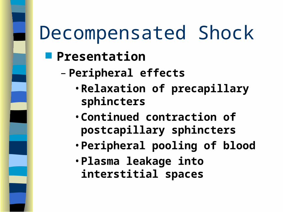
Decompensated Shock Presentation
– Peripheral effects• Relaxation of precapillary
sphincters• Continued contraction of
postcapillary sphincters• Peripheral pooling of blood• Plasma leakage into interstitial
spaces

Decompensated Shock
Presentation– Peripheral effects
• Continued anaerobic metabolism• Continued increase in extracellular
potassium• Rouleaux formations of RBCs
– “pile up like coins”
• Cold, gray, “waxy” skin

Decompensated Shock Presentation
– Listlessness, confusion, apathy, slow speech
– Tachycardia; weak, thready pulse– Decreased blood pressure– Moderate to severe orthostatic
hypotension– Decreased body temperature– Tachypnea

Irreversible Shock
Post-capillary sphincter relaxation
Loss of peripheral vascular resistance

Irreversible Shock Washout of accumulated products
• Hydrogen ion• Potassium• Rouleaux formations• Carbon dioxide
Rouleaux formations microembolize in lungs
Systemic metabolic acidosis occurs Cardiac Output decreases further

Irreversible Shock
Presentation– Confusion, slurred speech, unconscious– Slow, irregular, thready pulse– Falling BP; diastolic goes to zero– Cold, clammy, cyanotic skin– Slow, shallow, irregular respirations– Dilated, sluggish pupils– Severely decreased body temperature

Irreversible Shock Irreversible shock leads to:
– Renal failure– Hepatic failure– Disseminated intravascular
coagulation (DIC)– Multiple organ systems failure– Adult respiratory distress syndrome
(ARDS)– Death
Disseminated Intravascular Coagulation (DIC)
Decreased perfusion causes tissue damage/necrosis
Tissue necrosis triggers diffuse clotting Diffuse clotting consumes clotting factors Fibrinolysis begins Severe, uncontrolled systemic
hemorrhage occurs

Adult Respiratory Distress Syndrome (ARDS)
AKA: “Shock Lung”, “Da Nang Lung” Decreased perfusion damages alveolar
and capillary walls Surfactant production decreases Fluid leaks into interstitial spaces and
alveoli Gas exchange impaired Work of breathing increases
Shock Classifications
Hypovolemic Cardiogenic Vasogenic (Distributive) Neurogenic
Shock Classifications
Hypovolemic Causes– Hemorrhage
– Plasma
– Fluid & Electrolytes
– Endocrine

Shock Classifications Cardiogenic Causes
– Contractility
– Rate
– Obstructive (Preload/Afterload)• Tension pneumothorax• Pericardial tamponade• Pulmonary embolism• Severe Hypertension

Shock Classifications
Vasogenic (distributive)– Increased venous capacitance
– low resistance, vasodilation
• anaphylaxis
• sepsis
Shock Classifications
Neurogenic (spinal shock)– loss of spinal cord function below
site of injury
– loss of sympathetic tone
• cutaneous vasodilation
• relative bradycardia

Key Issues In Shock
Tissue ischemic sensitivity– Heart, brain, lung: 4 to 6 minutes– GI tract, liver, kidney: 45 to 60 minutes– Muscle, skin: 2 to 3 hours
Resuscitate Critical Tissues
First!

Key Issues In Shock Recognize & Treat during
compensatory phase
Best indicator of resuscitation
effectiveness = Level of Consciousness
Restlessness, anxiety, combativeness = Earliest
signs of shock

Key Issues In Shock
Falling BP = LATE sign of shock BP is NOT same thing as
perfusion Pallor, tachycardia, slow
capillary refill = Shock, until proven otherwise

Key Issues In Shock
Isolated head trauma does NOT cause shock
(“possible” in peds)
General Shock Management Airway
– Open, Clear, Maintained– Consider Intubation

General Shock Management
High concentration oxygen – Oxygen = Most Important Drug in Shock
Assist ventilation as needed – When in Doubt, Ventilate
• BVM Decompress Tension Pneumothorax
General Shock Management Establish venous access
– Replace fluid– Give drugs, as appropriate– Don’t delay definitive therapy
Maintain body temperature– Cover patient with blanket if needed– Avoid cold IV fluids
General Shock Management
Monitor– Mental Status
– Pulse
– Respirations
– Blood Pressure
– ECG
Hypovolemic Shock Control severe external bleeding Elevate lower extremities Avoid Trendelenburg Pneumatic anti-shock garment

Hypovolemic Shock Two large bore IV lines
– Infuse Lactated Ringer’s solution– Titrate BP to 90-100 mm Hg
Hypovolemic Shock Do NOT delay transport Start IVs enroute to hospital
Where does stabilization of critical
trauma occur?
Cardiogenic Shock
Supine, or head and shoulders slightly elevated
Do NOT elevate lower extremities

Cardiogenic Shock
Keep open line, micro-drip set Fluid challenge based on
cardiovascular mechanism and history– Titrate to BP ~ 90 mm Hg
Cardiogenic Shock Treat the underlying cause if possible Treat rate, then rhythm, then BP
Correct bradycardia or tachycardiaCorrect irregular rhythmsTreat BP
• Cardiac contractility– Dobutamine, Dopamine
• Peripheral resistance– Dopamine, Norepinephrine
Cardiogenic Shock Obstructive Shock
– Treat the underlying cause• Tension Pneumothorax• Pericardial Tamponade
– Isotonic fluids titrated to BP w/o pulmonary edema
– Control airway• Intubation
Shock Management
Avoid vasopressors until hypovolemia ruled out, or
corrected
Shock Management
Squeezing partially empty tank can cause ischemia, necrosis of
kidney and bowel

Vasogenic Shock
Consider need to assist ventilations Patient supine; lower extremities
elevated Avoid Trendelenburg
Vasogenic Shock
Infuse isotonic crystalloid
– “Top off tank” Consider PASG Consider possible hypovolemia Consider vasopressors

Vasogenic Shock
Maintain body temperature Hypothermia may occur

Vasogenic Shock Anaphylaxis
– Suppress inflammatory response• Antihistamines• Corticosteroids
– Oppose histamine response• Epinephrine
– bronchospasm & vasodilation
– Replace intravascular fluid• Isotonic fluid titrated to BP ~ 90 mm

Pneumatic AntiShock Garment (PASG)
Function– Primary effect is increased PVR– Hemorrhage control through
• Direct pressure• Fracture stabilization
– Increased intra-abdominal pressure– Little effect from autotransfusion

Pneumatic AntiShock Garment Indications
– Multiple lower extremity fractures
– Pelvic fractures
– Abdominal injuries
– Abdominal aortic aneurysm
– Refractory decompensated shock
Pneumatic Antishock Garment Contraindications
– Absolute• Pulmonary edema

Pneumatic Antishock Garment Contraindications
– Relative• Closed head injury• Thoracic hemorrhage• Impaled object (abdomen, chest?)• Pregnancy (abdominal section)• Evisceration• Ruptured diaphragm• Cardiogenic shock
Shock in Children
Small blood volume
– Increased hypovolemia risk Very efficient compensatory
mechanisms
– Failure may cause “sudden” shock Pallor, altered LOC, cool skin = shock
UPO

Shock in Children
Avoid massive fluid infusion
– Use 20 cc/kg boluses High surface to volume ratio
– Increased hypothermia risk
Shock in the Elderly Poor cardiovascular condition
– Rapid decompensation Sepsis more likely Hypoperfusion can cause:
– CVA– AMI– Seizures– Bowel Infarctions– Renal failure

Shock in the Elderly
Assessment more difficult– Peripheral vascular disease– Weak pulses– Altered sensorium– Hypertension masking hypoperfusion– Beta-blockers masking hypoperfusion
Fluid infusion may produce volume overload/CHF
Shock in OB Patients Pulse increases 10 to 15 bpm BP lower than in non-pregnant
patient Blood volume increased by 45%
– Slower onset of shock signs/ symptoms
Fluid resuscitation requires greater volume
Shock in OB Patients Oxygen requirement increased 10 to
20% Pregnant uterus may compress vena
cava, decreasing venous return to heart – Place women in late-term pregnancy on
left-side Fetus can be in trouble even though
mother looks well-perfused
Transport Considerations Indications for Rapid Transport Indications for Trauma Center
Transport Considerations for Air Medical
Transport


