

Highly bismuth-substituted, record-performance
magneto-optic garnet materials with low
coercivity for applications in integrated optics,
photonic crystals, imaging and sensing
Mohammad Nur-E-Alam,1,*
Mikhail Vasiliev,1 Viacheslav A. Kotov,
2 and Kamal
Alameh1,3
1Electron Science Research Institute, Edith Cowan University, 270 Joondalup Drive, Joondalup, WA, 6027,
Australia 2Institute of Radio Engineering and Electronics, Russian Academy of Sciences, 11 Mohovaya St, Moscow, 125009,
Russia 3Department of Nanobio Materials & Electronics, Gwangju Institute of Science and Technology, South Korea
Abstract: We report on the fabrication of radio frequency (RF) sputtered
Bi-substituted lutetium iron garnet films doped with aluminum and the
results of adjusting the properties of these films by means of co-sputtering
deposition using an additional bismuth oxide target. Very attractive optical,
magnetic and magneto-optical properties are achieved in these new
magneto-optic materials. The high-performance magnetically-soft thin-film
engineered materials synthesized have a wide range of potential
applications in next-generation integrated optics, magneto-photonics and
magnetic field sensors.
©2011 Optical Society of America
OCIS codes: (160.3820) Magneto-optical materials; (310.3840) Materials and process
characterization; (130.3130) Integrated optics materials
References and links
1. A. K. Zvezdin and V. A. Kotov, in Modern Magnetooptics and Magnetooptical Materials (Bristol, Institute of
Physics Publishing, and Philadelphia), ISBN 075030362X, 1997. 2. C. F. Buhrer, “Faraday Rotation and Dichroism of Bismuth Calcium Vanadium Iron Garnet,” J. Appl. Phys.
40(11), 4500–4502 (1969).
3. G. B. Scott and D. E. Lacklison, “Magnetooptic Properties and Applications of Bismuth Substituted Iron Garnets,” IEEE Trans. Magn. 12(4), 292–311 (1976).
4. T. Hibiya, Y. Morishige, and J. Nakashima, “Growth and Characterization of Liquid-Phase Epitaxial Bi-Substituted Iron Garnet Films for Magneto-Optic Application,” Jpn. J. Appl. Phys. 24, 1316–1319 (1985).
5. T. Okuda, N. Koshizuka, K. Hayashi, T. Takahashi, H. Kotani, and H. Yamamoto, “Epitaxial growth of Bi-
substituted yttrium iron garnet films by ion beam sputtering,” Advances in Magneto-Optics, Proc. Int. Symp. Magneto-Optics, J. Magn. Soc. Jpn. 11, Supplement S1, 179–182 (1987).
6. Y. H. Kim, J. S. Kim, S. I. Kim, and M. Levy, “Epitaxial Growth and Properties of Bi-Substituted Yttrium-Iron-
Garnet Films Grown on (111) Gadolinium-Gallium-Garnet Substrates by Using rf Magnetron Sputtering,” J. Korean Phys. Soc. 43(3), 400–405 (2003).
7. Y. Okamura, T. Kawakami, and S. Yamamoto, “Sputter epitaxy of cerium yttrium iron garnet films on
neodymium gallium garnet substrates,” J. Appl. Phys. 81(8), 5653–5655 (1997). 8. M. Gomi, T. Tanida, and M. Abe, “RF Sputtering of Highly Bi-substituted Garnet Films on Glass Substrates for
Magneto-Optic Memory,” J. Appl. Phys. 57(8), 3888–3890 (1985).
9. S. Kang, S. Yin, V. Adyam, Q. Li, and Y. Zhu, “Bi3Fe4Ga1O12 Garnet Properties and Its Application to Ultrafast Switching in the Visible Spectrum,” IEEE Trans. Magn. 43(9), 3656–3660 (2007).
10. S. Kahl, A. M. Grishin, S. I. Khartsev, K. Kawano, and J. S. Abell, “Bi3Fe5O12 Thin Film Visualizer,” IEEE
Trans. Magn. 37(4), 2457–2459 (2001). 11. M. Vasiliev, P. C. Wo, K. Alameh, P. Munroe, Z. Xie, V. A. Kotov, and V. I. Burkov, “Microstructural
characterization of sputtered garnet materials and all-garnet magnetic heterostructures: establishing the
technology for magnetic photonic crystal fabrication,” J. Phys. D Appl. Phys. 42(13), 135003 (2009).
#147538 - $15.00 USD Received 13 May 2011; revised 8 Jun 2011; accepted 12 Jun 2011; published 17 Jun 2011(C) 2011 OSA 1 July 2011 / Vol. 1, No. 3 / OPTICAL MATERIALS EXPRESS 413

12. A. K. Bandyopadhyay, S. E. Rios, S. Fritz, J. Garcia, J. Contreras, and C. J. Gutierrez, “Ion Beam Sputter-
Fabrication of Bi-YIG Films for Magnetic Photonic Applications,” IEEE Trans. Magn. 40(4), 2805–2807 (2004).
13. M. Vasiliev, M. N. Alam, V. A. Kotov, K. Alameh, V. I. Belotelov, V. I. Burkov, and A. K. Zvezdin, “RF
magnetron sputtered (BiDy)3(FeGa)5O12:Bi2O3 composite garnet-oxide materials possessing record magneto-optic quality in the visible spectral region,” Opt. Express 17(22), 19519–19535 (2009).
14. I. L. Lyubchanskii, N. N. Dadoenkova, M. I. Lyubchanskii, E. A. Shapovalov, and Th. Rasing, “Magnetic
photonic crystals,” J. Phys. D Appl. Phys. 36(18), R277–R287 (2003). 15. M. Vasiliev, K. Alameh, V. Belotelov, V. A. Kotov, and A. K. Zvezdin, ““Magnetic Photonic Crystals: 1-D
Optimization and Applications for the Integrated Optics Devices,” IEEE/OSA,” J. Lightwave Technol. 24(5),
2156–2162 (2006). 16. M. J. Steel, M. Levy, and R. M. Osgood, “High Transmission Enhanced Faraday Rotation in One-Dimensional
Photonic Crystals with Defects,” IEEE Photon. Technol. Lett. 12(9), 1171–1173 (2000).
17. M. Vasiliev, V. A. Kotov, K. E. Alameh, V. I. Belotelov, and A. K. Zvezdin, “Novel Magnetic Photonic Crystal Structures for Magnetic Field Sensors and Visualizers,” IEEE Trans. Magn. 44(3), 323–328 (2008).
18. M. Nur-E-Alam, M. Vasiliev, and K. Alameh, Nano-structured magnetic photonic crystals for magneto-optic
polarization controllers at the communication-band wavelengths,” Opt. Quantum Electron. 41(9), 661–669
(2009).
19. P. Tierno, F. Sagués, T. H. Johansen, and T. M. Fischer, “Colloidal transport on magnetic garnet films,” Phys.
Chem. Chem. Phys. 11(42), 9615–9625 (2009). 20. A. Abdelrahman, M. Vasiliev, K. Alameh, and P. Hannaford, “Asymmetrical two-dimensional magnetic lattices
for ultracold atoms,” Phys. Rev. A 82(1), 012320 (2010).
21. A. H. Eschenfelder, Magnetic Bubble Technology (Springer-Verlag, New York, ISBN 3–540–09822–4), 1980. 22. N. Adachi, K. Obata, T. Okuda, T. Machi, and N. Koshizuka, “Synthesis of Bi-Lu-substituted Iron Garnet Films
for Visualization of Magnetic Flux in High-Tc Superconductors,” Jpn. J. Appl. Phys. 41 (Part1, 10), 5986–5990
(2002). 23. M. Vasiliev, M. Nur-E-Alam, K. Alameh, P. Premchander, Y. T. Lee, V. A. Kotov, and Y. P. Lee, Annealing
behaviour and crystal structure of RF-sputtered Bi-substituted dysprosium iron-garnet films having excess co-sputtered Bi-oxide content,” J. Phys. D Appl. Phys. 44(7), 075002 (2011).
24. J. P. Krumme, V. Doormann, B. Strocka, and P. Willich, “Selected-area sputter epitaxy of iron-garnet films,” J.
Appl. Phys. 60(6), 2065–2068 (1986). 25. T. Mizumoto, S. Mashimo, T. Ida, and Y. Naito, “In-plane Magnetized Rare Earth Iron Garnet for a Waveguide
Optical Isolator Employing Nonreciprocal Phase Shift,” IEEE Trans. Magn. 29(6), 3417–3419 (1993).
1. Introduction
In recent years significant improvements have been achieved in the area of thin film materials
synthesis for various emerging optical applications and technologies. Rare-earth doped
materials (for example, various oxides and garnets) are very promising for the applications in
optical technologies such as planar optical waveguides, optical amplifiers and isolators.
Different types of materials containing rare-earth atoms have been investigated in detail and
used for various applications, but the rare-earth-substituted iron garnets, especially the Bi-
substituted rare-earth iron garnet materials are of high importance for a wide range of
applications due to their extra-ordinary magneto-optical (MO) properties and high
transparency in parts of visible and also across the entire near-infrared spectral range. Bi-
substituted iron garnet thin film materials are one of the best transparent MO materials
possessing the giant MO properties. Since 1960 Bi:IGs’ of different compositions containing
different metal dopants have been produced and their properties have been studied
extensively to realise their potential in various new and existing technologies [1–9]. Bi:IGs
are still considered to be the best materials for use in non-reciprocal optical components,
magnetic recording, magnetic field sensing and imaging as well as for applications in
magnetically-tunable photonic crystal devices. There exists a wide (and growing) range of
applications of epitaxial (monocrystalline) and also nanocrystalline MO garnet films of
various compositions and deposited onto different substrate types [8–21]. In a number of
important application areas, low-coercivity films with either the in-plane-oriented easy axis of
magnetization or having a strong in-plane magnetization component and at the same time
possessing very high MO quality are required. Multiple variations in the material properties
of garnets are required for different applications in integrated optics and in photonics, and
these can be engineered by adjusting the material composition. This work is devoted to the
#147538 - $15.00 USD Received 13 May 2011; revised 8 Jun 2011; accepted 12 Jun 2011; published 17 Jun 2011(C) 2011 OSA 1 July 2011 / Vol. 1, No. 3 / OPTICAL MATERIALS EXPRESS 414
synthesis and characterisation of highly Bi-substituted lutetium iron garnet thin films of
composition type (BiLu)3(FeAl)5O12 featuring a strong in-plane magnetization component
and magnetically-soft switching behavior. Previous work on the synthesis of monocrystalline
films of a similar composition type using a different technology has been reported by Adachi
et al in 2002 [22]. They reported on the preparation of (BiLu)3(FeGa)5O12 garnet films by the
liquid-phase epitaxy (LPE) technique, and their monocrystalline layers were also post-
annealed at high temperatures to obtain the in-plane magnetic anisotropy. The resulting in-
plane magnetized thin-film materials possessed the properties suitable for magnetic flux
visualization in high-Tc superconductors. LPE is the most commonly-used technology that
enables growth of monocrystalline Bi-substituted garnet films on various monocrystalline
garnet substrates, but it requires clean-room conditions and a range of complex process
equipment. In LPE, the achievable Bi substitution levels are rather limited, and the layer
growth occurs invariably on both substrate sides, which can limit the range of possible
applications of such films. RF magnetron sputtering is one of the ideal alternatives to produce
high-quality Bi-substituted iron garnet films, which however are poly- or nanocrystalline
[11]. RF magnetron sputtering is also suitable for the integration of garnet layers of
controllable composition into existing integrated-optic components and circuits. It is possible
to obtain very high MO quality (of the world-record standard) and also very small garnet
grain size of 40-50 nm in RF-sputtered films which are brought into the ferrimagnetic garnet
phase by means of high-temperature heat treatment [11,13,23]. Sputter epitaxy processes are
also possible, provided that very accurate lattice-matching is ensured and when using finely-
optimized process conditions and high substrate temperatures that ensure in situ crystal
growth during the deposition [7,24].
The methods for the calculation of the expected crystal lattice parameters of doped iron-
garnet materials containing various rare-earth and metal-ion substitutions have been described
in detail in [21]. For example, the cubic lattice parameter a of a garnet layer of composition
type described by the formula (BiLu)3(FeAl)5O12 can be predicted from the layer
stoichiometry by using the following Eq. (1).
( ) 12.376 0.0828 [ . .] 0.031 [ . .] 0.0741 [ . .]a A Bi f u Lu f u Al f u (1)
where f.u. (formula units) is the number of atoms of each corresponding element substituted
into the garnet lattice (the calculation is based on evaluating the effects of substituting each of
the atom types shown into the yttrium-iron garnet lattice of parameter 12.376 Å). We found,
using Eq. (1), that a garnet material with a composition described by the formula
Bi1.8Lu1.2Fe3.6Al1.4O12 is expected to have a lattice parameter of 12.384 Å and would therefore
represent a material engineered for almost-perfect lattice-matching with gadolinium gallium
garnet (GGG) substrates, which have a lattice parameter of 12.383 Å. High-crystalline-quality
iron-garnet materials with high Bi substitutions typically possess crystal lattice parameters
exceeding that of GGG significantly and have been deposited so far mostly onto specialized
and somewhat rare large-parameter substrate types, like GSGG. In addition, it is rather
difficult to obtain garnet-phase layers with Bi substitutions being as large as 1.8 f.u. using
LPE processes, however RF sputtering of such materials from oxide-mix-based targets has
been demonstrated successfully [13]. This has opened the way for the development of closely
substrate-matched and highly-Bi-substituted garnet layers exhibiting very strong specific
Faraday rotation and strong in-plane magnetization component (weak uniaxial magnetic
anisotropy) simultaneously.
The goal of this work is to investigate and compare the properties and the practicality of
these lattice-engineered garnet films sputtered onto GGG (111) and also the glass substrates
(Corning Eagle XG). To the best of our knowledge, no characterization data on the sputter-
deposited garnet material of this particular composition could be found in the literature
published to date. We report the results of optimizing the oven-annealing regimes as well as
#147538 - $15.00 USD Received 13 May 2011; revised 8 Jun 2011; accepted 12 Jun 2011; published 17 Jun 2011(C) 2011 OSA 1 July 2011 / Vol. 1, No. 3 / OPTICAL MATERIALS EXPRESS 415

on the optical, magnetic and magneto-optical properties of Bi1.8Lu1.2Fe3.6Al1.4O12 films, which
are found to be very attractive for various optical and magneto-optical applications in non-
reciprocal integrated optics, magneto-photonic crystals and waveguides, magnetic field
imaging and sensing devices. Experimental results confirm our hypothesis which states that
the co-sputtering approach (using an additional bismuth oxide target) will lead to improving
the MO quality of garnet films for this material type similarly to the results reported in
[13,23].
2. Materials Synthesis and Characterization Techniques
We fabricated highly Bi-substituted lutetium iron-aluminum garnet and also garnet-Bi2O3
nanocomposite layers of different thicknesses using RF magnetron sputtering technology.
The oxide-mix-based sputtering target (AJA Inc., USA) of nominal stoichiometry
Bi1.8Lu1.2Fe3.6Al1.4O12 and a separate Bi2O3 target were used to produce the amorphous thin
films of garnet-type stoichiometry. The targets were prepared using high-purity materials
(99.9% for the oxide-mix-based Bi1.8Lu1.2Fe3.6Al1.4O12 and 99.999% for Bi2O3). The target’s
composition was selected based on our previous observations of nearly-perfect stoichiometry
transfer from similar mixed-oxide garnet-type targets to the growing film layers in processes
using low (250 °C) substrate temperatures. Bi1.8Lu1.2Fe3.6Al1.4O12 garnet films were deposited
onto glass and GGG (111) substrates using a range of substrate temperatures between 250 and
680 °C, whilst the garnet-oxide composite films having different added volumes of extra
Bi2O3 were all prepared at 250 °C substrate temperature.
During the sputtering processes, we used low-pressure pure-argon (Ar) plasma; the details
of sputtering process conditions used are summarized in Table 1. The targets were always
pre-sputtered for 10-20 minutes before depositing the films onto the substrates to achieve
stable process conditions. The film thicknesses were monitored during the deposition
processes using in situ laser reflectometry. The film thicknesses were also re-measured after
the deposition using their transmission spectra obtained with a UV/visible spectrophotometer
and our thickness-fitting software [13].
Table 1. Sputtering Parameters and Process Conditions Used for the Deposition of
Magneto-Optic Bi1.8Lu1.2Fe3.6Al1.4O12 Garnet Layers and Garnet-Bismuth Oxide
Nanocomposite Derivatives
Sputtering targets composition Bi1.8Lu1.2Fe3.6Al1.4O12 and Bi2O3
Target size 3´ (diameter) with the material layer thickness of 1/8´´
Background Pressure P(base) < 1-2·106 Torr
Process gas and pressure Argon, P(Ar) = 1mTorr
RF power density at targets Typically 3.7 W/cm2 (170 W) for garnet and 0.22-0.55 W/cm2(10-25 W) for Bi2O3
Substrate-target distance 18-20 cm
Substrate temperature during
deposition
250-680 °C, typ. 250 °C
Substrate types Glass (Corning Eagle XG) and monocrystalline GGG (111)
Deposition rates 4-6 nm/min for garnet;
0.7-1.3 nm/min for Bi2O3
A conventional box-furnace-type oven system was used to run the annealing processes for
our as-deposited (amorphous) garnet and garnet-oxide thin films. We also performed the
annealing heat treatment at a range of different temperatures for one particular batch of
garnet-oxide composite thin films and evaluated the annealing effects on the optical and MO
properties of our garnet-oxide nanocomposite materials. The optical and MO performance of
#147538 - $15.00 USD Received 13 May 2011; revised 8 Jun 2011; accepted 12 Jun 2011; published 17 Jun 2011(C) 2011 OSA 1 July 2011 / Vol. 1, No. 3 / OPTICAL MATERIALS EXPRESS 416
thin film garnet materials was found to be critically dependent on the annealing temperature
and also the process duration used. The annealing processes were run for
Bi1.8Lu1.2Fe3.6Al1.4O12 garnet thin films in between a range of temperatures 620-700°C, with
3-5 °C/min temperature ramp-up and ramp-down rates, for a number of different annealing
durations. The 3°C/min temperature-ramp rate resulted in micro-crack-free film surfaces
observed in films sputtered onto both substrate types. Bi1.8Lu1.2Fe3.6Al1.4O12: Bi2O3 composite
films having different vol. % of extra bismuth oxide were subjected to annealing using a
range of temperatures in between 610 and 680 °C for different annealing process durations.
The annealed thin films were characterized optically, magnetically and magneto-optically by
deriving their absorption coefficient spectra and measuring the specific Faraday rotation at
several wavelengths. The Faraday rotation hysteresis loops were also measured to
characterize the magnetic switching properties. The specific Faraday rotation measurements
of films were performed using a Thorlabs PAX polarimeter system and an electromagnet, by
recording the azimuth directions of the polarization plane of polarized laser light transmitted
through samples. A transmission-mode polarization microscope (Leitz Orthoplan) was used
to observe the magnetic domain patterns of garnet films generated by the component of the
layers’ magnetization existing in the direction perpendicular to the film plane. Unlike the
(BiDy)3(FeGa)5O12 films studied by us previously in detail [13], the films of composition
type (BiLu)3(FeAl)5O12 were magnetically-soft, yet showed the high-contrast domain patterns
even after a brief contact with a strong permanent magnet (the materials possessed a low
remnant magnetization and did not remain in the monodomain state after being subjected to
the saturating field).
3. Results and Discussion
3.1 Properties of Sputtered Bi1.8Lu1.2Fe3.6Al1.4O12 Garnet Layers on Glass and GGG
Substrates
Bi1.8Lu1.2Fe3.6Al1.4O12 garnet layers of (1000 ± 20) nm thickness were deposited onto glass
and GGG substrates. The annealed high-quality thin garnet films were achieved after running
the optimized annealing treatments (1 hour at 650 °C for films deposited onto GGG
substrates and 3 hours at 630 °C for films on glass substrates). The materials demonstrated an
attractive combination of rather high specific Faraday rotation (confirming high Bi
substitution levels achieved) and low optical absorption across large parts of the visible
spectral range. Very good transparency was observed across the near-infrared range. We
derived the absorption coefficient spectrum of the material according to the technique
reported in Ref. [16]. Figure 1 shows the typical absorption spectrum of crystallized
Bi1.8Lu1.2Fe3.6Al1.4O12 layers deposited onto GGG (111) substrates, with the upper and lower
limits for the absorption coefficients shown. Similar spectra of absorption coefficient were
observed on the samples sputtered onto glass (Corning Eagle XG) substrate also. The
maximum (measured in optimally-annealed films on GGG substrates) values of Faraday
rotation per film thickness of this garnet material type were around 5.9 deg/µm at 532 nm, 1.6
deg/µm at 635 nm and 1.07 deg/µm at 660 nm, and the films also had relatively low
absorption, which led to high MO figures of merit.
#147538 - $15.00 USD Received 13 May 2011; revised 8 Jun 2011; accepted 12 Jun 2011; published 17 Jun 2011(C) 2011 OSA 1 July 2011 / Vol. 1, No. 3 / OPTICAL MATERIALS EXPRESS 417

Fig. 1. Derived absorption coefficient spectrum showing the upper (red color) and lower limits (brown color) of Bi1.8Lu1.2Fe3.6Al1.4O12 garnet films deposited onto GGG (111) substrates and
annealed at 650 °C for 1 h according to the methodology described in Section II. The data
points for the MO figure of merit measured using 532 nm, 635 nm and 660 nm light with associated error bars are shown in the inset.
These properties, together with their magnetically-soft behavior, make sputtered films of
composition Bi1.8Lu1.2Fe3.6Al1.4O12 very attractive for use in different magneto-optic
applications and in novel photonic components, for example in garnet waveguides [25]. We
measured the MO quality factors (2θF/α) of Bi1.8Lu1.2Fe3.6Al1.4O12 garnet layers deposited
onto GGG (111) substrates and obtained values of 13.9° ( ± 1.6°) at 532 nm, 15.7° ( ± 2°) at
635 nm and 12.7° ( ± 0.7°) at 660 nm; these values were lower by about 15-20% in films
deposited onto glass.
Figure 2 shows the hysteresis loops of specific Faraday rotation measured at 532 nm in
films sputtered onto GGG (111) and also glass substrates using 250 °C substrate temperature
(a, b) and also the same data for a film deposited onto GGG at 680 °C (c). The measured
coercive force for the films sputtered at 250 °C on GGG substrates was about 45 Oe, while
the coercivity of the films on glass substrates was near 100 Oe (Fig. 2 (a, b)). We observed a
much lower coercive force value of below 20 Oe in films on GGG substrates prepared at a
higher substrate temperature of 680 °C (Fig. 2 (c)). During hysteresis measurements, the
external magnetic field was applied in the direction perpendicular to the film plane, and
parallel to the light propagation direction. The almost-linear character of magnetization
curves observed below saturation indicates that a significant component of the film’s
magnetization lies in the film plane. However, the magnetization vectors of the films on both
substrate types also had a perpendicular component, which resulted in the observations of
maze-type magnetic domain patterns by polarization microscopy and also using magnetic
force microscopy (NT-MDT Nova Scanning Probe Nanolaboratory).
#147538 - $15.00 USD Received 13 May 2011; revised 8 Jun 2011; accepted 12 Jun 2011; published 17 Jun 2011(C) 2011 OSA 1 July 2011 / Vol. 1, No. 3 / OPTICAL MATERIALS EXPRESS 418

Fig. 2. Hysteresis loops of specific Faraday rotation measured at 532 nm in sputtered
Bi1.8Lu1.2Fe3.6Al1.4O12 garnet films deposited at 250 °C onto (a) GGG substrate (annealed for 1 h at 650 °C), (b) glass substrate (annealed for 3 h at 630 °C). Insets show the measured
coercive force, saturation field and the magnetic field sensitivity values at 532 and 635 nm
within the linear ranges of magnetization, and (c) hysteresis loop of specific Faraday rotation measured at 532 nm in sputtered Bi1.8Lu1.2Fe3.6Al1.4O12 garnet films of 650 nm deposited onto
GGG at 680 °C substrate temperature annealed for 3 h at 630 °C.
#147538 - $15.00 USD Received 13 May 2011; revised 8 Jun 2011; accepted 12 Jun 2011; published 17 Jun 2011(C) 2011 OSA 1 July 2011 / Vol. 1, No. 3 / OPTICAL MATERIALS EXPRESS 419

Within the linear magnetization range, a rather high Faraday-effect magnetic field sensitivity
(the ratio of increments of Faraday rotation to magnetic field) of up to 42.8 °/(cm·Oe) was
measured at 635 nm, which even exceeds the previously-reported value of 13 °/(cm·Oe)
measured in epitaxial (BiLu)3(FeGa)5O12 films obtained by LPE [22]. The domain structures
observed in our garnet and garnet-oxide composite thin films in the absence of externally
applied magnetic fields are shown in Fig. 3. An average domain width of about 1 micron was
observed in films of 1 µm thickness.
Fig. 3. Regular maze-type domains were observed in sputtered typical Bi1.8Lu1.2Fe3.6Al1.4O12
garnet films onto GGG deposited onto GGG substrate at (a) 250 °C T(sub) (annealed for 1 h @ 650 °C, (b) 680 °C T(sub) (annealed for 3 h @ 630 °C) and (c) Bi1.8Lu1.2Fe3.6Al1.4O12: Bi2O3
(4.5 vol. %) composite garnet-oxide films (annealed for 10 hrs @ 610 °C) using the
transmission-mode polarization microscope (Leitz Orthoplan) at high magnification (630 X).
The attractive properties of Bi1.8Lu1.2Fe3.6Al1.4O12 garnet material with magnetically-soft
behavior show great promise for the future development of different emerging types of
reconfigurable nano-photonic devices. Especially important is the possibility of obtaining
garnet films with in-plane magnetization, linear magnetization response and good magnetic
and MO properties on non-garnet substrates and without resorting to the use of complex
crystal growth technologies.
3.2. Properties of Co-sputtered Bi1.8Lu1.2Fe3.6Al1.4O12: Bi2O3 Nanocomposite Layers on Glass
and GGG Substrates
Bi1.8Lu1.2Fe3.6Al1.4O12: Bi2O3 nanocomposite layers having different volumetric fractions of
extra Bi2O3 (4.5-20 vol. %) were produced and then crystallized using a high temperature
annealing system. The optical and MO properties of all composite films were characterized.
Figure 4 shows the transmission spectra of several annealed garnet-oxide composite films of
1050 nm thickness sputtered onto both glass and garnet substrates measured using a UV/VIS
spectrophotometer (Beckman Coulter D 640 B). Non-uniformity effects were not observed
after the co-sputter deposition of amorphous oxide-mixed films or after running the annealing
heat treatment processes inside the oven. Significantly, lower absorption coefficients were
obtained in garnet-oxide composite films across the visible spectral region compared to
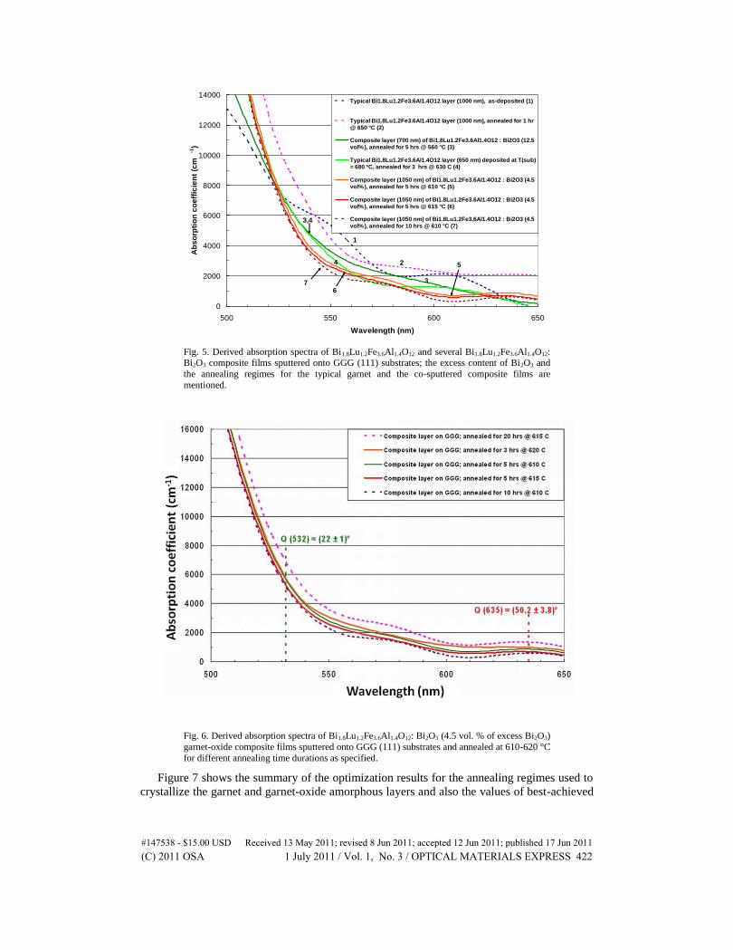
Bi1.8Lu1.2Fe3.6Al1.4O12 garnet layers as shown in Fig. 5. The addition of extra bismuth oxide
didn’t have much impact on the Faraday rotation of the films but it did improve the optical
quality noticeably, consequently improving the magneto-optic quality in terms of MO figure
of merit up to more than 50° at 635 nm.
#147538 - $15.00 USD Received 13 May 2011; revised 8 Jun 2011; accepted 12 Jun 2011; published 17 Jun 2011(C) 2011 OSA 1 July 2011 / Vol. 1, No. 3 / OPTICAL MATERIALS EXPRESS 420

Fig. 4. Transmission spectra of several 1050nm-thick Bi1.8Lu1.2Fe3.6Al1.4O12: Bi2O3 (4.5 vol. %)
composite garnet-oxide layers (samples from the same deposition batch) sputtered onto
monocrystalline GGG (111) and also onto glass (Corning Eagle XG) substrates and post-deposition annealed for 5 h at 610°C and at 615°C; the inset shows a schematic diagram of the
power transmission spectrum measurement using a UV/VIS spectrophotometer. The measured percentage of the incident optical power transmitted through the substrate/film system is
plotted (no additional normalization with respect to the blank substrate transmission was
applied).
The optical and MO properties of garnet materials were critically dependent on the
optimization of annealing process parameters, and the optimization of all annealing process
parameters for each garnet composition type was a time-consuming process since the
annealing behavior of films is strongly composition-dependent [13]. Bi1.8Lu1.2Fe3.6Al1.4O12:
Bi2O3 (4.5 vol. % excess bismuth oxide) composite films were annealed using many possible
approaches to thermal treatment, and it was found that the optimized annealing temperatures
were between 610 and 620 °C and the optimized time durations varied between 3 and 20
hours. Significant effects of the annealing temperature variation on both the optical and MO
properties were observed in this material type. The optimized absorption coefficient spectra
achieved in Bi1.8Lu1.2Fe3.6Al1.4O12: Bi2O3 (4.5 vol. % of excess Bi2O3) garnet-oxide composite
films sputtered onto GGG (111) substrates and annealed at 610-620 °C for different annealing
time durations are shown in Fig. 6.
#147538 - $15.00 USD Received 13 May 2011; revised 8 Jun 2011; accepted 12 Jun 2011; published 17 Jun 2011(C) 2011 OSA 1 July 2011 / Vol. 1, No. 3 / OPTICAL MATERIALS EXPRESS 421
0
2000
4000
6000
8000
10000
12000
14000
500 550 600 650
Wavelength (nm)
Ab
so
rpti
on
co
eff
icie
nt
(cm
-1)
Typical Bi1.8Lu1.2Fe3.6Al1.4O12 layer (1000 nm), as-deposited (1)
Typical Bi1.8Lu1.2Fe3.6Al1.4O12 layer (1000 nm), annealed for 1 hr
@ 650 °C (2)
Composite layer (700 nm) of Bi1.8Lu1.2Fe3.6Al1.4O12 : Bi2O3 (12.5
vol%), annealed for 5 hrs @ 560 °C (3)
Typical Bi1.8Lu1.2Fe3.6Al1.4O12 layer (650 nm) deposited at T(sub)
= 680 °C, annealed for 3 hrs @ 630 C (4)
Composite layer (1050 nm) of Bi1.8Lu1.2Fe3.6Al1.4O12 : Bi2O3 (4.5
vol%), annealed for 5 hrs @ 610 °C (5)
Composite layer (1050 nm) of Bi1.8Lu1.2Fe3.6Al1.4O12 : Bi2O3 (4.5
vol%), annealed for 5 hrs @ 615 °C (6)
Composite layer (1050 nm) of Bi1.8Lu1.2Fe3.6Al1.4O12 : Bi2O3 (4.5
vol%), annealed for 10 hrs @ 610 °C (7)
1
2
3,4
3
5
67
4
Fig. 5. Derived absorption spectra of Bi1.8Lu1.2Fe3.6Al1.4O12 and several Bi1.8Lu1.2Fe3.6Al1.4O12: Bi2O3 composite films sputtered onto GGG (111) substrates; the excess content of Bi2O3 and
the annealing regimes for the typical garnet and the co-sputtered composite films are
mentioned.
Fig. 6. Derived absorption spectra of Bi1.8Lu1.2Fe3.6Al1.4O12: Bi2O3 (4.5 vol. % of excess Bi2O3)
garnet-oxide composite films sputtered onto GGG (111) substrates and annealed at 610-620 °C
for different annealing time durations as specified.
Figure 7 shows the summary of the optimization results for the annealing regimes used to
crystallize the garnet and garnet-oxide amorphous layers and also the values of best-achieved
#147538 - $15.00 USD Received 13 May 2011; revised 8 Jun 2011; accepted 12 Jun 2011; published 17 Jun 2011(C) 2011 OSA 1 July 2011 / Vol. 1, No. 3 / OPTICAL MATERIALS EXPRESS 422

MO figures of merit (data points measured using a 635 nm plane-polarized laser source).
These experimental results provide a reliable source of data for further studies of this
interesting material, which has been synthesized for the first time. The best-achieved (so far)
MO performance characteristics of our garnet and garnet-oxide composite films for two
important wavelengths in the visible spectral region are shown in Fig. 8.
550
570
590
610
630
650
670
Material type
An
ne
ali
ng
te
mp
era
ture
re
gim
es
(°C
)
0
5
10
15
20
25
An
ne
ali
ng
pro
ce
ss
es
du
rati
on
(h
r)
Temperatures found suitable for annealing
Time durations used for running the annealing
processes at the temperatures specified
15
.7°
42
.8°
27
.5°
50
.2°
19
°
42
.8°
25
.6°
34
.5°
Bi 1
.8L
u1.2
Fe
3.6
Al 1
.4O
12
de
po
sit
ed
at
Tsu
b =
25
0 °
C
Bi 1
.8L
u1.2
Fe
3.6
Al 1
.4O
12
de
po
sit
ed
at
Tsu
b =
68
0 °
C
(BiL
u) 3
(Fe
Al)
5O
12
+ 4
.5 v
ol%
Bi 2
O3
(BiL
u) 3
(Fe
Al)
5O
12
+ 4
.5 v
ol%
Bi 2
O3
(BiL
u) 3
(Fe
Al)
5O
12
+ 4
.5 v
ol%
Bi 2
O3
(BiL
u) 3
(Fe
Al)
5O
12
+ 4
.5 v
ol%
Bi 2
O3
(BiL
u) 3
(Fe
Al)
5O
12
+ 4
.5 v
ol%
Bi 2
O3
(BiL
u) 3
(Fe
Al)
5O
12
+ 1
2.5
vo
l% B
i 2O
3
Fig. 7. The data points showing the summary of optimization of annealing temperature and
annealing processes duration used to crystallize the typical Bi1.8Lu1.2Fe3.6Al1.4O12 garnet layer
deposited at 250 °C and 680 °C substrates’ temperature and several composite Bi1.8Lu1.2Fe3.6Al1.4O12: Bi2O3 films of having excess Bi2O3 onto GGG (111) substrate.
13.9
11.9
22.1
22.17
20.85
18.03
20.6
42.8
50.2
42.8
34.5
25.6
27.5
15.7
0 20 40 60
Co
mp
osit
ion
typ
e
MO figure of merit (degrees)
635 nm MO figure of merit
532 nm MO figure of merit
(BiLu)3(FeAl)5O12 + 4.5 vol% Bi2O3
annealed for 3 hrs @ 620 °C
(BiLu)3(FeAl)5O12 + 4.5 vol% Bi2O3
annealed for 20 hrs @ 615 °C
(BiLu)3(FeAl)5O12 + 4.5 vol% Bi2O3
annealed for 5 hrs @ 615 °C
(BiLu)3(FeAl)5O12 + 4.5 vol% Bi2O3
annealed for 10 hrs @ 610 °C
(BiLu)3(FeAl)5O12 + 4.5 vol% Bi2O3
annealed for 5 hrs @ 610 °C
Bi1.8Lu1.2Fe3.6Al1.4O12
deposited at Tsub = 680 °C,
annealed for 3 hrs @ 630 °C
Bi1.8Lu1.2Fe3.6Al1.4O12
deposited at Tsub = 250 °C,
annealed for 1 hr @ 650 °C
Fig. 8. Measured quality factor in terms of figure of merit of typical Bi1.8Lu1.2Fe3.6Al1.4O12
garnet layer deposited at 250 °C and 680 °C substrates’ temperature and several best annealed
composite Bi1.8Lu1.2Fe3.6Al1.4O12: Bi2O3 films of having 4.5 vol. % excess Bi2O3 onto GGG (111) substrate.
The effects of adding bismuth oxide on the coercivity of the films sputtered onto both
GGG and glass substrates were observed, and the results are presented in Figs. 9 and 10.
Comparatively, lower coercive force values were measured in composite films sputtered onto
#147538 - $15.00 USD Received 13 May 2011; revised 8 Jun 2011; accepted 12 Jun 2011; published 17 Jun 2011(C) 2011 OSA 1 July 2011 / Vol. 1, No. 3 / OPTICAL MATERIALS EXPRESS 423

both types of substrates. We believe that better crystalline quality, lower coercive force
values and even higher magnetic field sensitivity can be achieved in our films sputtered onto
GGG substrates, if high-substrate-temperature deposition regime is optimized to achieve the
conditions suitable for epitaxial-quality layer growth (sputter epitaxy).
Note that two experimental setups were used to confirm the calibration accuracy of the
Thorlabs PAX polarimeter that was used for Faraday rotation measurements, namely (i) the
direct measurements of optical power transmitted through the sample and the use of an
analyzer rotated 45 degrees with respect to the polarisation direction of the incident laser
light, under various magnetization conditions, and (ii) a well-calibrated measurement setup
based on the detection of polarisation components. The measured Faraday rotations for both
setups were in excellent agreement. The Thorlabs PAX polarimeter had a high dynamic range
of 70 dB, a broad wavelength range, and an accuracy of ± 0.2°.
-7
-6
-5
-4
-3
-2
-1
0
1
2
3
4
5
6
7
-1500 -1000 -500 0 500 1000 1500
Magnetic field (Oe)
Fa
rad
ay
ro
tati
on
(d
eg
/ mm
)
Typical Bi1.8Lu1.2Fe3.6Al1.4O12 garnet film
on GGG; Hc = (45 +/- 5) Oe
Composite Bi1.8Lu1.2Fe3.6Al1.4O12: Bi2O3
(4.5 vol%) film on GGG; Hc = (30 +/- 5) Oe
-6
-5
-4
-3
-2
-1
0
1
2
3
4
5
6
-400 -300 -200 -100 0 100 200 300 400
Magnetic field (Oe)
Fa
rad
ay
ro
tati
on
(d
eg
/ mm
)
Hc = (45 +/- 5) Oeand Hsat = 350 Oe
Hc = (30 +/- 5) Oeand Hsat = 270 Oe
Fig. 9. Hysteresis loops of specific Faraday rotation measured at 532 nm in sputtered typical
Bi1.8Lu1.2Fe3.6Al1.4O12 layer on GGG (annealed for 1 h at 650 °C) and Bi1.8Lu1.2Fe3.6Al1.4O12: Bi2O3 (4.5 vol. %) composite garnet films deposited onto GGG substrate (annealed at 620 °C
for 3 h). Insets show the measured coercive force, and saturation field values within the linear
ranges of magnetization.
#147538 - $15.00 USD Received 13 May 2011; revised 8 Jun 2011; accepted 12 Jun 2011; published 17 Jun 2011(C) 2011 OSA 1 July 2011 / Vol. 1, No. 3 / OPTICAL MATERIALS EXPRESS 424

-6
-5
-4
-3
-2
-1
0
1
2
3
4
5
6
-1500 -1000 -500 0 500 1000 1500
Magnetic field (Oe)
Fa
rad
ay
ro
tati
on
(d
eg
/ mm
)
Typical Bi1.8Lu1.2Fe3.6Al1.4O12 garnet film
on glass; Hc = (100 +/- 5) Oe
Composite Bi1.8Lu1.2Fe3.6Al1.4O12 : Bi2O3
(12.5 vol%) film on glass; Hc =(80 +/- 5) Oe
-5
-4
-3
-2
-1
0
1
2
3
4
5
-500 -400 -300 -200 -100 0 100 200 300 400 500
Magnetic field (Oe)
Fa
rad
ay
ro
tati
on
(d
eg
/ mm
)
Hc = (100 +/- 5) Oeand Hsat = 400 Oe
Hc = (80 +/- 5) Oeand Hsat = 300 Oe
Fig. 10. Hysteresis loops of specific Faraday rotation measured at 532 nm in sputtered
Bi1.8Lu1.2Fe3.6Al1.4O12 garnet films on glass substrate (annealed for 3 h at 630 °C) and
Bi1.8Lu1.2Fe3.6Al1.4O12: Bi2O3 (12.5 vol. %) composite garnet films deposited onto glass
substrate (annealed at 560 °C for 5 h). Insets show the measured coercive force and saturation
field within the linear ranges of magnetization.
The surface morphology as well as surface magnetic field distribution topography of
Bi1.8Lu1.2Fe3.6Al1.4O12: Bi2O3 composite films having 4.5 vol. % and 12.5 vol. % of excess
bismuth oxide sputtered onto GGG substrates have been characterized using atomic force
microscopy (AFM) and magnetic force microscopy (MFM). Figure 11 shows the scanning
probe microscopy inspection results for garnet samples of composition Bi1.8Lu1.2Fe3.6Al1.4O12:
Bi2O3 (4.5 vol. %) presented as 3D images of the surface features and surface magnetic field
distribution (Fig. 11 (a and b)), and also shows the results for a Bi1.8Lu1.2Fe3.6Al1.4O12: Bi2O3
(12.5 vol. %) film presented as 2D images (Fig. 11(c, d)). The garnet samples were scanned
using semi-contact mode of probe-interaction to obtain the feedback-phase and also the
surface topography data simultaneously from the same scan area. The MFM cantilever tip
used was cobalt-coated to enable the magnetic-force interaction representation through the
phase of the cantilever feedback signal. Nano-crystalline surface microstructure and its
associated surface roughness features of the garnet films were observed from the obtained
high-contrast images extracted from the measured feedback-phase images and topography
data. The magnetic domains structure and the map of magnetic interaction force between the
cantilever tip and sample surface were also imaged.
#147538 - $15.00 USD Received 13 May 2011; revised 8 Jun 2011; accepted 12 Jun 2011; published 17 Jun 2011(C) 2011 OSA 1 July 2011 / Vol. 1, No. 3 / OPTICAL MATERIALS EXPRESS 425

Fig. 11. Scanning-probe (AFM/MFM) images of garnet-oxide composite thin films having 4.5 vol. % and 12.5 vol. % extra bismuth oxide sputtered onto GGG (111) substrates. (a-b) 3D
images showing the topography (5 × 5 µm sample area) of a 1050 nm thick
Bi1.8Lu1.2Fe3.6Al1.4O12:Bi2O3 (4.5 vol. %) composite film annealed for 5 h at 615 °C and its
surface magnetism features measured across a 25 × 25 µm sample area; (c-d) 2D AFM
topography (c) and (d) an AC magnetic force magnitude map (processed feedback phase
image) obtained from a 1.2 × 1.2 µm sample area of a Bi1.8Lu1.2Fe3.6Al1.4O12:Bi2O3 (12.5 vol. %) nanocomposite film annealed for 5 h at 580 °C. The black-white color palette of image (d)
represents the measured RMS strength of the AC magnetic interaction force between the tip
and surface, and the color map shown was obtained using a halved algebraic sum of the phase image data map obtained and its inverted phase image data map, so that only the magnitude of
the magnetic interaction force is represented. The white-colored pixels correspond to the
minima locations of the magnetic interaction force.
It is important to notice that Figs. 11(a, b) reveal that the addition of extra bismuth oxide
results in bismuth-rich MO garnet-phase grains surrounded by transparent non-magnetic
bismuth oxide regions. While the measured overall Faraday rotation of the composite film
was not improved, the measured overall optical transmission was considerably increased,
leading to significant increase in MO figure of merit.
To the best of our knowledge, this is the first report on the properties of this advanced MO
material type (garnet-oxide nanocomposites of class Bi1.8Lu1.2Fe3.6Al1.4O12:Bi2O3) appearing
in the literature published to date. Our work is ongoing and further results, especially on the
crystal structure and microstructural details of Bi1.8Lu1.2Fe3.6Al1.4O12 garnet layers and their
co-sputtered nanocomposite derivatives will be reported elsewhere.
4. Conclusion
We have studied the RF sputtering deposition and oven annealing processes required for the
manufacture of high-performance magneto-optic films of composition types
Bi1.8Lu1.2Fe3.6Al1.4O12 and (BiLu)3(FeAl)5O12:Bi2O3 on garnet and glass substrates. The
optical, magnetic and magneto-optical properties of a range of highly Bi-substituted lutetium
iron-aluminum garnet layers have been characterized in detail and reported for the first time.
#147538 - $15.00 USD Received 13 May 2011; revised 8 Jun 2011; accepted 12 Jun 2011; published 17 Jun 2011(C) 2011 OSA 1 July 2011 / Vol. 1, No. 3 / OPTICAL MATERIALS EXPRESS 426

Significantly improved magneto-optical figures of merit have been achieved using the co-
sputtering bismuth oxide-mixing approach, and the results confirm that this synthesis method
is suitable for a wide range of Bi-substituted MO garnet materials. The developed garnet and
garnet-oxide thin film materials possess a combination of properties which are highly
promising in regard to the future development of garnet waveguides, non-reciprocal
integrated-optics components as well as magnetic field imaging and sensing devices.
Acknowledgment
This research is supported by the Faculty of Computing, Health and Science, Edith Cowan
University. We also acknowledge the support provided by the Department of Nanobio
Materials and Electronics, Gwangju Institute of Science and Technology (South Korea).
#147538 - $15.00 USD Received 13 May 2011; revised 8 Jun 2011; accepted 12 Jun 2011; published 17 Jun 2011(C) 2011 OSA 1 July 2011 / Vol. 1, No. 3 / OPTICAL MATERIALS EXPRESS 427


