

HistologyStudy of TissuesEpithelial TissueConnective TissueNervous and Muscular Tissue Intercellular Junctions, Glands and MembranesTissue Growth, Development, Death and Repair
5-1

The Study of Tissues50 trillion cells of 200 different cell typesFour broad categories of tissues
epithelial tissue covers surfaces because cells are in contact lines hollow organs, cavities and ducts forms glands when cells sink under the surface
connective tissue material found between cells supports and binds structures together stores energy as fat provides immunity
nervous tissue cells that conduct electrical signals detects changes inside and outside the body responds with nerve impulses
muscular tissue cells shorten in length producing movement
Organ - structure with discrete boundaries that is composed of two or more tissue types
Histology (microscopic anatomy) – the study of tissues and how they are arranged into organs
5-2
The Primary Tissue ClassesTissue – a group of similar cells and cell products that arise from
the same region of the embryo and work together to perform a specific structural or physiological role in an organ.
Four primary tissues differ from one another in the: types and functions of their cells the characteristics of the matrix (extracellular material) the relative amount of space occupied by cells versus matrix
Matrix – (extracellular material) is composed of :fibrous proteinsa clear gel known as ground substance , tissue fluid, extracellular fluid
(ECF), interstitial fluid, or tissue gel
5-3
Embryonic TissuesHuman development begins as single cell the fertilized egg
divides to produce scores of identical, smaller cellsfirst tissues appear when these cells start to organize themselves into
layers first two, and then three strata
3 primary germ layersectoderm (outer)
gives rise to epidermis and nervous systemendoderm (inner)
gives rise to mucous membrane lining digestive and respiratory tracts, digestive glands, and among other things
mesoderm (middle) becomes gelatinous tissue - mesenchyme wispy collagen fibers and fibroblasts in gel matrix gives rise to muscle, bone, blood
5-4

Epithelial Tissue
5-5
Layers of closely adhering cellsCell sheet with upper surface
exposed to the environment or an internal body cavity
Apical side (free surface)Basal side (on basement membrane)
No blood vessels (avascular) underlying connective tissue supplies
oxygenRests on basement membrane
thin layer of collagen and adhesive proteins anchors epithelium to connective tissue
5-6
Types of Epithelium
• Covering and lining epithelium– epidermis of skin– lining of blood vessels and ducts– lining respiratory, reproductive, urinary & GI tract
• Glandular epithelium– secreting portion of glands– thyroid, adrenal, and sweat glands

Simple vs. Stratified EpitheliaSimple epithelium
contains one layer of cellsnamed by shape of cellsall cells touch the basement
membrane
5-7
• Stratified epithelium– contains more than one layer– named by shape of apical cells– some cells rest on top of others and do not touch basement membrane
Squamous Cuboidal Columnar
Simple
(a) Classes of epithelium
Pseudostratifiedcolumnar
Stratified
(b) Cell shapes
Simple EpitheliaFour types of simple epithelia
Three named for their cell shapes simple squamous (thin scaly cells) simple cuboidal (square or round cells) simple columnar (tall narrow cells)
Fourth type – pseudostratified columnar
not all cells reach the free surface shorter cells are covered over by taller ones looks stratified every cell reaches the basement membrane
Goblet cells – wineglass-shaped mucus secreting cells in simple columnar and pseudostratified epithelia
5-8
5-9
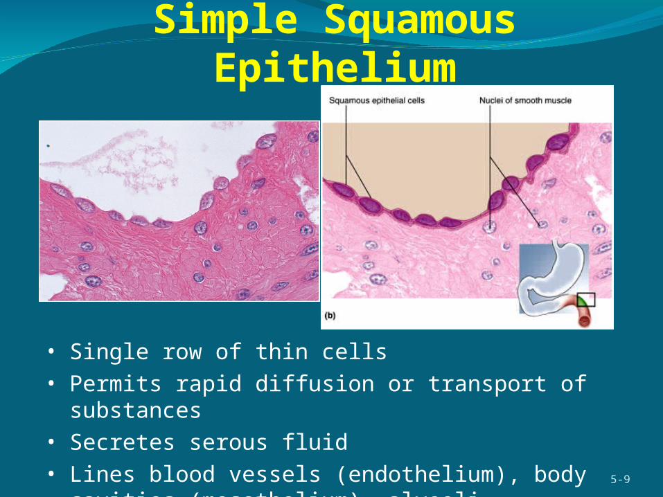
• Single row of thin cells• Permits rapid diffusion or transport of substances• Secretes serous fluid• Lines blood vessels (endothelium), body cavities (mesothelium),
alveoli
Simple Squamous Epithelium

Simple Cuboidal Epithelium
Single layer of square or round cellsAbsorption and secretion, mucus production and movementLiver, thyroid, mammary and salivary glands, bronchioles, and
kidney tubules
5-10
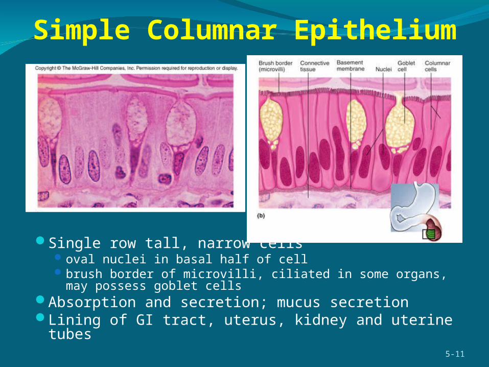
Single row tall, narrow cellsoval nuclei in basal half of cellbrush border of microvilli, ciliated in some organs, may possess goblet
cellsAbsorption and secretion; mucus secretion Lining of GI tract, uterus, kidney and uterine tubes
5-11
Simple Columnar Epithelium
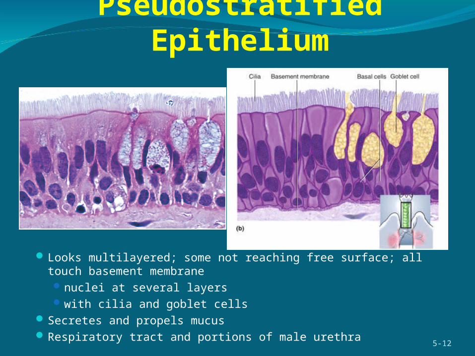
Pseudostratified Epithelium
Looks multilayered; some not reaching free surface; all touch basement membranenuclei at several layerswith cilia and goblet cells
Secretes and propels mucus Respiratory tract and portions of male urethra
5-12

Stratified Epithelia
5-13
More than one layer of cells Named for shape of surface cells
exception is transitional epitheliumDeepest cells on basement membraneVariations
keratinized epithelium has surface layer of dead cellsnonkeratinized epithelium lacks the layer of dead cells

Keratinized Stratified Squamous
Multiple cell layers with cells becoming flat and scaly toward surface
Epidermis; palms and soles heavily keratinizedResists abrasion; retards water loss through skin; resists
penetration by pathogenic organisms 5-14

Nonkeratinized Stratified Squamous
Same as keratinized epithelium without the surface layer of dead cells
Tongue, oral mucosa, esophagus and vaginaResists abrasion and penetration of pathogens
5-15
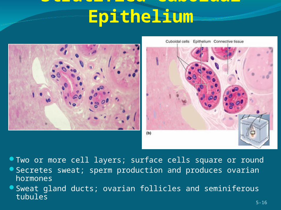
Stratified Cuboidal Epithelium
Two or more cell layers; surface cells square or roundSecretes sweat; sperm production and produces ovarian
hormonesSweat gland ducts; ovarian follicles and seminiferous tubules
5-16
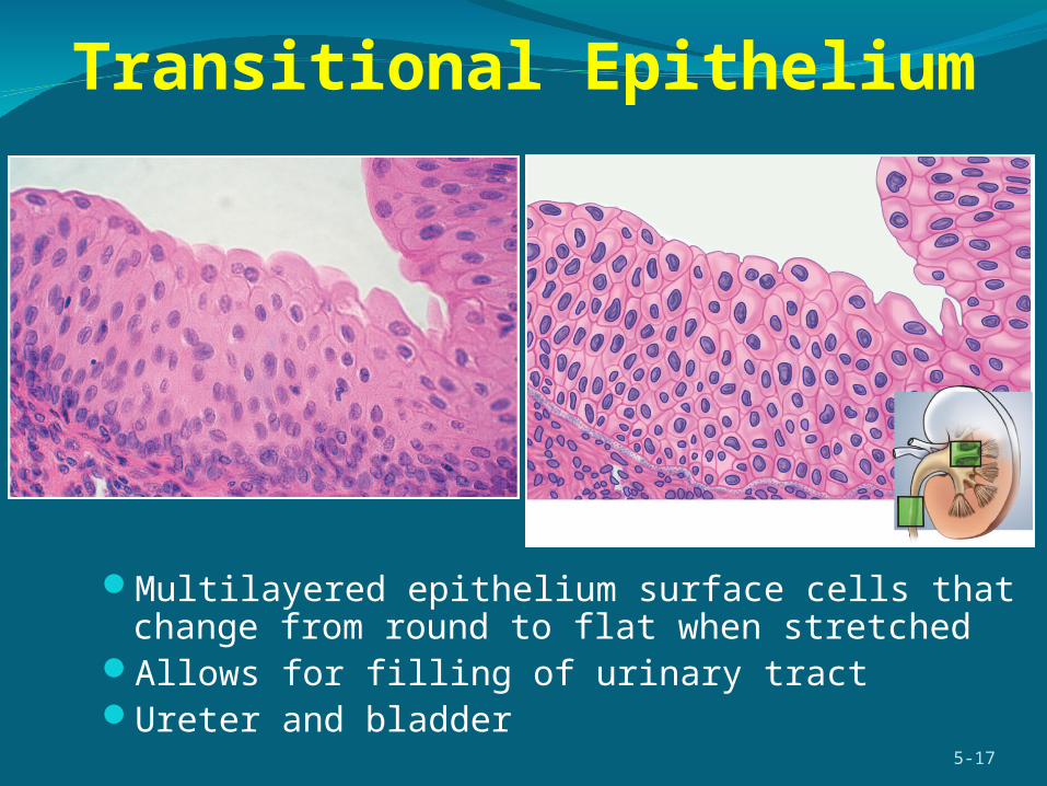
Transitional Epithelium
Multilayered epithelium surface cells that change from round to flat when stretched
Allows for filling of urinary tractUreter and bladder
5-17
Endocrine and Exocrine GlandsGland – cell or organ that secrete substances for use elsewhere in the body or
releases them for elimination from the body composed of epithelial tissue in a connective tissue framework and capsule
Exocrine glands - maintain their contact with the body surface by way of a duct (epithelial tube that conveys secretion to surface) sweat, mammary and tear glands
Endocrine glands - lose their contact with the surface and have no ducts hormones – secretion of endocrine glands secrete (hormones) directly into blood thyroid, adrenal and pituitary glands
Some organs have both endocrine and exocrine function liver, gonads
Unicellular glands – found in epithelium that is predominantly nonsecretory can be endocrine or exocrine mucus-secreting goblet or endocrine cells of stomach and small intestine
5-18
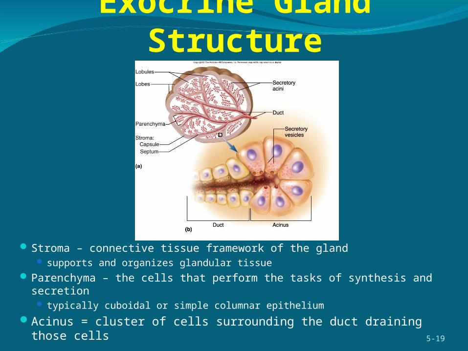
Exocrine Gland Structure
Stroma – connective tissue framework of the gland supports and organizes glandular tissue
Parenchyma – the cells that perform the tasks of synthesis and secretion typically cuboidal or simple columnar epithelium
Acinus = cluster of cells surrounding the duct draining those cells5-19

Duct
Secretory portion
Compound acinar Compound tubuloacinarSimple coiled tubular
Example: Sweat gland
Example: Mammary gland
Example: PancreasKey
Types of Exocrine Glands
Simple - unbranched ductCompound - branched ductShape of gland
tubular – duct and secretory portion have uniform diameter acinar - secretory cells form dilated sac (acinus or alveolus) tubuloacinar - both tubular and acinar portions
5-20
Figure 5.30

Types of SecretionsSerous glands
produce thin, watery secretions perspiration, milk, tears and digestive juices
Mucous glandsproduce glycoprotein, mucin, that absorbs water to form a
sticky secretion called mucusgoblet cells – unicellular mucous glands
Mixed glands contain both cell types and produce a mixture of the two types
of secretionsCytogenic glands
release whole cells, sperm and egg cells5-21
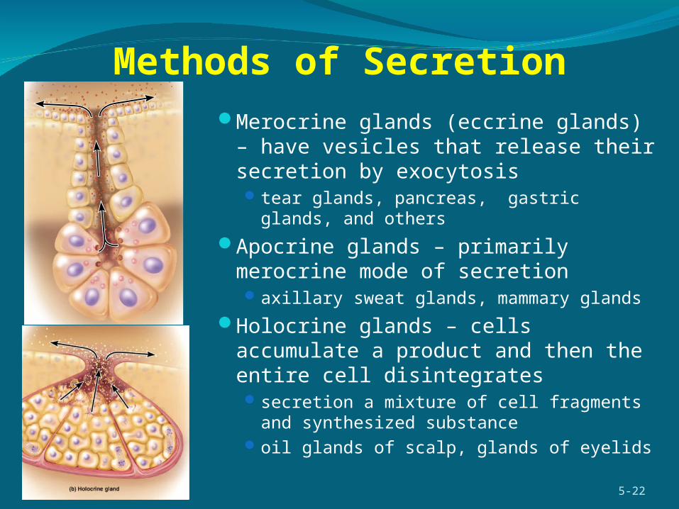
Methods of SecretionMerocrine glands (eccrine glands) – have
vesicles that release their secretion by exocytosis tear glands, pancreas, gastric glands, and others
Apocrine glands – primarily merocrine mode of secretionaxillary sweat glands, mammary glands
Holocrine glands – cells accumulate a product and then the entire cell disintegrates secretion a mixture of cell fragments and
synthesized substanceoil glands of scalp, glands of eyelids
5-22
Connective TissueConnective tissue – a type of tissue in which cells usually
occupy less space than the extracellular material
Binds organs to each other
Support and protect organs
Most cells of connective tissue are not in direct contact with each otherseparated by extracellular material
Highly vascular – richly supplied with blood vessels
Most abundant, widely distributed, and histologically variable of the primary tissues
5-23
Functions of Connective TissueBinding of organs – tendons and ligaments
Support – bones and cartilage
Physical protection – cranium, ribs, sternum
Immune protection – white blood cells attack foreign invaders
Movement – bones provide lever system
Storage – fat, calcium, phosphorus
Heat production – metabolism of brown fat in infants
Transport - blood5-24
Components of Fibrous Connective Tissue
CellsBlast type cells = retain ability to divide & produce matrix
(fibroblasts, chondroblasts, & osteoblasts)Cyte type cells = mature cell that can not divide or produce
matrix (chondrocytes & osteocytes)Macrophages develop from monocytes
engulf bacteria & debris by phagocytosisPlasma cells develop from B lymphocytes
produce antibodies that fight against foreign substancesMast cells produce histamine that dilate small BVAdipocytes (fat cells) store fat
5-25

Components of Fibrous Connective Tissue
FibersCollagenous fibers
most abundant of the body’s proteins – 25% tough, flexible, and resist stretching tendons, ligaments, and deep layer of the skin are mostly collagen less visible in matrix of cartilage and bone
Reticular fibers thin collagen fibers coated with glycoprotein form framework of such organs as spleen and lymph nodes
Elastic fibers thinner than collagenous fibers branch and rejoin each other made of protein called elastin allows stretch and recoil yellow fibers – fresh elastic fibers 5-26

Components of Fibrous Connective Tissue
Ground substanceusually a gelatinous to rubbery consistency resulting from three classes of
large moleculesglycosaminoglycans (GAG)
long polysaccharide composed of unusual disaccharides called amino sugars and uronic acid
play important role of regulating water and electrolyte balance in the tissues chondroitin sulfate – most abundant GAG
in blood vessels and bone responsible for stiffness of cartilage
hyaluronic acid – viscous, slippery substance that forms an effective lubricant in joints and constitutes much of the vitreous body of the eyeball
proteoglycan gigantic molecule shaped like a test-tube brush forms thick colloids that creates strong structural bond between cells and
extracellular macromolecules – holds tissues togetheradhesive glycoproteins – bind components of tissues together
5-27

Types of Fibrous Connective TissueLoose connective tissue
much gel-like ground substance between cells
types areolar reticular
Dense connective tissuefibers fill spaces between cellstypes vary in fiber orientation
dense regular connective tissue dense irregular connective tissue
5-28

Loose Connective-Areolar TissueLoosely organized fibers, abundant blood
vessels, and a lot of seemingly empty space
Possess all six cell types
Fibers run in random directions mostly collagenous, but elastic and reticular also present
Found in tissue sections from almost every part of the body surrounds blood vessels and nerves
Nearly every epithelium rests on a layer of areolar tissue blood vessels provide nutrition to epithelium and waste
removal ready supply of infection fighting leukocytes that move about
freely in areolar tissue
5-29
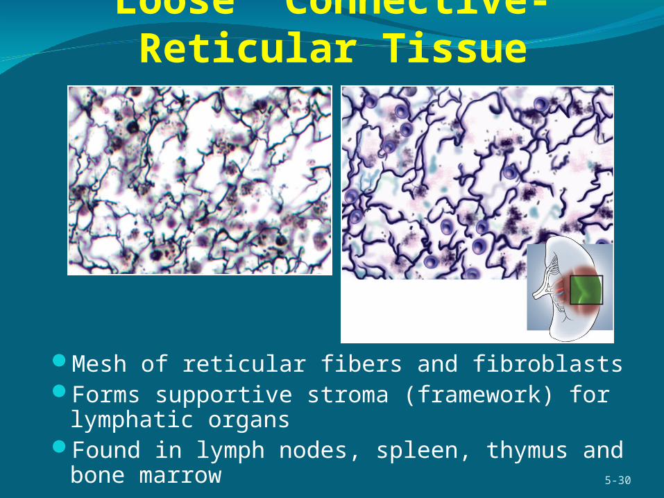
Loose Connective-Reticular Tissue
Mesh of reticular fibers and fibroblastsForms supportive stroma (framework) for lymphatic organsFound in lymph nodes, spleen, thymus and bone marrow
5-30

Dense Regular Connective Tissue
Densely, packed, parallel collagen fiberscompressed fibroblast nuclei
Tendons attach muscles to bones and ligaments hold bones together
5-31
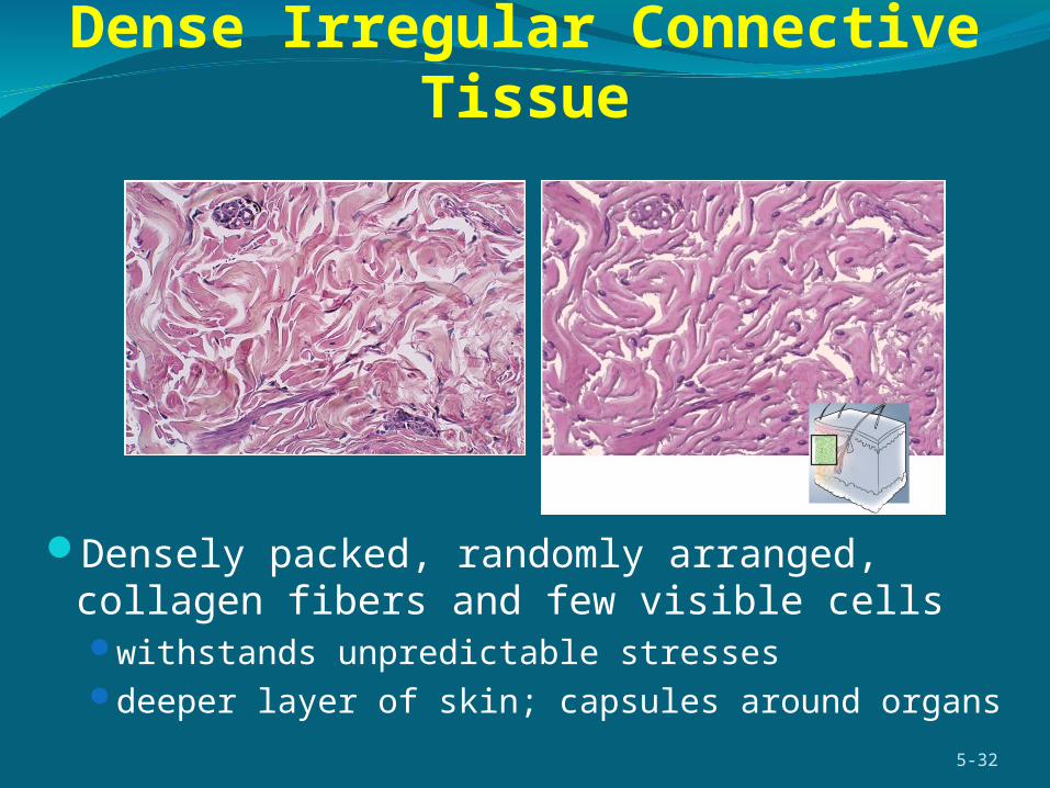
Dense Irregular Connective Tissue
Densely packed, randomly arranged, collagen fibers and few visible cellswithstands unpredictable stressesdeeper layer of skin; capsules around organs
5-32

Adipose Tissue Adipose tissue (fat) – tissue in which adipocytes are the dominant cell type
Space between adipocytes is occupied by areolar tissue, reticular tissue, and blood capillaries
5-33
Fat is the body’s primary energy reservoir The quantity of stored triglyceride and the number of
adipocytes are quite stable in a person fat is recycled continuously to prevent stagnation new triglycerides are constantly synthesized and stored old triglycerides are hydrolyzed and released into
circulation
Provides thermal insulation Anchors and cushions organs such as eyeball, kidneys Contributes to body contours – female breast and hips On average, women have more fat than men Too little fat can reduce female fertility Most adult fat is called white fat Brown fat – in fetuses, infants, children – a heat
generating tissue
CartilageSupportive connective tissue with flexible, rubbery matrix
Chondroblasts produce matrix and surround them selves until they become trapped in little cavities (lacunae)
Chondrocytes – cartilage cells in lacunae
Perichondrium – sheath of dense irregular connective tissue that surrounds elastic and most hyaline cartilage (not articular cartilage) contains a reserve population of chondroblasts that contribute to cartilage growth
throughout life
No blood vessels diffusion brings nutrients and removes wastes heals slowly
Matrix rich in chondroitin sulfate and contain collagen fibers
Types of cartilage vary with fiber types hyaline cartilage, fibrocartilage and elastic cartilage
5-34

Hyaline Cartilage
Clear, glassy microscopic appearance because of unusual fineness of the collagen fibers
Usually covered by perichondriumArticular cartilage, costal cartilage, trachea, larynx, fetal skeletonEases joint movement, holds airway open, moves vocal cords during speech
5-35

Elastic Cartilage
Cartilage containing elastic fibers Covered with perichondriumProvides flexible, elastic support
external ear and epiglottis5-36
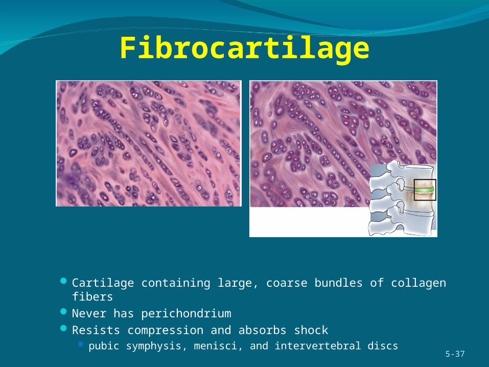
Cartilage containing large, coarse bundles of collagen fibers Never has perichondriumResists compression and absorbs shock
pubic symphysis, menisci, and intervertebral discs5-37
Fibrocartilage
Bone‘Bone’ has two meanings:
an organ of the body; femur, mandible; composed of multiple tissue types
bone tissue – osseous tissue – makes up most of the mass of bone
Two forms of osseous tissue spongy bone - spongy in appearance
delicate struts of bone - trabeculae covered by compact bone found in heads of long bones and in middle of flat bones such as the
sternumcompact bone – denser calcified tissue with no visible spaces
more complex arrangement cells and matrix surround vertically oriented blood vessels in long bones
5-38

Bone Tissue-Compact bone
Osteon – central canal and its surrounding lamellaeOsteocytes – mature bone cells that occupy the lacunaeCanaliculi – delicate canals that radiate from each lacuna to its
neighbors, and allows osteocytes to contact each otherPeriosteum – tough fibrous connective tissue covering of the bone
as a whole
5-39
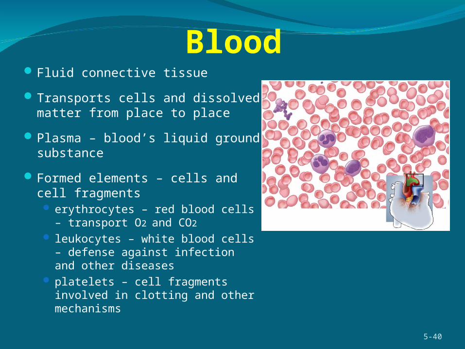
BloodFluid connective tissue
Transports cells and dissolved matter from place to place
Plasma – blood’s liquid ground substance
Formed elements – cells and cell fragments erythrocytes – red blood cells – transport
O2 and CO2
leukocytes – white blood cells – defense against infection and other diseases
platelets – cell fragments involved in clotting and other mechanisms
5-40
Excitable Tissues Muscular & Nervous Tissue
Excitability – a characteristic of all living cellsdeveloped to highest degree in nervous and muscular tissues
Membrane potential – electrical charge difference (voltage) that occurs across the plasma membranes is the basis for their excitationrespond quickly to outside stimulus by means of changes in
membrane potentialnerves – changes result in rapid transmission of signals to
other cellsmuscles – changes result in contraction, shortening of the cell
5-41

Nervous TissueConsists of neurons (nerve cells)
detect stimulirespond quicklytransmit coded information
rapidly to other cells
and neuroglia (glial)protect and assist neurons‘housekeepers’ of nervous
system
Neuron partsneurosoma (cell body)dendritesaxon (nerve fiber)
5-42
Muscular TissueMuscular tissue – elongated cells that are specialized
to contract in response to stimulation
Primary job is to exert physical force on other tissues and organs
Creates movements involved in body and limb movement, digestion, waste elimination, breathing, speech, and blood circulation
Important source of body heat
Three types of muscle: skeletal, cardiac, and smooth5-43
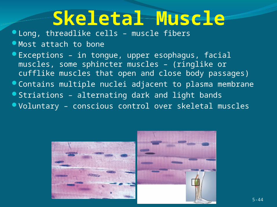
Skeletal MuscleLong, threadlike cells – muscle fibersMost attach to boneExceptions – in tongue, upper esophagus, facial muscles, some
sphincter muscles – (ringlike or cufflike muscles that open and close body passages)
Contains multiple nuclei adjacent to plasma membraneStriations – alternating dark and light bandsVoluntary – conscious control over skeletal muscles
5-44
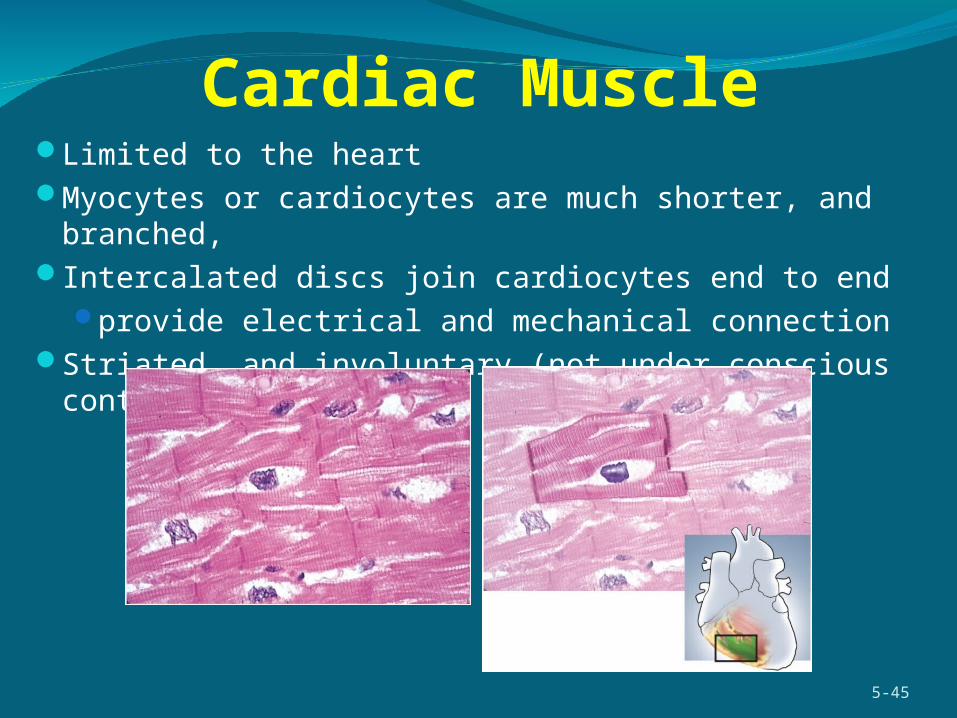
Cardiac MuscleLimited to the heartMyocytes or cardiocytes are much shorter, and branched, Intercalated discs join cardiocytes end to end
provide electrical and mechanical connectionStriated, and involuntary (not under conscious control)
5-45
Smooth Muscle
5-46
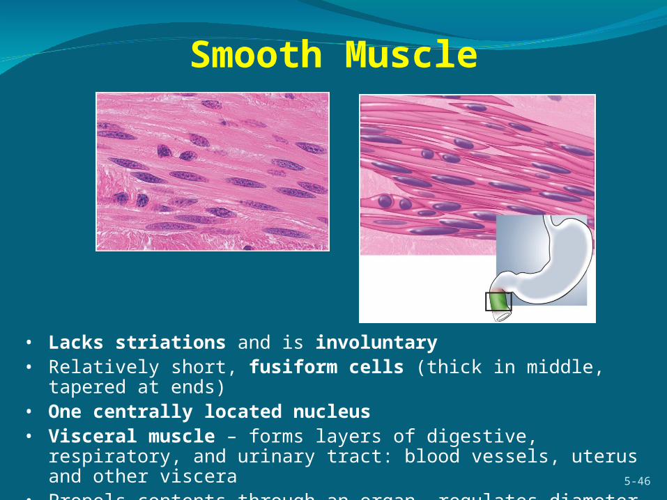
• Lacks striations and is involuntary• Relatively short, fusiform cells (thick in middle, tapered at ends)• One centrally located nucleus• Visceral muscle – forms layers of digestive, respiratory, and urinary tract:
blood vessels, uterus and other viscera• Propels contents through an organ, regulates diameter of blood vessels

Intercellular Junctions
Intercellular junctions – connections between one cell and anotherAll cells (except blood and metastatic cancer cells) are anchored to each other or
their matrix by intercellular junctionsResist stress and communicate with each other
5-47
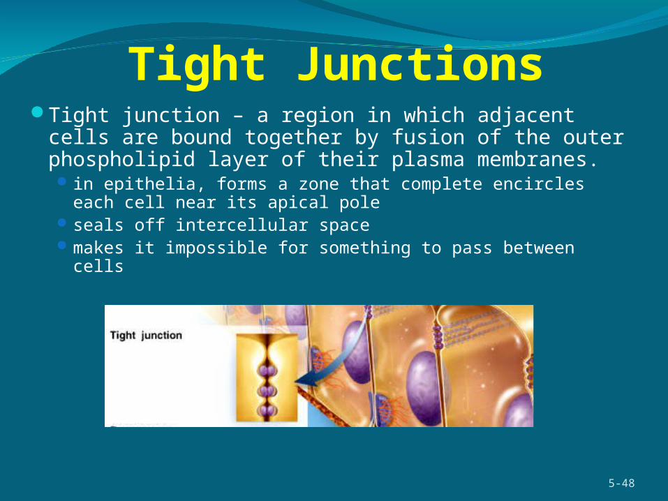
Tight JunctionsTight junction – a region in which adjacent cells are bound
together by fusion of the outer phospholipid layer of their plasma membranes. in epithelia, forms a zone that complete encircles each cell near its
apical pole seals off intercellular spacemakes it impossible for something to pass between cells
5-48
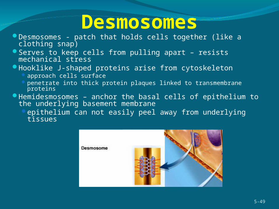
DesmosomesDesmosomes - patch that holds cells together (like a clothing snap)Serves to keep cells from pulling apart – resists mechanical stressHooklike J-shaped proteins arise from cytoskeleton
approach cells surfacepenetrate into thick protein plaques linked to transmembrane proteins
Hemidesmosomes – anchor the basal cells of epithelium to the underlying basement membraneepithelium can not easily peel away from underlying tissues
5-49
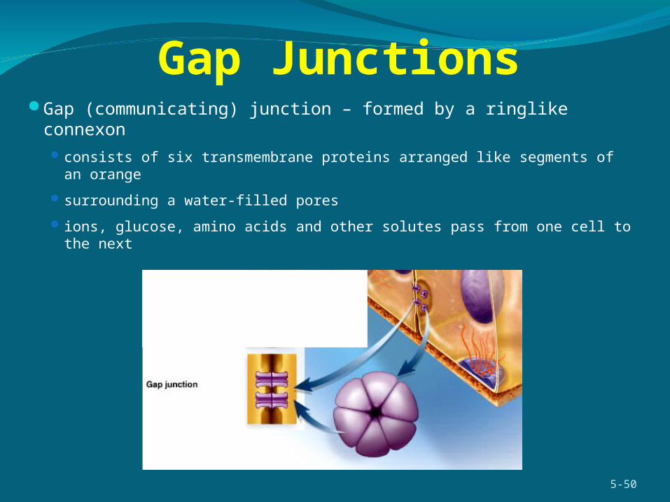
Gap JunctionsGap (communicating) junction – formed by a ringlike connexon
consists of six transmembrane proteins arranged like segments of an orange
surrounding a water-filled pores
ions, glucose, amino acids and other solutes pass from one cell to the next
5-50
MembranesMembranes – line body cavities and cover their viscera
Cutaneous membrane - the skin – largest membrane in the body stratified squamous epithelium (epidermis) over connective tissue (dermis) relatively dry layer serves protective function
Mucous membrane (mucosa) – lines passageways open to the external environment
Serous membrane (serosa) - internal membrane simple squamous epithelium over areolar tissue produces serous fluid that arises from bloodcovers organs and lines walls of body cavities
endothelium lines blood vessels and heart mesothelium line body cavities (pericardium, peritoneum and pleura)
Synovial membrane - lines joint cavitiesconnective tissue layer only, secretes synovial fluid
5-51

Mucous Membranes (Mucosa)
Lines passages that open to the external environment digestive, respiratory, urinary, and reproductive tracts
Consists of two to three layers: epithelium lamina propria – areolar connective tissue muscularis mucosae – smooth muscle layer
Absorptive, secretory, and protective functionsCovered with mucus
5-52
Mucous coat
Cilia
Basement membrane
Collagen fibers
Fibroblast
Muscularis mucosae
Elastic fibers
Blood vessel
Ciliated cells of pseudostratified epithelium
Mucin in goblet cellEpithelium
Lamina propria
Mucous membrane (mucosa)
Tissue GrowthTissue growth – increasing the number of cells or the
existing cells grow larger
Hyperplasia - tissue growth through cell multiplication
Hypertrophy - enlargement of preexisting cellsmuscle grow through exerciseaccumulation of body fat
Neoplasia – development of a tumor (neoplasm)benign or malignantcomposed of abnormal, nonfunctional tissue
5-53

Changes in Tissue TypesTissues can change types
Differentiationunspecialized tissues of embryo become specialized mature
types mesenchyme to muscle
Metaplasiachanging from one type of mature tissue to another
simple cuboidal tissue of vagina before puberty changes to stratified squamous after puberty
pseudostratified columnar epithelium of bronchi of smokers to stratified squamous epithelium
5-54
Stem Cells
5-55
Undifferentiated cells with developmental plasticityEmbryonic stem cells
totipotent (any cell type possible) source = cells of very early embryo
Pluripotent (tissue types only possible) source = cells of inner cell mass of embryo
Adult stem cells (undifferentiated cells in tissues of adults)multipotent (bone marrow producing several blood cell
types)unipotent (only epidermal cells produced)
Tissue RepairRegeneration - replacement of dead or damaged cells
by the same type of cell as beforerestores normal functionskin injuries and liver regenerate
Fibrosis - replacement of damaged cells with scar tissue holds organs togetherdoes not restore normal function
severe cuts and burns, healing of muscle injuries, scarring of lungs in tuberculosis
5-56

Tissue Shrinkage and DeathAtrophy – shrinkage of a tissue through a loss in cell size or number
senile atrophy through normal agingdisuse atrophy from lack of use (astronauts)
Necrosis – premature, pathological death of tissue due to trauma, toxins, or infections infarction – sudden death of tissue when blood supply is cut offgangrene – tissue necrosis due to insufficient blood supplydecubitus ulcer – bed sore or pressure sore
pressure reduces blood flow to an area a form of dry gangrene
gas gangrene - anaerobic bacterial infectionApoptosis - programmed cell death
normal death of cells that have completed their function and best serve the body by dying and getting out of the way
5-57

Tissue EngineeringTissue engineering – artificial production of tissues and
organs in the lab for implantation in the human bodyframework of collagen or biodegradable polyester fibersseeded with human cellsgrown in “bioreactor” (inside of mouse)
supplies nutrients and oxygen to growing tissue
Skin grafts already availableresearch in progress on heart valves, coronary arteries, bone,
liver, tendonshuman outer ear grown on back of mouse and recent
replacement of urinary bladder wall sections
5-58

5-59
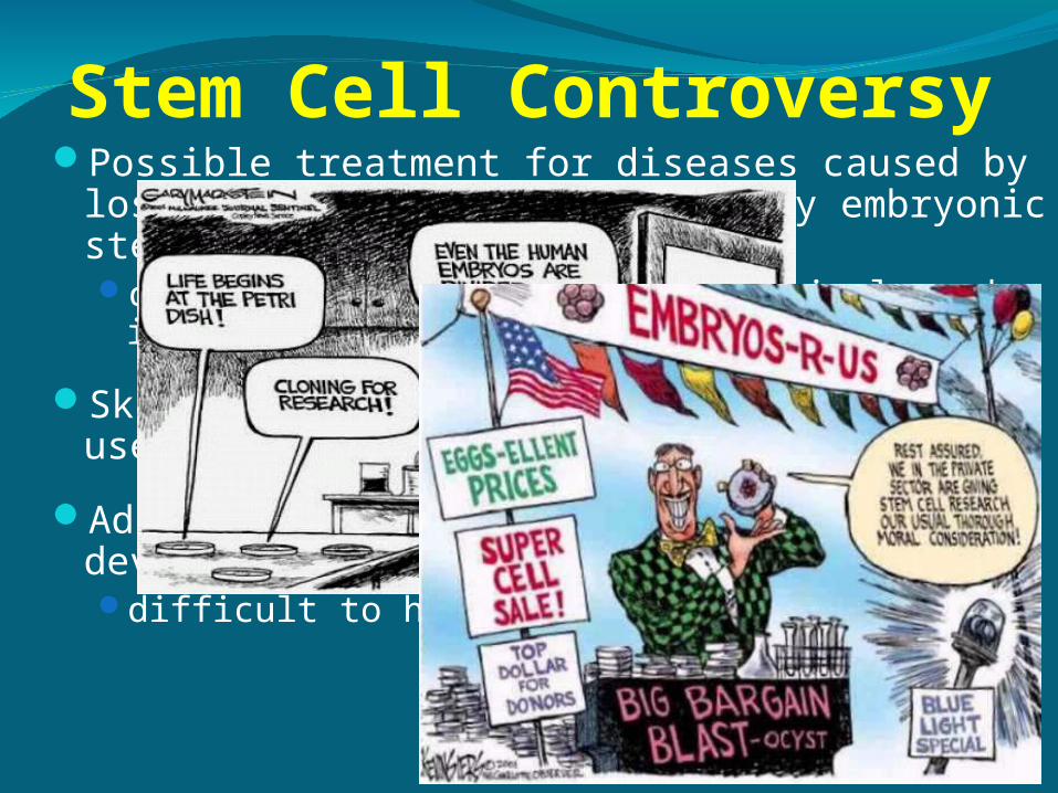
Possible treatment for diseases caused by loss of functional cell types by embryonic stem cellscardiac muscle cells, injured spinal cord, insulin-secreting cells
Skin and bone marrow stem cells have been used in therapy for years
Adult stem cells have limited developmental potentialdifficult to harvest and culture
Stem Cell Controversy
5-60


