

IMMUNOLOGY AT STANFORD UNIVERSITY
History of narcolepsy at Stanford University
Emmanuel J. M. Mignot
Published online: 14 May 2014
� The Author(s) 2014. This article is published with open access at Springerlink.com
Abstract Although narcolepsy was first described in the late nineteenth century in Germany and France, much of the
research on this disorder has been conducted at Stanford University, starting with Drs. William C. Dement and Christian
Guilleminault in the 1970s. The prevalence of narcolepsy was established, and a canine model discovered. Following the
finding in Japan that almost all patients with narcolepsy carry a specific HLA subtype, HLA-DR2, Hugh Mac Devitt, F.
Carl Grumet, and Larry Steinman initiated immunological studies, but results were generally negative. Using the narco-
leptic canines, Dr. Nishino and I established that stimulants increased wakefulness by stimulating dopaminergic trans-
mission while antidepressants suppress cataplexy via adrenergic reuptake inhibition. A linkage study was initiated with Dr.
Grumet in 1988, and after 10 years of work, the canine narcolepsy gene was cloned by in 1999 and identified as the
hypocretin (orexin) receptor 2. In 1992, studying African Americans, we also found that DQ0602 rather than DR2 was a
better marker for narcolepsy across all ethnic groups. In 2000, Dr. Nishino and I, in collaboration with Dr. Lammers in the
Netherlands, found that hypocretin 1 levels in the cerebrospinal fluid (CSF) were undetectable in most cases, establishing
hypocretin deficiency as the cause of narcolepsy. Pursuing this research, our and Dr. Siegel’s group, examining postmortem
brains, found that the decreased CSF hypocretin 1 was secondary to the loss the 70,000 neurons producing hypocretin in the
hypothalamus. This finding revived the autoimmune hypothesis but attempts at demonstrating immune targeting of hyp-
ocretin cells failed until 2013. At this date, Dr. Elisabeth Mellins and I discovered that narcolepsy is characterized by the
presence of autoreactive CD4? T cells to hypocretin fragments when presented by DQ0602. Following reports that
narcolepsy cases were triggered by vaccinations and infections against influenza A 2009 pH1N1, a new pandemic strain
that erupted in 2009, our groups also established that a small epitope of pH1N1 resembles hypocretin and is likely involved
in molecular mimicry. Although much remains to be done, these achievements, establishing hypocretin deficiency as the
cause of narcolepsy, demonstrating its autoimmune basis, and showing molecular mimicry between hypocretin and
sequences derived from a pandemic strain of influenza, are likely to remain classics in human immunology.
Keywords Narcolepsy � Cataplexy � HLA � MHC � DQB1*06:02 � Autoimmune disease
Early years
Starting in the late nineteenth century, narcolepsy has been
recognized as a unique syndrome distinct from epilepsy
[1]. It was shown to include severe daytime sleepiness and
cataplexy, defined as sudden episodes of muscle weakness
triggered by emotions, typically laughing or joking. The
first case reports in a bookbinder and a cooper (barrel
maker and seller), published by Westphal in 1877 [2] and
Gelineau in 1880 [3], are still strikingly similar to cases we
see today, although onset was late (in adulthood) unlike
most cases we see today (childhood and adolescent). As an
example of cataplexy, Gelineau’s patient reported col-
lapsing at the Zoo of the ‘‘Jardin des Plantes,’’ while
observing monkeys making faces. Symptoms recognized
E. J. M. Mignot (&)
Stanford University Center for Sleep Sciences, 3165 Porter
Drive, #2178, Palo Alto, CA 94304, USA
e-mail: [email protected]
Emmanuel J. M. Mignot
123
Immunol Res (2014) 58:315–339
DOI 10.1007/s12026-014-8513-4
soon after included sleep paralysis, vivid dreaming, hyp-
nagogic hallucinations, disturbed nocturnal sleep, and
weight gain [4–6]. When cataplexy was present, most cli-
nicians were of the opinion the syndrome was a discrete
disease entity with a specific pathophysiology [7], some-
thing time has proven to be correct, as cataplexy is the best
predictor of an absence of hypocretin in the cerebrospinal
fluid (CSF) of patients with narcolepsy [8]. Table 1 reports
on the most important milestones of narcolepsy research
from these early years to today.
Flu-hypothalamic connection and Von Economo’s
visionary work
Although the cause of narcolepsy, a loss of hypocretin cell
in the hypothalamus, was not known until recently, insight
came from the work of Constantin Von Economo [9].
Constantin Van Economo was a true European before its
time. Of Greek-Macedonian descent, he was born in Braila,
now Romania in 1876 and raised in Trieste, now Italy. As
citizen of the Austro-Hungarian Empire, he was sent to
Vienna to study mechanical engineering, but rapidly
switched to medicine. Travelling across Europe and
studying with the best, he finally settled back in Vienna at
the Clinic for Psychiatry and Nervous Diseases.
In the 1918–1923, immediately following the 1918
devastating Spanish flu, an H1N1 flu epidemic that killed
over 50 million individuals [10], another seasonal epi-
demic occurred where patients had severe encephalitis,
with high mortality rates and significant brain pathology
including ‘‘non-purulent, non-hemorrhagic acute inflam-
mation on the gray matter.’’ Lymphocytic infiltrates, also
often perivascular, are noted with edema and occasional
area of necrosis (lesions described as not unlike in
poliomyelitis). A major symptom of the disease was
extreme sleepiness (thus the name encephalitis letharg-
ica), a symptom often associated with ophthalmoplegia.
Other presentations of the disease included insomnia or
reversal of the sleep/wake cycle, movement disorders, and
psychiatric symptoms. Following the somnolent-ophthal-
moplegic form, many subjects improved but had residual
Parkinson’s disease [9].
That the encephalitis lethargica epidemic subsided after a
few years, and that it occurred so soon after the 1918 flu
epidemic, has long suggested that the epidemic was con-
nected to the flu, although this has been highly debated. A
similar secondary epidemic ‘‘Noma’’ was reported the years
following the 1889–1890 epidemic flu in Italy [9]. Further,
more encephalitis lethargica cases were reported in the
western (New Zealand) versus eastern (United States)
Samoan Islands, where a stricter quarantine against the 1918
flu was observed [11]. Others have however recently sug-
gested that the epidemic was due to another, unrelated virus,
with some recent pathophysiological support in favor of an
enterovirus [12]. As is often the case, the passage of time,
and a lack of samples are the worst enemies of investigation,
making it difficult to determine what happened.
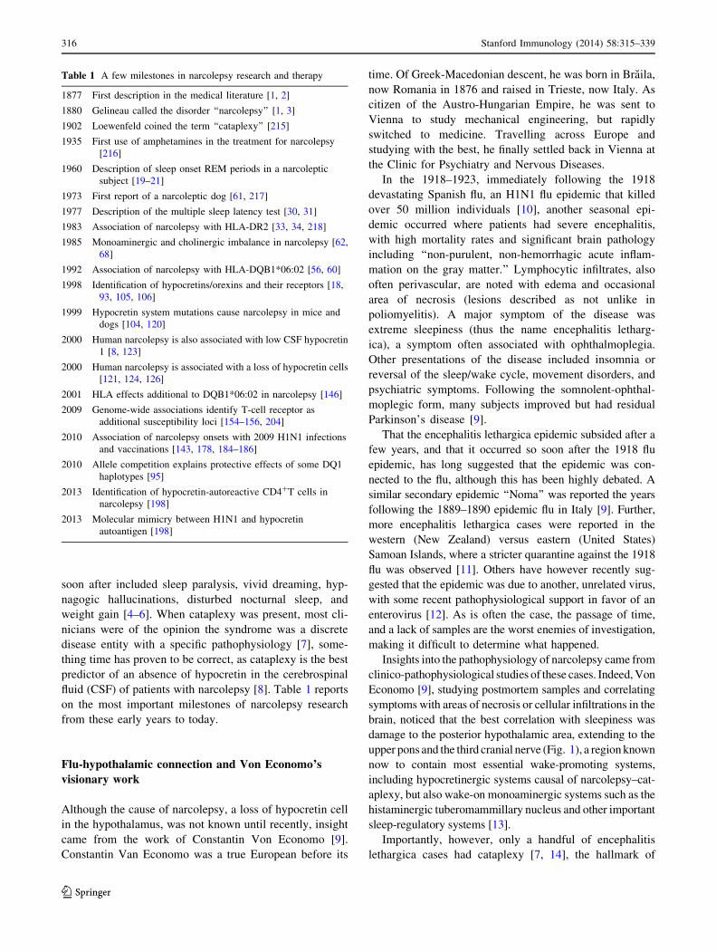
Insights into the pathophysiology of narcolepsy came from
clinico-pathophysiological studies of these cases. Indeed, Von
Economo [9], studying postmortem samples and correlating
symptoms with areas of necrosis or cellular infiltrations in the
brain, noticed that the best correlation with sleepiness was
damage to the posterior hypothalamic area, extending to the
upper pons and the third cranial nerve (Fig. 1), a region known
now to contain most essential wake-promoting systems,
including hypocretinergic systems causal of narcolepsy–cat-
aplexy, but also wake-on monoaminergic systems such as the
histaminergic tuberomammillary nucleus and other important
sleep-regulatory systems [13].
Importantly, however, only a handful of encephalitis
lethargica cases had cataplexy [7, 14], the hallmark of
Table 1 A few milestones in narcolepsy research and therapy
1877 First description in the medical literature [1, 2]
1880 Gelineau called the disorder ‘‘narcolepsy’’ [1, 3]
1902 Loewenfeld coined the term ‘‘cataplexy’’ [215]
1935 First use of amphetamines in the treatment for narcolepsy
[216]
1960 Description of sleep onset REM periods in a narcoleptic
subject [19–21]
1973 First report of a narcoleptic dog [61, 217]
1977 Description of the multiple sleep latency test [30, 31]
1983 Association of narcolepsy with HLA-DR2 [33, 34, 218]
1985 Monoaminergic and cholinergic imbalance in narcolepsy [62,
68]
1992 Association of narcolepsy with HLA-DQB1*06:02 [56, 60]
1998 Identification of hypocretins/orexins and their receptors [18,
93, 105, 106]
1999 Hypocretin system mutations cause narcolepsy in mice and
dogs [104, 120]
2000 Human narcolepsy is also associated with low CSF hypocretin
1 [8, 123]
2000 Human narcolepsy is associated with a loss of hypocretin cells
[121, 124, 126]
2001 HLA effects additional to DQB1*06:02 in narcolepsy [146]
2009 Genome-wide associations identify T-cell receptor as
additional susceptibility loci [154–156, 204]
2010 Association of narcolepsy onsets with 2009 H1N1 infections
and vaccinations [143, 178, 184–186]
2010 Allele competition explains protective effects of some DQ1
haplotypes [95]
2013 Identification of hypocretin-autoreactive CD4?T cells in
narcolepsy [198]
2013 Molecular mimicry between H1N1 and hypocretin
autoantigen [198]
316 Stanford Immunology (2014) 58:315–339
123
narcolepsy, although atypical atonia could have been
missed in the context of the more complex clinical picture.
Other clinico-anatomical correlations made by Von Eco-
nomo included a correlation between damage in the ante-
rior preoptic hypothalamus with insomnia (a region known
now to contain preoptic sleep-promoting GABAergic sys-
tems) [9]. Other investigators had noted prior that cases of
secondary narcolepsy were often associated with tumors
located close to the third ventricle [7, 15].
Sleep onset REM sleep as a feature of narcolepsy
The discovery of rapid eye movement (REM) sleep by
Aserenski and Kleitman in Chicago in 1953 opened the
area of modern sleep research [16]. In parallel with this
work, Jouvet described ‘‘paradoxical sleep’’ pointing out
that a pervasive atonia with brief bursts of phasic activity
was present during this stage of sleep [17]. William C
Dement, who trained as a psychiatrist and was a graduate
student in Kleitman’s laboratory when REM sleep was
discovered, became interested in dreaming and reported the
common association of this phenomenon with REM sleep
[18]. From these observations and the clinical descriptions
of narcolepsy, it became quickly evident that narcolepsy
involved abnormal REM sleep. Working with Alan
Rechschaffen, Dement described that unlike controls who
typically entered their first REM sleep period 90 min after
sleep onset, patients with narcolepsy often went directly
into REM sleep during nighttime sleep testing, a phe-
nomenon we call sleep onset REM periods (SOREMPs)
[19, 20]. A similar finding was also reported by Vogel et al.
[21]. Subsequent studies, still valid today, found that only
50 % of cases entered REM sleep within 15 min of sleep
onset during nocturnal sleep studies, limiting its usefulness
as a clinical test [22].
The Stanford Sleep Clinic and first narcolepsy
prevalence studies
William C. Dement joined Stanford University in 1963 [23,
24]. Seeking narcoleptic subjects for his studies, he con-
ducted one of the first prevalence studies for the condition
and also started a small sleep clinic to see these patients in
1964. He identified many patients within the San Francisco
Bay area using newspaper advertisements and a description
of the syndrome [25]. By considering the number of cases
that responded to the advertisement and readerships of the
add, he estimated the prevalence at 0.07 %, a figure
remarkably similar to the currently accepted prevalence of
0.03–0.05 %, established through dozens of well-designed
population-based studies across the world [26, 27]. Dement
was surprised by the unexpectedly high frequency and saw
many patients as the result of this study, most of whom
discovered their condition thanks to the advertisement.
However, population size was not sufficient to support a
narcolepsy-only clinic, and clinical activity stopped in 1965.
In 1970, Dr. Christian Guilleminault joined the clinic with a
primary interest in sleep-disordered breathing and coined the
term obstructive sleep apnea [23, 24, 28]. Sustained clinical
activity resulted, and the Stanford Sleep Clinic became a
beacon for the field, eventually leading to the establishment
of sleep medicine as a distinct medical specialty [29].
The new Sleep Disorders Clinic introduced all-night
polysomnographic examination of patients with sleep-rela-
ted complaints, medical responsibility and management of
the patient, and objective assessment of the relationship
between nighttime sleep and daytime function. For the lat-
ter, Dement and Carskadon developed the multiple sleep
latency test (MSLT), which remains the standard diagnostic
measure of daytime sleepiness [23, 30]. In this test, patients
or volunteers are asked to nap every 2 h (while staying
awake in between) and the mean sleep latency measured as
an objective measure of daytime sleepiness. Using the
MSLT, Richarsdon, Mitler, and others found that narco-
leptic patients often have naps containing REM sleep in
contrast to controls [31, 32]. They found that at least two
SOREMP and a short mean sleep latency is a reliable
objective test for narcolepsy [31, 32]. Together with the
Fig. 1 Van Economo’s sleep regulatory centers, marked by the
dotted line in the transitional region from the diencephalon to the
mesencephalon. Aq aqueduct, Hy hypophysis, J infundibulum, N.
occulomot.: third cranial nerve. O optic chiasm, Th thalamus, V3 and
V4 ventricles. Marked by parallel oblique vertical lines (posterior
hypothalamus and upper brainstem): region whose affection produces
sleep; marked by horizontal lines (anterior hypothalamic regions):
region whose affection produces insomnia from Van Economo [9]
Stanford Immunology (2014) 58:315–339 317
123
demonstration of REM sleep onset during nocturnal sleep
[22], the MSLT is still the most commonly used diagnostic
test for narcolepsy.
First HLA association studies in narcolepsy and early
immunological studies
In 1983, as part of a program to search for HLA associa-
tions in orphan disorders, a weak association was recog-
nized with HLA-Bw35 in Japanese patients [33]. At the
time, HLA type was defined using panels of autologous
antibodies and not molecular typing. Following on these
findings, Juji and Honda found that 100 % of Japanese
narcolepsy–cataplexy patients carried HLA-DR2 and DQ1
versus 25 % of controls [34, 35]. This was rapidly con-
firmed across the world [36–39], including at Stanford
University [40], where nonetheless very rare DR2-negative
cases were identified, causing controversy between the
Stanford group and Japan [35, 40, 41]. Current results
indicate that although the association between hypocretin
deficiency and HLA is extremely high (98 %), rare
exceptions have been documented (see below).
The result was nonetheless remarkable: over 95 % of
cases with cataplexy carried DR2, DQ1 versus 25 % in
general Caucasian and Japanese populations [37, 42].
Using another technique, mixed leukocyte culture (MLC),
DR2 antigens were found to be heterogeneous, including
Dw12, Dw2, Dw21, and Dw22 subtypes [43, 44]. All
Japanese patients carried Dw2, which is the dominant DR2
subtype in Caucasians (25 %), but a more minor antigen in
Japanese (8–10 %), where Dw12 is more common [44, 45].
Restriction fragment length polymorphism (RFLP) studies
with DQ probes performed in the laboratory of Hugh
McDevitt also confirmed this finding, showing a unique
pattern of association correlating with Dw2 but not Dw12
[46, 47]. As will be seen later, this pattern differentiates
DRB1*15:01, DQA1*01:02, DQB1*06:02 versus
DRB1*15:02, DQA1*01:03, DQB1*06:01, the two major
DR2 and DQ1 haplotypes in the Japanese population [48].
The HLA genes, also called major histocompatibility
(MHC) genes, are located on human chromosome 6. As
noted by Hugh McDevitt [49] at Stanford, MHC poly-
morphisms are essential contributors to genetic diversity in
the immune response, allowing more diverse epitope pre-
sentation across individuals. Although HLA polymor-
phisms modulate immune responses to infections, these
polymorphisms were rapidly shown to be most strongly
associated with autoimmune diseases [50]. Considering
this hypothesis for narcolepsy, researchers studied inflam-
mation around disease onset [51], or tried to identify brain-
specific autoantibodies [52], but found no evidence for
autoimmunity [53, 54]. As all results were consistently
negative, researchers speculated that HLA DR2 was per-
haps only a linkage marker for a yet unknown sleep gene
located in this region of the genome. As genetic mapping
and characterization of the HLA region progressed, this
second hypothesis became less and less likely [55]. As
described below, in 1992–1997, studies demonstrated
clearly that the closely linked HLA-DQB1*06:02 and
DQA1*01:02 loci (forming the DQ0602 heterodimer)
rather than DR2 was the best marker for narcolepsy, and
that the association signal rapidly decreased on both sides
of the HLA-DQ locus [56–58]. Sequencing of these HLA
genes also showed no abnormalities [59, 60].
Canine narcolepsy
In 1972, Dement presented video recordings of patients
with narcolepsy–cataplexy at an educational exhibit dur-
ing the annual convention of the American Medical
Association in San Francisco [24]. A member of the
audience, on the veterinary faculty at University of
California Davis, noticed the resemblance with a canine
patient he had seen with a provisional diagnosis of
refractory epilepsy. Unfortunately, the dog had been
euthanized, but a video was available, showing the sim-
ilarity with human narcolepsy, with the dog collapsing but
awake when excited by food or other activities. After a
national search and contacting veterinarians across North
America, ‘‘Monique,’’ a French poodle was identified and
donated to Stanford [61]. In the following few years, Drs.
Dement and Mitler visited veterinarians in more than 50
cities and spoke at many colleges of veterinary medicine
in the United States. Small numbers of animals of various
breeds with narcolepsy were identified, and a small canine
colony was established [62]. Poodles and Beagles were
bred (backcrosses included) but genetic transmission was
not established in these breeds [62, 63], later shown to
have hypocretin deficiency [64].
In 1975, two affected Doberman littermates and one
unrelated Doberman with narcolepsy were donated to the
colony [62, 63]. Breeding these animals led to the first suc-
cessful genetic transmission of narcolepsy, with a litter of
affected animals born at Stanford on July 29, 1976. Multiple
cases of Labradors with narcolepsy were subsequently
reported and, with the help of Dr. Cavalli-Sforza, the trait
found to be transmitted as a single autosomal recessive gene
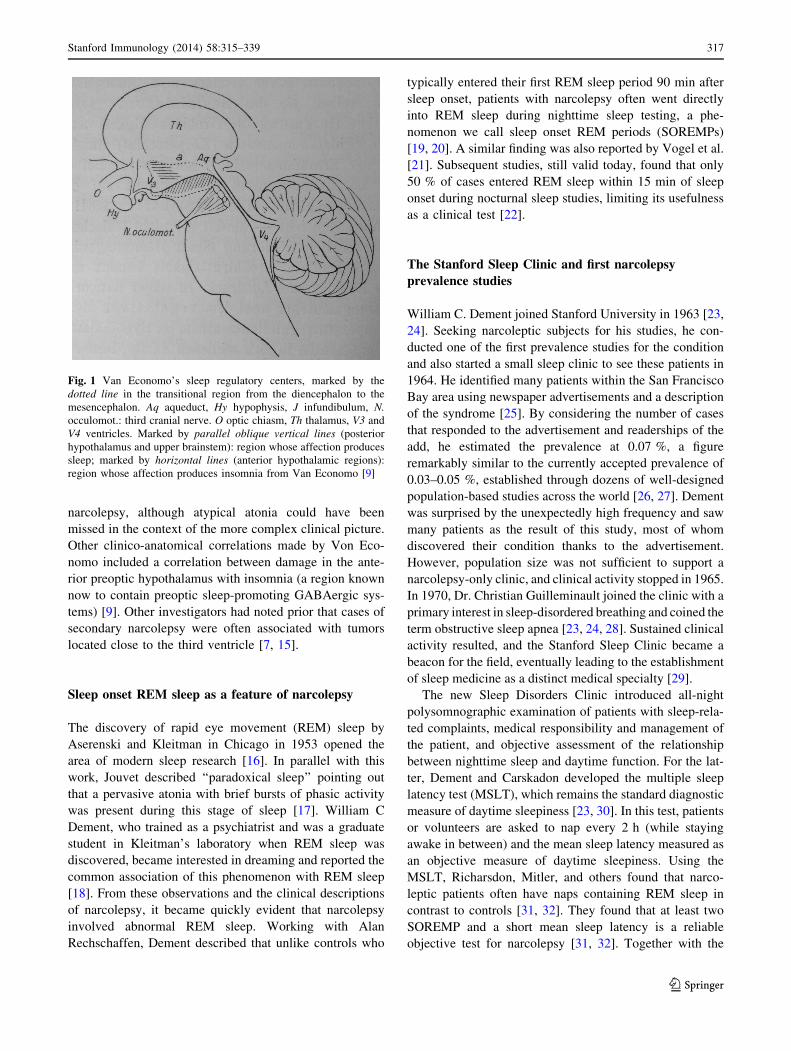
[62, 63]. Canine narcolepsy (Fig. 2) was characterized in
detail at the clinical level [65–69] but many investigators
refused to believe these animals had narcolepsy.
In the late 1980s, following the HLA discovery in
humans [34], efforts were made to see whether a similar
MHC association could be found in canine narcolepsy,
whether the genetic form or the sporadic form. Studies
318 Stanford Immunology (2014) 58:315–339
123
using mixed leukocyte culture [70] and RFLPs in canine
narcolepsy did not identify shared MHC DR or DQ genes
[71], as in humans. Interestingly, whereas the lack of
association in autosomal recessive narcolepsy is not sur-
prising considering its pathophysiology (mutations in the
hypocretin receptor 2, see below), it is still unclear why
sporadic canine narcolepsy, a disorder commonly associ-
ated with low CSF hypocretin as in humans [64], does not
show a clear MHC DR and DQ association.
Neurochemical and pharmacological studies in canine
narcolepsy suggest a downstream imbalance
of monoaminergic and cholinergic systems,
and dysregulated dopaminergic transmission
in the amygdala
In the 1970s, the study of monoaminergic and cholinergic
transmission in the regulation of sleep emerged as a leading
research avenue [72]. Thanks to pioneering transection
experiments by Jouvet [17], the pons was considered as a
logical first candidate region for a narcolepsy abnormality.
The levels of acetylcholine, various monoamines, metab-
olites, and receptors were measured in the cerebrospinal
fluid and various brain regions of narcoleptic animals and
humans [73–81]. These led to the general hypothesis of a
pontine monoaminergic–cholinergic imbalance in narco-
lepsy [62]. In this model, narcolepsy was the result of
cholinergic hyperactivity and monoaminergic hypoactivity
in the pons, a concept paralleling the then fashionable
Hobson and McCarley model for REM sleep regulation
[82, 83]. A primary role of the amygdala was also pro-
posed, based on the observation of consistent dopaminergic
abnormalities in this brain region [80, 81]. The involve-
ment of this structure is attractive conceptually, as it may
explain why cataplexy is triggered by emotions.
Working with Tom Kilduff, Craig Heller, and others, Dr.
Nishino and I next conducted a systematic pharmacological
dissection of the mode of action of the then commonly pre-
scribed narcolepsy treatments, stimulants for sleepiness, and
antidepressants for cataplexy [68]. This led us to demonstrate
that presynaptic activation of adrenergic transmission
mediates the anticataplectic effects of antidepressants [84,
85], a finding that still has application today, as serotonin
norepinephrin reuptake inhibitors (SNRI) such as venla-
faxine or pure adrenergic reuptake inhibitors, such as ato-
moxetine, are still used to treat human cataplexy [5]. We also
established that wake-promoting effects of amphetamine
stimulants and modafinil are mediated by a presynaptic
activation of dopaminergic transmission [85–87]. The idea
that modafinil was acting through dopaminergic reuptake
inhibition was contested by Lafon Laboratories of France
(the original inventor of modafinil) and then Cephalon (the
company which owned modafinil until recently) and most
other scientists, until recent studies by Volkow et al. con-
firmed our data in vivo [88]. Other studies emphasized the
importance of cholinergic hypersensitivity in the basal
forebrain area [89], mesolimbic dopaminergic hypoactivity
[90] as important pathophysiological abnormalities in canine
narcolepsy. Although these data were informative and doc-
umented a large overlap between REM sleep regulation and
narcolepsy pathophysiology, it was not moving us forward
the true cause of the condition.
Transethnic studies demonstrate that DQ0602 is
the culprit narcolepsy-associated allele
Faced with a lack of evidence for autoimmunity [53, 54],
efforts focused on fine mapping the genetic association in
the HLA DR region. The HLA DR and DQ region is
compact, containing in sequence the DRA gene (practically
monomorphic), accessory DRB genes (DRB 3, 4, 5 genes
present in some but not all haplotypes), the DRB1 gene (a
very polymorphic gene), an intergenic segment of about
Fig. 2 Narcoleptic dogs of the colony, a sporadic case with
hypocretin deficiency (a) and two familial cases with hypocretin
receptor 2 mutations (b). The dog on the left was later shown to have
low CSF hypocretin, like human narcolepsy [64]. The bottom dog on
the right is having a complete attack of cataplexy, an episode of
complete muscle paralysis while awake that has been triggered by the
excitement of playing with his littermate
Stanford Immunology (2014) 58:315–339 319
123
40 kb, and finally the polymorphic DQA1 and DQB1 loci,
genes separated by approximately 12 kb. Alpha genes and
beta genes encode heterodimers, with the product of the
DRA gene principally partnering with DRB1 to form the
DRab molecule, while DQA1 and DQB1 encode proteins
that dimerize to produce DQab molecules.
In Caucasians and Japanese, linkage disequilibrium
between DQ and DR is so strong that practically all
([99 %) DQB1*06:02 alleles are linked with DQA1*01:02
and DRB1*15:01 (DR2) (these alleles are in full linkage
disequilibrium), making it impossible to distinguish whe-
ther the effect was mediated by DR or DQ. In the early
1990s, however, studies in control African Americans
revealed additional diversity in DR–DQ haplotypes, so that
in this ethnic group DQB1*06:02 was found not only with
DRB1*15:01, but also with DRB1*11:01 and more rarely
DRB1*12:02 (all haplotypes containing DQA1*01:02) [57,
91, 92]. This, together with a study that had suggested less
DR2 positivity in African American patients with narco-
lepsy [93], led us to test HLA-DR and DQ associations in
African American controls and patients.
In 1992, to our surprise, we found that all African
American patients with narcolepsy had DQA1*01:02 and
DQB1*06:02 [56, 60]; however, DR was not DRB1*15
(DR2) in many instances, indicating that the primary
association was with HLA-DQ, not DR, and more partic-
ularly the DQab heterodimer DQ0602 encoded by
DQA1*01:02 and DQB1*06:02. In subsequent studies,
screening hundreds of patients, we discovered a number of
very rare cases of DQB1*06:02-positive subjects that car-
ried various DRB1 alleles such as DRB1*03:01, DR8del,
DRB1*08:01, DRB1*08:06, DRB1*16:01 that are only
exceptionally found in controls [57, 94], confirming the
enrichment of these rare DQ0602 haplotypes as well in
narcolepsy. Finally, in a recent study, we found that
DRB1*15:01 alone, in the context of the DRB1*15:01,
DQA1*01:02, DQB1*06:01, a frequent haplotype in South
China, does not predispose to narcolepsy [95]. To our
knowledge, the study in 1992 was the first to take advan-
tage of ethnic diversity in fine mapping a disease poly-
morphism [56, 60], something we have used to our
advantage in many other subsequent studies.
The canine narcolepsy gene is the hypocretin receptor 2
In the early part of the previous century, human narcolepsy
was frequently believed to be a familial disorder, but more
recent studies have shown that it is not a simple genetic
disorder. Monozygotic twins are most frequently discor-
dant for narcolepsy, indicating a requirement for environ-
mental factors to trigger narcolepsy onset [96]. Indeed,
familial clustering of narcolepsy–cataplexy is the exception
rather than the rule. Only 1–2 % of the first-degree rela-
tives of patients with narcolepsy have narcolepsy–cata-
plexy [96–99], although there is suggestion that as many as
4 % of first relatives have milder symptoms without cata-
plexy. This indicates a 20- to 40-fold increased risk when
compared to the general population [96]. The complex
genetics involving HLA, other genes, and environmental
factors was consistent with an autoimmune basis with an
unknown target.
The complex picture in human narcolepsy led us to
focus our genetic studies on canines. In contrast to the
human situation, narcolepsy is a simple autosomal reces-
sive disorder in Dobermans and Labradors [63], thus
making positional cloning, a technique first used in 1986 in
humans, theoretically possible in this species. Backcrosses
were performed and a genetic linkage study initiated in
1989 with Frank C. Grumet. Our first focus was to exclude
potential candidate genes. Canine narcolepsy was shown
not to be associated [70, 71] or tightly linked with dog
leukocyte antigen (DLA) polymorphisms [100], suggesting
canine narcolepsy gene was not an MHC gene. Additional
candidate genes and minisatellite probes were used in a
second stage. Using a candidate gene approach, a RFLP
band cross-reacting with the human immunoglobulin l-
switch segment on a Southern blot was shown to com-
pletely cosegregate with canine narcolepsy in 1991 [100],
leading to a LOD score of 7.2. This result initially sug-
gested an immunoglobulin/immune involvement in canine
narcolepsy. Further studies however demonstrated that this
linkage marker was coincidentally a cross-reacting
sequence of no functional significance [101].
Considering the relatively small number of animals
tested, the actual narcolepsy gene was likely to be located
at a large genetic distance from our initial l-switch-like
marker. Chromosome walking in the vicinity of the iden-
tified marker was difficult using available phage and cos-
mid genomic libraries, which have small genomic inserts
making chromosome walking very slow and impractical. In
1997, Robin Li built a large insert bacterial artificial library
(BAC) genomic library in collaboration with Dr. Peter De
Jong [102]. The technique of fluorescence in situ hybrid-
ization (FISH) was also established in our laboratory, and
the canine narcolepsy marker was found to be located on
dog chromosome 12 [103], which also contains the dog
leukocyte antigen (DLA) locus, but separated by a large
genomic distance.
Using the newly available BAC library, we began chro-
mosome walking in earnest. In this process, high-density
gridded library filters are hybridized with DNA probes
derived from BAC end sequences through the polymerase
chain reaction (PCR). The new clones are then isolated,
verified through PCR and FISH methods, their ends
sequenced, and the filters rehybridized to extend the so-
320 Stanford Immunology (2014) 58:315–339
123

called contig of overlapping large clones. In parallel, new
polymorphic linkage markers are isolated from new BAC
clones by creating ‘‘mini libraries’’ and hybridization with
microsatellite repeats [i.e., (GAAA)n]. These markers are
then tested in canine backcrosses to confirm genetic linkage
and map possible recombinant animals, which refine the map
location of the mutation [104]. Also, in parallel, BAC end
sequences are also analyzed using the BLAST algorithm to
identify putative known genes. In 1998, an end-sequence of
the BAC clone containing the l-switch-like marker was
shown to contain Myosin VI (MYO6), a gene known to map
on the long arm of human chromosome 6 (6q13).
The finding that both DLA and MYO6 were on the same
dog and human chromosomes led us to suspect a large
region of conserved synteny between dog chromosome 12
and human chromosome 6. This result was a turning point
as it gave us direct access to the emerging human and
mouse maps in the region. Human expressed sequenced
tags (ESTs) known to map between MHC and MYO6 in
humans were then used as probes to screen the canine BAC
library. These allowed us to identify new seed BAC clones
within the large critical interval from which to extend and
merge our contigs. The resulting canine BAC clones were
then hybridized on canine metaphase spreads to verify
localization onto dog chromosome 12 [104]. Together with
chromosome walking and microsatellite marker development
and genetic testing in backcrosses, the process was refined
until the canine narcolepsy gene was flanked in a small
genetic segment known to contain only two potential genes.
These two genes were tested for potential abnormalities and
an abnormal RFLP hybridization pattern observed with one
of the two ESTs, the hypocretin receptor 2 gene (HCRTR2)
(Fig. 3) [104]. Further analysis then demonstrated that in
both Labradors and Dobermans with autosomal recessive
narcolepsy, the hypocretin receptor transcripts were dis-
rupted by distinct exon splicing mutations (Fig. 3) [104].
The Ling Lin et al. [104] report was the first to implicate
hypocretins/orexins in the cause of canine narcolepsy.
Hypocretin (also called orexin) deficiency as the cause
of human narcolepsy
Hypocretins/orexins were identified almost simultaneously
by DeLecea et al. [105] and Sakurai et al. [106] in 1998. Luis
De Lecea, Kaare Gautvick, and Tom Kilduff identified and
characterized the preprohypocretin transcript (clone 35)
[107] in the laboratory of Dr. Gregory Sutcliffe using a
directional tag PCR subtraction technique [107]. Their aim
was to identify novel hypothalamic-specific transcripts. The
identified hypocretin gene was shown to be only expressed in
the lateral hypothalamus and to encode a precursor molecule
Fig. 3 Mutations found in
narcoleptic Dobermans and
Labradors. a In Dobermans, a
large SINE insertion upstream
of exon4 causes exon skipping
and non-functional HCRT2
receptors (Lin et al. [104] ). In
Labradors with familial
narcolepsy, a different mutation
causes exon 6 skipping and has
similar effects. b Western RLFP
blots of BAC clones (probed
with a HCRT2 human EST)
derived from an heterozygous
narcoleptic Doberman
containing the critical region of
interest. As can be seen, a
different pattern is found in
clones derived from narcolepsy-
mutated versus control
chromosomes, suggesting a
significant difference
surrounding HCRT2
Stanford Immunology (2014) 58:315–339 321
123

for two related peptides having a possible homology with
secretin (this weak homology is disputed by others). Based
on the selective expression of the gene in the lateral hypo-
thalamus and their homology with the gut hormone secretin,
the peptides were called hypocretins 1 and 2 by Luis DeL-
ecea [105], who also demonstrated neuroexcitatory proper-
ties for hypocretin 2 and suggested a possible role in feeding
regulation based on the neuroanatomical localization in the
lateral hypothalamus [105].
The existence of hypocretins was independently con-
firmed by Sakurai et al. [106] in the laboratory of Masashi
Yanagisawa a few weeks later. These authors also identified
and mapped two receptors for these peptides (HCRTR1 and
HCRTR2). In this elegant work, a series of orphan G-Pro-
tein-coupled receptors (e.g., receptor genes having no
identified endogenous ligand identified) were expressed in
cell lines (the ‘‘orphanage’’) and the resulting cell lines used
to screen high-pressure liquid chromatography (HPLC)
purified tissue fractions for biological activity [106]. Cell
lines containing the orphan receptor HFGAN72 (later shown
to be the hypocretin receptor 1) were found to strongly react
with purified brain fractions. These fractions were shown to
evoke a calcium transient, suggesting the activation of the
G-Protein-coupled receptor by an endogenous ligand. The
resulting activity was purified and shown to be a 33 amino
acid peptide that Sakurai et al. called orexin A [106].
Another weaker activity was also isolated and shown to be a
28 aminoacid peptide sharing 13/28 aminoacid identity with
orexin A; this second peptide was called orexin B [106].
Both peptides were then shown to be processed from the
same precursor, a transcript identical to DeLecea’s previ-
ously reported preprohypocretin mRNA molecule [105].
Hypocretins 1 and 2 and orexin A and B are thus identical
with the caveat that DeLecea reported a 6 amino acid longer
sequence for hypocretin 1 versus orexin A. The latter author
mentioned that the N-terminal of the hypocretin 1 peptide
could not yet be established at the time [105]. Sakurai et al.
[106] also noted that hypocretin 2/orexin B had a lower
affinity for the hypocretin receptor 1 and found that another
unknown EST had high nucleotide homology with
HFGAN2. This receptor was expressed in CHO cell lines
and was shown to bind and mobilize calcium in the presence
of hypocretins 1 and 2. This second receptor was called the
orexin receptor 2 (hypocretin receptor 2 as the gene name).
The discrete localization of these peptides in the lateral
hypothalamus suggested a role for hypocretins in feeding
behavior. In their initial publication, Sakurai et al. [106]
reported a stimulation of feeding after central administration
of hypocretins/orexins and an increased preprohypocretin
mRNA expression after fasting, leading them to select the
name ‘‘orexin.’’ The authors speculated that a main physio-
logical function for these molecules could thus be the regu-
lation of energy homeostasis [106]. Subsequent work
indicated variable effects on feeding, and more effects on
metabolism and activity [108–114]. Neuroanatomical work
indicating widespread projections for hypocretin neurons in
the entire brain and spinal cord also suggested more complex
physiological functions [115, 116]. Of note, dense projec-
tions to monoaminergic cell groups such as the locus coe-
ruleus [117], the raphe [118], and tuberomammilary nuclei
[119] suggested a possible involvement in sleep regulation.
In 1999, a few weeks after canine narcolepsy was shown to
be due to hypocretin receptor mutations, a knockout mouse
for the preprohypocretin gene was described and shown to
have sleep abnormalities reminiscent of narcolepsy [120],
thus independently indicating a role for hypocretin in the
sleep disorder.
The potential role of hypocretin gene mutations in
human narcolepsy was almost immediately investigated by
Juliette Faraco in our group [121]. Not surprisingly, con-
sidering that narcolepsy was genetically complex and
likely autoimmune, mutation screening in the preprohyp-
ocretin and hypocretin receptor genes yielded few positive
results. In only one case was a highly suspicious mutation
found, a L [ R polar amino acid substitution in the
hydrophobic polyleucine track of the signal peptide. The
clinical picture was very atypical, combining undetectable
CSF hypocretin 1, DQ0602 negativity, and very early onset
at 6 months of age [122]. Unfortunately, the father DNA
was not available to confirm a de novo mutation, although
transfection studies of the mutant allele in neural cells
revealed abnormal processing/trafficking, with elaboration
of a tubular-like cellular compartment and resulting tox-
icity, explaining the likely dominant phenotype. No mouse
model was ever generated to fully confirm this hypothesis.
In parallel with this work, hypocretin 1 levels were first
measured in the cerebrospinal fluid (CSF) of 9 narcoleptic
subjects and 8 controls by Nishino et al. [123]. Seven
narcoleptic subjects were found to have undetectable
hypocretin 1 levels. Two narcoleptic patients had normal
levels of hypocretin 1, respectively. Hypocretin 1 levels
were detectable in all controls. This result suggested that
human narcolepsy was caused by a deficiency in hypo-
cretin production [123]. A simple explanation was that
hypocretin-producing cells are destroyed by an autoim-
mune process in HLA-associated narcolepsy. Only hundred
thousand cells in the hypothalamus produce these peptides,
and a discrete lesion in this area might not have been
detected in previous neuropathological studies.
Indeed, two studies were quickly published demon-
strating the loss of hypocretinergic cells in human narco-
lepsy brain tissue, supporting this hypothesis. In one study
performed by Christelle Peyron, in situ hybridization of the
perifornical hypothalamus and peptide radioimmunoassay
measurements in six human brains indicated a global loss
of hypocretins, without signs of inflammation in all human
322 Stanford Immunology (2014) 58:315–339
123
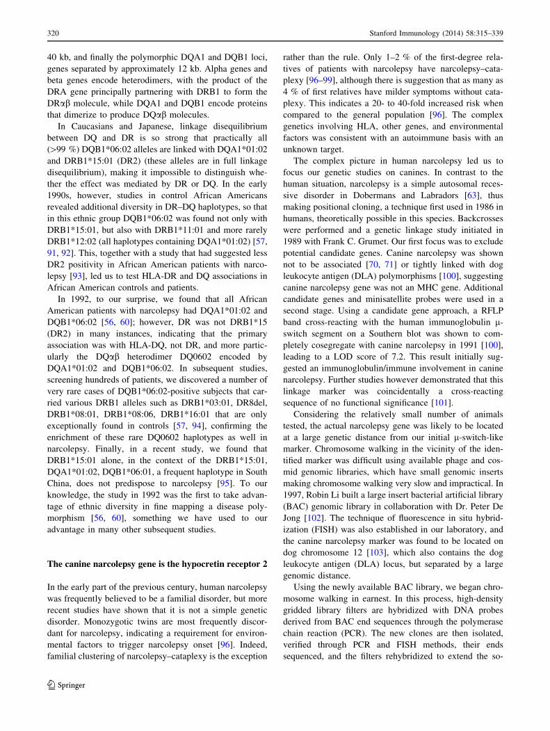
cases examined [121] (Fig. 4). The second study, published
a few weeks later, used immunocytochemistry and found a
85–95 % reduction in the number of hypocretin neurons in
4 narcolepsy brains (one without cataplexy) [124]. In both
studies, melanin-concentrating hormone (MCH) neurons,
which are intermixed with hypocretin cells in the normal
brain, were not affected [121, 124], indicating that cell loss
was relatively specific for hypocretin neurons. One study
suggested gliosis, while the other found no clear evidence.
Further studies using NARP and dynorphin, two markers
colocalized with hypocretin in the posterior hypothalamus,
also found decreased staining for these markers, indicating
cell loss rather than lack of expression of hypocretin alone
[125, 126]. Interestingly, two recent studies also found a
compensatory increase in histaminergic cell number in the
tuberomammilary nucleus in postmortem brains, indicating
significant remodeling of wake-promoting systems fol-
lowing hypocretin cell loss [127, 128].
Further HLA association and immunopathology studies
in narcolepsy
The finding of hypocretin cell loss in narcolepsy, together with
the demonstration that HLA-DQ0602 was mostly responsible
for the association signal within the HLA region in the disease,
rekindled the hypothesis of autoimmunity, with hypocretin
cells as the logical target. Surprisingly, however, autoanti-
bodies targeting hypocretin peptides were not found [129–
131], and immunostaining of hypothalamic tissue with human
narcolepsy sera did not reveal autoantibodies targeting co-
localized antigens on these neurons [132–135].
Several red herring findings were made. Passive transfer
experiments of human sera in mice have been published
[136–138], suggesting the presence of functional autoan-
tibodies with modulating effects on spontaneous colonic
migrating motor complex contractions or reactions of
rodent bladder strips to muscarinic stimulation, but we
have tried and could not replicate the finding (data not
shown). These investigators also suggested that peripheral
injections of human narcolepsy sera in mice caused nar-
colepsy symptoms, but when we attempted replication, all
had freezing seizures that may have been confused with
narcolepsy events. Two of the 5 treated animals died, and
hypocretin neurons were intact postmortem in all animals,
including the 3 animals after recovery. Further, blinded
sera samples were sent in Australia for the bladder strip
assays, but opposite results to the initial findings were
returned to us. Using a BAC-based transgenic animal
model expressing a flag-tagged poly(A)-binding protein
(Pabpc1) cDNA sequence in hypocretin neurons, Cvetko-
vic-Lopes et al. [139] isolated transcripts believed to be
A
B
10 mm 10 mm
50µm 50µm
Fig. 4 Hypocretin cell loss as the cause of narcolepsy, as demonstrated by in situ hybridization (a), from Peyron et al. [121], f fornix) and
hypocretin peptide immunocytochemistry (b), from Thannickal et al. [124] ). Right narcolepsy samples, left control brains
Stanford Immunology (2014) 58:315–339 323
123
increased in hypocretin cells, including the protein Tribbles
homologue 2 (Trib2). The authors went on to demonstrate
increase Trib2 autoantibodies in recent onset narcolepsy
cases, a result that was replicated by our group and a
Japanese study using sera samples from subjects collected
in the 1990–2000s, and some cross-reactivity of sera with
hypocretin neurons.
Unfortunately, however, these authors may have been on
the right track for the wrong reasons. Further studies using a
similar approach but another mRNA-binding protein than
Pabpc1, the protein P10, found that few of the genes
expressed in hypocretin neurons as reported by Cvetkovic-
Lopes et al. [139], including Trib2, were enriched in hypo-
cretin neurons [140]. The result of the well-validated P10
technique was also confirmed by our own multiple expres-
sion array studies [141, 142]. Pursuing this line of investi-
gation, we also found that TRIB2 autoantibodies were
generally absent in more recent narcolepsy samples [143]. It
is our hypothesis that TRIB2 autoantibodies may have
marked a coinfection present together with a narcolepsy
trigger in some cases with onset notably in the 1990s and
2000s, a result substantiated by the finding of a correlation
between A/H1N1 and TRIB2 autoantibody levels in a recent
study [144]. Interestingly, a recent study, reminiscent of the
older Australian studies mentioned above, reported that local
injections of purified immunoglobulins of narcolepsy-
TRIB2-positive individuals but not controls, produced hyp-
ocretin cell lesions and narcolepsy symptoms [137]. Careful
reading of this manuscript however does not support the
conclusion of the study, as no hypocretin cell count statistics
are provided, only an exemplar hypothalamic section
showing widespread local cell loss that would be much larger
than just hypocretin cell loss. Further, the authors report on
‘‘narcolepsy-like immobilization attacks’’ without associ-
ated EEG studies in 6 animals, which may well have been
seizures considering their mean duration (66–464 s), much
longer than typically reported in murine cataplexy (2–10 s)
[120]. This brief discussion exemplifies the difficulties for
others not in the field to make sense of a confusing literature.
Only time will tell on whether or not the Tribbles story will
hold on to scrutiny.
The absence of immunological findings led us to pursue
the characterization of the HLA signal in narcolepsy.
Sequencing studies of the HLA-DQ region, as well as
studies of microsatellite markers in the region, indicated
that no other gene was present in the susceptibility interval,
and that the effect was in the DQ region [55, 57, 58].
Pursuing studies across multiple ethnic groups, a strikingly
consistent pattern emerged. Indeed, not only was DQ0602
(the combination of DQA1*01:02 and DQB1*06:02), a
near prerequisite for developing narcolepsy, but individuals
homozygous for DQ0602 were at approximately 2–3 times
greater risk of developing narcolepsy [95, 145–147],
suggesting that the amount of DQ0602 heterodimer
increased risk as well [148]. Intriguingly, we also found
that DQ0602/DQB1*03:01 were also at increased risk
versus other combinations [95, 146, 147], an effect difficult
to explain as it occurred in the context of multiple DQa-
associated alleles (DQA1*03:01, DQA1*03:02,
DQA1*05:05 and DQA1*06:01), suggesting it was not
mediated via a DQa/b heterodimers. This effect was con-
firmed in trios using transmission disequilibrium tests, a
design where power is enhanced by the removal of alleles
that are located together with DQ0602 in DQ0602-positive
parents and thus never transmitted [149].
In addition to this effect, protective effects of
DQB1*05:01, DQB1*06:01, DQB1*06:03, and other DQ1
alleles that are non-DQ0602 were found [95, 146, 147, 149–
153]. DQ1 is a broad DQ subtype that includes the DQa alleles
encoded by DQA1*01 and DQb alleles encoded by DQB1*05
and 06 subtypes. These DQ1 alleles, unlike those of the other
broad DQ groups (DQ2, 3, and 4), are ‘‘compatible’’ with each
other, meaning that they have sequence similarity and proper
folding as selected by invariant chain binding (in contrast to
non-DQ1 subtypes such as DQ2 and DQ3 are generally
compatible with each other). Estimating relative risk, we
noted that risk of DQ0602/other DQ1 was about one-half of
DQ0602/other, indeed suggesting that there is competition of
transencoded DQ1 alleles that are non-DQ0602, reducing the
amount of DQ0602, and thus risk, a phenomenon we called
allele competition [95, 153] (Fig. 5).
Genome-wide association studies (GWAS) in human
narcolepsy indicate association with T-cell receptor loci
(TCR) and other autoimmune associated loci
Whereas the decades spanning 1985–2005 saw the emer-
gence of positional cloning as a powerful tool to isolate
highly penetrant disease genes, the release of the first
complete human genome sequence in the early 2000 led in
2005 to more systematic, large-scale genome-wide asso-
ciation studies (GWAS) where up to a million single-
nucleotide polymorphisms can be tested at once in a sub-
ject. This allowed investigators to better describe the
genetic architecture underlying multigenic disorders. Using
this technique in narcolepsy in 2009, we found that the
disease was strongly associated not only with HLA, but
also with a specific polymorphism in the TCR alpha gene
[154]. Although genetic risk was not high (OR * 2) when
compared to effects found with HLA polymorphisms, the
finding was nonetheless remarkable as it further demon-
strated a role of the immune system in narcolepsy. It was
also unusual, as none of the other autoimmune disorders
that have been subjected to GWAS analysis have TCR loci
as susceptibility factor.
324 Stanford Immunology (2014) 58:315–339
123

Further studies in larger and larger samples that also
included other ethnic groups, notably Chinese, Japanese, and
African Americans, were conducted and led to the identifi-
cation of other associated genes, most known to be involved
in other autoimmune diseases [155–157]. Other associated
loci included the TCR beta-gene a partner of TCRa; TNFSF4
(also called OX40L) a costimulatory receptor for T-cell
activation involved in Lupus [158], Crohn’s disease [159],
rheumatoid arthritis [160], and celiac disease; Cathepsin H,
an enzyme likely involved in antigen processing and asso-
ciated with Type 1 diabetes [161]; ZNF365, a transcription
factor associated with inflammatory bowel disease (IBD)
[159] and atopic dermatitis [162]; IL10RB-IFNAR1, a
region associated with IBD [163].
Of additional interest was the finding of an association
within the PPAN-P2RY11-EIF3G gene region, 10 kb from
the DNA methylase gene 1 (DNMT1) [156]. This finding
was notable as this gene region was not known to be
associated with other autoimmune diseases, although
P2RY11, an ATP receptor, regulates cell death, notably in
immune cell subsets. Interestingly, in a parallel exome
sequencing project of rare dominant phenotypes with nar-
colepsy, we found that a rare disease associating late-onset
narcolepsy with deafness, cerebellar ataxia, and dementia
(ADCA–DN) was secondary to mutation in exon 21 of the
DNMT1 gene, resulting in late-onset neurodegeneration,
with a likely effect on hypocretin cells [164]. Further
mapping of the GWAS signal confirmed location within
P2RY11-EIF3G and not extending to DNMT1, although
regulatory elements for the latter could still lie within the
nearby region. In favor of this hypothesis, although
P2RY11 is a pseudogene in rodent, the syntenic block
containing PPAN-P2RY11-EIF3G-DNMT1 synteny is
conserved from zebrafish through mammals.
Overall, these GWAS association studies did not reveal
any smoking gun evidence, but strongly confirmed that the
etiology of narcolepsy was likely autoimmune. A more
detailed analysis of the pathway suggested a primary
importance of HLA-DQ0602 presentation to CD4? cells
and T-cell mediation.
Rare HLA-DQB1*06:02-negative subjects with primary
narcolepsy
The issue of whether or not HLA-negative subjects with
narcolepsy had a true disease, i.e., hypocretin deficiency,
has been debated since the discovery of the HLA-DR2
association in narcolepsy. The discovery that DQB1*06:02
was a better marker than DR2, notably in African Ameri-
cans, helped resolved some of this debate. Similarly, one
DQB1*06:02-negative subject with low CSF hypocretin
and very early onset (6 months) is likely secondary to a
damaging hypocretin mutation in its signal peptide, but
these findings still left a few unexplained cases with usual
childhood or adolescent onset, hypocretin deficiency, and
DQB1*06:02 negativity.
To further our understanding of these exceptionally rare
cases, which we estimate represent approximately 2 % of
cases and vary in frequency across countries (maybe more
frequent in Italy), we further characterized 8 such cases
with documented low CSF hypocretin through exome
sequencing and full HLA typing. Interestingly, we found
that 4 of 8 cases carried DPB1*09:01, a subtype that should
have been rare in this multiethnic sample (*5 %) [165].
This result suggests that another heterodimer, possibly
DPA1*0201/DPB1*09:01, may also play a role in excep-
tionally rare cases of autoimmune hypocretin deficiency.
Relative Risk 0.5Relative Risk 1.0 (ref.)Relative Risk 2.0
Fig. 5 Allele competition model explaining HLA-DQ effects in
narcolepsy. HLA-DQB10602 is almost a prerequisite for developing
narcolepsy, probably because it can binds the culprit HCRT epitope.
In addition, consistent effects are observed across multiple studies,
with HLA-DQ0602 dosage/amount influencing risk of developing the
disease. For example, subjects homozygous for DQ0602 have twofold
higher risk of developing narcolepsy in comparison with most
DQ0602 heterozygotes. At the opposite, DQ0602 heterozygotes that
have other DQ1 alleles in trans that can heterodimerize with the DQaand DQb alleles of DQB0602 have a twice lower risk, as predicted
from the allele competition model [95, 145, 147]
Stanford Immunology (2014) 58:315–339 325
123
The role of upper airway infections in triggering
narcolepsy
Starting in the mid-2000s, narcolepsy became increasingly
recognized, giving us the opportunity to study patients closer
to onset, thanks to faster diagnosis [166]. Blood samples
could also be collected closer to onset for biological analysis.
In particular, we saw more and more young children with a
recent onset, and as these cases often have an explosive and
rapid evolution, we were able to query circumstances sur-
rounding onset. We noted a number of reports of a past
history of streptococcus infections (i.e., strep throat), which
resulted in tonsillectomy and hospitalization in some cases,
and in one case with suspected pediatric autoimmune neu-
ropsychiatric disorders associated with streptococci (PAN-
DAS) [167]. Streptococcus infections were interesting as
they are known to be associated with onset of rheumatic heart
fever, Syndenham’s Chorea [168], two other autoimmune
diseases (although these have decreased in frequency in the
western world with the use of antibiotics), and PANDAS, a
more controversial psychiatric entity. Further, older studies
had suggested an association of narcolepsy with antistrep-
tolysin-O (ASO) and anti DNAse B, two markers of recent
streptococcus infection, although this was not replicated
[169–171]. Intriguingly, increased ASO has also been
reported in recent cases of encephalitis lethargica [172],
which still occur today at low frequency (although differ-
ential diagnosis with anti-NMDA encephalitis may be
difficult).
Still searching for the elusive autoantigen, we conducted
Western blot studies of selected rat brain regions, stained with
narcolepsy sera, and noted a frequent pattern of cross-reac-
tivity with a 58-kDa protein in many samples [173]. In parallel
with this work, we decided to re-evaluate whether recent
narcolepsy samples had increased titers of ASO, arguing that
past studies may have been variable because distance to onset
was not controlled [169–171]. Antibodies against Helico-
bacter pylori [Anti Hp IgG] were also tested, as this infection
had been suggested to be involved in the triggering of idio-
pathic thrombocytopenic purpura, another autoimmune dis-
ease. To our surprise, we found that high ASO titers were
found more frequently in patients within 1 or 3 years of onset,
compared to age-matched controls or patients with long-
standing disease [174]. This finding was interesting as a par-
allel epidemiological study also found increased risk of
developing narcolepsy when reporting past streptococcus
infections [175]. In additional studies, we also discovered that
the 58-kDa cross-reactive protein is protein disulfide isom-
erase (PDI), an abundant protein with pleiotropic metabolic,
immunologic, and thrombotic effects [173, 176].
In parallel with this work, starting in 2000, a strong
collaboration between Stanford and Beijing University was
established. In 2004, as the world federation for sleep
society met in Zuhai, publicity surrounding narcolepsy led
to increased case recognition in China and referral to the
Beijing University sleep center. Interestingly, the large
majority of cases diagnosed at the center in Beijing were
children (70 %) [177], many with abrupt onset, a pattern
that we attributed to increased ascertainment and vigilance
due to the one child policy in China; many more adults,
approximately 400,000 Chinese narcolepsy subjects,
should be present in the general population of this country
based on the established prevalence. As onset in these
children was extremely clear and could be generally dated
to the exact month if not week by parents, we decided to
examine whether a seasonal pattern of onset was present, as
would be predicted based on our suspicion of an associa-
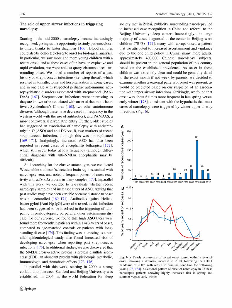
tion with upper airway infections. Strikingly, we found that
onset was about 6 times more frequent in late spring versus
early winter [178], consistent with the hypothesis that most
cases of narcolepsy were triggered by winter upper airway
infections (Fig. 6).
0
50
100
150
200
250
1998 2000 2001 2002 2003 2004 2005 2006 2007 2008 2009 2010 2011 2012
5 3 414 14
411
35
13
33
51
201
49
31
Num
ber
of r
ecen
t ons
et c
ases
/yea
r
0
0.05
0.1
0.15
0.2
0.25
% o
f yea
rly r
ecen
t ons
et c
ases
A
B
Fig. 6 a Yearly occurrence of recent onset (onset within a year of
onset) showing a dramatic increase in 2010, following the H1N1
pandemic of 2009, with return to baseline condition the following
years [178, 184]. b Seasonal pattern of onset of narcolepsy in Chinese
narcoleptic patients showing highly increased risk in spring and
summer versus early winter
326 Stanford Immunology (2014) 58:315–339
123
The Pandemrix and pandemic 2009 H1N1 influenza
tragedy
In the spring of 2010, a number of events converged to
indicate that a specific trigger, likely the 2009 H1N1
pandemic influenza, had a significant effect in increasing
onset of narcolepsy in young children. To review the
background, in the spring of 2009, a new strain of influenza
A H1N1 of likely swine origin appeared in Mexico,
spreading rapidly in humans, and affecting young adults
with a high reported case fatality rate of 0.4 % [179]. This
caused alarm, as with such a high mortality rates, millions
of death were likely worldwide when the new virus would
hit the world population the following winter. Faced with
such a threat, the World Health Organization (WHO) and
other organizations encouraged vaccine makers to initiate
large-scale production of vaccines targeting the new strain,
which had not been included in the 2009–2010 regular
trivalent season flu vaccine [180]. To generate these vac-
cines, almost all manufacturers used a A/California/7/2009
(H1N1)-pdm09-like reassortant virus containing hemag-
glutinin HA type 1, neuraminidase NA type 1 (thus H1N1),
and polymerase basic 1 (PB1) proteins from A/California/
7/2009, on a backbone H1N1 virus PR8, derived from an
older, A/Puerto Rico/8/1934, H1N1 virus [181].
As predicted, p 2009 H1N1 spread rapidly and became
the dominant influenza strain the following winter. Fortu-
nately, however, mortality was not as high as anticipated,
ranging closer to that of a regular seasonal flu [182, 183].
Soon after, in the spring of 2010, we noted that a much
higher number of children with recent onset were referred
to our center when compared to prior years [143]. Further,
in China, the 2010 spring and summer were exceptional,
documenting a 3–5 times increase in the number of chil-
dren with narcolepsy when compared to prior years, a peak
that appeared 4–6 months after the peak of H1N1 infec-
tions [178, 184].
In parallel with this and perhaps most strikingly, in both
Finland [185, 186] and Sweden [187–189], cases of
childhood onset narcolepsy were reported a few months
following vaccination with a particular pH1N1 vaccine
formulation called Pandemrix, documenting a *tenfold
increased risk of developing narcolepsy following vacci-
nation [143]. Other studies confirmed that this particular
vaccine had similar effects in Norway [190], England
[191], France [192], and Ireland [193, 194], although it is
important to realize that only *1/15,000 children vacci-
nated with Pandemrix ever develop narcolepsy (including
DQ0602 siblings and in at least one case a discordant twin).
Pandemrix is a unique vaccine, manufactured in Dres-
den by Glaxosmithkline (GSK) using a Fluarix manufac-
turing process to isolate surface antigens (typically
purifying mostly the HA protein, which is dosed at 3.75 lg
H1 in this vaccine) [181, 193, 195]. In addition, a specific
adjuvant, AS03A, a mix of squalene (10.68 mg), DL-a-
tocopherol (11.86 mg), and polysorbate 80 (4.85 mg), was
added. The AS03A adjuvant is potent at stimulating CD4?
T-cell responses [196], and it is clear that vaccine efficacy
was high; only one injection found to be sufficient to obtain
high coverage as measured by the hemagglutinin inhibition
assay notably when geometric mean titer are compared to
other vaccines (an assay measuring antibodies targeting the
HA protein) [181].
Other vaccines that have been used were manufactured
using different protocols to isolate surface antigens and/or
different adjuvants. Arepandrix, a vaccine also produced
by GSK but in Laval, Quebec, is identical to Pandemrix,
except that a slightly different process of isolation of sur-
face antigens (the Flulaval process) was used [193, 195].
Focetria, a Novartis vaccine, is another vaccine relatively
similar to Pandemrix. It uses a MF59 adjuvanted, con-
taining 9.75 mg of squalene and 7.5 lg of H1 and
1.175 mg of polysorbate, and contains a more pure H1
preparation [181, 193, 195]. Arepandrix, which has been
used in Canada, has recently been found to increase the risk
of narcolepsy, but more weakly, 1.5- to 3-fold [197].
Although no study has been formally done, Focetria has not
been reported to trigger many cases of narcolepsy.
In the United States, only non-adjuvanted or live
attenuated vaccines have been used. Of interest is the fact
all seasonal trivalent split or subunit vaccines that have
been used since 2009 still contain A/California/7/2009
(H1N1)-pdm09-like reassortant as one of the three strains
covered. Although this has not been formally studied and
sporadic cases have been reported, the effect on narcolepsy
risk for these vaccines is likely either protective, inexistent,
or weakly predisposing. Certainly, no strong signal has
been reported to cause alarm.
In summary, it appears that in the spring and summer of
2009, a larger than usual number of childhood cases was
observed in China and probably in other countries inde-
pendent of any vaccination. In addition, cases of narco-
lepsy in children also occurred in reaction to Pandemrix,
although overall risk was small. The effects of other
pH1N1 vaccines were either much milder or nonexistent.
Hypocretin as the culprit autoantigen
and demonstration of CD41 T-cells reactive
to hypocretin when presented by DQ0602
In view of our genome-wide association data indicating
T-cell receptor associations and the lack of detectable
autoantibodies in serum, we next focused our investiga-
tions on T-cell reactivity, starting with hypocretin (HCRT)
as the possible culprit autoantigen. We elected to use
Stanford Immunology (2014) 58:315–339 327
123
enzyme-linked immunoSpot (ELISpots) as the technique of
choice as it is one the most sensitive tests to detect rare
autoreactive T-cell clones. This test measures the activa-
tions of T cells by antigens presented by antigen-presenting
cells (APCs) through the local trapping of secreted cyto-
kines on antibody-coated wells, creating ‘‘spots’’ that can
be revealed every time a cell is activated. Prior unpublished
studies in our laboratory used this test with full hypocretins
1/2 sequences and peripheral blood mononuclear cells
(PBMCs) or dendritic cells as APCs had not been suc-
cessful (unpublished data), showing multiple spots and no
differentiation between control and narcolepsy.
We thus decided to increase the specificity by sepa-
rating CD4? and CD8? T cells, smaller peptide frag-
ments, and APCs carrying only HLA-DQ0602. To create
a DQ0602-specific APC cell line, Mellins et al. trans-
fected T2 cells lacking the expression of both MHC class
I and class II with DQA1*01:02 and DQB1*06:02, cre-
ating a T2.DQ602 cell line [198]. We then used purified
HLA-DQ0602 and biotin-labeled EBV490–503 as a known
ligand, to scan overlapping 15 mers covering the entire
preprohypocretin peptide for binding. Using this tech-
nique, we could identify and test a total of 10 core
sequences with binding to DQ0602 for presentation to
CD4? T cells. Additional experiments used more classic,
autologous monocyte-derived dendritic cells as APCs
were also performed, starting with CD4? T cells since
those are those recognizing HLA class II molecules such
as DQ0602 [198].
Intriguingly, in most cases, HCRT binders presented to
DQ0602 produced CD4? T-cell reactivity in both controls
and patients, a finding we believe may not occur in vivo,
for example because these hypocretin peptide fragments
may never be processed by APC for presentation [198]. For
three binders, however, no reactivity was recorded in
controls, suggesting that T cells reactive to these fragments
are either anergized or absent, probably as a result of tol-
erance. One of these fragments was HCRT1–13, a known
signal peptide binder that had been crystalized with
DQ0602 [199]. The two other fragments were homologous
C-terminal end regions of the secreted hypocretin 1 and
hypocretin 2 peptides, HCRT56–68 and HCRT87–99, regions
required for the activation of hypocretin receptors [200].
When presented to narcolepsy versus control CD4? T cells,
a differential activation in narcolepsy but not controls was
found with HCRT56–68 and HCRT87–99, suggesting these
may be involved in the pathophysiology of hypocretin cell
loss in narcolepsy. C-amidated, functional fragments of the
secreted hypocretin 1 and hypocretin 2 peptides were also
able to produce the same effect [198]. A total of approxi-
mately 50 patients and 50 DQ0602 controls were finally
tested with the same antigens, including discordant
monozygotic twins and siblings vaccinated with Pandemrix
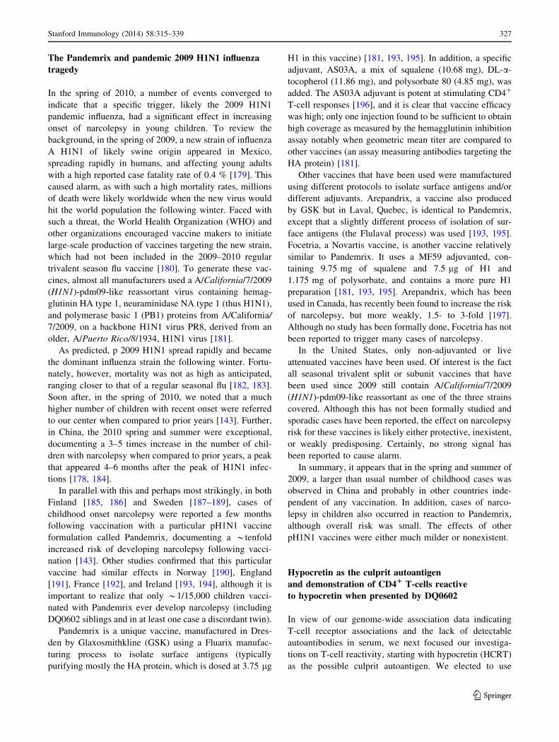
with and without narcolepsy. In all cases, the test predicted
narcolepsy with high specificity (100 %) and sensitivity
(*90 %), indicating diagnostic value (Fig. 7). Using other
cytokines, we also found the T-cell response to be con-
sistent with a Th1 and Th17 response, as usually found in
other autoimmune diseases [198].
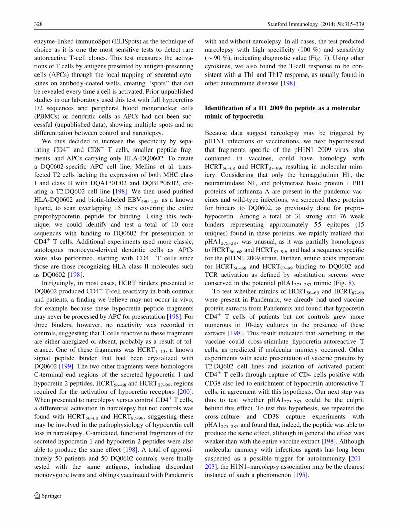
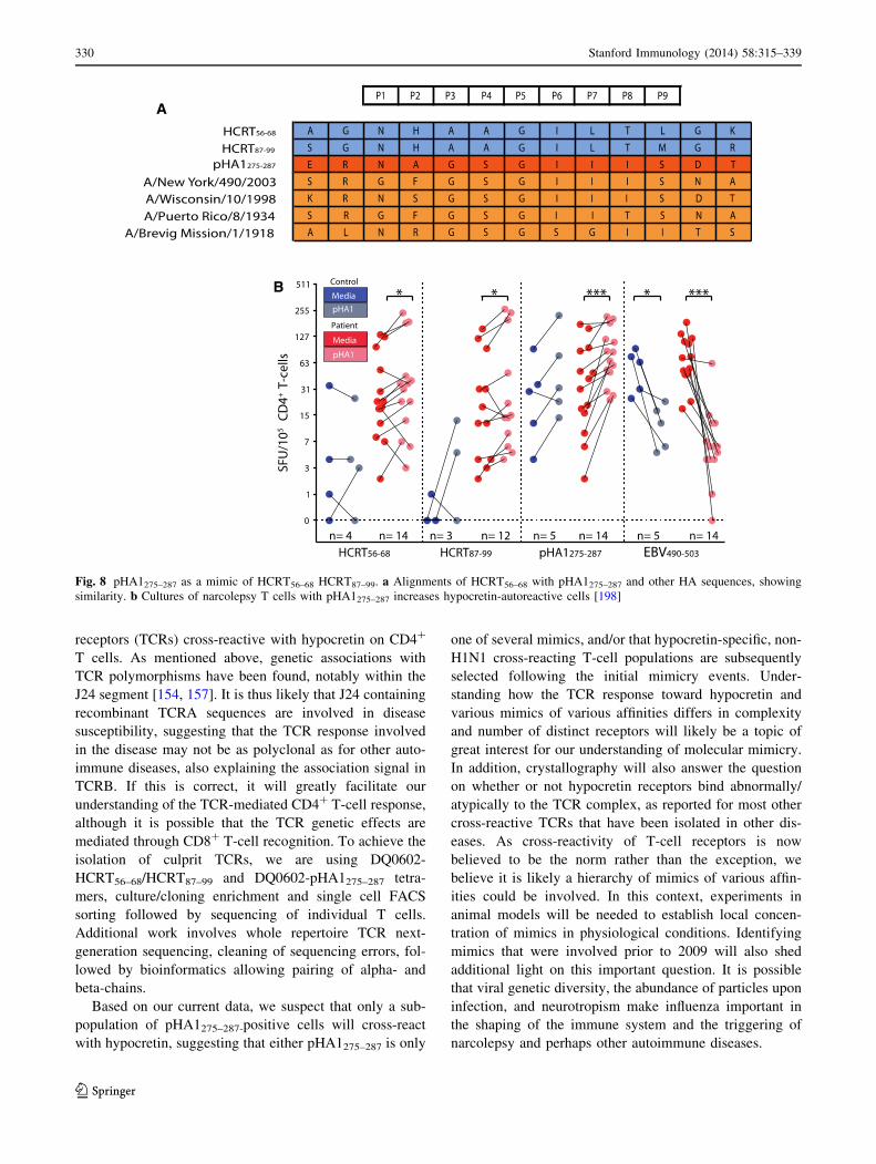
Identification of a H1 2009 flu peptide as a molecular
mimic of hypocretin
Because data suggest narcolepsy may be triggered by
pH1N1 infections or vaccinations, we next hypothesized
that fragments specific of the pH1N1 2009 virus, also
contained in vaccines, could have homology with
HCRT56–68 and HCRT87–99, resulting in molecular mim-
icry. Considering that only the hemagglutinin H1, the
neuraminidase N1, and polymerase basic protein 1 PB1
proteins of influenza A are present in the pandemic vac-
cines and wild-type infections, we screened these proteins
for binders to DQ0602, as previously done for prepro-
hypocretin. Among a total of 31 strong and 76 weak
binders representing approximately 55 epitopes (15
uniques) found in these proteins, we rapidly realized that
pHA1275–287 was unusual, as it was partially homologous
to HCRT56–68 and HCRT87–99, and had a sequence specific
for the pH1N1 2009 strain. Further, amino acids important
for HCRT56–68 and HCRT87–99 binding to DQ0602 and
TCR activation as defined by substitution screens were
conserved in the potential pHA1275–287 mimic (Fig. 8).
To test whether mimics of HCRT56–68 and HCRT87–99
were present in Pandemrix, we already had used vaccine
protein extracts from Pandemrix and found that hypocretin
CD4? T cells of patients but not controls grew more
numerous in 10-day cultures in the presence of these
extracts [198]. This result indicated that something in the
vaccine could cross-stimulate hypocretin-autoreactive T
cells, as predicted if molecular mimicry occurred. Other
experiments with acute presentation of vaccine proteins by
T2.DQ602 cell lines and isolation of activated patient
CD4? T cells through capture of CD4 cells positive with
CD38 also led to enrichment of hypocretin-autoreactive T
cells, in agreement with this hypothesis. Our next step was
thus to test whether pHA1275–287 could be the culprit
behind this effect. To test this hypothesis, we repeated the
cross-culture and CD38 capture experiments with
pHA1275–287 and found that, indeed, the peptide was able to
produce the same effect, although in general the effect was
weaker than with the entire vaccine extract [198]. Although
molecular mimicry with infectious agents has long been
suspected as a possible trigger for autoimmunity [201–
203], the H1N1–narcolepsy association may be the clearest
instance of such a phenomenon [195].
328 Stanford Immunology (2014) 58:315–339
123
Mediation of hypocretin cell killing
Although our results explain much, and may be the first
clear demonstration of molecular mimicry in humans,
several questions remain unanswered. First and foremost,
CD4? T cells are not likely to be the ultimate cell popu-
lation causing hypocretin cell destruction. In most other
cases, CD4? T cells are acting as helper T cells, directing
antibody formation by B cells and coordinating a CD8?
T-cell response that is cytotoxic and creates the cell
destruction. In narcolepsy, we found no evidence of anti-
body involvement, a phenomenon we hypothesize is due to
the fact hypocretin is specific to the brain. In the peripheral
circulation, B cell help through class II presentation of
antigens directs the production of antibodies. It may be that
hypocretin presentation by HLA DQB0602 on B cells to
autoreactive CD4 cells does not occur because B cells are
not commonly present in the central nervous system unless
the blood–brain barrier is very much disrupted. Although B
cells can traffic to the brain through the integrin system as
T cells, it is unclear whether this occurs under normal
conditions, as most of the studies supporting this have been
done in patients with multiple sclerosis. If this hypothesis is
correct, autoimmune diseases directed against brain-
specific antigens may have less B-cell involvement, pro-
viding there is no blood–brain barrier breakdown.
A more likely hypothesis may be the involvement of a
CD8? T cytotoxic cell population, although it is possible
CD4? T cells themselves or a novel mechanism specific to
the brain are involved, for example involving natural Killer
cells or microglia phagocytosis. Whatever the mechanism
should be, the specificity of the cell loss likely involves the
recognition of antigens specific of hypocretin cells. It is
notable that our CD4? T-reactive cells can recognize
truncated fragment of the secreted neurotransmitter [198], a
product available broadly in the interstitial field. Local
concentration gradients of hypocretin, other epitopes, or
other antigens could possibly play a role. Further studies in
this direction is likely to teach us much on how the immune
system treats neurons, a population of cells that is partic-
ular as it cannot express HLA class II and is not easily
replaceable.
T-cell receptors involved and other mimics
A key experimental next step toward our understanding of
narcolepsy is the identification of the culprit T-cell
A
C D
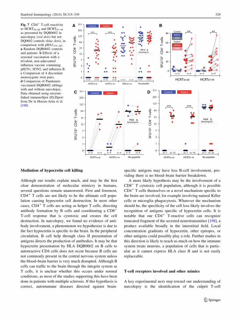
BFig. 7 CD4? T-cell reactivity
to HCRT56–68 and HCRT87–99
as presented by DQB0602 in
narcolepsy (red dots) but not
DQ0602 controls (blue dots), in
comparison with pHA1275–287.
a Random DQB0602 controls
and patients. b Effects of a
seasonal vaccination with a
trivalent, non-adjuvanted
influenza vaccine containing
pH1N1, H3N2, and influenza B.
c Comparison of 4 discordant
monozygotic twin pairs.
d Comparison of Pandemrix
vaccinated DQB0602 siblings
with and without narcolepsy.
Data obtained using enzyme-
linked immunoSpot (ELISpot)
from De la Herran-Arita et al.
[198]
Stanford Immunology (2014) 58:315–339 329
123
receptors (TCRs) cross-reactive with hypocretin on CD4?
T cells. As mentioned above, genetic associations with
TCR polymorphisms have been found, notably within the
J24 segment [154, 157]. It is thus likely that J24 containing
recombinant TCRA sequences are involved in disease
susceptibility, suggesting that the TCR response involved
in the disease may not be as polyclonal as for other auto-
immune diseases, also explaining the association signal in
TCRB. If this is correct, it will greatly facilitate our
understanding of the TCR-mediated CD4? T-cell response,
although it is possible that the TCR genetic effects are
mediated through CD8? T-cell recognition. To achieve the
isolation of culprit TCRs, we are using DQ0602-
HCRT56–68/HCRT87–99 and DQ0602-pHA1275–287 tetra-
mers, culture/cloning enrichment and single cell FACS
sorting followed by sequencing of individual T cells.
Additional work involves whole repertoire TCR next-
generation sequencing, cleaning of sequencing errors, fol-
lowed by bioinformatics allowing pairing of alpha- and
beta-chains.
Based on our current data, we suspect that only a sub-
population of pHA1275–287-positive cells will cross-react
with hypocretin, suggesting that either pHA1275–287 is only
one of several mimics, and/or that hypocretin-specific, non-
H1N1 cross-reacting T-cell populations are subsequently
selected following the initial mimicry events. Under-
standing how the TCR response toward hypocretin and
various mimics of various affinities differs in complexity
and number of distinct receptors will likely be a topic of
great interest for our understanding of molecular mimicry.
In addition, crystallography will also answer the question
on whether or not hypocretin receptors bind abnormally/
atypically to the TCR complex, as reported for most other
cross-reactive TCRs that have been isolated in other dis-
eases. As cross-reactivity of T-cell receptors is now
believed to be the norm rather than the exception, we
believe it is likely a hierarchy of mimics of various affin-
ities could be involved. In this context, experiments in
animal models will be needed to establish local concen-
tration of mimics in physiological conditions. Identifying
mimics that were involved prior to 2009 will also shed
additional light on this important question. It is possible
that viral genetic diversity, the abundance of particles upon
infection, and neurotropism make influenza important in
the shaping of the immune system and the triggering of
narcolepsy and perhaps other autoimmune diseases.
A
B
Fig. 8 pHA1275–287 as a mimic of HCRT56–68 HCRT87–99. a Alignments of HCRT56–68 with pHA1275–287 and other HA sequences, showing
similarity. b Cultures of narcolepsy T cells with pHA1275–287 increases hypocretin-autoreactive cells [198]
330 Stanford Immunology (2014) 58:315–339
123

Breakdown of immune tolerance
Another important question in the field of autoimmune
disease is how and why is tolerance toward autoantigens
brakes. The fact one of our polymorphism is located close
to the IL10RB suggests the importance of inhibitory sig-
nals [204]. Whether or not hypocretin is expressed in the
thymus is unknown, and examining the frequency of naıve
cells cross-reactive to hypocretin epitopes may shed light
on the role of central tolerance. Similarly, regulatory T
cells could be involved and, if identified, corresponding
TCRs were sequenced and compared to autoreactive TCRs.
It is of interest that in many of our culture conditions
notably with PBMCs, enrichment of hypocretin cross-
reactive cells seems more difficult when the cells are cul-
tured with hypocretin in comparison with the pHA1275–287
mimic. Figure 9 describes a potential roadmap to study the
pathophysiology of narcolepsy.
Why did Pandemrix have such a strong effect
in Northern Europe?
The finding of a mimic inside the H1 protein, pHA1275–287,
does not entirely explain why Pandemrix, as opposed to
other Pandemic vaccines, was particularly potent at trig-
gering narcolepsy. Two major explanations could be
involved, differences in vaccine composition or in the
target population of vaccinees [195, 198]. With respect to
vaccine composition differences, HA is the main protein
dosed in all vaccines and reported as reference for com-
parison; other viral components are present in lower
amounts [181, 193]. A several other vaccines have a higher
concentration of HA by dose than Pandemrix, it is sur-
prising that HA would be the culprit.
One possible explanation could be the effect of the
AS03A adjuvant at promoting CD4? T-cell response [196],
as it is possible this adjuvant is more potent than MF59
(squalene) alone [181], or has more effect on blood–brain
barrier permeability. This explanation however does not
explain why Arepandrix was weaker than Pandemrix at
increasing narcolepsy risk (OR = 1.5–3) [197], as it con-
tained the same AS03A adjuvant and presumably the same
HA concentration. Interestingly, however, as the antigen
preparation protocol is different between Pandemrix (Flu-
arix protocol), Arepandrix (Flulaval protocol), and Focetria
(Agrippal Protocol) [205], additional mimics on proteins
others than HA could be involved (i.e., NA, PB1, or less
probably antigens in the backbone that could work in
conjunction with pHA1275–287). Prior studies have clearly
shown these proteins to be differently purified in these
protocols, with the Agrippa process producing more pure
HA than the fluarix process [205] (no information is
available on the flulaval process). It is also possible that the
various inactivation processes used in these protocols, or
differences in excipients, produce differential antigenic
Fig. 9 Pathophysiological model for narcolepsy and road map for
future experiments. Narcolepsy may result from the succession of
unlikely events, starting with a predisposing genetic background,
stochastic events leading to the generation of potential pathogenic
naıve T cells, inadequate central tolerance, cross-stimulation with
various environmental mimics at the wrong developmental time (in
relation to other prior infections), the absence of peripheral tolerance,
penetration in the central nervous system of culprit T cells, molecular
mimicry, and destruction of hypocretin neurons by CD8? T cells or
other mechanisms. Other epitopes may also be involved, notably
those implicating CD8? T cells
Stanford Immunology (2014) 58:315–339 331
123

modification (oxidation, etc.) that could play a role. We are
pursuing these hypotheses.
Other factors not related to the vaccine itself could also
have played a role. Indeed, even in children and with
Pandemrix, absolute risk remains low, 1/15,000 vaccinees,
so that many other factors (environmental, stochastic, or
genetic) in addition to Pandemrix are likely to be needed
for a subject to develop narcolepsy. As the various vaccines
have been used in different populations, population-spe-
cific differences could make it seem like various vaccines
have different effects. One possible difference could
involve genetic polymorphisms enriched in Northern Eur-
ope. In a recent study, we found evidence for a genome-
wide significant difference in a SNP located between the
DR and DQ genes when comparing Chinese patients who
developed narcolepsy prior versus after September 2009
[157]. Nonetheless, odds ratio for such a polymorphism
remained low, and there is no evidence of strong popula-
tion differences for this SNP across Caucasians.
A second aspect could involve coinfections and timing of
vaccination in relation to how the H1N1 pandemic itself
unfolded in various countries. Prior data suggest that other
infections, such as streptococcus, may have played an
additional role in some population [174, 175], although it
could just be a secondary association [206]. Regarding H1N1
infection by themselves (a factor known to increase risk
based on our Chinese data [178, 184] ), peak vaccination
occurred 2–3 weeks following the major wave of infection in
most of the signal countries [207]; thus, a double hit could
have played a role in either direction (protection or adding
predisposition by increasing the immune response).
To address this issue, a virus-specific NS1 B-cell epitope
not present in the vaccine backbone was identified and
antibodies directed against it evaluated in Western blots
[208]. As reactivity was only found in subjects after doc-
umented natural infections but not in post-Pandemrix
cases, the authors concluded a double hit is unlikely.
Although tantalizing, we believe that exploring only one
epitope is not sufficient to conclude, considering HLA
dependence of these effects. It is also possible that the
narcolepsy immune response may not implicate the NS1
epitope in infected subjects because of immunodominance
differences in the response. Interestingly, preliminary data
in Sweden suggest that Pandemrix had less effects on
narcolepsy in subjects in the North of the country, where
infections came before the vaccination, possibly suggesting
that infections could have been protective. In summary, the
potential role of coinfections is uncertain especially con-
sidering the fact about the same timing of vaccination in
relation to natural pandemic infection occurred in Canada
with Arepandrix and in Scandinavia with Arepandrix.
Nonetheless, Focetria was more used in Southern Europe,
for example in Italy, when H1N1 infections likely reached
epidemic proportion later in the year. A third possibility
explaining variability in response to the various adjuvanted
vaccines is target population and how many adults versus
children were vaccinated and in what sequence [181, 207].
Finally, and maybe most importantly, these populations
are all distinct in their past immunological history and
original antigenic sin is likely to have played a role. As
young children were mostly at risk, it is possible that past
exposure to other prior influenza strain or other infections
mitigates risk, maybe simply because in these cases, a
reactivation of memory cells that are targeting a prior
immunodominant peptide common to prior strains reduces
the probability of engaging T cells specific of new mimics
present in pH1N1 in a naıve immune response. Alterna-
tively, new epitopes could reengage a population of
memory cells that have seen a prior antigen and can be
cross-reactive. We believe that the study of cross-reactive
T-cell populations in naıve and memory compartments of
controls of various origin and age, for example with hyp-
ocretin tetramers, will answers some of these questions.
Conclusion
Since my arrival at Stanford in the late 1980s, I have been
blessed with the unique collaborative ambience of the Uni-
versity and its immunology program. Trained as a molecular
pharmacologist and a psychiatrist, I was lucky to find col-
laborators such as F. Carl Grumet at the Blood Bank who,
like me, thought that positionally cloning the canine narco-
lepsy gene was not only possible but also the logical next
step. Collaborators such as Lucas Cavalli Sforza in genetics
and Joachim Hallmayer open my eyes to the power of human
genetics. Moving into immunology, I was exposed to the best
and the brightest, starting with deep HLA and immunoge-
netic training, and then to general immunology with Mark
Davis, Carl Grumet, Hugh Mac Devitt, Betsy Mellins, Larry
Steinman, and others. The small size of the University, the
accessibility of its leader, the excellence and diversity of its
faculty, and a willingness to dare and collaborate outside of
your field and comfort zone is what made Stanford unique,
notably in immunology.
Our current results indicate that narcolepsy, a disease
caused by hypocretin cell loss, is associated with autore-
active CD4? T cells recognizing fragments of hypocretin
when presented by DQ0602, an HLA allele strongly
associated with the disease. As the cause of the symptoms
is the loss of hypocretin, we believe this population of
autoreactive CD4? T cells is very likely within the causal
pathway for narcolepsy. Influenza A, notably 2009 pH1N1,
is a likely environmental trigger of the autoreactive CD4?
responses. Using two independent techniques, we found
that Pandemrix pH1N1 viral extracts were able to activate
332 Stanford Immunology (2014) 58:315–339
123
hypocretin-autoreactive cells, suggesting that molecular
mimics were present in the vaccine and the wild-type virus.
One particular mimic, pHA1275–287, was identified within
the virus as a likely culprit for the cross-reactivity. Toge-
ther with other data, these findings suggest that influenza
can be the trigger of autoimmune responses, notably
narcolepsy.
In the future, we plan to explore how hypocretin cells
are being destroyed, study where and why central and
peripheral tolerance toward hypocretin is broken in the
disease, identify other potential mimics and explore pop-
ulation- and vaccine-specific effects that may be involved
in precipitating narcolepsy. Isolation and characterization
of culprit T cell is ongoing to demonstrate mimicry.
Eventually, animal models will be needed to demonstrate
pathological effects of specific T-cell clones so that Koch’s
criteria of causation are met [203].
Additional work will include the study of cases without
cataplexy or of mild hypersomnia cases present in the
population, and immune therapeutic attempts in subjects
identified before the hypocretin cell loss is complete and
irreversible. Non-open-labeled studies using IVIg have
produced mixed results [209–214], not surprisingly con-
sidering the absence of current evidence for B-cell
involvement. We believe that these studies will teach us
much regarding vaccine safety and autoimmunity in gen-
eral, notably when it is directed against neurons. A likely
advantage of narcolepsy as a model for autoimmunity is it
relative simplicity: the involvement of a single primary
HLA heterodimer, possibly a single CD4? T-cell epitope
sequence derived from secreted hypocretin peptides, and a
likely relatively focused, oligoclonal causative T-cell
response.
In parallel with immune-related work, the expected
introduction of hypocretin antagonists (called dual orexin
receptor antagonists, DORA) as hypnotics will revive
interest in this target for drug development. Hypocretin
does not cross the blood–brain barrier well, and although
creating brain penetrating agonists for peptide receptor can
be challenging, it is likely a matter of time before such
compounds become available for treating daytime sleepi-
ness and narcolepsy.
Acknowledgments Most of this research was funded by NIH center
Grant NS-23724 and by gifts from patients. We thank the hundreds of
students, staff members, researchers, veterinarians, patients, and
medical doctors who over the years supported this research, con-
tributing experiments, samples, administrative time, and financial
support. I am particularly grateful to Bill Dement and Christian Gu-
illeminault, my sleep mentors; Seiji Nishino, my collaborator in
canine research; Mali Einen, my patient coordinator; and finally Ling
Lin and Juliette Faraco, who have been pillars of the laboratory since
its inception, and without whom this research would have been
impossible.
Open Access This article is distributed under the terms of the
Creative Commons Attribution License which permits any use, dis-
tribution, and reproduction in any medium, provided the original
author(s) and the source are credited.
References
1. Schenck CH, Bassetti CL, Arnulf I, Mignot E. English transla-
tions of the first clinical reports on narcolepsy and cataplexy by
Westphal and Gelineau in the late 19th century, with com-
mentary. J Clin Sleep Med. 2007;3(3):301–11.
2. Westphal C. Eigenthumliche mit Einschlafen verbundene An-
falle. Arch Psychiat. 1877;7:631–5.
3. Gelineau JBE. De la narcolepsie. Gazette des hopitaux. 1880;53:
626–8.
4. Yoss RE, Daly DD. Narcolepsy. Med Clin N Am. 1960;44:
953–68.
5. Mignot EJ. A practical guide to the therapy of narcolepsy and
hypersomnia syndromes. Neurotherapeutics. 2012;9(4):739–52.
doi:10.1007/s13311-012-0150-9.
6. Plazzi G, Serra L, Ferri R. Nocturnal aspects of narcolepsy with
cataplexy. Sleep Med Rev. 2008;12(2):109–28. doi:10.1016/j.
smrv.2007.08.010.
7. Daniels L. Narcolepsy. Medicine. 1934;13(1):1–122.
8. Mignot E, Lammers GJ, Ripley B, Okun M, Nevsimalova S,
Overeem S, et al. The role of cerebrospinal fluid hypocretin
measurement in the diagnosis of narcolepsy and other hyper-
somnias. Arch Neurol. 2002;59(10):1553–62.
9. Von Economo C. Encephalitis Lethargica. London: Oxford
Medical Publications, Humphrey Milford, Oxford University
Press; 1931.
10. Taubenberger JK, Baltimore D, Doherty PC, Markel H, Morens
DM, Webster RG et al. Reconstruction of the 1918 influenza
virus: unexpected rewards from the past. MBio. 2012;3(5).
doi:10.1128/mBio.00201-12.
11. Ravenholt RT, Foege WH. 1918 influenza, encephalitis leth-
argica, parkinsonism. Lancet. 1982;2(8303):860–4.
12. Dourmashkin RR, Dunn G, Castano V, McCall SA. Evidence
for an enterovirus as the cause of encephalitis lethargica. BMC
Infect Dis. 2012;12:136. doi:10.1186/1471-2334-12-136.
13. Mignot E. Why we sleep: the temporal organization of recovery.
PLoS Biol. 2008;6(4):e106. doi:10.1371/journal.pbio.0060106.
14. Symonds CP, Oxf MD, Lond FRCP. Narcolepsy as a symptom
of encephalitis lethargica. Lancet. 1922;173:1214–5.
15. Aldrich MS, Naylor MW. Narcolepsy associated with lesions of
the diencephalon. Neurology. 1989;39(11):1505–8.
16. Aserinski E, Kleitman N. Regularly occurring periods of eye
motility, and concomitant phenomena, during sleep. Science.
1953;118:273–4.
17. Jouvet M. Recherche sur les structures nerveuses et les me-
canismes responsables des differentes phases du sommeil
physiologique. Arch Ital Biol. 1962;100:125–206.
18. Dement W, Wolpert EA. The relation of eye movement, body
motility, and external stimuli to dream content. J Exp Psychol.
1958;55:543–53.
19. Rechtschaffen A, Wolpert EA, Dement WC, Mitchell SA, Fisher
C. Noctural sleep of narcoleptics. Electroencephalogr Clin
Neurophysiol. 1963;15:599–609.
20. Rechschaffen A, Dement W. Studies on the relation of narco-
lepsy, cataplexy and sleep with low voltage random EEG
activity. In: Kety S, Evarts E, Williams H, editors. Sleep and
altered states of consciousness. Baltimore: Williams and Wil-
kins; 1967. p. 488–505.
Stanford Immunology (2014) 58:315–339 333
123

21. Vogel G. Studies in psychophysiology of dreams III. The dream
of narcolepsy. Arch Gen Psychiatry. 1960;3:421–8.
22. Andlauer O, Moore IV H, Jouhier L, Drake C, Peppard PE, Han F
et al. Nocturnal REM sleep latency for identifying patients with
narcolepsy/hypocretin deficiency. JAMA Neurol. 2013;70(7):
891–902.
23. Dement WC. A personal history of sleep disorders medicine.
J Clin Neurophysiol. 1990;7(1):17–47.
24. Dement WC. The history of narcolepsy and other sleep disor-
ders. J Hist Neurosci. 1993;2(2):121–34. doi:10.1080/09647049
309525558.
25. Dement WC, Carskadon M, Ley R. The prevalence of narco-
lepsy II. Sleep Res. 1973;2:147.
26. Silber MH, Krahn LE, Olson EJ, Pankratz VS. The epidemiol-
ogy of narcolepsy in Olmsted County, Minnesota: a population-
based study. Sleep. 2002;25(2):197–202.
27. Ohayon MM, Priest RG, Zulley J, Smirne S, Paiva T. Prevalence
of narcolepsy symptomatology and diagnosis in the European
general population. Neurology. 2002;58(12):1826–33.
28. Pelayo R, Hodgson N, Guilleminault C. The history of sleep
medicine. Handb Clin Neurol. 2010;95:547–56. doi:10.1016/
S0072-9752(08)02134-9.
29. Shepard JW Jr, Buysse DJ, Chesson AL Jr, Dement WC,
Goldberg R, Guilleminault C, et al. History of the development
of sleep medicine in the United States. J Clin Sleep Med. 2005;
1(1):61–82.
30. Carskadon MA, Dement WC, Mitler MM, Roth T, Westbrook
PR, Keenan S. Guidelines for the multiple sleep latency test
(MSLT): a standard measure of sleepiness. Sleep. 1986;9(4):
519–24.
31. Richardson GS, Carskadon MA, Flagg W, Van den Hoed J, Dement
WC, Mitler MM. Excessive daytime sleepiness in man: multiple
sleep latency measurement in narcoleptic and control subjects.
Electroencephalogr Clin Neurophysiol. 1978;45(5):621–7.
32. Mitler MM, Van den Hoed J, Carskadon MA, Richardson G,
Park R, Guilleminault C, et al. REM sleep episodes during the
multiple sleep latency test in narcoleptic patients. Electroen-
cephalogr Clin Neurophysiol. 1979;46(4):479–81.
33. Honda Y. Introduction to ‘‘ HLA in narcolepsy’’. In: Honda Y,
Juji T, editors. HLA in narcolepsy. New York: Springer; 1988.
p. 1–10.
34. Juji T, Satake M, Honda Y, Doi Y. HLA antigens in Japanese
patients with narcolepsy. All the patients were DR2 positive.
Tissue Antigens. 1984;24(5):316–9.
35. Honda Y. Clinical features of narcolepsy: Japanese experience.
In: Honda Y, Juji T, editors. HLA in narcolepsy. New York:
Sringer; 1988. p. 24–57.
36. Billiard M, Seignalet J. Extraordinary association between
HLA-DR2 and narcolepsy. Lancet. 1985;1:226–7.
37. Mueller-Eckhardt G, Meier-Ewert K, Schendel DJ, Reinecker
FB, Multhoff G, Mueller-Eckhardt C. HLA and narcolepsy in a
German population. Tissue Antigens. 1986;28(3):163–9.
38. Montplaisir J, Poirier G. HLA in narcolepsy in Canada. In:
Honda Y, Juji T, editors. HLA in narcolepsy. New York:
Springer; 1988. p. 97–107.
39. Roth B, Nevsimalova S, Sonka K, Docekal P, Schulz H, Geisler P,
et al. A study of occurrence of HLA DR2 in 124 narcoleptics:
clinical aspects. Arch Suiss Neurol et de Psychiat. 1988;139(4):
41–51.
40. Guilleminault C, Holloman J, Grumet C, Kilduff T, McDevitt
HO, Dement WC, et al. HLA-DR2 and the narcolepsy syn-
drome: the Stanford experience. In: Honda Y, Juji T, editors.
HLA in narcolepsy. New York: Springer; 1988. p. 108–113.
41. Guilleminault C, Grumet C. HLA-DR2 and narcolepsy: not all
narcoleptic-cataplectic patients are DR2. Hum Immunol.
1986;17(1):1–2.
42. Matsuki K, Honda Y, Juji T. Diagnostic criteria for narcolepsy
and HLA-DR2 frequencies. Tissue Antigens. 1987;30(4):
155–60.
43. Marcadet A, Gebuhrer L, Betuel H, Seignalet J, Freidel AC,
Confavreux C, et al. DNA polymorphism related to HLA-DR2
Dw2 in patients with Narcolepsy. Immunogenetics. 1985;22:
679–83.
44. Naohara T, Honda Y, Juji T. Positive HLA-Dw2 in Narcolepsy.
In: Honda Y, Juji T, editors. HLA in narcolepsy. New York:
Springer; 1988. p. 121–9.
45. Uryu N, Maeda M, Nagata Y, Matsuki K, Juji T, Honda Y, et al.
No difference in the nucleotide sequence of the DQ beta beta 1
domain between narcoleptic and healthy individuals with DR2,
Dw2. Hum Immunol. 1989;24(3):175–81.
46. Holloman JD, Bell JI, Kilduff TS, Dement WC, Guilleminault
C. HLA-DR restriction-fragment-length polymorphisms in nar-
colepsy. J Neurosci Res. 1987;18:239–44.
47. Inoko H. DNA typing in Narcolepsy. In: Honda Y, Juji T, edi-
tors. HLA in narcolepsy. New York: Springer; 1988. p. 137–49.
48. Serjeantson SW, Gao X, Hawkins BR, Higgins DA, Yu YL.
Novel HLA-DR2-related haplotypes in Hong Kong Chinese
implicate the DQB1*0602 allele in susceptibility to multiple
sclerosis. Eur J Immunogenet. 1992;19(1–2):11–9.
49. McDevitt HO, Tyan ML. Genetic control of the antibody
response in inbred mice. Transfer of response by spleen cells
and linkage to the major histocompatibility (H-2) locus. J Exp
Med. 1968;128(1):1–11.
50. Dausset J. Correlation between histocompatibility antigens and
susceptibility to illness. Prog Clin Immunol. 1972;1:183–210.
51. Matsuki K, Juji T, Honda Y. Immunological features of narco-
lepsy in Japan. In: Honda Y, Juji T, editors. HLA in narcolepsy.
New York: Springer; 1988. p. 150–7.
52. Rubin RL, Hajdukovich RM, Mitler MM. HLA DR2 association
with excessive somnolence in narcolepsy does not generatize to
sleep apnea and is not accompanied by systemic autoimmune
abnormalities. Clin Immunol Immunipathol. 1988;49:149–58.
53. Mignot E, Guilleminault C, Grumet FC, Dement WC. Is nar-
colepsy an autoimmune disease? In: Smirne S, Francesci M,
Ferini-Strambi L, Zucconi M, editors. Proceedings of the third
Milano international symposium, ‘‘sleep, hormones, and the
immune system’’, Sept 18–19. Milan: Masson; 1992. p. 29–38.
54. Carlander B, Eliaou JF, Billiard M. Autoimmune hypothesis in
narcolepsy. Neurophysiol Clin. 1993;23:15–22.
55. Ellis MC, Hetisimer AH, Ruddy DA, Hansen SL, Kronmal GS,
McClelland E, et al. HLA class II haplotype and sequence
analysis support a role for DQ in narcolepsy. Immunogenetics.
1997;46(5):410–7.
56. Matsuki K, Grumet FC, Lin X, Gelb M, Guilleminault C,
Dement WC, et al. DQ (rather than DR) gene marks suscepti-bility to narcolepsy. Lancet. 1992;339(8800):1052.
57. Mignot E, Kimura A, Lattermann A, Lin X, Yasunaga S, Mu-
eller-Eckhardt G, et al. Extensive HLA class II studies in 58
non-DRB1*15 (DR2) narcoleptic patients with cataplexy. Tissue
Antigens. 1997;49(4):329–41.
58. Kadotani H, Faraco J, Mignot E. Genetic studies in the sleep
disorder narcolepsy. Genome Res. 1998;8(5):427–34.
59. Lock CB, Welsh KI, Parkes JD, Briggs DC, Waughan RW, Van
Dam M. HLA in narcolepsy–cataplexy in the United Kingdom.
In: Honda Y, Juji T, editors. HLA in narcolepsy. New York:
Springer; 1988. p. 76–88.
60. Mignot E, Lin X, Arrigoni J, Macaubas C, Olive F, Hallmayer J,
et al. DQB1*0602 and DQA1*0102 (DQ1) are better markers
than DR2 for narcolepsy in Caucasian and black Americans.
Sleep. 1994;17(8 Suppl):S60–7.
61. Mitler MM, Boysen BG, Campbell L, Dement WC. Narcolepsy–
cataplexy in a female dog. Exp Neurol. 1974;45(2):332–40.
334 Stanford Immunology (2014) 58:315–339
123
62. Baker TL, Foutz AS, McNerney V, Mitler MM, Dement WC.
Canine model of narcolepsy: genetic and developmental deter-
minants. Exp Neurol. 1982;75(3):729–42.
63. Foutz A, Mitler M, Cavalli-Sforza L, Dement WC. Genetic
factors in canine narcolepsy. Sleep. 1979;1(4):413–21.
64. Ripley B, Fujiki N, Okura M, Mignot E, Nishino S. Hypocretin
levels in sporadic and familial cases of canine narcolepsy.
Neurobiol Dis. 2001;8(3):525–34.
65. Kushida CA, Baker TL, Dement WC. Electroencephalographic
correlates of cataplectic attacks in narcoleptic canines. Electro-
encephalogr Clin Neurophysiol. 1985;61:61–70.
66. Lucas EA, Foutz AS, Dement WC, Mitler MM. Sleep cycle
organization in narcoleptic and normal dogs. Physiol Behav.
1979;23(4):737–43.
67. Lucas EA, Foutz AS, Mitler MM, Brown D, Dement WC.
Multiple sleep latency test in normal and narcoleptic canines.
Soc Neurosci Abstr. 1978;6:541.
68. Nishino S, Mignot E. Pharmacological aspects of human and
canine narcolepsy. Prog Neurobiol. 1997;52(1):27–78.
69. Mitler MM, Dement WC. Sleep studies on canine narcolepsy:
pattern and cycle comparisons between affected and normal
dogs. Electroencephalogr Clin Neurophysiol. 1977;43(5):691–9.
70. Dean RR, Kilduff TS, Dement WC, Grumet FC. Narcolepsy
without unique MHC class II antigen association: studies in the
canine model. Hum Immunol. 1989;25:27–35.
71. Motoyama M, Kilduff TS, Lee BSM, Dement WC, McDevitt
HO. Restriction fragment length polymorphism in canine nar-
colepsy. Immunogenetics. 1989;29:124–6.
72. Karczmar AG, Longo VG, De Carolis S. A pharmacological
model of paradoxical sleep: the role of cholinergic and mono-
aminergic systems. Physiol Behav. 1970;5:175–82.
73. Aldrich MS, Hollingsworth Z, Penney JB. Dopamine-receptor
autoradiography of human narcoleptic brain. Neurology. 1992;
42:410–5.
74. Aldrich MS, Prokopowicz G, Ockert K, Hollingsworth Z, Penny
JB, Albin RL. Neurochemical studies of human narcolepsy:
alpha-adrenergic receptor autoradiography of human narcoleptic
brain and brainstem. Sleep. 1994;17:598–608.
75. Aldrich MS. The neurobiology of narcolepsy–cataplexy. Prog
Neurobiol. 1993;41:533–41.
76. Bowersox S, Kilduff T, Faul K, Dement WC, Ciaranello RD.
Brain dopamine receptor levels elevated in canine narcolepsy.
Brain Res. 1987;402:44–8.
77. Boehme R, Baker T, Mefford I, Barchas J, Dement WC, Ci-
aranello R. Narcolepsy: cholinergic receptor changes in an
animal model. Life Sci. 1984;34:1825–8.
78. Faull KF, Zeller-DeAmicis LC, Radde L, Bowersox SS, Baker
TL, Kilduff TS, et al. Biogenic amine concentrations in the
brains of normal and narcoleptic canines: current status. Sleep.
1986;9(1):107–10.
79. Kilduff T, Bowersox SS, Kaitin KI, Baker TL, Ciaranello RD,
Dement WC. Muscarinic cholinergic receptors and the canine
model of narcolepsy. Sleep. 1986;9:102–7.
80. Mefford IN, Baker TL, Boehme R, Foutz AS, Ciaranello RD,
Barchas JD, et al. Narcolepsy: biogenic amine deficits in an
animal model. Science. 1983;220:629–32.
81. Miller JD, Faull KF, Bowersox SS, Dement WC. CNS mono-
amines and their metabolites in canine narcolepsy: a replication
study. Brain Res. 1990;509:169–71.
82. Hobson JA, McCarley RW, Wyzinski PW. Sleep cycle oscilla-
tion: reciprocal discharge by two brainstem neuronal groups.
Science. 1975;189:55–8.
83. McCarley RW, Hobson JA. Neuronal excitability modulation
over the sleep cycle: a structural and mathematical model.
Science. 1975;189(4196):58–60.
84. Mignot E, Renaud A, Nishino S, Arrigoni J, Guilleminault C,
Dement WC. Canine cataplexy is preferentially controlled by
adrenergic mechanisms: evidence using monoamine selective
uptake inhibitors and release enhancers. Psychopharmacology.
1993;113(1):76–82.
85. Nishino S, Arrigoni J, Shelton J, Dement WC, Mignot E.
Desmethyl metabolites of serotonergic uptake inhibitors are
more potent for suppressing canine cataplexy than their parent
compounds. Sleep. 1993;16(8):706–12.
86. Mignot E, Nishino S, Guilleminault C, Dement WC. Modafinil
binds to the dopamine uptake carrier site with low affinity.
Sleep. 1994;17(5):436–7.
87. Nishino S, Mao J, Sampathkumaran R, Shelton J. Increased
dopaminergic transmission mediates the wake-promoting effects
of CNS stimulants. Sleep Res Online. 1998;1(1):49–61.
88. Volkow ND, Fowler JS, Logan J, Alexoff D, Zhu W, Telang F,
et al. Effects of modafinil on dopamine and dopamine trans-
porters in the male human brain: clinical implications. JAMA.
2009;301(11):1148–54. doi:10.1001/jama.2009.351.
89. Nishino S, Tafti M, Reid MS, Shelton J, Siegel JM, Dement
WC, et al. Muscle atonia is triggered by cholinergic stimulation
of the basal forebrain: implication for the pathophysiology of
canine narcolepsy. J Neurosci. 1995;15(7 Pt 1):4806–14.
90. Reid MS, Tafti M, Nishino S, Sampathkumaran R, Siegel JM,
Dement WC, et al. Local administration of dopaminergic drugs
into the ventral tegmental area modulate cataplexy in the nar-
coleptic canine. Brain Res. 1996;733:83–100.
91. Fernandez-Vina M, Moraes JR, Moraes ME, Miller S, Stasny P.
HLA class II haplotypes in Amerindians and in black North and
South Americans. Tissue Antigens. 1991;38:235–7.
92. Behar E, Lin X, Grumet FC, Mignot E. A new DRB1*1202
allele (DRB1*12022) found in association with DQA1*0102
and DQB1*0602 in two black narcoleptic subjects. Immuno-
genetics. 1995;41(1):52.
93. Neely SE, Rosenberg RS, Spire JP, Antel J, Arnason W.
Familial narcolepsy and HLA antigens. Ann Neurol. 1986;20(1):
168.
94. Mignot E, Lin X, Hesla PE, Dement WC, Guilleminault C,
Grumet FC. A novel HLA DR17, DQ1 (DQA1-0102/DQB1-
0602 positive) haplotype predisposing to narcolepsy in Cauca-
sians. Sleep. 1993;16(8):764–5.
95. Han F, Lin L, Li J, Dong SX, An P, Zhao L, et al. HLA-DQ
association and allele competition in Chinese narcolepsy. Tissue
Antigens. 2012;80(4):328–35. doi:10.1111/j.1399-0039.2012.
01948.x.
96. Mignot E. Genetic and familial aspects of narcolepsy. Neurol-
ogy. 1998;50(2 Suppl 1):S16–22.
97. Guilleminault C, Mignot E, Grumet FC. Familial patterns of
narcolepsy. Lancet. 1989;2(8676):1376–9.
98. Honda Y, Asaka C, Tanimura M, Furusho T. A genetic study of
narcolepsy and excessive daytime sleepiness in 308 families
with a narcolepsy or hypersomnia proband. In: Guilleminault C,
Lugaresi E, editors. Sleep/wake disorders: natural history, epi-
demiology and long-term evolution. New York: Raven Press;
1983. p. 187–99.
99. Nevsimalova S, Mignot E, Sonka K, Arrigoni JL. Familial
aspects of narcolepsy–cataplexy in the Czech Republic. Sleep.
1997;20(11):1021–6.
100. Mignot E, Wang C, Rattazzi C, Gaiser C, Lovett M, Guille-
minault C, et al. Genetic linkage of autosomal recessive canine
narcolepsy with a mu immunoglobulin heavy-chain switch-like
segment. Proc Natl Acad Sci USA. 1991;88(8):3475–8.
101. Mignot E, Bell RA, Rattazzi C, Lovett M, Grumet FC, Dement
WC. An immunoglobulin switchlike sequence is linked with
canine narcolepsy. Sleep. 1994;17(8 Suppl):S68–76.
Stanford Immunology (2014) 58:315–339 335
123
102. Li R, Mignot E, Faraco J, Kadotani H, Cantanese J, Zhao B,
et al. Construction and characterization of an eightfold redun-
dant dog genomic bacterial artificial chromosome library.
Genomics. 1999;58(1):9–17.
103. Li R, Faraco JH, Lin L, Lin X, Hinton L, Rogers W, et al.
Physical and radiation hybrid mapping of canine chromosome
12, in a region corresponding to human chromosome 6p12–q12.
Genomics. 2001;73(3):299–315.
104. Lin L, Faraco J, Li R, Kadotani H, Rogers W, Lin X, et al. The
sleep disorder canine narcolepsy is caused by a mutation in the
hypocretin (orexin) receptor 2 gene. Cell. 1999;98(3):365–76.
105. De Lecea L, Kilduff TS, Peyron C, Gao X-B, Foye PE, Dan-
ielson PE, et al. The hypocretins: hypothalamus-specific pep-
tides with neuroexcitatory activity. Proc Natl Acad Sci USA.
1998;95:322–7.
106. Sakurai T, Amemiya A, Ishii M, Matsuzaki I, Chemelli RM,
Tanaka H, et al. Orexins and orexin receptors: a family of
hypothalamic neuropeptides and G protein-coupled receptors
that regulate feeding behavior. Cell. 1998;92:573–85.
107. Gautvik KM, de Lecea L, Gautvik VT, Danielson PE, Tranque
P, Dopazo A, et al. Overview of the most prevalent hypothala-
mus-specific mRNAs, as identified by directional tag PCR
subtraction. Proc Natl Acad Sci USA. 1996;93(16):8733–8.
108. Edwards CM, Abusnana S, Sunter D, Murphy KG, Ghatei MA,
Bloom SR. The effect of the orexins on food intake: comparison
with neuropeptide Y, melanin-concentrating hormone and gal-
anin. J Endocrinol. 1999;160(3):R7–12.
109. Haynes AC, Jackson B, Overend P, Buckingham RE, Wilson S,
Tadayyon M, et al. Effects of single and chronic intracerebro-
ventricular administration of the orexins on feeding in the rat.
Peptides. 1999;20(9):1099–105.
110. Ida T, Nakahara K, Katayama T, Murakami N, Nakazato M.
Effect of lateral cerebroventricular injection of the appetite-
stimulating neuropeptide, orexin and neuropeptide Y, on the
various behavioral activities of rats. Brain Res. 1999;821:526–9.
111. Lubkin M, Stricker-Krongrad A. Independent feeding and met-
abolic actions of orexins in mice. Biochem Biophys Res Com-
mun. 1998;253(2):241–5.
112. Yamanaka A, Sakurai T, Katsumoto T, Yanagisawa M, Goto K.
Chronic intracerebroventricular administration of orexin-A to
rats increases food intake in daytime, but has no effect on body
weight. Brain Res. 1999;849(1–2):248–52.
113. Girault EM, Yi CX, Fliers E, Kalsbeek A. Orexins, feeding, and
energy balance. Prog Brain Res. 2012;198:47–64. doi:10.1016/
B978-0-444-59489-1.00005-7.
114. Rolls A, Schaich Borg J, de Lecea L. Sleep and metabolism: role
of hypothalamic neuronal circuitry. Best Pract Res Clin Endo-
crinol Metab. 2010;24(5):817–28. doi:10.1016/j.beem.2010.08.
002.
115. Peyron C, Tighe DK, van den Pol AN, de Lecea L, Heller HC,
Sutcliffe JG, et al. Neurons containing hypocretin (orexin)
project to multiple neuronal systems. J Neurosci. 1998;18(23):
9996–10015.
116. Nambu T, Sakurai T, Mizukami K, Hosoya Y, Yanagisawa M,
Goto K. Distribution of orexin neurons in the adult rat brain.
Brain Res. 1999;827:243–60.
117. Horvath TL, Peyron C, Diano S, Ivanov A, Aston-Jones G,
Kilduff TS, et al. Hypocretin (orexin) activation and synaptic
innervation of the locus coeruleus noradrenergic system. J Comp
Neurol. 1999;415(2):145–59.
118. Brown RE, Sergeeva O, Eriksson KS, Haas HL. Orexin A
excites serotonergic neurons in the dorsal raphe nucleus of the
rat. Neuropharmacology. 2001;40(3):457–9.
119. Eriksson KS, Sergeeva O, Brown RE, Haas HL. Orexin/hypo-
cretin excites the histaminergic neurons of the tuberomammil-
lary nucleus. J Neurosci. 2001;21(23):9273–9.
120. Chemelli RM, Willie JT, Sinton CM, Elmquist JK, Scammell T,
Lee C, et al. Narcolepsy in orexin knockout mice: molecular
genetics of sleep regulation. Cell. 1999;98(4):437–51.
121. Peyron C, Faraco J, Rogers W, Ripley B, Overeem S, Charnay
Y, et al. A mutation in a case of early onset narcolepsy and a
generalized absence of hypocretin peptides in human narcoleptic
brains. Nat Med. 2000;6(9):991–7.
122. Nevsimalova S, Vankova J, Sonka K, Faraco J, Rogers W, O-
vereem S, et al. Hypocretin (orexin) deficiency in narcolepsy–
cataplexy. Sb Lek. 2000;101(4):381–6.
123. Nishino S, Ripley B, Overeem S, Lammers GJ, Mignot E.
Hypocretin (orexin) deficiency in human narcolepsy. Lancet.
2000;355(9197):39–40.
124. Thannickal TC, Moore RY, Nienhuis R, Ramanathan L, Gulyani
S, Aldrich M, et al. Reduced number of hypocretin neurons in
human narcolepsy. Neuron. 2000;27(3):469–74.
125. Blouin AM, Thannickal TC, Worley PF, Baraban JM, Reti IM,
Siegel JM. Narp immunostaining of human hypocretin (orexin)
neurons: loss in narcolepsy. Neurology. 2005;65(8):1189–92.
doi:10.1212/01.wnl.0000175219.01544.c8.
126. Crocker A, Espana RA, Papadopoulou M, Saper CB, Faraco J,
Sakurai T et al. Concomitant loss of dynorphin, NARP, and
orexin in narcolepsy. Neurology. 2005;65(8):1184–8.
127. Valko PO, Gavrilov YV, Yamamoto M, Reddy H, Haybaeck J,
Mignot E, et al. Increase of histaminergic tuberomammillary
neurons in narcolepsy. Ann Neurol. 2013;74(6):794–804.
doi:10.1002/ana.24019.
128. John J, Thannickal TC, McGregor R, Ramanathan L, Ohtsu H,
Nishino S, et al. Greatly increased numbers of histamine cells in
human narcolepsy with cataplexy. Ann Neurol. 2013;74(6):
786–93. doi:10.1002/ana.23968.
129. Tanaka S, Honda Y, Inoue Y, Honda M. Detection of autoan-
tibodies against hypocretin, hcrtrl, and hcrtr2 in narcolepsy:
anti-Hcrt system antibody in narcolepsy. Sleep. 2006;29(5):
633–8.
130. Black JL 3rd, Silber MH, Krahn LE, Fredrickson PA, Pankratz
VS, Avula R, et al. Analysis of hypocretin (orexin) antibodies in
patients with narcolepsy. Sleep. 2005;28(4):427–31.
131. Black JL 3rd, Silber MH, Krahn LE, Avula RK, Walker DL,
Pankratz VS, et al. Studies of humoral immunity to prepro-
hypocretin in human leukocyte antigen DQB1*0602-positive
narcoleptic subjects with cataplexy. Biol Psychiatry.
2005;58(6):504–9. doi:10.1016/j.biopsych.2005.04.026.
132. Martinez-Rodriguez JE, Sabater L, Graus F, Iranzo A, San-
tamaria J. Evaluation of hypothalamic-specific autoimmunity in
patients with narcolepsy. Sleep. 2007;30(1):27–8.
133. Knudsen S, Mikkelsen JD, Jennum P. Antibodies in narcolepsy–
cataplexy patient serum bind to rat hypocretin neurons. Neuro-
Report. 2007;18(1):77–9. doi:10.1097/WNR.0b013e328010
baad.
134. Overeem S, Verschuuren JJ, Fronczek R, Schreurs L, den Her-
tog H, Hegeman-Kleinn IM, et al. Immunohistochemical
screening for autoantibodies against lateral hypothalamic neu-
rons in human narcolepsy. J Neuroimmunol. 2006;174(1–2):
187–91. doi:10.1016/j.jneuroim.2006.02.010.
135. Overeem S, Black JL 3rd, Lammers GJ. Narcolepsy: immuno-
logical aspects. Sleep Med Rev. 2008;12(2):95–107. doi:10.
1016/j.smrv.2007.07.010.
136. Smith AJ, Jackson MW, Neufing P, McEvoy RD, Gordon TP. A
functional autoantibody in narcolepsy. Lancet. 2004;364(9451):
2122–4. doi:10.1016/S0140-6736(04)17553-3.
137. Katzav A, Arango MT, Kivity S, Tanaka S, Givaty G, Agmon-
Levin N, et al. Passive transfer of narcolepsy: anti-TRIB2
autoantibody positive patient IgG causes hypothalamic orexin
neuron loss and sleep attacks in mice. J Autoimmun. 2013;45:
24–30. doi:10.1016/j.jaut.2013.06.010.
336 Stanford Immunology (2014) 58:315–339
123

138. Jackson MW, Reed JH, Smith AJ, Gordon TP. An autoantibody
in narcolepsy disrupts colonic migrating motor complexes.
J Neurosci. 2008;28(49):13303–9. doi:10.1523/JNEUROSCI.
4489-08.2008.
139. Cvetkovic-Lopes V, Bayer L, Dorsaz S, Maret S, Pradervand S,
Dauvilliers Y, et al. Elevated Tribbles homolog 2-specific
antibody levels in narcolepsy patients. J Clin Invest. 2010;
120(3):713–9. doi:10.1172/JCI41366.
140. Dougherty JD, Schmidt EF, Nakajima M, Heintz N. Analytical
approaches to RNA profiling data for the identification of genes
enriched in specific cells. Nucleic Acids Res. 2010;38(13):
4218–30. doi:10.1093/nar/gkq130.
141. Honda M, Eriksson KS, Zhang S, Tanaka S, Lin L, Salehi A,
et al. IGFBP3 colocalizes with and regulates hypocretin (orex-
in). PLoS ONE. 2009;4(1):e4254. doi:10.1371/journal.pone.
0004254.
142. Eriksson KS, Mignot E. T-box 3 is expressed in the adult mouse
hypothalamus and medulla. Brain Res. 2009;1302:233–9.
doi:10.1016/j.brainres.2009.08.101.
143. Dauvilliers Y, Montplaisir J, Cochen V, Desautels A, Einen M, Lin
L, et al. Post-H1N1 narcolepsy–cataplexy. Sleep. 2010;33(11):
1428–30.
144. Lind A, Ramelius A, Olsson T, Arnheim-Dahlstrom L, Lamb F,
Khademi M et al. A/H1N1 antibodies and TRIB2 autoantibodies
in narcolepsy patients diagnosed in conjunction with the Pan-
demrix vaccination campaign in Sweden 2009-2010. J Autoim-
mun. 2014. doi:10.1016/j.jaut.2014.01.031.
145. Pelin Z, Guilleminault C, Risch N, Grumet FC, Mignot E. HLA-
DQB1*0602 homozygosity increases relative risk for narcolepsy
but not disease severity in two ethnic groups. US Modafinil in
Narcolepsy Multicenter Study Group. Tissue Antigens. 1998;
51(1):96–100.
146. Mignot E, Lin L, Rogers W, Honda Y, Qiu X, Lin X, et al.
Complex HLA-DR and -DQ interactions confer risk of narco-
lepsy–cataplexy in three ethnic groups. Am J Hum Genet. 2001;
68(3):686–99.
147. Hong SC, Lin L, Lo B, Jeong JH, Shin YK, Kim SY, et al.
DQB1*0301 and DQB1*0601 modulate narcolepsy suscepti-
bility in Koreans. Hum Immunol. 2007;68(1):59–68. doi:10.
1016/j.humimm.2006.10.006.
148. Weiner Lachmi K, Lin L, Kornum BR, Rico T, Lo B, Aran A,
et al. DQB1*06:02 allele-specific expression varies by allelic
dosage, not narcolepsy status. Hum Immunol. 2012;73(4):
405–10. doi:10.1016/j.humimm.2012.01.004.
149. Mignot E, Lin L, Li H, Thomson G, Lathrop M, Thorsby E et al.
13th International Histocompatibility Working group for nar-
colepsy joint report. In: Hansen J, Dupont B, editors. Immuno-
biology of human MHC. Proceedings of the 13th International
Histocompatibility Workshop and Congress (Seattle, WA 2004).
Seattle: IHWG Press; 2006. p. 782–7.
150. Hohjoh H, Terada N, Honda Y, Juji T, Tokunaga K. Negative
association of the HLA-DRB1*1502-DQB1*0601 haplotype
with human narcolepsy. Immunogenetics. 2001;52(3–4):
299–301.
151. Hor H, Kutalik Z, Dauvilliers Y, Valsesia A, Lammers GJ,
Donjacour CE, et al. Genome-wide association study identifies
new HLA class II haplotypes strongly protective against nar-
colepsy. Nat Genet. 2010;42(9):786–9. doi:10.1038/ng.647.
152. Tafti M, Hor H, Dauvilliers Y, Lammers GJ, Overeem S, Mayer
G, et al. DQB1 locus alone explains most of the risk and pro-
tection in narcolepsy with cataplexy in Europe. Sleep. 2014;
37(1):19–25. doi:10.5665/sleep.3300.
153. Ollila HM, Fernandes-Vina M, Mignot E. Alelle competition in
narcolepsy. Sleep. 2014;in press.
154. Hallmayer J, Faraco J, Lin L, Hesselson S, Winkelmann J,
Kawashima M, et al. Narcolepsy is strongly associated with the
T-cell receptor alpha locus. Nat Genet. 2009;41(6):708–11.
doi:10.1038/ng.372.
155. Faraco J, Lin L, Kornum BR, Kenny EE, Trynka G, Einen M,
et al. ImmunoChip study implicates antigen presentation to T
cells in narcolepsy. PLoS Genet. 2013;9(2):e1003270. doi:10.
1371/journal.pgen.1003270.
156. Kornum BR, Kawashima M, Faraco J, Lin L, Rico T, Hesselson
S et al. Common variants in P2RY11 are associated with nar-
colepsy. Nat Genet. 2011;43(1).
157. Han F, Faraco J, Dong XS, Ollila HM, Lin L, Li J. Genome wide
analysis of narcolepsy in china implicates novel immune loci
and reveals changes in association prior to versus after the 2009
H1N1 influenza pandemic. PLoS Genet. 2013;9(10):e1003880-
e. doi:10.1371/journal.pgen.1003880.
158. Manku H, Langefeld CD, Guerra SG, Malik TH, Alarcon-Ri-
quelme M, Anaya JM, et al. Trans-ancestral studies fine map the
SLE-susceptibility locus TNFSF4. PLoS Genet. 2013;9(7):
e1003554. doi:10.1371/journal.pgen.1003554.
159. Jostins L, Ripke S, Weersma RK, Duerr RH, McGovern DP, Hui KY,
et al. Host-microbe interactions have shaped the genetic architecture
of inflammatory bowel disease. Nature. 2012;491(7422):
119–24. doi:10.1038/nature11582.
160. Kim K, Bang SY, Lee HS, Cho SK, Choi CB, Sung YK, et al.
High-density genotyping of immune loci in Koreans and Euro-
peans identifies eight new rheumatoid arthritis risk loci. Ann
Rheum Dis. 2014;. doi:10.1136/annrheumdis-2013-204749.
161. Cooper JD, Smyth DJ, Smiles AM, Plagnol V, Walker NM,
Allen JE, et al. Meta-analysis of genome-wide association study
data identifies additional type 1 diabetes risk loci. Nat Genet.
2008;40(12):1399–401. doi:10.1038/ng.249.
162. Hirota T, Takahashi A, Kubo M, Tsunoda T, Tomita K, Sakashita
M, et al. Genome-wide association study identifies eight new
susceptibility loci for atopic dermatitis in the Japanese population.
Nat Genet. 2012;44(11):1222–6. doi:10.1038/ng.2438.
163. Moran CJ, Walters TD, Guo CH, Kugathasan S, Klein C, Turner
D, et al. IL-10R polymorphisms are associated with very-early-
onset ulcerative colitis. Inflamm Bowel Dis. 2013;19(1):115–23.
doi:10.1002/ibd.22974.
164. Winkelmann J, Lin L, Schormair B, Kornum BR, Faraco J,
Plazzi G, et al. Mutations in DNMT1 cause autosomal dominant
cerebellar ataxia, deafness and narcolepsy. Hum Mol Genet.
2012;21(10):2205–10. doi:10.1093/hmg/dds035.
165. Han F, Schormair B, Pizza F, Ollila HM, Nevsimalova S, Jen-
num P et al. HLA DQB1*06:02 negative narcolepsy with hyp-
ocretin/orexin deficiency. Sleep. 2014;in press.
166. Hecht M, Lin L, Kushida CA, Umetsu DT, Taheri S, Einen M,
et al. Report of a case of immunosuppression with prednisone in
an 8-year-old boy with an acute onset of hypocretin-deficiency
narcolepsy. Sleep. 2003;26(7):809–10.
167. Snider LA, Swedo SE. PANDAS: current status and directions
for research. Mol Psychiatry. 2004;9(10):900–7. doi:10.1038/sj.
mp.4001542.
168. Cardoso F. Sydenham’s chorea. Handb Clin Neurol. 2011;100:
221–9. doi:10.1016/B978-0-444-52014-2.00014-8.
169. Billiard M, Laaberki M, Reygrobellet C, Seignalet J, Brissaud L,
Besset A. Elevated antibodies to streptococcal antigens in nar-
coleptic subjects. Sleep Res. 1989;201.
170. Montplaisir J, Poirer G, Lapierre O, Montplaisir S. Streptococcal
antibodies in narcolepsy and idiopathic hypersomnia. Sleep Res.
1989;18:271.
171. Mueller-Eckhardt G, Meier-Ewart K, Schiefer HG. Is there an
infectious origin of narcolepsy? Lancet. 1990;335(8686):424.
172. Dale RC, Church AJ, Surtees RA, Lees AJ, Adcock JE, Harding
B, et al. Encephalitis lethargica syndrome: 20 new cases and
evidence of basal ganglia autoimmunity. Brain. 2004;127(Pt 1):
21–33. doi:10.1093/brain/awh008.
Stanford Immunology (2014) 58:315–339 337
123
173. Aran A, Weiner K, Lin L, Finn LA, Greco MA, Peppard P, et al.
Post-streptococcal auto-antibodies inhibit protein disulfide
isomerase and are associated with insulin resistance. PLoS ONE.
2010;5(9):e12875. doi:10.1371/journal.pone.0012875.
174. Aran A, Lin L, Nevsimalova S, Plazzi G, Hong SC, Weiner K,
et al. Elevated anti-streptococcal antibodies in patients with
recent narcolepsy onset. Sleep. 2009;32(8):979–83.
175. Koepsell TD, Longstreth WT, Ton TG. Medical exposures in
youth and the frequency of narcolepsy with cataplexy: a popu-
lation-based case-control study in genetically predisposed peo-
ple. J Sleep Res. 2009;19(1 Pt 1):80–6. doi:10.1111/j.1365-
2869.2009.00756.x.
176. Aran A, Lin L, Finn LA, Weiner K, Peppard P, Young T, et al.
Post-streptococcal antibodies are associated with metabolic
syndrome in a population-based cohort. PLoS ONE. 2011;6(9):
e25017. doi:10.1371/journal.pone.0025017.
177. Han F, Lin L, Li J, Aran A, Dong SX, An P, et al. Presentations
of primary hypersomnia in Chinese children. Sleep. 2011;
34(5):627–32.
178. Han F, Lin L, Warby SC, Faraco J, Li J, Dong SX, et al. Nar-
colepsy onset is seasonal and increased following the 2009
H1N1 pandemic in China. Ann Neurol. 2011;70(3):410–7.
doi:10.1002/ana.22587.
179. Fraser C, Donnelly CA, Cauchemez S, Hanage WP, Van Ker-
khove MD, Hollingsworth TD, et al. Pandemic potential of a
strain of influenza A (H1N1): early findings. Science. 2009;
324(5934):1557–61. doi:10.1126/science.1176062.
180. Girard MP, Tam JS, Assossou OM, Kieny MP. The 2009 A
(H1N1) influenza virus pandemic: a review. Vaccine. 2010;
28(31):4895–902. doi:10.1016/j.vaccine.2010.05.031.
181. Broadbent AJ, Subbarao K. Influenza virus vaccines: lessons
from the 2009 H1N1 pandemic. Curr Opin Virol. 2011;1(4):
254–62. doi:10.1016/j.coviro.2011.08.002.
182. Belongia EA, Irving SA, Waring SC, Coleman LA, Meece JK,
Vandermause M, et al. Clinical characteristics and 30-day out-
comes for influenza A 2009 (H1N1), 2008–2009 (H1N1), and
2007–2008 (H3N2) infections. JAMA. 2010;304(10):1091–8.
doi:10.1001/jama.2010.1277.
183. Viboud C, Simonsen L. Global mortality of 2009 pandemic
influenza A H1N1. Lancet Infect Dis. 2012;12(9):651–3. doi:10.
1016/S1473-3099(12)70152-4.
184. Han F, Lin L, Li J, Dong XS, Mignot E. Decreased incidence of
childhood narcolepsy 2 years after the 2009 H1N1 winter flu
pandemic. Ann Neurol. 2012. doi:10.1002/ana.23799.
185. Nohynek H, Jokinen J, Partinen M, Vaarala O, Kirjavainen T,
Sundman J, et al. AS03 adjuvanted AH1N1 vaccine associated
with an abrupt increase in the incidence of childhood narcolepsy
in Finland. PLoS ONE. 2012;7(3):e33536. doi:10.1371/journal.
pone.0033536.
186. Partinen M, Saarenpaa-Heikkila O, Ilveskoski I, Hublin C, Linna
M, Olsen P, et al. Increased incidence and clinical picture of
childhood narcolepsy following the 2009 H1N1 pandemic vac-
cination campaign in Finland. PLoS ONE. 2012;7(3):e33723.
doi:10.1371/journal.pone.0033723.
187. Bardage C, Persson I, Ortqvist A, Bergman U, Ludvigsson JF,
Granath F. Neurological and autoimmune disorders after vacci-
nation against pandemic influenza A (H1N1) with a monovalent
adjuvanted vaccine: population based cohort study in Stockholm,
Sweden. BMJ. 2011;343:d5956. doi:10.1136/bmj.d5956.
188. Szakacs A, Darin N, Hallbook T. Increased childhood incidence of
narcolepsy in western Sweden after H1N1 influenza vaccination.
Neurology. 2013;80(14):1315–21. doi:10.1212/WNL.0b013
e31828ab26f.
189. Persson I, Granath F, Askling J, Ludvigsson JF, Olsson T,
Feltelius N. Risks of neurological and immune-related diseases,
including narcolepsy, after vaccination with Pandemrix: a
population- and registry-based cohort study with over 2 years of
follow-up. J Intern Med. 2014;275(2):172–90. doi:10.1111/joim.
12150.
190. Heier MS, Gautvik KM, Wannag E, Bronder KH, Midtlyng E,
Kamaleri Y, et al. Incidence of narcolepsy in Norwegian children
and adolescents after vaccination against H1N1 influenza A. Sleep
Med. 2013;14(9):867–71. doi:10.1016/j.sleep.2013.03.020.
191. Miller E, Andrews N, Stellitano L, Stowe J, Winstone AM,
Shneerson J, et al. Risk of narcolepsy in children and young
people receiving AS03 adjuvanted pandemic A/H1N1 2009
influenza vaccine: retrospective analysis. BMJ. 2013;346:f794.
doi:10.1136/bmj.f794.
192. Dauvilliers Y, Arnulf I, Lecendreux M, Monaca Charley C,
Franco P, Drouot X, et al. Increased risk of narcolepsy in chil-
dren and adults after pandemic H1N1 vaccination in France.
Brain. 2013;36(Pt 8):2486–96. doi:10.1093/brain/awt187.
193. Barker CI, Snape MD. Pandemic influenza A H1N1 vaccines and
narcolepsy: vaccine safety surveillance in action. Lancet Infect
Dis. 2014;14(3):227–38. doi:10.1016/S1473-3099(13)70238-X.
194. Wijnans L, Lecomte C, de Vries C, Weibel D, Sammon C, Hviid
A, et al. The incidence of narcolepsy in Europe: before, during,
and after the influenza A(H1N1)pdm09 pandemic and vaccina-
tion campaigns. Vaccine. 2013;31(8):1246–54. doi:10.1016/j.
vaccine.2012.12.015.
195. Ahmed SS, Schur PH, Macdonald NE, Steinman L. Narcolepsy,
2009 A(H1N1) pandemic influenza, and pandemic influenza
vaccinations: what is known and unknown about the neurolog-
ical disorder, the role for autoimmunity, and vaccine adjuvants.
J Autoimmun. 2014. doi:10.1016/j.jaut.2014.01.033.
196. Moris P, van der Most R, Leroux-Roels I, Clement F, Drame M,
Hanon E, et al. H5N1 influenza vaccine formulated with AS03 A
induces strong cross-reactive and polyfunctional CD4 T-cell
responses. J Clin Immunol. 2011;31(3):443–54. doi:10.1007/
s10875-010-9490-6.
197. Montplaisir J, Petit D, Quinn MJ, Ouakki M, Deceuninck G,
Desautels A, Mignot E, De Wals P. Risque de narcolepsie associee
au vaccin influenza pandemique adjuvante 2009 au Quebec.
Congres de l’Association des epidemiologistes de langue franc-
aise et de la Societe francaise de sante publique, Bordeaux, 17–19
octobre 2013.
198. De la Herran-Arita AK, Kornum BR, Mahlios J, Jiang W, Lin L,
Hou T, et al. CD4? T cell autoimmunity to hypocretin/orexin and
cross-reactivity to a 2009 H1N1 influenza A epitope in narcolepsy.
Sci Transl Med. 2013;5(216):216ra176. doi:10.1126/scitransl
med.3007762.
199. Siebold C, Hansen BE, Wyer JR, Harlos K, Esnouf RE,
Svejgaard A, et al. Crystal structure of HLA-DQ0602 that pro-
tects against type 1 diabetes and confers strong susceptibility to
narcolepsy. Proc Natl Acad Sci USA. 2004;101(7):1999–2004.
doi:10.1073/pnas.0308458100.
200. Heifetz A, Barker O, Morris GB, Law RJ, Slack M, Biggin PC.
Toward an understanding of agonist binding to human orexin-1
and orexin-2 receptors with G-protein-coupled receptor model-
ing and site-directed mutagenesis. Biochemistry. 2013;52(46):
8246–60. doi:10.1021/bi401119m.
201. Gough SC, Simmonds MJ. The HLA region and autoimmune
disease: associations and mechanisms of action. Curr Genomics.
2007;8(7):453–65. doi:10.2174/138920207783591690.
202. Varrin-Doyer M, Spencer CM, Schulze-Topphoff U, Nelson PA,
Stroud RM, Cree BA, et al. Aquaporin 4-specific T cells in
neuromyelitis optica exhibit a Th17 bias and recognize Clos-
tridium ABC transporter. Ann Neurol. 2012;72(1):53–64.
doi:10.1002/ana.23651.
203. Denman AM, Rager-Zisman B. Viruses and autoimmune dis-
eases—adapting Koch’s postulates. Autoimmun Rev. 2004;3(5):
355–61. doi:10.1016/j.autrev.2004.03.001.
338 Stanford Immunology (2014) 58:315–339
123

204. Han F, Faraco J, Dong X, Ollila H, Lin L, Li J et al. Genome
wide analysis of narcolepsy in China implicates novel immune
loci and reveals changes in association prior to versus after the
2009 H1N1 influenza pandemic. PLoS Genet. 2013;9(10):
e1003880. doi:10.1371/journal.pgen.1003880.
205. Chaloupka I, Schuler A, Marschall M, Meier-Ewert H. Com-
parative analysis of six european influenza vaccines. Eur J Clin
Microbiol Infect Dis. 1996;15:121–7.
206. Jean C, Louie JK, Glaser CA, Harriman K, Hacker JK, Aranki F,
et al. Invasive group A streptococcal infection concurrent with
2009 H1N1 influenza. Clin Infect Dis. 2010;50(10):e59–62.
doi:10.1086/652291.
207. ECDC. European Centre for Disease Prevention and Control:
Narcolepsy in association with pandemic influenza vaccination
(a multi-country European epidemiological investigation).
Stockholm, Sept 2012. ISBN:978-92-9193-388-4.
208. Melen K, Partinen M, Tynell J, Sillanpaa M, Himanen SL, Sa-
arenpaa-Heikkila O, et al. No serological evidence of influenza
A H1N1pdm09 virus infection as a contributing factor in
childhood narcolepsy after Pandemrix vaccination campaign in
Finland. PLoS ONE. 2013;8(8):e68402. doi:10.1371/journal.
pone.0068402.
209. Dauvilliers Y, Abril B, Mas E, Michel F, Tafti M. Normalization
of hypocretin-1 in narcolepsy after intravenous immunoglobulin
treatment. Neurology. 2009;73(16):1333–4. doi:10.1212/WNL.
0b013e3181bd1344.
210. Dauvilliers Y, Carlander B, Rivier F, Touchon J, Tafti M.
Successful management of cataplexy with intravenous immu-
noglobulins at narcolepsy onset. Ann Neurol. 2004;56(6):905–8.
doi:10.1002/ana.20339.
211. Knudsen S, Biering-Sorensen B, Kornum BR, Petersen ER,
Ibsen JD, Gammeltoft S, et al. Early IVIg treatment has no
effect on post-H1N1 narcolepsy phenotype or hypocretin defi-
ciency. Neurology. 2012;79(1):102–3. doi:10.1212/WNL.
0b013e31825dce03.
212. Plazzi G, Poli F, Franceschini C, Parmeggiani A, Pirazzoli P,
Bernardi F, et al. Intravenous high-dose immunoglobulin treatment
in recent onset childhood narcolepsy with cataplexy. J Neurol.
2008;255(10):1549–54. doi:10.1007/s00415-008-0983-7.
213. Valko PO, Khatami R, Baumann CR, Bassetti CL. No persistent
effect of intravenous immunoglobulins in patients with narcolepsy
with cataplexy. J Neurol. 2008;255(12):1900–3. doi:10.1007/
s00415-008-0996-2.
214. Fronczek R, Verschuuren J, Lammers GJ. Response to intra-
venous immunoglobulins and placebo in a patient with narco-
lepsy with cataplexy. J Neurol. 2007;254(11):1607–8. doi:10.
1007/s00415-007-0594-8.
215. Lowenfeld L. Uber Narkolepsie. Munch Med Wochenschr.
1902;49:1041–5.
216. Prinzmetal M, Bloomberg W. The use of benzedrine for the
treatment of narcolepsy. J Am Med Assoc. 1935;105:2051–4.
217. Knecht CD, Oliver JE, Redding R, Selcer R, Johnson G. Narco-
lepsy in a dog and a cat. J Am Vet Med Assoc. 1973;162(12):
1052–3.
218. Honda Y, Asake A, Tanaka Y, Juji T. Discrimination of nar-
colepsy by using genetic markers and HLA. Sleep Res. 1983;
12:154.
Stanford Immunology (2014) 58:315–339 339
123


