

7/29/2019 Immunoglobu in Transport Across Polarized Epithelial Cells
http://slidepdf.com/reader/full/immunoglobu-in-transport-across-polarized-epithelial-cells 1/12
IMMUNOGLOBULIN TRANSPORT ACROSS
POLARIZED EPITHELIAL CELLS
IgA, IgG and IgM are transported acrossepithelial cells in a receptor-mediated processknown as transcytosis. In addition toneutralizing pathogens in the lumen of thegastrointestinal, respiratory and urogenitaltracts, these antibody receptor complexes arenow known to mediate intracellular neutralization of pathogens and might also beimportant in immune activation and tolerance.Recent studies on the intracellular transportpathways of antibody receptor complexes and Antibody stimulated receptor mediatedtranscytosis are providing new insight into thenature and regulation of endocytic pathways.The MUCOSAE of the respiratory, gastrointestinal
and urogenital tracts are covered by epithelial cells.Epithelial cells are found as sheets that haveTIGHT JUNCTIONS,which effectively divide thecell surface into two distinct domains — the apicaland basolateral surfaces. Functionally, epithelialcells regulate the flow of ions and solutes, detectand respond to stimuli from the externalenvironment and form a protective barrier thatseparates the apical aspect of the mucosa from theunderlying tissue. These functions are vital for
both the development and maintenance of organ
function.The normal function and architecture of mucosal surfaces are constantly challenged by the
presence of ingested and inhaled toxins, parasites, bacteria and viruses, which after invasion or disruption of the epithelium, results in infectionand disease.To protect these sensitive surfaces, the
body has developed a complex set of nonspecificand specific mechanisms to neutralize pathogens.These nonspecific mechanisms include the actionof mucosal secretions such as mucus, acid,lactoferrin and lysozyme, all of which are known
to inhibit pathogenic activity. Moreover, thecarbohydrate-rich glycocalyx that covers the apicalsurface together with the tight junctions betweenneighbouring cells act as barriers against pathogeninvasion2. The specific defence reactions includecellular (T LYMPHOCYTES) andimmunoglobulin (Ig)-mediated responses. IgG,IgA and IgM (BOX 1) are secreted from both thesystemic lymphoid tissue and the body’s lymphoid
tissue that is distributed underneath the epithelium(BOX 2). They are then transported across theepithelium by TRANSCYTOSIS and are secretedinto the lumen where they prevent pathogenicinvasion. In addition, in humans, the IgG that is
present in the mother’s blood supply is transported
by transcytosis across the placenta to the fetus, or in rats, the IgG in milk is transported across the gutepithelium of newborns where they enter theoffspring’s circulatory system. Although receptor-mediated transcytosis was first described for theimmunoglobulins, transcytosis is now recognizedas a principal mechanism for protein delivery tothe apical surface, and by which the polarity of epithelial cells is maintained. All epithelial cellsshow transcytosis and, in some cells, it is the mainroute to the apical surface. The focus of this reviewis on the function and transcytotic transport of
polymeric IgA and IgM, and monomeric IgG.These three classes of Igs are closely involved inmucosal immunity and their function depends, inlarge part, on transcellular transport acrossepithelial barriers.
Transport of polymeric IgA and IgMDimeric IgA (dIgA) is the principal class of immunoglobulin found in mucosal and exocrinegland secretions. dIgA is produced locally byactivated B LYMPHOCYTES (plasma cells) thatreside in the mucosal LAMINA PROPRIA. IgA-secreting B cells can be activated in both T-celldependent and T-cell-independent mechanisms(BOX 2). The latter is important in producingantibodies that are protective against thecommensal bacteria that populate the gut and
probably represent a primitive form of specificimmune defence. T-cell-independent activation islikely to involve ANTIGEN presentation bydendritic cells that have recently been shown todirectly sample the intestinal microflora. Owing tothe vast surface area of the intestinal tract, and itsconstant interaction with both endogenous floraand external microbes, the highest levels of dIgAin the body are secreted from this organ. dIgA isalso found in saliva, COLOSTRUM, breast milk and tears. dIgA is composed of two monomeric
IgA subunits and a polypeptide J chain (BOX 1),although trimers and tetramers are also found. IgMis also present in mucosal secretions where it iscomposed of pentamers of IgM monomers that areassembled with an associated J chain (BOX 1).Following secretion, dIgA or pentameric IgM bindto the polymeric immunoglobulin receptor ( pIgR )that is located on the basolateral surface of theepithelial cells that form the mucosa. For simplicity,we will focus on the transport of dIgA,although similar mechanisms are involved in IgM
transport. The pIgR is a transmembrane proteinwith an extracellular ligandbinding region — arranged as five extracellular domains of 100 – 110amino acids each, that share similarity to the
7/29/2019 Immunoglobu in Transport Across Polarized Epithelial Cells
http://slidepdf.com/reader/full/immunoglobu-in-transport-across-polarized-epithelial-cells 2/12
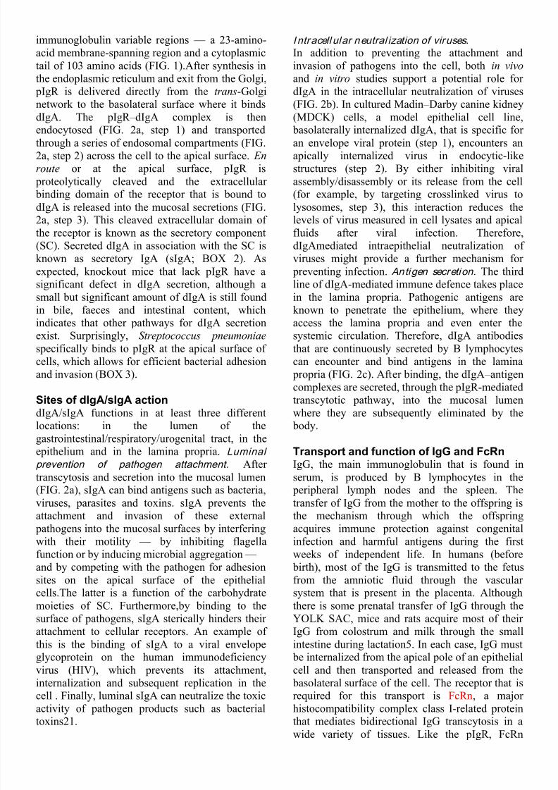
immunoglobulin variable regions — a 23-amino-acid membrane-spanning region and a cytoplasmictail of 103 amino acids (FIG. 1).After synthesis inthe endoplasmic reticulum and exit from the Golgi,
pIgR is delivered directly from the trans-Golginetwork to the basolateral surface where it bindsdIgA. The pIgR – dIgA complex is thenendocytosed (FIG. 2a, step 1) and transportedthrough a series of endosomal compartments (FIG.2a, step 2) across the cell to the apical surface. En
route or at the apical surface, pIgR is proteolytically cleaved and the extracellular binding domain of the receptor that is bound todIgA is released into the mucosal secretions (FIG.2a, step 3). This cleaved extracellular domain of the receptor is known as the secretory component(SC). Secreted dIgA in association with the SC isknown as secretory IgA (sIgA; BOX 2). As
expected, knockout mice that lack pIgR have asignificant defect in dIgA secretion, although asmall but significant amount of dIgA is still foundin bile, faeces and intestinal content, whichindicates that other pathways for dIgA secretionexist. Surprisingly, Streptococcus pneumoniae
specifically binds to pIgR at the apical surface of cells, which allows for efficient bacterial adhesionand invasion (BOX 3).
Sites of dIgA/sIgA action
dIgA/sIgA functions in at least three differentlocations: in the lumen of thegastrointestinal/respiratory/urogenital tract, in theepithelium and in the lamina propria. Luminal
prevention of pathogen attachment. After transcytosis and secretion into the mucosal lumen(FIG. 2a), sIgA can bind antigens such as bacteria,viruses, parasites and toxins. sIgA prevents theattachment and invasion of these external
pathogens into the mucosal surfaces by interferingwith their motility — by inhibiting flagella
function or by inducing microbial aggregation — and by competing with the pathogen for adhesionsites on the apical surface of the epithelialcells.The latter is a function of the carbohydratemoieties of SC. Furthermore,by binding to thesurface of pathogens, sIgA sterically hinders their attachment to cellular receptors. An example of this is the binding of sIgA to a viral envelopeglycoprotein on the human immunodeficiencyvirus (HIV), which prevents its attachment,internalization and subsequent replication in the
cell . Finally, luminal sIgA can neutralize the toxicactivity of pathogen products such as bacterialtoxins21.
I ntracell ular neutral ization of viruses.
In addition to preventing the attachment andinvasion of pathogens into the cell, both in vivo
and in vitro studies support a potential role for dIgA in the intracellular neutralization of viruses(FIG. 2b). In cultured Madin – Darby canine kidney(MDCK) cells, a model epithelial cell line,
basolaterally internalized dIgA, that is specific for an envelope viral protein (step 1), encounters anapically internalized virus in endocytic-likestructures (step 2). By either inhibiting viralassembly/disassembly or its release from the cell(for example, by targeting crosslinked virus tolysosomes, step 3), this interaction reduces thelevels of virus measured in cell lysates and apicalfluids after viral infection. Therefore,dIgAmediated intraepithelial neutralization of viruses might provide a further mechanism for
preventing infection. Antigen secretion. The thirdline of dIgA-mediated immune defence takes placein the lamina propria. Pathogenic antigens areknown to penetrate the epithelium, where theyaccess the lamina propria and even enter thesystemic circulation. Therefore, dIgA antibodiesthat are continuously secreted by B lymphocytescan encounter and bind antigens in the lamina
propria (FIG. 2c). After binding, the dIgA – antigencomplexes are secreted, through the pIgR-mediatedtranscytotic pathway, into the mucosal lumen
where they are subsequently eliminated by the body.
Transport and function of IgG and FcRnIgG, the main immunoglobulin that is found inserum, is produced by B lymphocytes in the
peripheral lymph nodes and the spleen. Thetransfer of IgG from the mother to the offspring isthe mechanism through which the offspringacquires immune protection against congenitalinfection and harmful antigens during the first
weeks of independent life. In humans (before birth), most of the IgG is transmitted to the fetusfrom the amniotic fluid through the vascular system that is present in the placenta. Althoughthere is some prenatal transfer of IgG through theYOLK SAC, mice and rats acquire most of their IgG from colostrum and milk through the smallintestine during lactation5. In each case, IgG must
be internalized from the apical pole of an epithelialcell and then transported and released from the
basolateral surface of the cell. The receptor that is
required for this transport is FcRn, a major histocompatibility complex class I-related proteinthat mediates bidirectional IgG transcytosis in awide variety of tissues. Like the pIgR, FcRn

7/29/2019 Immunoglobu in Transport Across Polarized Epithelial Cells
http://slidepdf.com/reader/full/immunoglobu-in-transport-across-polarized-epithelial-cells 3/12
belongs to the Fc family of receptors that areinvolved in many cellular functions, includingregulation of lymphocyte proliferation anddifferentiation, phagocytosis of IgGcoated particlesand the release of inflammatory agents (BOX 1).FcRn is a heterodimer that is composed of a 50-kDa transmembrane -chain subunit that isnoncovalently associated with the 15-kDa 2-microglobulin subunit (FIG. 1). FcRn has a 50-amino-acid cytosolic domain that contains sortingsignals that direct the intracellular trafficking of the receptor. The crystal structure of FcRn has
been solved at a 2.2-Å resolution. The 2-microglobulin subunit is required for FcRnfunction, as 2-microglobulin knockout mice havedefects in IgG transport . The physiologicalactivity and metabolism of IgG, like dIgA and itsreceptor pIgR, depend on the intracellular
trafficking of FcRn. IgG binds FcRn with highaffinity at slightly acidic pH (below 6.5) but withlow affinity at neutral pH. In the intestine, placentaand yolk sac, FcRn transports IgG from the apicalto the basolateral surface. In the acidicenvironment of the rat small intestine, binding of IgG to FcRn occurs at the apical plasma membraneof ENTEROCYTES (FIG. 3a, step 1). In the yolk sac and placenta,where the pH is neutral, IgG isendocytosed through fluid-phase endocytosis, andthe binding of IgG to FcRn occurs in the acidic
environment of the apical endosome (FIG. 3a, step2). Regardless of the mechanism of ligandinternalization, FcRn – IgG complexes are delivered
by transcytosis to the basolateral surface of the cellwhere the neutral pH promotes the dissociation of IgG from its receptor (FIG. 3a, step 3).
Role of FcRn in regulating IgG catabolism.
In addition to mediating the transfer of maternalIgG, FcRn is also important in regulating theamount of IgG in serum.As described more than 30
years ago, the rate of IgG turnover increases as theamount of IgG in serum rises.Turnover occurs inendothelial cells and involves a saturable processthat is probably receptor mediated. FcRn is a likelycandidate as it is localized to the endothelial cellsof small arterioles and capillaries in muscle andliver, and the serum half-lives of IgG in 2-microglobulin-deficient mice are abnormally lowin comparison to control animals. The putativemechanisms for FcRn-mediated IgG homeostasis
are shown in FIG. 3b. In endothelial cells, IgG istaken up by fluid-phase endocytosis and deliveredto endosomes (FIG. 3b, step 1). The fate of endocytosed Ig varies depending on theconcentration of internalized IgG — which is
directly proportional to the concentration inserum.At modest levels of IgG, most of the ligand
binds FcRn and it is either recycled (step 2) or delivered by transcytosis to the basolateral surface,where the neutral pH promotes the dissociation andrelease into the interstitial space (FIG. 3b, step3).When IgG levels are high, the fate of internalized Ig is different. Once FcRn binding issaturated, the non-receptor-bound IgG is deliveredalong with other fluid-phase cargo to thelysosomes, where it is then degraded (FIG. 3b, step4). Therefore, IgG levels in serum are governed bythe saturable nature of the intracellular FcRn – IgGinteraction.
FcRn in immune activation and tolerance.
FcRn is expressed in both adult human intestinalepithelial cells and cultured intestinal T84 cell
monolayers, where it transcytoses in bothdirections, and has led to the proposal of a newfunction for FcRn. This mechanism is depicted inFIG. 3c. It entails the fluid-phase internalizationand transport of IgG from the interstitial space tothe intestinal lumen, where it is released intosecretions (FIG. 3c, steps 1 and 2).After bindingwith its cognate antigen in the lumen, the IgG –
antigen complexes are transcytosed in the oppositedirection, delivering immune complexes to thelamina propria for subsequent induction of immune
activation or tolerance (FIG. 3c, steps 3 and 4).Data to support this model were recently obtainedfor luminal-to-serosal transport of a chimeric
protein containing the hormone erythropoietin — which stimulates red-blood-cell formation — attached to the Fc fragment of IgG1. Theerythropoietin – Fc chimaera is transported acrossthe intestinal barrier of newborn mice or therespiratory epithelium of adult mice in a FcRn-dependent reaction that stimulates production of red-blood-cell progenitor. FcRn-mediated IgG –
antigen immunosurveillance might be particularlyimportant in the lower respiratory and femalegenital tracts of humans in which the concentrationof IgG is greater than IgA. However, the relativecontribution of sIgA or IgG in immunoregulationin these or other tissues remains to be determined.
Transcytotic pathways in polarizedepithelial cells Intracellular trafficking of
pIgR – dIgA.
The intracellular pathway for transcytosis of pIgR –
dIgA complexes has been extensively studied in polarized MDCK cells (transfected with pIgR cDNA), rat liver hepatocytes and hepatocyte-derived cell lines.As described above, the initial

7/29/2019 Immunoglobu in Transport Across Polarized Epithelial Cells
http://slidepdf.com/reader/full/immunoglobu-in-transport-across-polarized-epithelial-cells 4/12
step in this process is internalization throughclathrin-coated pits of pIgR – dIgA from the
basolateral/sinusoidal surface of the cell (FIG. 4,step 1). This requires two cytoplasmic tyrosineresidues at amino-acid positions 668 and 734 of
pIgR43. Rapid endocytosis also requires serine726, a principal site for pIgR phosphorylation44.Internalized pIgR – dIgA is delivered to peripherallylocalized basolateral early endosomes (FIG. 4, step1). The small GTPase Rab5 and its effector
protein, the early endosome antigen 1 (EEA1), areassociated with this compartment. pIgR – dIgAcomplexes, once segregated from fluid-phasemarkers (FIG. 4, step 2a), are concentrated in themembrane-rich tubular extensions of thiscompartment, along with receptors that willeventually recycle and return to the
basolateral/sinusoidal surface (FIG. 4, step 2b and
3a). Most of the pIgR – dIgA complexes, and asignificant fraction of basolateral recycling proteins such as TRANSFERRIN (Tf ) and itsreceptor, are subsequently delivered in an actin-and microtubule-dependent manner to a
perinuclear compartment that extends into theapical cytoplasm ofMDCK cells (FIG. 4, step 2c).This compartment is variously described as therecycling endosome, common endosome or common recycling endosome (CRE). The CRE hasa tubular morphology, receives membrane-
associated cargo that is internalized from both theapical and basolateral poles of the cell (FIG. 4, step2c and 7a), and is likely to be the primary stationfor segregating apically destined pIgR – dIgA fromreceptors, like the Tf receptor, that will recycle
back to the basolateral pole of the cell (FIG. 4, step3a).An equivalent paracentriolar compartment isobserved in rat liver hepatocytes and in thehepatocytederived cell line WIF-B. Rab17, anepithelial-specific Rab GTPase, is localized to asimilar compartment in Eph4 mammary gland
epithelial cells and regulates basolateral recyclingof Tf and its receptor. In MDCK cells, Rab17 islocalized to a subapical tubular-vesicular compartment that contains transcytosing pIgR –
dIgA, and overexpression of Rab17 inhibits pIgR –
dIgA transcytosis55. Whether Rab17 alsoassociates with the CRE of MDCK cells or other compartments (see below) remains to bedetermined. The final steps in pIgR – dIgAtranscytosis are controversial and the subject of ongoing investigation (BOX 4). One hypothesis is
that, after exit from the CRE, pIgR – dIgA istransported to a distinct compartment known as theapical recycling endosome (ARE) in MDCK cellsor subapical compartment (SAC) in hepatocytes
(FIG. 4, step 3c). The ARE/SAC is proposed tofunction as the final compartment that regulates thedelivery of intracellular components to the apical
plasma membrane. In this regard, the smallGTPases Rab11 and Rab25 are associated with thiscompartment and regulate transcytotic traffic andapical recycling. Following exit from the ARE(FIG. 4, step 4), the pIgR is delivered to the apicalcell surface. Either en route to, or at the apical
plasma membrane, the pIgR is proteolyticallycleaved by a thiol-dependent proteinase, releasingthe receptor and bound components into secretions(FIG. 4, step 5).However, in MDCK cells, a largefraction of pIgR – dIgA escapes cleavage and isinternalized from the apical pole of the cell,whereit is delivered to apical early endosomes (FIG. 4,step 6). It is then subsequently delivered to theCRE or Rab11-positive elements of the ARE (FIG.
4, step 7a), from where it is recycled. Delivery tothe ARE is a microtubule-dependent process. Lessthan 10% of apically internalized pIgR – dIgA istranscytosed and basolaterally released. Efficientapical recycling ensures that most of the pIgR –
dIgA complex is eventually cleaved and releasedinto secretions.
I ntracell ular trafficking of FcRn – IgG.
Unlike the pIgR, FcRn is known to undergotranscytosis in both directions. In addition, a
fraction of receptor – ligand complexes can recycleat the cell surface or be transported to lysosomeswhere they are degraded. The pathway for apical-to-basolateral transcytosis of FcRn – IgG has beenexplored in cultured epithelial cells, in the fetalyolk sac of rats, human placenta and in fetalenterocytes. In the latter, FcRn is concentrated atthe base of apical microvilli in coated pits, and, asdescribed above, ligand binding to the receptor occurs in the acidic pH of the intestinallumen.Next, the receptor – ligand complex is
internalized by a process that is dependent on adistinct dileucine and unique tryptophaninternalization motif, and is delivered to a
pleiomorphic tubulovesicular compartment thatunderlies the apical plasma membrane (FIG. 5,step 1). The location, morphology and presence of
both membrane and fluid-phase markers indicatethat this compartment is an apical early endosome.In the absorptive endoderm of the fetal yolk sac,FcRn is not found at the surface but, instead, islocalized to subapical tubulovesicular apical early
endosomes. In this tissue, IgG is internalized byfluid-phase endocytosis, which then binds to FcRnin the acidic lumen of this apical endocyticcompartment (FIG. 5, step 2). Similarly, in MDCK

7/29/2019 Immunoglobu in Transport Across Polarized Epithelial Cells
http://slidepdf.com/reader/full/immunoglobu-in-transport-across-polarized-epithelial-cells 5/12
cells that express functional FcRn, apicallyinternalized IgG is delivered to apical earlyendosomes, where it binds FcRn61. FcRn – IgG isthen segregated from non-receptorbound fluid-
phase markers, which are delivered to lateendosomes and lysosomes (FIG. 5, step 3a) and iseither recycled (step 3b) or delivered from the AEE(FIG. 5, step 3c) to a further set of tubulovesicular endosomes. These are located in the perinuclear region as well as subjacent to the lateral surfaces of the cell (FIG. 5, step 3c), and might representelements of the Tf-rich CRE. In enterocytes,coated vesicles (100-nm diameter) are observedand probably serve as exocytic carriers to the
basolateral cell surface (FIG. 5, step 4).Interestingly, 60-nm diameter coated vesicles arealso observed in MDCK cells that bud off the CREand are involved in recycling Tf – receptor
complexes to the basolateral pole of the cell68.These vesicles might serve a similar purpose in thedelivery of FcRn – IgG to the basolateral surface of MDCK cells. It is probable that basolateral-to-apical transcytosis of FcRn – IgG will follow a
pathway similar to that described for the pIgR, butthis remains to be determined (FIG. 5, steps 5 –
8).Now that functional FcRn has been expressed inMDCK cells, it should be possible to morecarefully compare the compartments involved inFcRn traffic versus those involved in pIgR traffic.
Regulation of transcytosisRegulators of pI gR – dIgA transcytosis.
Transcytosis of pIgR – dIgA has been the subject of considerable study and is regulated/modulated bythe cytoskeleton, Rho family GTPases, RabGTPases (including Rab3b, Rab11, Rab17 andRab25), the heterotrimeric G-proteinGs75,76,TAP/ p115, calmodulin,
phosphatidylinositol- 3-kinases, SNAREs(including syntaxin 3, SNAP-25 and cellubrevin),
protein kinase C85,86, p62yes, phospholipaseC88, cyclic AMP, intracellular calcium, agentsthat alter organellar pH, association with lipidmicrodomains, brefeldin A91, receptor
phosphorylation of serine 664, receptor dimerization and ligand binding. This section willhighlight some new and emerging mechanisms of regulating pIgR – dIgA transcytosis.
L igand-dependent stimulation of pIgR traf fi c.
Analysis of pIgR – dIgA traffic has providedimportant insights into receptor sorting and theintracellular compartments that are involved in this
process. In addition, the pIgR provides importantinformation about how receptor signaling can
modulate the extent and rate of endocytic traffic.Although empty pIgR — that is, pIgR with noligand bound — undergoes constitutivetranscytosis, dIgA binding stimulates receptor transcytosis both in vitro and in vivo, although thismight not be true for human pIgR. Nevertheless, insome species, this is an important mechanism thatallows the epithelium to adjust the rate of sIgAformation according to the amount of availabledIgA. Ligand-stimulated transcytosis is coupledwith, and requires the production of, secondarymessengers, including inositol 1,4,5-triphosphate(Ins(1,4,5)P3) and free intracellular Ca2+. Recentobservations indicate that the generation of thesesignaling molecules depends on the Src family
protein tyrosine kinase p62yes (FIG. 6). p62yes co-immunoprecipitates with the pIgR in an endosomalfraction that is enriched in transcytosing pIgR.
Transcytosis of dIgA into bile is significantlyimpaired in knockout animals that lack p62yesexpression, which indicates that this kinase isrequired for efficient transcytosis87. Interaction of the pIgR and p62yes occurs through the carboxylterminus of the pIgR (amino-acid residues 727 –
736), although the interaction is likely to beindirect. The probable target of p62yes is
phospholipase C, which is phosphorylatedfollowing dIgA binding and is required for ligand-stimulated Ins(1,4,5)P3 production and the
production of diacylglycerol (DAG)88.Ins(1,4,5)P3, in turn, stimulates the release of calcium from apically localized intracellular stores,and ligandstimulated transcytosis can be blocked
by the Ins(1,4,5)P3 receptor antagonistxestospongin C97. The requirement for theseupstream signalling events can be bypassed by thetreatment of cells with ionomycin, a Ca2+ionophore. The rise of intracellular Ca2+, oupledwith the production of DAG, promotes theactivation and association of protein kinase C
epsilon (PKC) with pIgR – dIgA endosomes.PKCstimulates pIgR – dIgA transcytosis,although its mechanism of action remains to bedetermined. Two further findings are worth noting.First, the p62yes signalling cascade — whichmight be initiated at the basolateral plasmamembrane or in the basolateral early endosomes — is rapidly activated following the binding of dIgAto the receptor, and is transmitted across thecell,where it stimulates dIgA transcytosis at a later stage in the transcytotic pathway (CRE or ARE)88,97. Second, ionomycin treatment has noeffect on pIgR transcytosis unless the receptor first
binds dIgA ligand at the basolateral pole of the celland dimerizes. This would indicate that a further
7/29/2019 Immunoglobu in Transport Across Polarized Epithelial Cells
http://slidepdf.com/reader/full/immunoglobu-in-transport-across-polarized-epithelial-cells 6/12
‘sensitization’ step is required for ligand-stimulated transcytosis. This latter step involves anarginine at position 657 of the cytoplasmic domainof the receptor, and sensitization is blocked whenthis residue is mutated to a non-phosphorylatablealanine (Ala) (R657A). This sensitization step hasrecently been characterized and is likely to involvethe Rab3b GTPase. The Rab3 subfamily (Rab3a –
d) regulates Ca2+-stimulated exocytosis in severalcell types, including neurons and endocrine cells.Rab3b is also expressed in epithelial cells. Of significance, Rab3b interacts directly with theempty pIgR, but less so with the ligand-boundreceptor. Rab3b binds to residues 655 – 668 in thecytoplasmic domain of the pIgR, and ligand-induced dissociation of Rab3b is blocked in theR657A mutant, as described above. Furthermore,ligand-induced dissociation is not observed if dIgA
is bound to pIgR at the apical pole of the cell, andis also prevented by treatment with tyrosine kinaseinhibitors or calcium chelators, or by expression of a dominant-active Rab3b mutant (Rab3bQ81L)that is locked in the active GTP-bound state.As
predicted, expression of Rab3bQ81L inhibitsligand-stimulated transcytosis of pIgR, presumably
because the GTPase is unable to undergohydrolysis and dissociate from the receptor.As inneurons, Rab3 negatively regulates exocytictraffic. These results indicate that receptors such as
pIgR can directly interact with their owntrafficking. The endocytic compartments in whichRab3b associates and dissociates, and whether ligand-bound pIgR acts as a Rab3b exchangefactor or GTPase activating protein in this reaction,remains to be determined.
Role of the cytoskeleton in pI gR traf fi c.
Transcytosis of the pIgR is also governed by thecytoskeleton. Microtubules are required for theefficient transfer of pIgR – dIgA from basolateral
early endosomes to the more apical endocyticcompartments (CRE and ARE) 69, 70. Themolecular motor that drives this process isunknown, but is likely to be dynein or anunconventional kinesin motor that would propelvesicles along the microtubules that line the lateralsurfaces of the cell and are oriented with their minus ends towards the apical pole of the cell. Inaddition to microtubules, actin is also important in
pIgR traffic. pIgR – dIgA transcytosis issignificantly slowed in cytochalasin-D-treated
MDCK cells. Cytochalasin D inhibits an earlyevent in pIgR – dIgA transcytosis that precedes themicrotubuledependent step and impairs transit
between the basolateral endosomes and the more
apical endosomal compartments. Interestingly,cytochalasin D only affects transcytosis of ligand-occupied receptor, as unoccupied receptor transcytosis is unaffected by cytochalasin Dtreatment (R.R and G.A, unpublishedobservations). In fact, filamentous actin isassociated with pIgR – dIgAlabelled endosomes atthe base of polarized MDCK cells. Ligand bindingmight facilitate transcytosis by promotingendocytic vesicle propulsion as a result of ACTINCOMET formation, and by stimulating movement
by myosin motors, or by promoting actin turnover.
Regulation of pIgR – dIgA transcytosis by Rho
family GTPases.
In addition to Rab family GTPases, the Rho familyGTPases RhoA, Rac1 and Cdc42 also govern
basolateral- to-apical transcytosis of pIgR – dIgA.
Rho family GTPases were originally identified ascoupling extracellular signalling cues to changes inthe actin cytoskeleton. In addition, it is now knownthat they regulate several cellular functions,including endocytic and biosynthetic traffic.Expression of a dominant-active RhoA mutant(RhoAV14) stimulates both basolateral and apicalinternalization of pIgR – dIgA complexes,whereasexpression of dominant-negative RhoAN19 has theopposite effect. Expression of RhoAV14 alsomarkedly slows pIgR – dIgA transcytosis by
trapping pIgR – dIgA in basolateral earlyendosomes and slowing its exit from thiscompartment. RhoAV14 expression has little effecton apical recycling of the receptor – ligandcomplex. Rac1 also regulates basolateral andapical internalization of pIgR – dIgA, but in anopposite manner to RhoA.Expression of dominant-active Rac1V12 slows pIgR – dIgA endocytosis,whereas expression of dominant-negative Rac1stimulates internalization. Rac1V12 expression
blocks transcytosis by trapping pIgR – dIgA in a
central aggregate composed of elements of theCRE and ARE.Apical recycling is similarlyimpacted by the expression of Rac1V12. Finally,Cdc42 also regulates pIgR – dIgA internalizationand transcytosis. Expression of either dominant-active Cdc42V12 or dominant- negativeCdc42N17 slows pIgR – dIgA internalization and
basolateral-to-apical transcytosis. These resultsindicate that Rho family GTPases might regulateseveral steps in pIgR – dIgA transcytosis. Thedownstream effectors of these GTPases remain to
be defined, although modulation of the actincytoskeleton could regulate transcytosis of both
pIgR – dIgA and FcRn – IgG (see below).

7/29/2019 Immunoglobu in Transport Across Polarized Epithelial Cells
http://slidepdf.com/reader/full/immunoglobu-in-transport-across-polarized-epithelial-cells 7/12
Regulation of FcRn –IgG transcytosisBy contrast to pIgR – dIgA transport, far less isunderstood about the regulation of FcRn – IgGtranscytosis. Phosphorylation of FcRn regulatestranscytosis of this receptor. Serine (Ser) 313 andSer319, in the cytoplasmic domains of FcRn, arethe main sites for receptor phosphorylation incultured renal inner-medullary collecting- ductcells that are transfected with this receptor. WhenSer313 is mutated to an Ala residue (S313A),apical- to-basolateral transcytosis, but not
basolateral-toapical, is significantly slowed andimpaired. The S319A mutation slows transcytosis,
but to a lesser degree than the S313Amutant.Mutation of Ser313 to an aspartic acidresidue — which mimics the negative charge of a
phosphate group — has the predicted effect andstimulates apical-to-basolateral transcytosis.
Increased apical recycling of the S313A receptor isobserved,which indicates that phosphorylationmight regulate entry into the transcytotic pathway.By contrast to the pIgR, where phosphorylation of Ser664 is thought to inactivate a basolateral sortingsignal thereby promoting apical delivery92,
phosphorylation of S313 in FcRn might create a basolateral sorting signal that promotes basolateraldelivery of this receptor. Recent evidence indicatesthat FcRn – IgG transcytosis is also modulated byagents that either perturb the cytoskeleton, alter the
pH of intracellular compartments or impair intracellular signalling cascades. Theactindisrupting agent, cytochalasin D, slows
basolateral-toapical transcytosis of FcRn – IgG inBeWo choriocarcinoma cells, but has no effect onthe apical-to-basolateral transcytosis of thereceptor99. The effect of the microtubule-depolymerizing agent nocodazole depends on thecell type, having either no effect, blocking just
basolateral-to-apical transcytosis or blockingtranscytosis in both directions. Monensin, which
increases the pH of intracellular organelles, blocksFcRn transcytosis in both directions. Other agentsthat impair FcRn – IgG transcytosis include the
phosphatidylinositol-3-kinase antagonistswortmannin and LY294002, and the calmodulinantagonist W-7, which selectively affects
basolateral-to-apical transcytosis of FcRII – FcRnchimaeras100. With the recent development of several in vitro model systems, it should now be
possible to dissect further the signalling pathwaysthat regulate the FcRn – IgG transcytotic pathway.
Summary and conclusionsIgs are important in preventing bacterial and viralinfections by specifically interacting with and
neutralizing pathogens. Igs such as IgA, IgG andIgM cannot simply diffuse across the epithelial
barriers that are present in the mucosa and placenta, but instead are actively transported by thetwo receptors, pIgR and FcRn. The pIgR bindsdIgA and pentameric IgM at the basolateral surfaceof the epithelial cell and transports these Igs in the
basolateral-to-apical direction. This transport ishighly regulated and involves the cytoskeleton,several Rab GTPases and some signallingcascades, including those that are dependent onligand binding. The compartments that areinvolved in pIgR – dIgA transcytosis and theregulation of the transcytotic pathway remainactive areas of research.At the apical plasmamembrane, the pIgR is proteolytically cleaved,releasing bound dIgA/IgM into secretions.Although there can be some apical recycling, the
pIgR effectively undergoes one round of traffic before it is used. Further areas of active researchinclude the use of sIgA for immunotherapy, the useof pIgR as a means of targeted gene delivery toepithelial cells and the role of pIgR – dIgA in virusneutralization and antigen secretion101 – 103.Transportation of IgG is mediated by FcRn, and,like pIgR – dIgA transport, involves transcellular transport. However, there are some differences.First, IgG binding to the FcRn can either occur atthe surface or in endosomes, and this reflects, in
part, the low-pH-dependent binding of IgG toFcRn. Second, transport can occur in both theapical-to-basolateral and basolateral-to-apicaldirection. Third, release of IgG from FcRn is
primarily dependent on pH-dependent changes inreceptor – ligand affinity, and not proteolyticcleavage. Fourth, FcRn can probably undergoseveral cycles of transcytosis before it is turnedover. Similar to pIgR – dIgA transport, FcRn – IgGtranscytosis is also likely to be highly regulated.Because cell-culture models for FcRn transport
were only developed in the past few years, our understanding of those compartments that areinvolved and of the mechanisms of regulation,considerably lags behind that of pIgR – dIgAtransport. However, current data indicate theinvolvement of the cytoskeleton and signaltransduction cascades. FcRn also has an importantfunction in regulating IgG catabolism. Finally,FcRn – IgG, like pIgR – dIgA, has been proposed to
be important in intracellular eutralization andrecent data indicate that it could be important in
immune activation and tolerance. These are areasof research that are sure to be further explored inthe near future.

7/29/2019 Immunoglobu in Transport Across Polarized Epithelial Cells
http://slidepdf.com/reader/full/immunoglobu-in-transport-across-polarized-epithelial-cells 8/12
Immunoglobulins are Y-shaped heteromeric proteincomplexes that are composed of two light chains (Mr = 25 kD) and two heavy chains (Mr = 55 kD) in thecase of IgG, and are produced and secreted by plasmacells. The light chains interact with the amino terminusof the heavy chains to form two arms — the Fab
portion — which contain the antigenbinding sites attheir tips. The carboxyl termini of the heavy chainscombine to form a stalk region called the Fc domain
that allows for interactions with molecules such ascomplement or Fc receptors.Mammals have fivedifferent classes of immunoglobulins: IgA, IgD, IgE,IgG and IgM. These vary depending on the type of heavy chain used and their ability to form multimers of Y-shaped complexes. Regardless of immunoglobulinclass, each Y-shaped complex contains either or light chains that associate with heavy chains in a 1:1fashion. Immunoglobulins bind to cells by their interactions with Fc receptors. The bestunderstood Fcreceptors are FcRn and FcR (which bind IgG), FcR
(which binds IgE) and pIgR105 (which binds polymericIgA and IgM). FcRn and pIgR are involved in thetransport of immunoglobulins,whereas FcRn alsocontrols the half-life of IgG in serum. FcR and FcR are signalling receptors that, following binding of IgGor IgE, respectively, regulate various forms of endocytosis including phagocytosis, antibody
production and inflammatory responses. For example,the binding of IgE – antigen complexes to cell-surfaceFcRs on mast cells stimulates the release of histamine,which accounts for the itchiness and rednessthat is associated with allergic reactions. There are three
isoforms of FcR (FcRI, FcRII and FcRIII) andtwo isoforms of FcR (FcRI and FcRII). For moredetails, see.
sIgA is a specific defence mechanism that pathogensencounter as they come into contact with mucosalsurfaces102. Following secretion by plasma cells (step1), dIgA binds to the pIgR and is transported across the
epithelium and released at the apical pole of the cell bound to the secretory component (SC) (steps 2 – 4). Insecretions, sIgA prevents bacterial and viral adhesionand invasion by aggregating and immobilizing these
pathogens (step 5), effectively neutralizing their abilityto cause infection. Neutralized pathogens are cleared bygut peristalsis or by the mucociliary escalator of therespiratory tract. In the intestine, pathogens are sampled
by specialized M cells (step 6),which transport antigensto underlying antigenpresenting cells, includingmacrophages and dendritic cells (step 7). In turn, thesecells activate T-helper lymphocytes (step 8),which
produce interleukins that stimulate the production of polymeric IgA by plasma cells (step 9).A T-cellindependent mechanism also exists that mightinvolve dendritic cells (step 10). Topicalimmunotherapy involves the delivery of sIgA or sIgMto exposed surfaces, such as the mucosa,where theyneutralize pathogens103. The trick is to deliver largequantities of these antibodies over an extended periodof time in a form that is resistant to degradation. SCenhances the lifetime of secretory antibodies byslowing proteolysis and turnover. sIgA can be producedin transgenic tobacco plants that produce a light chain,a hybrid heavy chain, a J chain andSC106.Recombinant sIgA is capable of preventing oralcolonization by Streptococcus mutans whenadministered orally107.Because glycosylation andassembly of sIgA are likely to differ between plants and

7/29/2019 Immunoglobu in Transport Across Polarized Epithelial Cells
http://slidepdf.com/reader/full/immunoglobu-in-transport-across-polarized-epithelial-cells 9/12
mammals,more recent attempts have employedmammalian cell systems in which monoclonal IgAs,specific for particular pathogens, are combined with SCin vitro103. In addition, systems are being developed inwhich a single mammalian cell is transfected withcDNAs for heavy and light chains (specific for a
particular epitope), J chain and SC, providing thecomponents necessary to build large quantities of
specific sIgAs103. The passive administration of theserecombinant antibody complexes might be useful incases for which effective vaccines do not exist,when or if antibiotic resistance occurs, or when new pathogensare found.
The pIgR is a type I membrane protein that has alarge extracellular region arranged in five domains(that are homologous to the variablelike domainsof the Ig superfamily), a transmembrane region
and a 103-amino-acid cytoplasmic region. TheFcRn is a type I membrane protein that is relatedto MHC class I molecules. It is a heterodimer
composed of a smaller 2-microglobulin subunit
and is associated with a larger -subunit consisting
of three extracellular domains (1 –3) related tothe constant-like domains of the Ig superfamily, atransmembrane region and a 50-amino-acidcytoplasmic tail. MHC, major histocompatibilitycomplex; pIgR, polymeric immunoglobulinreceptor.
Figure 2 | Functions of dIgA/sIgA and pIgR.a | Formation of sIgA. dIgA is secreted by the
plasma cells, where it binds to the pIgR at thebasolateral pole of secretory epithelial cells. dIgAis transported through a series of endosomalcompartments (step 1) en route to the apicalplasma membrane domain (step 2), where aproteinase cleaves the large extracellular domainof the receptor, thereby releasing it, bound todIgA, into secretions (step 3). dIgA, in associationwith the cleaved receptor fragment (also known assecretory component, SC) form sIgA. Insecretions, sIgA interacts withantigens/pathogens, neutralizing their ability to
cause disease. b | Intracellular virus neutralization.pIgR –dIgA complexes are delivered to endocyticcompartments (step 1) where they encounter infecting viruses (step 2) or newly synthesizedviral membrane proteins. The interaction of pIgR –dIgA with the virus prevents virusassembly/disassembly and the exit from the cell,possibly by targeting pIgR –dIgA –virus complexesto lysosomes (step 3) where they are degraded. c| Antigen secretion. Pathogenic antigens thatpenetrate the epithelium and enter the laminapropria are bound to pIgR –dIgA, internalized (step
1), transported across the cell (step 2) andreleased at the apical pole of the cell (step 3)where they are cleared. dIgA, dimeric IgA; pIgR,polymeric immunoglobulin receptor; sIgA,secretory IgA.
Box 3 | Use of pIgR as a portal for bacterial
adherence and invasion into epithelial cells The pIgR is important in transporting secretoryimmunoglobulins that prevent bacterial and viraladherence and replication. Surprisingly, it is used bysome pathogenic bacteria as a mechanism to bind toand invade epithelial cells. Streptococcus pneumoniae
adheres to and invades the epithelium of the upper andlower respiratory tract causing sepsis,meningitis and pneumonia.Adhesion of S. pneumoniae
to cells involves several potential adhesins, including

7/29/2019 Immunoglobu in Transport Across Polarized Epithelial Cells
http://slidepdf.com/reader/full/immunoglobu-in-transport-across-polarized-epithelial-cells 10/12
the phosphorylcholine-binding protein (CbpA) and ahighly related protein, SpsA.Bacteria that lack CbpAshow decreased adhesion to epithelial cells and, as aresult, are less likely to colonize thenasopharynx.Unexpectedly, the receptor for both of these adhesins is likely to be the extracellular domain of the human pIgR14.Anti-pIgR antibodies can prevent
bacterial adhesion to nasopharyngeal cells,whereas the
upregulation of pIgR expression, by treating cells withinterferon-γ, stimulates bacterial adhesion andinvasion.MDCK cells transfected with the human pIgR are permissive for bacterial adhesion andinvasion,whereas adhesion and uptake in nontransfectedcells is slight. Interestingly, apically internalized
bacteria are transferred across the epithelium,wherethey are released across the basolateral pole of the cell,leaving the epithelium intact. Finally, bacterial sepsis issignificantly diminished in mice lacking p62yes, akinase involved in pIgR transcytosis.
Figure 3 | Functions of IgG and FcRn.a | Transport of IgG. In the suckling rats smallintestine where the extracellular pH is mildly acidic(pH 6.0 –6.5) — IgG binds to FcRn located onenterocytes, where it is endocytosed (step 1). Incells, such as those that line the yolk sac — wherethe pH of the extracellular fluid is neutral — IgG isinternalized by fluid-phase endocytosis (step 2). Inthis case, binding to FcRn occurs in the acidic
environment of the early endosome. FcRn –
IgGcomplexes are transported to the basolateral cellsurface (step 3), where the neutral pH at theserosal side of the tissue promotes liganddissociation and secretion. b | Regulation of IgGcatabolism. In endothelial cells, IgG is taken up byfluid-phase endocytosis and delivered toendosomes (step 1), where it interacts with FcRn.Ligand — bound to receptor — is either recycledback to the apical plasma membrane where it isreturned to blood (step 2), or transported to, andreleased at, the basolateral pole of the cell (step
3). When IgG concentrations are high and bindingto FcRn becomes limiting, unbound IgG isdelivered along with bulk fluid to lysosomes whereit is degraded (step 4). c | FcRn –IgG in immuneactivation and tolerance. IgG is endocytosed at the
basolateral pole of the cell (step 1) andtransported by FcRn to the apical pole of the cell,where it is released into secretions (step 2).Following binding to antigen, the IgG –antigencomplexes, internalized by fluid-phaseendocytosis or through their interactions with FcRn(step 3), are transcytosed in the opposite direction(step 4), delivering immune complexes to the
lamina propria for subsequent induction of immuneactivation or tolerance (step 5).
Figure 4 | Transcytotic pathway of pIgR –dIgA.dIgA binds to pIgR at the basolateral surface of the cell where it is internalized along with fluid-phase markers and basolateral recycling proteinssuch as Tf and its receptor (TfR) (step 1).Endocytosed cargo is delivered to Rab5- andEEA1-positive basolateral early endosomes(BEE). In this compartment several sorting events

7/29/2019 Immunoglobu in Transport Across Polarized Epithelial Cells
http://slidepdf.com/reader/full/immunoglobu-in-transport-across-polarized-epithelial-cells 11/12
occur, including the transport of fluid-phase cargo(step 2a) to Rab7- and Rab9-positive lateendosomes (LE) and lysosomes (not shown), thereturn of a significant fraction of basolateralrecycling proteins to the basolateral cell surface(step 2b), and the delivery of most of pIgR –dIgAand the remaining Tf –receptor complexes to theRab17-positive common recycling endosome
(CRE) (step 2c). In the CRE pIgR –
dIgA is sortedfrom any remaining basolateral recycling proteins(step 3a). The fate of pIgR –dIgA at this point is notclear. One hypothesis is that it is delivered directlyto the apical plasma membrane (step 3b; see BOX4). An alternative hypothesis is that pIgR –dIgA istransported to the Rab11-, Rab17- and Rab25-positive elements of the apical recyclingendosome (ARE) (step 3c). From here is itdelivered to the apical plasma membrane (step 4)where proteolytic cleavage releases the ligand-binding portion of the receptor and associated
dIgA into secretions (step 5). Some pIgR –
dIgAescapes cleavage and is internalized along withfluid-phase markers from the apical pole of the cell(step 6) where it is delivered to Rab5- and EEA1-positive apical early endosomes (AEE). pIgR –dIgAis recycled to the apical surface through the CREor ARE (step 7a), whereas fluid-phase markersare delivered to late endosomes (LE) (step 7b).dIgR, dimeric IgA; pIgR, polymeric immunoglobulinreceptor; Tf, transferrin;TfR, transferrin receptor.
Box 4 | How many compartments are involved
in dIgA transcytosis?At present, it is not clear how many endocyticcompartments are involved in pIgR – dIgAtranscytosis.Mathematical modelling has led to thehypothesis that transcytosing dIgA is delivered fromcommon recycling endosomes (CREs) to the apical cellsurface. An alternative model proposes that transcytosisrequires passage through the apical recycling endosome(ARE)/subapical compartment, before arrival at theapical plasma membrane. We argue that the latter model fits the available data better for the followingreasons. First, after sorting from transferrin (Tf) –
receptor complexes in the CRE, IgA accumulatesunderneath the apical plasma membrane in long tubular endosomes and vesicular elements,which have a C-shaped morphology and might represent transcytoticvesicles. Importantly, Tf is not observed in this regionof the cell. Second, the Rab11 GTPase might be acompartmental-specific marker of the ARE.AlthoughRab11 is found on the trans-Golgi network (TGN) andTf-rich recycling endosomes of nonpolarized CHOcells, there is little overlap between endogenous Rab11and the endocytic compartments that are accessed by
endogenous Tf – receptor complexes in MDCK cells46,51. The small amount of Rab11 that has beenreported to be associated with recycling endosomes islikely to reflect Rab11-dependent cargo transport
between CRE and the ARE. Significantly, expression of
either dominant-negative or dominant-active mutants of Rab11 disrupt pIgR – dIgA transcytosis and apicalrecycling, but have no effect on sorting or recycling of internalized Tf – receptor complexes. Third, another RabGTPase, Rab25, that has a similar distribution toRab11, is also associated with the ARE, and regulatestranscytotic traffic, but not Tf recycling. Fourth, and
perhaps most compellingly, the pH of ARE elements is
6.5,whereas that of the Tf-rich CRE is pH 5.8, a factthat is hard to reconcile with a single compartmentmodel48. In this regard, expression of the acid-activatable influenza virus M2 protein,which localizesto the ARE, alters pIgR – dIgA transcytosis and apicalrecycling but has no effect on sorting or recycling of Tf89. Further research is necessary to resolve thiscontroversy.
Figure 5 | Transcytotic pathways of FcRn –IgG.In enterocytes from new-born rats, IgG binding to
FcRn occurs at the apical cell surface (step 1),whereas in the endoderm of the fetal yolk sac IgGis internalized by fluid-phase endocytosis (step 2)and encounters FcRn in the AEE. In the AEE,FcRn –IgG is sorted from fluid-phase markers,

7/29/2019 Immunoglobu in Transport Across Polarized Epithelial Cells
http://slidepdf.com/reader/full/immunoglobu-in-transport-across-polarized-epithelial-cells 12/12
which are delivered to LE (step 3a) and lysosomes(not shown), and is either delivered to the ARE(step 3b) or delivered to the CRE through the AEE(step 3c). In the CRE, FcRn –IgG is sorted into 60 –100-nm coated vesicles that probably serve asexocytic carriers to the basolateral cell surface(step 4). FcRn can also transport IgG in thebasolateral-to-apical direction, although the
intracellular pathway remains to be defined. Alikely possibility is that IgG, internalized byfluidphase endocytosis (step 5), binds to FcRn inthe acidic pH of the BEE. FcRn –IgG either recycles (step 6a), or is delivered to the CRE (step6b), ARE (step 7) and apical plasma membrane(step 8) where ligand dissociation and releaseoccurs. AEE, apical early endosome; BEE,basolateral early endosome; LE, late endosomes.
Figure 6 | Model for ligand-stimulatedtranscytosis of pIgR –dIgA.Ligand binding to pIgR (step 1) promotes receptor dimerization (step 2). Either at the plasmamembrane or in endosomes the tyrosine kinasep62yes associates with the distal end of thecytoplasmic domain of the receptor (aa 727 –736)
(step 3). This interaction is likely to be mediated byan adaptor protein. p62yes phosphorylates
PLC(step 4), stimulating this protein to formDAG and Ins(1,4,5)P3. p62yes activity rapidlydiminishes following ligand binding, which mightreflect dissociation of the kinase from the pIgR –dIgA complex. Ins(1,4,5)P3 diffuses across the cellwhere it binds to Ins(1,4,5)P3 receptors located at
the apical pole of the cell, where it promotes therelease of Ca2+ from intracellular stores such asthe ER (step 5). Active GTP-bound Rab3b directlyassociates with the proximal end of the pIgR (aa655 –668) (step 6). The GEF that promotesGDP/GTP exchange and activation of Rab3b isunknown, as is the compartment where Rab3b –pIgR association occurs, although it is likely to bethe CRE or ARE. The association of Rab3b withpIgR –dIgA is probably transient, as little Rab3b isfound associated with this complex at steadystate. The presence of elevated intracellular Ca2+
and DAG stimulates the activation and associationof PKCwith endosomes (step 7). Activated
PKCstimulates pIgR –dIgA transcytosis,however, the mechanism of action has not beenidentified. Ligand-stimulated transcytosis is alsodependent on Rab3b hydrolysis of its GTP,although the GAP involved in this process isunknown. One possible candidate is the pIgRitself. GTP hydrolysis promotes Rab3bdissociation and is permissive for ligand-stimulated apical release of pIgR –dIgA complexes(step 8). DAG, diacylglycerol; dIgA, dimeric IgA;
ER, endoplasmic reticulum; GAP, GTPase-activating protein; GEF, guanine nucleotideexchange factor; Ins(1,4,5)P3, inositol-1,4,5-triphosphate; pIgR, polymeric immunoglobulin
receptor; PKC, protein kinase C; PLC,
phospholipase C; PP1, 4-amino-5-(4-methylphenyl)-7-(t-butyl)pyrazolo[3,4-d]pyrimidine.


