

Immunotoxicology in Marine Invertebrates:
Effects of Manganese on Immune Response
Carolina Oweson
Faculty of Science
Department of Marine Ecology
Akademisk avhandling för filosofie doktorsexamen i marin zoologi vid Göteborgs Universitet, som enligt beslut vid naturvetenskapliga fakulteten kommer att försvaras offentligt fredagen den 5e juni 2009, kl. 10.00 i Föreläsningssalen, Sven Lovén Centrum för Marina Vetenskaper - Kristineberg, Fiskebäckskil. Examinator: Prof. Michael Thorndyke, Institutionen för Marin Ekologi, Göteborgs Universitet Fakultetsopponent: Dr. Elisabeth Dyrynda, School of Life Sciences, Heriot-Watt University, Riccarton, Edinburgh, UK

Cover by Andreas Ribbung
Printed by Intellecta Infolog AB
© Carolina Oweson
ISBN 91-89677-43-9 http://hdl.handle.net/2077/19653

Abstract Manganese, Mn, is an abundant element in nature, particularly in soft bottom sediments of the oceans and in bedrock. The metal is predominantly bound to the sediment in the colloid state, MnO2. Eutrophication caused by the high nutrient load in coastal waters together with over-fishing cause cascade effects in the ecosystem increasing the algal blooms and enhancement of hypoxic condition over large bottom areas. During hypoxic events MnO2 is reduced and released into the bottom water as bioavailable ions, Mn2+. Mn is essential for several metabolic and enzymatic processes and is necessary for both animals and plants. Elevated levels though, are toxic and severe effects on the nervous system have been known for long. In addition, previous studies have shown an impaired immune system of the bottom living lobster, Nephrops norvegicus, when exposed to concentrations that are realistic to find in nature. In this study I aimed to investigate if immunotoxic effects of manganese are general also for other marine invertebrates.
It is widely accepted that invertebrates do not have a documented so called adaptive immune response. They lack the genes, proteins and cells for the highly specific recognition and the long-term memory as found in vertebrates. Invertebrates primarily rely on the innate immune system to effectively combat a wide array of microbial pathogens. The innate immune system comprises of a first line of defence systems such as coagulation and melanization reactions, often followed by cellular reactions such as phagocytosis, encapsulation and production of antimicrobial substances. Many innate immune reactions are highly evolutionary conserved and are found throughout the whole animal kingdom. In aquatic invertebrates the open coelom or semi-open haemal circulatory system continuously expose them to potential pathogens and their immune response has proved to be exceptionally efficient in pathogen elimination as witnesses by the invertebrates’ evolutionary success.
In this thesis species from three different phyla within the Bilaterians were investigated; the Norway lobster, Nephrops norvegicus (Crustacea), the blue mussel Mytilus edulis (Mollusca) and the common sea star, Asterias rubens (Echinodermata), differing in preferred habitats, feeding behaviour and somewhat in their strategies of immune defence. Studies were made on molecular, cellular and organism levels. On molecular and cellular levels we investigated the effects of manganese on the renewal of haemocytes (proliferation and differentiation of new cells), manganese effects on viability of haemocytes and the stress responses measured in both haemocytes and haematopoietic tissue. On the whole organism we investigated the effect of manganese on the ability for the animals to clear their cavity form injected bacteria.
The results of this thesis show that Mn in concentrations found in bottom waters affects the immune system of marine invertebrates differently. In N. norvegicus the metal severely suppresses the number of circulating haemocytes by inducing apoptosis, programmed cell death. The impaired immunity made them more susceptible to infections, which was also found in M. edulis. In A. rubens the same Mn concentration seemed to have a stimulating effect (hormesis) on the haematopoiesis which increased the number of circulating haemocytes. Although manganese was shown stressful to the haemocytes and affected their ability to phagocyte, the increased number of haemocytes compensates these impairments. There was seemingly a negative correlation between the accumulation of the metal in the tissues of the animals and their ability to eliminate bacteria. Although Mn does not cause chronic effects on immunity, the expanding areas with bioavailable Mn might have an impact on species composition since some invertebrates become more susceptible to infections. Keywords: Invertebrates, immune system, haemocytes, manganese (Mn),
immunotoxicology, Crustacea, Mollusca, Echinodermata


Till Mamma & Pappa


Allt ordnar sig alltid till det bästa


Immunotoxicology in Marine Invertebrates: Effects of Manganese on Immune Response
Carolina Oweson
This doctoral thesis is produced as a collection of papers. The papers are throughout the thesis referred to by their Roman numerals. The papers are appended at the end of the thesis. Paper I Oweson, C., Baden, S. P., Hernroth, B. E. (2006). Manganese induced
apoptosis in haematopoietic cells of Nephrops norvegicus (L.). Aquatic Toxicology 77:322-328.
Paper II Oweson, C., Sköld, H., Pinsino, A., Matranga, V., Hernroth, B. (2008).
Manganese effects on the haematopoietic cells in Asterias rubens (L.). Aquatic Toxicology 89:75-81.
Paper III Oweson, C., Li, C., Söderhäll, I., Hernroth, B. (2009). Effects of
hypoxia and manganese on haematopoiesis in the common sea star, Asterias rubens (L.). Manuscript.
Paper IV Oweson, C. and Hernroth, B. (2009). A comparative study on the
influence of manganese on the bactericidal response of marine invertebrates. Manuscript. Submitted to Fish and Shellfish Immunology; FSIM-S-09-00134[1].

CONTENTS
1. INTRODUCTION 1
1.1. Invertebrate immune systems 1
1.2. Animals studied 5
1.2.1. Crustacea 6
1.2.2. Mollusca 7
1.2.3. Echinodermata 7
1.3. Manganese and Hypoxia 8
2. AIM OF THE THESIS 11
3. METHODOLOGICAL CONSIDERATION 12
3.1. Animal handling 12
3.2. Cell viability 13
3.3. Cell proliferation 13
3.4. Cell differentiation 14
3.5. Apoptosis 14
3.6. Stress response 15
3.7. Functional response 16
3.7.1. Phagocytosis test 16
3.7.2. Bactericidal capacity 17
4. MAIN RESULTS AND DISCUSSION 19
5. CONCLUSIONS 23
ACKNOWLEDGMENT 25
REFERENCES 26
SVENSK SAMMANFATTNING 31


1
1. INTRODUCTION
The immune system, within all animals, is based on two fundamental systems:
recognition, to distinguish between self and non-self, and effector systems. Through
evolution species have developed sophisticated solutions to manage invading threats
like infectious microbes, i.e. pathogens, and other non-self molecules. The character
of the immune system of a species reflects its surrounding environment. The
immune actions in different animals are dependant on their way of living and how
they have evolved together with their threats. Thus, their susceptibility to
environmental stressors may differ.
1.1. Invertebrate immune systems
In general invertebrates have an open or semi-open circulatory system and aquatic
invertebrates live in continuous contact with potential pathogens (Auffret & Oubella,
1997; Canesi et al., 2002). This makes them dependent on minute reaction of defence
and coagulation mechanisms. They have an immune defence based on activities of
the blood cells in their body fluid, which entrap foreign particles (Ratcliffe et al.;
1984, Chia & Xing, 1996; Johansson & Söderhäll, 1989; Söderhäll & Cerenius, 1998).
In the open circulatory systems of e.g. echinoderms, blood is called coelomic fluid
and the blood cells are called coelomocytes. In the semi open circulatory systems of
e.g. arthropods, the blood is on the other hand called haemolymph and the blood
cells haemocytes. To make it easier for the reader the blood and blood cells are,
when discussed in general, in this thesis referred to as haemolymph and haemocytes.
It is widely accepted that invertebrates do not have a documented so called
adaptive immune response. They lack the genes, proteins and cells for the highly
specific recognition and the long-term memory as found in vertebrates (Flajnik & Du
Pasquier, 2004). To effectively combat a wide array of microbial pathogens,
invertebrates primarily rely on the innate immune system. The innate immune system
is comprised of a first line of defence systems such as coagulation and melanization
reactions, often followed by cellular reactions such as phagocytosis, encapsulation
and production of antimicrobial substances. Many innate immune reactions are
highly evolutionary conserved and are found throughout the whole animal kingdom
(Hoffmann & Reichhart, 2002). The immune defence, based on humoral and cellular
actions, is proven exceptionally efficient in pathogen elimination as witnessed by the
invertebrates’ evolutionary success (Haine et al. 2008). The innate immune system

2
employs germline-encoded pattern recognition receptors (PRRs) to identify invading
pathogens. The receptors are able to identify non-self by pathogen-associated
molecular patterns (PAMPs). These molecules, for example lipopolysaccarides (LPS),
peptidoglucans and β-1-3-glucans, stimulate the immune system unspecifically since
they are present on the surface of large groups of bacteria and other microorganisms
(Medizhitov & Janeway, 2002; Steiner, 2004). Especially peptidoglucans (PGNs) are
excellent targets for recognition by the eukaryotic immune system, because PGN is
an essential cell wall component of virtually all bacteria and it is not present in
eukaryotic cells (Rosenthal & Dziarski, 1994). PGN is especially abundant in Gram-
positive bacteria, in which it accounts for almost half the cell wall mass. In Gram-
negative bacteria, a relatively thin PGN layer surrounds the cytoplasmic membrane
underneath the LPS-containing outer membrane that is also a unique molecule to be
recognized (Doyle & Dziarski, 2001).
The innate immunity uses a set of sensors to recognize foreign patterns as
told earlier, which are found either intracellular, on cell surfaces or excreted in the
haemolymph of the host for an instant reaction (Steiner, 2004). The recognition
receptors of the innate immune system induce the effector system of the immunity.
The most frequently studied pattern recognition receptors is the peptidoglucans
recognition proteins, PGRPs in insects, which can lead to both cellular and humoral
responses. The cellular responses include phagocytosis or encapsulation and
degranulation of haemocytes resulting in release of cytotoxic substances. Examples
of humoral responses include activation of proteins constitutively present in the
haemolymph, such as the prophenoloxidase- and coagulation cascades, as well as
activation of intracellular signalling pathways that stimulate production of different
defence proteins, for example antimicrobial peptides (AMPs) (these different
responses are explained further below) (Hoffmann & Reichhart, 2002; Cerenius &
Söderhäll, 2004; Kurata et al., 2006). All species comprise these different responses to
a certain extent, but threats in the species environment have evolved changes in
strategies.
Phagocytosis refers to engulfment of entities of an individual cell. It is a highly
conserved cellular response and occurs in all metazoan and many protozoan phyla. It
is the primary reaction of haemocytes to small particles and targets bacteria, yeast and
apoptotic cells (Yokoo et al., 1995). Further, encapsulation is the immune response
against foreign bodies too large for phagocytosis by a single cell. It refers to the
3
formation of multicellular nodules following a massive bacteria infection or larger
invading objects such as nematodes (Lackie, 1988).
The prophenoloxidase activating system, ProPO-AS, can distinguish minute
amount of lipopolysaccarides (LPS), peptidoglucans or β-1,3 glucans from bacteria
or fungi. ProPO-AS is an antimicrobial cascade reaction in invertebrates, generating
melanin in the cuticle, haemolymph and tissue (Fig. 1). Melanin physically shields the
intruding organism and constrains the infection. Important in the formation of
melanin is the production of its cytotoxic intermediates, for example quinone
(Cerenius & Söderhäll, 2004). When recognition receptors on the surface of
semigranular and granular haemocytes are activated, the cell releases the ProPO-AS
from granules through the degranulation process. Once outside the haemocyte
complex pattern recognition proteins activate the ProPO-AS and a proteolytic
cascade is initiated resulting in the cleavage of ProPO to the active enzyme
phenoloxidase, PO, (Kan et al., 2008; Kim et al., 2008; Cerenius et al., 2008). The PO
enzyme starts a complex stepwise pathway to melanization (Smith & Söderhäll, 1983;
Söderhäll & Cerenius, 1998). The intermediary cytotoxic compounds are also needed
for cell communication to initiate further activities in haemocytes, such as
phagocytosis and encapsulation, for example peroxinectin (Jiravanichhpaisal et al.,
2006). Production of melanin and its intermediates prevents growth of
microorganisms by inhibiting proteinases and chitinases (Söderhäll & Cerenius, 1992;
Söderhäll & Cerenius, 1998; Johansson et al., 2000). Recent research has clarified that
activation of the proPO-AS in insects is "cross talking" with the activation of AMP
synthesis through the Toll-pathway (Kan et al., 2008; Kim et al., 2008; Cerenius et al.,
2008).
Wound healing and coagulation are essential processes in invertebrates since
many invertebrates have an open circulatory system, and must therefore instantly seal
wounds to prevent body fluid imbalance. Many invertebrates also have the ability to
regenerate lost parts of their bodies, which is preceded by a rapid closure of the cut,
particularly evident in echinoderms (Smith, 1981; Smith, 1991; Gurther et al., 2008).

4
The secretion of antimicrobial peptides is generated through different pathways,
where two different pathways have been thoroughly described, the Toll pathway and
the ImD pathway (Hoffmann & Reichhart, 2002; Dziarski, 2004). The Toll pathway
in insects is primarily stimulated by infections of Gram+ bacteria and fungi (Michel et
al., 2001). Interaction of PGN on bacteria with host PGRP activates proteases
cleaving of an extracellular cytokine-like protein called Spätzle, which serves as an
endogenous activator of the membrane bound Toll-receptor. Activation of the Toll-
receptor initiates a signal transduction pathway resulting in translocation of the two
transcription factors Dif to the nucleus which initiates transcription of Drosomycin, a
gene encoding an antifungal peptide, and some other AMPs (Hoffmann & Reichhart,
2002; Weber et al., 2003; Steiner, 2004). The second system, the Toll-independent
ImD pathway, is mediated through transmembrane host PGRPs reacting on Gram-
bacteria and certain Gram+ bacilli. The PGRPs act as receptors or co-receptors for
these bacteria (Hoffmann & Reichhart, 2002; Werner et al., 2003). Activation of this
pathway results in a general humoral response, through the transcription factor Relish,
comprising a number of AMPs predominated by the Diptericin, lacking in the Toll
PAMPs Peptidoglucans β-1,3glucan
LPS
Serine Proteinase Cascade
ProPO, Prophenoloxidase
O2
Phenol Quinone Melanization
PO, Phenoloxidase
PRPs Pattern
Recognition Receptors
Figure 1. The prophenoloxidase-activating system, ProPO-AS, in crustaceans. The proPO-AS is confined to semigranular- and granular cells in haemolymph and is triggered by minute amount of LPS, peptidoglucans or β-1,3-glucans (Modified after Söderhäll & Cerenius, 1998).
5
pathway. Both Dif and Relish are members of the Rel family of transcription factors,
which are similar to the mammalian NF-κB.
1.2. Animals studied
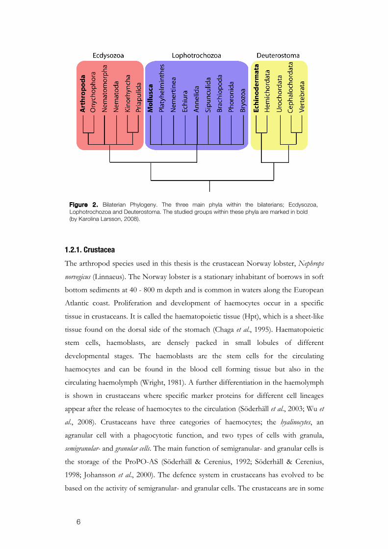
The species studied in this thesis are from three different phyla within the Bilaterians:
Arthropoda, Mollusca and Echinodermata, differing in preferred habitats, feeding
behaviour and somewhat in their strategies of immune defence (Fig. 2). There are
differences in mobilization and activation of the immune defence between these
groups of invertebrates. For example, the filter feeding mussels have developed an
immune system based on phagocytosis, probably since they constantly interact with
foreign particles and thus also pathogens (Cheng, 1969; Canesi et al., 2002). The
immune mechanisms of crustaceans rely mostly on a clotting and melanization
systems (Söderhäll & Cerenius, 1992, 1998) since they are more likely to be injured
and in need of a fast clotting system. Likewise, the echinoderms often get injured due
to predation and need a fast system for preventing blood loss, wound healing and
regeneration of tissues. The immune defence of the three invertebrate phyla studied
in this thesis is briefly summarized as follows: The circulating haemocytes of various
invertebrates are morphologically and functionally diverse. The different types of
haemocytes are mainly well characterized in arthropods, for example in Drosophila
melanogaster (Crozatier et al., 2007) and Pacifastacus leniusculus (Johansson et al., 2000;
Wu et al., 2008), while for many species characterization is not completed. The major
classification of haemocytes in invertebrates is the presence or absence of
cytoplasmic granules. The granules contain a range of hydrolytic enzymes including
proteinases, glucosidases and sulphatases (Pipe, 1997) and are described as
lysosomes.
6
1.2.1. Crustacea
The arthropod species used in this thesis is the crustacean Norway lobster, Nephrops
norvegicus (Linnaeus). The Norway lobster is a stationary inhabitant of borrows in soft
bottom sediments at 40 - 800 m depth and is common in waters along the European
Atlantic coast. Proliferation and development of haemocytes occur in a specific
tissue in crustaceans. It is called the haematopoietic tissue (Hpt), which is a sheet-like
tissue found on the dorsal side of the stomach (Chaga et al., 1995). Haematopoietic
stem cells, haemoblasts, are densely packed in small lobules of different
developmental stages. The haemoblasts are the stem cells for the circulating
haemocytes and can be found in the blood cell forming tissue but also in the
circulating haemolymph (Wright, 1981). A further differentiation in the haemolymph
is shown in crustaceans where specific marker proteins for different cell lineages
appear after the release of haemocytes to the circulation (Söderhäll et al., 2003; Wu et
al., 2008). Crustaceans have three categories of haemocytes; the hyalinocytes, an
agranular cell with a phagocytotic function, and two types of cells with granula,
semigranular- and granular cells. The main function of semigranular- and granular cells is
the storage of the ProPO-AS (Söderhäll & Cerenius, 1992; Söderhäll & Cerenius,
1998; Johansson et al., 2000). The defence system in crustaceans has evolved to be
based on the activity of semigranular- and granular cells. The crustaceans are in some
Figure 2. Bilaterian Phylogeny. The three main phyla within the bilaterians; Ecdysozoa, Lophotrochozoa and Deuterostoma. The studied groups within these phyla are marked in bold (by Karolina Larsson, 2008).

7
areas highly infected by the dinoflagellate, Hematodinium spp., which is a parasite
invading the haemocoel and connective tissue of most organs and dissolve the
muscle tissue (Field & Appleton, 1995; Messick & Shields, 2000). In fisheries, this
parasite causes economical losses of great value every year.
1.2.2. Mollusca
The mollusc Mytilus edulis (Linnaeus) or the common blue mussel is widespread along
the European coastline and lives on hard- and sandy bottoms at 0-10 m. As filter
feeders a substantial portion of the diet of molluscs is microorganisms (ZoBell et al.
1938). Thus, filter feeding results in concentrations of potential pathogens, but
bacteria in large numbers may persist without causing diseases in the animal. Adult
molluscs have an efficient defence against pathogens, but stress may comprise the
host and outbreaks of different bacterial diseases caused by e.g. the most common
Vibrios and Pseudomonas (Olafsen et al. 1993). The site of haematopoiesis in Mytilus
edulis is currently unknown, but in related organisms such as snails haemocytes are
produced in small nodes, primarily in the epithelial cells lining the pericardium
(Sminia, 1974). Haemocyte mitosis in molluscs seems also to occur in haemolymph
(Mayrand et al., 2005). The immune defence of M. edulis has evolved to be specialized
on phagocytosis and has very efficient antimicrobial peptides (Mitta et al., 1999;
Wootton et al., 2003). The role of granular cells within bivalves is phagocytosis as
well as encapsulation of microbes. After engulfment the phagosomes fuse with
lysosomes and the microbes are sequestered in the acidic phago-lysosome by the
enzymes, reactive metabolites and antimicrobial peptides (Cheng, 1983; Pipe, 1992;
Winston et al., 1996). In molluscs, three different categories of haemocytes are found
and all of them are able to phagocyte although one of them, the eosinophilic, seems
to be more prominent (Pipe et al., 1997; Dyrynda et al., 1997).
1.2.3. Echinodermata
Asterias rubens (Linnaeus) is the common sea star in European waters and lives on
hard or soft bottoms at depth between 0 - 200 m. Studies on echinoderm species
reveal that their immune system is based on the phagocytotic activity of the immune
cells (Coteur et al., 2002). They also have a simplified complement system (Smith et al.
2001) and bacteria-inducible transcription factors including a NF-κB homologue
(Pancer et al. 1999). The coelomic fluid of A. rubens possesses large populations of
8
circulating cells. The circulating cells in A. rubens have not been named in a universal
way. The same type of cells can be called different names in different literature.
Phagocytes constitute the predominated sub-population, comprising approximately
80-95% of the population of coelomocytes (Pinsino et al., 2007). These cells can be
transformed to petaloid and filopodial forms. It is found that coelomocytes in A.
rubens have the ability to form networks and fuse to syncytic formations when non-
self organisms are invading the coelomic fluid (Holm et al., 2008). In addition, there
are also amoebocytes, so called because of their ability to migrate within tissue and
vibratile cells present in the coelomic fluid (Smith, 1981). The coelomocytes are able
to efficiently clear bacteria from the coelomic cavity and in case of injury they take
part in wound healing by migrating to the injured site, prevent bleeding by clotting
and interact with the extracellular matrix during the healing process (Smith, 1981;
Dybas & Frankboner, 1986). The recruitment of circulating coelomocytes is not fully
understood. The coelomic epithelium has been suggested as one of the most
probable potential source of the coelomocytes of echinoderms (Munoz-Chapuli et al.,
2005) but also the axial organ (Leclerc et al., 1987) and the Tiedemanns’s body have
been suggested as well as the possibility of self-replication of the circulating
coelomocytes (Ratcliffe & Rowely, 1979). All three of these tissues have shown
mitogenic response to LPS, which further indicate their role as haematopoietic
tissues (Holm et al., 2008). Pathogen-induced mortalities of echinoderms, in
particular of sea urchins, have been reported from several places (Jangoux, 1990).
Mass mortalities of the sea star, Acanthaster planci, attributed to a sporozoan have
been found in the Pacific Ocean (Zann et al, 1990).
1.3. Manganese and Hypoxia
Many naturally occurring compounds are increasing in distribution and concentration
due to anthropogenic activities. These substances can reach toxic levels and may
affect the immune system of living organisms. Manganese, Mn, is an abundant
element in nature, particularly in soft bottom sediments of the oceans and in
bedrock. The metal is predominantly bound to the sediment in a four-valent colloid
state, MnO2. However, during hypoxic conditions, lower than 16 % O2 saturation
that can occur during periods of days to weeks in the bottom water (Baden et al.,
1990; Pihl et al., 1991), MnO2 is reduced and released into its bioavailable state, Mn2+,
and can reach toxic levels in benthic biota (Hall et al., 1996). There have been reports

9
of measured Mn concentration increased by a factor of 1000 (Trefry et al., 1984).
Along the Swedish west coast the Mn2+ fraction can increase and reach 19-20 mg L-1
in the bottom waters (Magnusson et al., 1996). Mn2+ re-oxidizes only on particles and
the bioavailable fraction may therefore stay in the water column for quite some time
even after hypoxia.
Eutrophication caused of the high input of nutrients in coastal waters
together with over-fishing cause cascade effects in the ecosystem increasing the algal
blooms and enhances hypoxic condition in large bottom areas (Casini et al., 2008;
Diaz & Rosenberg, 2008). The seasonal hypoxia is increasing along the Swedish and
European coastline (Diaz & Rosenberg, 1995; Diaz & Rosenberg, 2008) and thus
also the level of bioavailable Mn (Fig. 3.).
Manganese (Mn) is an essential trace metal accumulating especially in mitochondria
in both animals and plants. The metal is involved in metabolic processes as a
cofactor or activator of different enzymatic reactions, e.g. electron transfer reactions
and phosphorylation (Simkiss & Taylor, 1989). Mn can however act as a toxicant to
organisms when the concentrations are elevated and start affecting neuromuscular
transmission by interacting with mitochondrial Ca2+ and disturbing the ion balance in
muscle membranes (Gavin et al., 1999). Ionic Mn can also cross the blood-brain
barrier and interfere with chemical synapse functions. The fact that Mn has an effect
on the central nervous system has been known for long and a symptom called
Manganism, similar to Parkinson’s disease can be expressed (Iregren, 1990; Verity,
1999).
Figure 3. The distribution of documented hypoxic areas in 2008. Diaz & Rosenberg, 2008.

10
Detoxification through metallothioneins known to regulate the sequestration
and the metabolism of a variety of metals such as cadmium (Cd) and copper (Cu)
might not be the pathway for elimination of Mn (Viarengo, 1985). The intracellular
pathways of Mn have been studied in the yeast Saccharomyces cerevisiae (Cizewski-
Culotta et al., 2005) and include widely conserved transport proteins. When Mn
occurs in excess the cell minimizes the uptake by degradation of a transport protein,
SMF1. The export is regulated through the Golgi apparatus by a secretory pathway
known as PMR1. Both these pathways are also used for Ca transport. Detoxification
could also happen through entrapment of the metal by lysosomes (Temara et al.,
1998; Sterling et al., 2007).
Earlier studies of N. norvegicus reveal that manganese accumulates primarily in
the nervous tissue, but also in the haemolymph, where it accumulates three times the
exposure concentration. It was shown to reach neurotoxic levels in the bottom living
N. norvegicus (Baden & Neil, 1998; Holmes et al., 1999; Baden & Eriksson 2006).
Recent studies have revealed that a surplus of Mn affected several immunological
processes of N. norvegicus (Hernroth et al., 2004). Hernroth and co-workers found that
proliferation and maturation of haemocytes in N. norvegicus are inhibited. One of the
observations is a decreased number of circulating haemocytes.

11
2. AIM OF THE THESIS
The main objective of this study has been to explore the effects of exposure to
manganese (Mn) on immunological mechanisms of marine invertebrates and the
consequences for the animals’ defence against microorganisms. The overall
hypothesis is that Mn2+ accumulation in haemolymph causes defective mechanisms in
haematopoiesis and suppresses the activation of immune response with increased
prevalence for microbial infection as a result. The studies were intended to clarify
similar/dissimilar influences from manganese exposure in concentrations reported
from field conditions on the immune systems of selected invertebrate species from
different phyla.
The specific aims were:
Paper I Investigate potential mechanisms behind the lowered number of
haemocytes, haemocytopenia, caused by manganese, in N. norvegicus.
Focus was on whether apoptosis or necrosis contribute to the
haemocytepenia.
Paper II Compare A. rubens, to earlier studies on N. norvegicus. Mechanistic and
functional responses were considered, in order to get a broad view of
the effects of Mn as a stressor to echinoderms.
Paper III Investigate effects of exposure to Mn in combination with hypoxia on
the proliferation and maturation of A. rubens coelomocytes.
Paper IV A comparative study of clearance rate of the bacterium, Vibrio
parahaemolyticus, injected in three different species, N. norvegicus, A.
rubens and M. edulis exposed to Mn. In addition, potential acute or
chronic effects of elevated concentration of Mn were investigated.

12
3. METHODOLOGICAL CONSIDERATION
All papers include analysis of the actual level of Mn in haemolymph from Mn
exposed and unexposed animals, which makes it possible to draw conclusions that
the reason for change is elevated Mn levels. Likewise, the number of circulating
coelomocytes or haemocytes in all animals are routinely analysed to check for
possible changes when exposed to increased levels of Mn, which is a fundamental
hypothesis in the thesis. Analyses of interest from the specific paper are presented
below.
3.1. Animal handling
The three studied phyla of invertebrates were collected outside the Sven Lovén
Centre of Marine Sciences – Kristineberg, formally known as Kristineberg Marine
Research Station in the Gullmar Fjord situated at the Swedish west coast. Animals
were maintained in basins supplied with running seawater of ambient temperature
and salinity and were fed regularly until acclimatized and used for the experiments.
The specimens of Asterias rubens and Mytilus edulis were collected by scuba divers, A.
rubens at the depth of 5-15 m and M. edulis at 0.5-2 m. Nephrops norvegicus were caught
in creels by local fishermen at about 60 m depth. All animals used for the study were
of similar size within each group; A. rubens 10-12 cm across, from arm tip to most
distant arm tip, M. edulis 5-7 cm across the shell, and N. norvegicus 5-8 cm length over
carapax and the group was a random mixture of gender.
During time of experiment the lobsters and mussels were kept in containers
with seawater allowing mussels 0.5 L per individual and the lobsters about 50 L per
individual. The containers used where continuously mixed and aerated through
bubbling of the water. To simulate the hiding burrows lobsters naturally use, plastic
tubes were available in their tanks. Sea stars, on the other hand, are very delicate to
handle in a laboratory environment and we could not use a continuous flow-through
system when exposing the animals to manganese. To be able to expose sea stars to
controlled Mn concentrations they were placed in 3.5 l glass aquaria on a slowly
moving mixing table fulfilling the demand of oxygen without bubbling. The water
was exchanged daily and the animals were not fed during the experiment.
During manganese exposure Mn is dissolved in filtered seawater at
appropriate nominal concentrations, achieved by using manganese(II)chloride
tetrahydrate (GR, Merck, Germany). Animals used as controls were treated in the

13
same way but in seawater without Mn. When examining the effects of hypoxia on the
sea star A. rubens, the same type of containers were used as when exposing the
animals to manganese, but sealed. Oxygen levels between 14 - 16 % saturation were
achieved by aeration with nitrogen gas in the sealed containers and controlled with
oxygen meters (Oxi 340, WTW), continuously logged with Achat II Software. Lower
saturation levels would be irrelevant in this study, since such oxygen depletion would
subordinate the effects of the immune system in the animals.
3.2. Cell viability
Viability of the circulating haemocytes is an important indicator for studying the
functionality of the cells. If the cells are less competent than under normal
conditions, the whole system is most likely less efficient. To investigate the
cytotoxicity of Mn on haemocytes two different methods were used based on; a)
Metabolic activity, examining calorimetrically the ability of cells to convert
tetrazolium to formazan through dehydrogenase activity (Mosmann, 1983) and b)
Cell membrane integrity, investigating the ability of haemocytes to exclude Trypan
Blue. The tetrazolium test gives a good view of how vital the cells are and is used in
both Papers I and II. When doing the tetrazolium test in vitro in Paper I, we had
difficulties with Mn complex binding to the anticoagulation buffer, since it contained
EDTA, but since we did the Trypan Blue test in parallel the outcome of the results
could be verified. To avoid the complex binding in Paper II, we did not use any
anticoagulation buffer and diluted coelomocytes in coelomic fluid after concentrating
them.
3.3. Cell proliferation
Increased cell proliferation in the haematopoietic tissue is a way to compensate for
loss in number of circulating haemocytes and could as well be a strategy to
compensate for loss of viability of the haemocytes. Cell proliferation was not
increased in N. norvegicus, which would be a normal reaction to the decrease in
circulating haemocytes (Hernroth et al. 2004). In A. rubens the number of circulating
haemocytes increased radically when exposed to Mn. In order to investigate the
influence of Mn on cell proliferation of the circulating haemocytes and of coelomic
epithelium, which is regarded as a source of haemocyte, renewal (Muñoz-Chápuli et
al., 2005) two different methods were used. Proliferation was compared between Mn

14
exposed and un-exposed sea stars by microscopical determination of the ratio of
nuclei in mitotic stages found in cells from coelomic epithelium, used in Paper II.
However, mitotic nucleus could sometimes be hard to judge. The second method
used was to get a less subjective view of mitotic stages of haematopoietic cells, and
mitosis was traced and compared by using the substitute nucleotide, 5-Bromo-2´-
deoxyuridine, BrdU. BrdU-substitutes for thymidine in S-phase of replicating cells
and this was detected with a specific antibody, used in Papers II and III. Both
methods, the Mitotic index and the BrdU-incorporation, indicated that Mn induced
proliferation of cells in the HPT of A. rubens.
3.4. Cell differentiation
Runx-homologous molecules are a family of transcription factors defined by a highly
conserved DNA binding Runt-domain (Rennert et al., 2003; Stricker et al., 2003).
Runx genes are in generally known to be involved in the transcriptional control of
developmental processes (Wheeler et al., 2000; Coffman, 2003), but the Runt gene in
invertebrates is also determining the haematopoietic cell fate of granular cells (Tracey
& Speck, 2000; Reviewed by Coffman, 2003). Hernroth et al. (2004) studied the Runt
gene by using c-DNA-probe and in situ hybridization technique to examine the effect
of manganese on differentiation of haematopoietic cells of N. norvegicus. To
investigate whether manganese and hypoxia have an effect on differentiation of
haemocytes in A. rubens, the expression of the Runt gene was quantified with Real-
Time Polymerase Chain Reaction (qRT-PCR) technique. Since the Runt gene in A.
rubens had not been sequenced before, homology cloning and sequencing was done
before designing specific Runt primers and the sequence was annotated to BLAST
algorithm at the National Centre for Biotechnology Information
(http://www.ncbi.nlm.gov/blast). Analysis of the data from the different exposure
groups was made with comparative quantification. The qRT-PCR has advantages
since the analysis gives a quantitative measurement of the Runt expression compared
to the semi-quantitative in situ hybridization technique.
3.5. Apoptosis
In N. norvegicus the number of haemocytes drastically decreased when the animals
were exposed to Mn. Hernroth et al. (2004) suggested that Mn inhibited the
proliferation, which normally would increase upon such losses. Other possible

15
reasons for the heamocytopenia could be increased necrosis or apoptosis of both
circulating and proliferating haematopoietic cells. By distinguish between apoptosis
and necrosis in Paper I we aimed to judge the degree of Mn toxicity to the cells.
Agents that can cause apoptosis at low doses could cause necrosis by inhibiting vital
metabolic processes at high doses (Raffray & Cohen, 1997). Cell death caused by
necrosis involves a catastrophic failure of cellular homeostasis, uncontrolled,
degrading enzymatic reactions and cell leakage, which could initiate inflammatory
reactions in mammalian systems (Alison & Sarraf, 1995; Raffray & Cohen, 1997).
Apoptosis is a gene-derived cell suicide process, found in virtually all metazoan
organisms, to eliminate unwanted or damaged cells. During apoptosis the integrity of
the cellular organelles and plasma membrane is maintained and the fragments are
eliminated through non-traumatic phagocytic clearance (Steller, 1995; Jacobson et al.,
1997; Raff, 1998). Apoptosis is in general characterized by generation of DNA
fragments that can be recognized through detecting their specific single strand breaks
or their typical migration on agarose gel. Both these methods were used in this study
to analyze dose and time dependent induction of apoptosis. DNA fragmentation
assay, called TUNEL (TdT-mediated dUTP Nick-end Labelling), where a fluorecein-
labeled probe is complementary to specific end sequences was used to identify the
strand breaks specific to apoptotic fragments. The other test used was a DNA-ladder
assay, identifying apoptosis specific DNA fragmentation when separated on agarose
gel, forming a so-called DNA-ladder (Wyllie, 1980).
Initially, a pilot study was performed to investigate Mn-induced apoptosis in
circulating haemocytes. Due to experimental difficulties recognized as interference
between auto-florescence of the haemocytes and the green dye fluorecein-labeled
probe, the experiment was instead performed on cells from the Hpt. Both methods,
TUNEL and DNA-ladder assays, indicated that Mn induced apoptosis.
3.6. Stress response
When, in Paper II, testing whether a stress response is induced in A. rubens, two
different methods were performed. One indication of induced stress in animals is
increased levels of the so-called heat shock proteins (Hsp). The heat shock proteins
are a family of ubiquitous expressed proteins, which help to process misfolded and
damaged polypeptide chains and support maturation by functioning as a chaperone
protein (Bukau et al. 1998). Hsp70, one protein within this family, is an indicator of

16
stress, since it is upregulated when exposed to a functional or environmental stressor
(Matranga et al., 2000; Pinsino et al., 2007; Holm et al., 2008). A specific antibody
against Hsp70 was used as a stress marker in haemocytes and coelomic epithelium.
Another attempt of measuring the stress levels induced in animals was done
by using the rather new technique, OxyBlot. Previously, protein carbonyls have been
used for investigating oxidative damage of proteins due to environmental stress
(Almroth et al., 2005). Protein oxidation was analyzed by measuring the levels of
dinitrophenylhydrazone derivates of protein carbonyls, by separation with gel
electrophoresis and identification through blotting procedure and a specific antibody.
This registers the endpoint protein at oxidative damage, which indicates irreversible
damage of the proteins. We used Western Blot technique for both analyses. Hsp70
was also detected through immunohistochemistry on tissue sections.
3.7. Functional response
It is of great importance to investigate if increased levels of Mn affect the functional
responses in animals since it would be effect their survival in nature. In Paper II
effect on phagocytosis was investigated in vitro and in Paper IV the bactericidal
capacity after in vivo injection of bacteria was studied.
3.7.1. Phagocytosis assay
Paper II includes a test of how successful haemocytes from Mn exposed sea stars are
to phagocyte dead yeast cells marked with fluorescence, FITC, compared to that of
unexposed sea stars. The haemocytes in coelomic fluid were incubated with FITC-
marked yeast. The fluorescence of yeast cells that are not engulfed by haemocytes are
then quenched with Trypan Blue, which can enter only dead cells through their
insufficient cell membrane. The yeast engulfed by active cells is then still fluorescent
and detectable with a fluorometer.
First we intended to apply this method on a variety of organisms also
including animals from the Baltic Sea. The method was tested on M. edulis, Macoma
baltica and Saduria entomon. Mn effect on total haemocyte number was counted. Some
problems occurred when trying to optimize the phagocytosis assay for the different
animals. Since the haemocytes for most of the animals were decreasing in number
when exposed to Mn it became difficult to get a proper number for the assay. We
tried to concentrate the number of haemocytes through centrifugation, but since we

17
wanted to avoid using EDTA as an anticoagulant because it might bind Mn and
change the ion concentration of the metal in the assay, it was impossible to avoid
clotting of cells. However, the haemocytes of A. rubens were sufficient without
concentration and thus this in vitro phagocytosis experiment was used to compare the
phagocytic index only on haemocytes from Mn exposed and un-exposed sea stars.
3.7.2. Bactericidal capacity
In Paper IV the whole focus of the study was on how effective animals were in
defending themselves from a sub-lethal dose of a pathogen injected after Mn
exposure when compared to unexposed animals. The study was made on N.
norvegicus, M. edulis and A. rubens, and the pathogen used was a bacterium, Vibrio
parahaemolyticus. We used V. parahaemolyticus as a model organism since the coastal
water is their natural habitat and they have the ability to infect fish and shellfish (De
Paola et al., 1998, Colwell & Hug, 2001). Studies from Eiler et al. (2006) found V.
parahaemolyticus in Skagerrak and the Baltic Sea to the Gulf of Bothnia. There are
reports on increased spreading with increased temperature in water (Ra Londe,
2006). The bacteria were isolated from mussels sampled when water temperature was
approximately 20 °C in the area outside the Sven Lovén Centre - Kristineberg.
The appropriate concentration of the bacteria was determined for each
species to ensure that the dose was not lethal but still detectable with the viable count
method. The animals where first exposed to Mn for 5 days before being injected with
V. parahaemolyticus. Samples of haemolymph and the digestive gland were then taken
from animals in a time course. The fluid was streaked out and incubated on agar
plates. When analyzing viable counts during the first hours post injection we could
see high variances between individuals. Since the goal was to recognize potential
differences between the animals exposed to Mn and the un-exposed rather than the
clearance kinetics we decided to avoid such an early investigation. We could see that
the clearance from the fluid was quite fast and thus we decided to also include the
digestive glands of the animals. Viable counts were determined to compare the
bactericidal capacity of the different groups. To investigate if manganese has a
prolonged effect after time of exposure a recovery study was performed. The same
procedure was repeated after a recovery period of 3 days in water without Mn
additive after first being exposed during 5 days to manganese. Samples of
haemolymph and the digestive gland were taken after 24h.

18
Vibrios are known to enter a viable but non-culturable (VBNC) stage (Wang
& Gu, 2005) when encountering non-favourable conditions. Since only culturable V.
parahaemolyticus were investigated in this study those that might be VBNC would be
missed. However, it was assumed that the bacteria were equally affected by the
environmental conditions and thus viable counts were judged as a satisfactory
method.

19
4. MAIN RESULTS AND DISCUSSION
Measuring the concentration of Mn after exposure reveals an uptake and
accumulation in the haemolymph and in the digestive gland of the animals, but
varied between the different species. These studies have demonstrated that after 5
days of exposure the levels of Mn in haemolymph of the animals are in steady state
with the surrounding water in A. rubens, the accumulation is significantly higher in M.
edulis and increases almost 3 fold in N. norvegicus. The accumulation of Mn in the
digestive glands of the tested species gives a different picture. Here, the uptake of
Mn in A. rubens and N. norvegicus was slower reaching a lower concentration of Mn
than in the blood while in M. edulis Mn accumulated to a similar level as found in the
haemolymph (Paper IV). The differences in accumulation of Mn observed between
the species seem to reflect the different immune response.
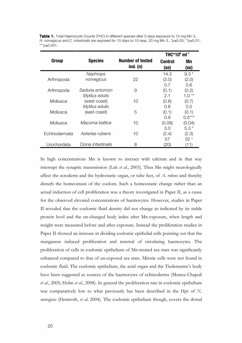
When exposed to Mn in concentrations relevant to what is found in nature,
15 mg Mn L-1 (Magnusson et al., 1996), the number of haemocytes was affected in all
tested animals, although the alteration differs between the animals. Both N. norvegicus
and M. edulis showed reduced numbers of haemocytes after Mn exposure. Opposite
to these findings, A. rubens significantly increased its circulating haemocytes. The
reduction in circulating haemocytes was in agreement with the results from earlier
studies on N. norvegicus (Hernroth et al. 2004) and we could see similar results in pilot
studies when testing the effect of Mn on M. baltica, S. entomon and Ciona intestinalis
(Table 1.). The contradictory results from A. rubens are a very interesting discovery.
The numbers of circulating haemocytes of A. rubens have previously been shown to
be quite stable despite changes in salinity and temperature and as well to Cd exposure
(Coteur et al. 2004, 2005). It indicates that the relatively low uptake in A. rubens
initiates a stimulating effect of the immune system. This stimulating effect, hormesis,
on the haemocyte numbers of A. rubens might have responded differently if the Mn
dose was higher than we used. This was not relevant in our study since we wanted to
investigate the effects of Mn concentrations occurring in nature. We were not able to
see a hormesis effect on N. norvegicus when exposed to lower concentrations (Paper
I).
20
THC*106 ml-1
Group
Species
Number of tested ind. (n)
Control (se)
Mn (se)
Arthropoda
Nephrops norvegicus
22
14.3 (3.0)
9.3 * (2.0)
Arthropoda
Saduria entomon
9
0.7 (0.1)
0.6 (0.2)
Mollusca
Mytilus edulis (west coast)
10
2.1 (0.8)
1.0 ** (0.7)
Mollusca
Mytilus edulis (east coast)
5
0.8 (0.1)
0.5 (0.1)
Mollusca
Macoma baltica
10
0.9 (0.08)
0.5*** (0.04)
Echinodermata
Asterias rubens
10
3.0 (2.4)
5.3 * (2.3)
Urochordata
Ciona intestinalis
8
57 (20)
32 * (11)
In high concentrations Mn is known to interact with calcium and in that way
interrupt the synaptic transmission (Luk et al., 2003). Thus Mn might neurologically
affect the ectoderm and the hydrostatic organ, or tube feet, of A. rubens and thereby
disturb the homeostasis of the coelom. Such a homeostatic change rather than an
actual induction of cell proliferation was a theory investigated in Paper II, as a cause
for the observed elevated concentrations of haemocytes. However, studies in Paper
II revealed that the coelomic fluid density did not change as indicated by its stable
protein level and the un-changed body index after Mn-exposure, when length and
weight were measured before and after exposure. Instead the proliferation studies in
Paper II showed an increase in dividing coelomic epithelial cells pointing out that the
manganese induced proliferation and renewal of circulating haemocytes. The
proliferation of cells in coelomic epithelium of Mn-treated sea stars was significantly
enhanced compared to that of un-exposed sea stars. Mitotic cells were not found in
coelomic fluid. The coelomic epithelium, the axial organ and the Tiedemanns’s body
have been suggested as sources of the haemocytes of echinoderms (Munoz-Chapuli
et al., 2005; Holm et al., 2008). In general the proliferation rate in coelomic epithelium
was comparatively low to what previously has been described in the Hpt of N.
norvegicus (Hernroth, et al. 2004). The coelomic epithelium though, covers the dorsal
Table 1. Total Haemocyte Counts (THC) in different species after 5 days exposure to 15 mg Mn /L. N. norvegicus and C. intestinalis are exposed for 10 days to 10 resp. 20 mg Mn /L. *p≤0.05; **p≤0.01; ***p≤0.001.

21
part of the entire coelomic cavity of the animals and given the large size its
contribution of renewal of coelomocytes should be significant.
When analyzing hypoxia treated animals, A. rubens, in Paper III, there were
no changes of proliferation in cells from the coelomic epithelium nor change in
amount of circulating haemocytes. Though when exposed to manganese, a 4 fold
increase in proliferation was found in both groups, Mn and Mn and hypoxia in
combination which showed that Mn rather than hypoxia stimulated the proliferation..
Studies on differentiation of these cells, explored by the expression of the Runt gene,
showed a dramatic synergistic effect of Mn in combination with hypoxia. Since Runt
is expressed in higher levels when haematopoietic cells differentiate to granular cells,
this might be an indication of a change in composition of haemocytes.
Different cell types are most probably different in their resistance to Mn,
which might generate toxicant tissue selectively. Hirata (2002) found that the viability
of a neuronal cell line (PC2), in terms of its ability to convert tetrazolium to
formazan by mitochondrial dehydrogenase, was significantly reduced when kept in
culture and exposed to 5 and 55 mg l-1 of Mn for 48 h. Such an effect on haemocytes
could not be shown in present the studies on Norway lobster. The viability was not
reduced when the haemocytes were exposed in vitro or in the in vivo study in Paper I,
although the animals accumulated more than twice the exposure concentration of
Mn. The ability of the haemocytes to exclude Trypan blue, which was also tested in
Paper I, did confirm the maintenance of their cell membrane integrity.
The results from the viability tests on N. norvegicus in Paper I showed that
necrosis was most likely not the explanation to haemocyte depletion, but apoptosis
was. The apoptotic cells amplified in stem cells with increased Mn concentration
when tested with the TUNEL-assay. The degree of apoptosis was related to both
time of exposure and concentration. The DNA-ladder assay did also show a
tendency to increased fragmentation related to concentration of Mn. However, after
five days of exposure using the DNA-ladder assay, only the highest exposure
concentration, 20 mg Mn L-1, elicited a pronounced apoptotic fragmentation.
Apoptosis is a single cell event and the detection level for a DNA-ladder formation
might not be reached at the lower concentrations and the shorter exposure time.
Furthermore, typical apoptotic bodies were observed in the microscope when
analyzing both kinds of cells. Thus, it was concluded that apoptosis of the circulating
haemocytes and their precursor cells obviously contributed to the haemocytopenia of
22
lobsters that was found after Mn exposure. Contrary to the findings in lobsters,
studies in Paper II establish that Mn did decrease the viability of haemocytes in A.
rubens when tested through the same analysis. These findings point out that even
though the cellular number increases significantly in the sea star, the conditions of
the cells seemed negatively affected. However, the viability assay does not give
enough information concerning possible negative effects on the animal’s
immunological response in terms of host-parasite interactions. At a cellular level
however, there were negative effects of Mn on the phagocytic capacity of
coelomocytes in A. rubens (Paper II) as the capacity to engulf yeast particles was
significantly reduced with approximately 6 %. It has earlier been found that
cadmium, Cd, does have a negative effect on the immune system in A. rubens (Coteur
et al., 2005) since, they found a reduction in phagocytic activity although no
differences in haemocyte numbers.
The study on the bactericidal capacity when injected with V. parahaemolyticus
(Paper IV) showed a rapid clearance of the haemolymph. Thus it was assumed that
the bacteria were either killed or translocated to other tissues. The digestive gland in
both M. edulis and N. norvegicus appear to be a sink for the tested bacterium, especially
so in N. norvegicus. This has been reported before in crustaceans and bivalves (Sahoo
et al., 2007; Williams et al., 2009). The bacteria might be translocated to the digestive
gland by phagocytotic cells, which have previously been reported (Fontaine &
Lightner, 1974; Aldrich et al., 1995) or most probably transported through the
haemolymph since they were culturable throughout the experiment. A. rubens on the
other hand did not show the same translocation as the other two tested species. It
seems like the echinoderm can compensate for the negative effect of manganese on
phagocytic activity through the induced proliferation of coelomocytes. It was
obvious that the Mn-exposed sea stars have a better ability to clear the coelomic fluid
and their digestive gland from V. parahaemolyticus compared to that of the other
species. The phagocytic capacity of the digestive gland in A. rubens might be more
efficient due to the larger organ compared to the other tested animals.
A. rubens and M. edulis might not represent species found in areas frequently
exposed to elevated levels of Mn. The study does however demonstrate an
accumulation of Mn in different species and effects on the immune system and
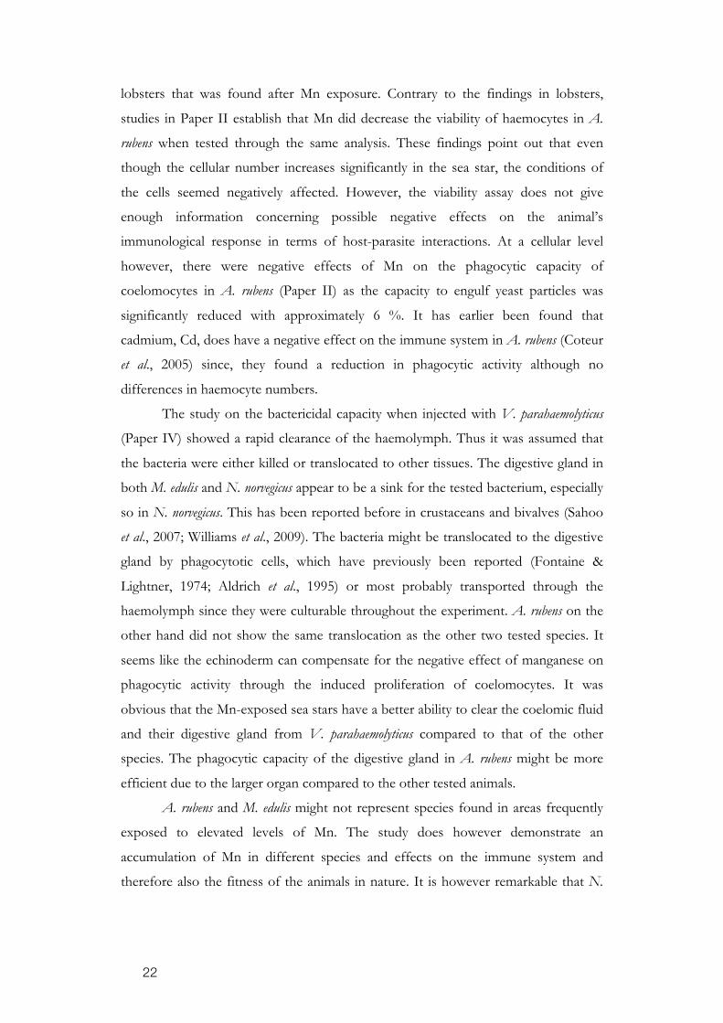
therefore also the fitness of the animals in nature. It is however remarkable that N.
23
norvegicus, living in an environment with recurrent increase in Mn concentration,
seems to be the least prepared to cope with the problem.
5. CONCLUSIONS
This thesis has shown immune suppressive effects of manganese exposure, in both
mechanistic and functional responses, in concentrations realistic to find in bottom
waters. The species were not similar in response, however. Taken together; these
results showed that Mn exposure significantly affects fundamental immune reactions
in species within the studied phyla pointing out the potential harm also for other
organisms. In N. norvegicus the metal severely suppresses the numbers of haemocytes
Figure 4. The effects of manganese exposure on the immune systems of the three studied species; Nephrops norvegicus, Mytilus edulis and Asterias rubens. The effect on differentiation in A. rubens is in combination with hypoxia. The light grey box at the bottom describes effects from a previous study (Hernroth et al., 2004).

24
by inducing apoptosis. The impaired immunity made them more susceptible to
infections. Other invertebrates, such as M. edulis, responded in a similar way as the
lobsters. A. rubens reacted to the same Mn concentration with a stimulating effect on
the haematopoiesis which increased the numbers of haemocytes. Although
manganese was shown stressful to the haemocytes and affected their ability to
phagocyte, the high numbers compensate these impairments. There was seemingly a
negative correlation between the accumulation of the metal in the tissues of the
animals and their ability to eliminate bacteria. Manganese interferes with
proliferation, differentiation and apoptosis, whereby the number of circulating
haemocytes is affected. Animals with a lowered cell number are inferior to cope with
invasive microbes.
Deficient immune systems increase the prevalence for infections and are of
utmost ecological importance. Mobilization and activation of a functional immune
system is of great concern for the fitness of all animals and the effects of Mn
reported here should be considered in a broader immunotoxicological perspective.
Although Mn does not cause chronic effects on immunity the expanding areas with
bioavailable Mn might have an impact on species composition since some become
more susceptible to infections.

25
ACKNOWLEDGMENT
I would like to thank the following organizations for financial support to this thesis: The Swedish Research Council for Environment, Agricultural Science and Spatial Planning (FORMAS), the Memory Foundation of Birgit and Birger Wåhlström, the Memory Foundation of Lars Hierta, the Memory Foundation of Carl Tryggers, the Memory Foundation of Wilhelm and Martina Lundgren and the Scientific Foundations of the Royal Swedish Academy of Science. Jag vill börja med att tacka mina tre handledare; Bodil Hernroth, Irene Söderhäll & Susanne Pihl-Baden, för att jag fick chansen att göra det här projektet. Ni har varit otroligt bra alla tre och kompletterar varandra på ett strålande sätt. Bodil, du är värd ett särskilt stort tack! Det har varit fantastiskt att få lära mig inte allt du kan, men en del. Ditt engagemang är inspirerande. Utan dig Bodil, hade jag inte börjat fundera på att doktorera och alldeles säkert inte lyckats få ihop en färdig avhandling. Du är en klippa! Jag vill också tacka Helen för att det är kul att jobba och snacka med dig. Olga, thank you for all the help with the qPCR. We did it, finally… Kristina, det är skönt att vi är två hårdhudingar som snorklar på höst & vinter. Jag tackar självklart alla andra på JämFys i Uppsala och fram för allt alla på Kristineberg som jag inte jobbat tillsammans med, men som ser till att det är kul att vara på jobbet ändå. Stor kram till hela gänget i Lyset: Emma, Ida, Andreas, Sandra, Kikki, Hannah, Linda, Lene, Josefin, Kenta, Maj, Ulrika, Erika, Linus, Pia, Karl, Annelie, Cicci, Marina, Maria, Martin, Olivia, Soffan och Tobias och den bästa sommargästen, Andreas. Det är skönt att ni finns, särskilt de dagar vi själva får se till att stan vaknar till. Big up to Glen Trash! Det var väl någon dag där i solgasset på Klubban eller på gamla Belone som min marina karriär och allt annat började. Ni är skönaste gänget! Mina Uptown grrls: Anna, Anna, Joel, Zandra & Karo. Var ska jag börja? Ni är helt fantastiska! Tack för att jag alltid får komma och bo hos er när jag dyker upp. Tack för allt ni fixar. Tack för allt snick-snack på dagar & sena kvällar. Kram till Marie också, även om du inte bor i Uppsala längre. Ni är mina bästa vänner. Och Karo, vi har aldrig tråkigt… jo, en gång . En extra stor kram till dig! Jag vill ge en jättestor kram till min familj . Tack till Syrran, Andreas, Freja & Tora. Snart kommer vi äntligen kunna ses mer igen. Jag ser fram emot det. Ett särskilt tack till Andreas för hjälpen med att få min bok så fin. Tack Mamma & Pappa för allt stöd och hjälp på vägen. Det har känts skönt att komma och koppla av hemma hos er ibland. Den största kramen av alla går till Pelle. Det har varit otroligt skönt att du har funnits med och peppat mig. Jag är så glad att det är Du&Jag. Tack för allt du gjort. Nu är det äntligen slutpendlat. Jag kan packa upp väskan och vi får bo ihop – for real. Det kommer bli kalasbra!

26
REFERENCES Aldrich, H.C., McDowell, L.M., Tamplin, M.L., Frase, C., Murphree, R., Jackson, J.K. 1995.
Detection of Vibrio vulnificus and Vibrio cholerae O1 in oyster tissue using immunoelectron microscopy. J. Shellfish Res. 14:493-99.
Alison, M.R., Sarraf, C.E., 1995. Apoptosis: Regulation and relevance to toxicology. Hum. Exp. Toxicol. 14, 234-247.
Almroth, B.C., Sturve, J., Berglund, Å., Förlin, L., 2005. Oxidative damage in eelpout (Zoarces viviparus), measured as protein carbonyls and TBARS, as biomarkers. Aquat. Toxicol. 73:171–180.
Auffret, M., Oubella, R., 1997. Haemocyte aggregation in the oyster Crassostrea gigas: in vitro measurement and experimental modulations by xenobiotics. Comp Biochem Physiol. 118:705-712.
Baden, S.P., Loo, L-O., Pihl, L., Rosenberg, R., 1990. Effects of eutrophication on benthic communities including fish: Swedish west coast. Ambio 3, 113-122.
Baden, S.P., Neil, D.M., 1998. Accumulation of manganese in the haemolymph, nerve and muscle tissue of Nephrops norvegicus (L.) and its effect on neuromuscular performance. Comp. Biochem. Physiol. 119, 351-359.
Baden, S.P., Eriksson, S.P., Gerhardt, L., 1999. Accumulation and elimination kinetics of manganese from different tissues of the Norway lobster Nephrops norvegicus (L.). Aquat. Toxicol. 46:127-137.
Baden, S.P., Eriksson, S.P., 2006. Role, routes and effects of manganese in Crustaceans. Oceanogr. Mar. Biol. Ann. Rev. 44:61–83.
Bukau, B., Horwich, A.L., 1998. The Hsp70 and Hsp60 Chaperone Machines. Cell. 92(3): 351-366.
Candida Carnevali, M.D. 2006. Regeneration in Echinoderms: repair, growth, cloning. ISJ. 3: 64-76.
Canesi, L., Gallo, G., Gavioli, M., Pruzzo, C. 2002. Bacteria- hemocyte Interactions and Phagocytosis in Marine Bivalves. Micro Res and Tech. 57:469-476.
Casini, M., Lövgren, J., Hjelm, J., Cardinale, M., Molinero, J-C., Kornilovs, G. 2008. Multi-level trophic cascades in a heavily exploited open marine ecosystem. Proc. R. Soc. B. doi:10.1098/rspb.2007.1752. Published online.
Cerenius, L., Söderhäll, K. 2004. The prophenoloxidase-activating system in invertebrates. Imm. Rev. 198(1):116-126.
Cerenius, L., Lee, B.L., Söderhäll, K. 2008. The proPO-system: pros and cons for its role in invertebrate immunity. Trends Immunol. 29(6):263-71.
Chaga, O., Lignell, M., Söderhäll, K., 1995. The haematopoietic cells of the freshwater crayfish, Pacifastacus leniusculus. Anim. Biol. 4:59-70.
Cheng, T.C., A.S. Thakur, and E. Rifkin. 1969. Phagocytosis and an internal defense mechanism in the mollusca: With an experimental study of the role of leucocytes in the removal of inc particles in Littorina Scabra Linn. Proc. Symp. Mollusca, Bangalore II: 546-566.
Cheng TC. 1983. The role of lysosomes in molluscan inflammation. Am. Zool. 23:129-44. Chia, F.S., Xing, J., 1996. Echinoderm haemocytes. Zool. Studies. 35:231-254. Cizewski Culotta V, Yang M, Hall MD. 2005. Manganese transport and trafficking: Lessons
learned from Saccharomyces cerevisiae. Euc cell 4(7):1159-1165. Coffman, J. A. 2003. Runx transcription factors and the developmental balance between cell
proliferation and differentiation. Cell Biol. Int. 27(4):315-324. Colwell, R., Hug, A., 2001. Marine ecosystems and cholera. Hydrobiologia. 460: 141-145. Coteur, G, DeBecker, G., Warnau, M., Jangoux, M., Dubois, Ph., 2002. Differentation of
immune cells challenged by bacteria in the common European starfish, Asterias rubens (Echinodermata). European J Cell Biol. 81:413-418.
Coteur, G., Corriere, N., Dubois, Ph., 2004. Environmental factors influencing the immune responses of the common European starfish (Asterias rubens). Fish Shellfish Immunol. 16:51-63.

27
Coteur, G., Gillian, D., Pernet, Ph., Dubois, Ph., 2005. Alteration of cellular immune responses in the seastar Asterias rubens following dietary exposure to cadmium. Aquat Toxicol. 73:418-421.
Crozatier M, Meister M. 2007. Drosophila haematopoiesis. Cell Microbiol. May;9(5):1117-26. Epub 2007 Mar 29. Review. PubMed PMID: 17394559.
De Paola, A., Motes, M.L., Chan, A.M., Suttle, C.A., 1998. Phages infecting Vibrio vulnificus are abundant and diverse in oysters (Crassostrea virginica) collected from the Gulf of Mexico. Appl Environ Microbiol. 64(1):346-351.
Diaz, R.J., Rosenberg, R., 1995. Marine benthic hypoxia. A review of its ecological effects and the behavioural responses of benthic macrofauna. Oceanogr. Mar. Biol. Ann. Rev. 245-303.
Diaz, R.J., Rosenberg, R., 2008. Spreading dead zones and consequences for marine ecosystems. Science. 321:926-929.
Doyle, RJ., Dziarski, R. 2001. The bacterial cell: peptidoglucans. In: Sussman, M. (Ed.) Molecular Medical Microbiology. Academic Press, London, pp. 137-153.
Dybas, L., Frankboner, P.V., 1986. Holothurian survival strategies: Mechanisms for the maintenance of the bacteriostatic environment in the coelomic cavity of the sea cucumberm Parastichopus californicus. Dev. Comp. Immunol. 10:311-330.
Dyrynda EA, Pipe RK, Ratcliff NA. 1997. Sub-populations of haemocytes in the adult and developing marine mussel, Mytilus edulis, identified by use of monoclonal antibodies. Cell Tissue Res. 289:527-36
Dziarski, R. 2004. Peptidoglycan recognition proteins (PGRPs). Mol Immunol. 40:877-886. Eiler, A., Johansson, M., Bertilsson, S., 2006. Environmental influences on Vibrio
populations in northern temperate and boreal coastal waters (Baltic and Skagerrak Seas). Appl Environ Microbiol. 72(9):6004-6011.
Field, R. H., Appelton, P. L. 1995. A Hematodinium-like dinoflagellate infection of the Norway lobster Nephrops norvegicus: observations on pathology and progression of infection. Dis Aquat Org 22, 115-128.
Flajnik, M. F., Du Pasquier, L. 2004. Evolution of innate and adaptive immunity: can we draw a line? Trends Immunol. 25:640-644.
Fontaine, C.T., Lightner, D.V. 1974. Observations on the phagocytosis and elimination of carmine particles injected into the abdominal musculature of the white shrimp, Penaeus setiferus. Invertebr. Pathol. 24:141-148.
Gavin, C.E., Gunter, K.K., and Gunter, T.E., 1999. Manganese and calcium transport in mitochondria: implications for manganese toxicity. Neurotoxicol. 20, 445-453.
Gurther, G.C., Werner, S., Barrandon, Y., Longaker, M.T. 2008. Wound repair and regeneration. Nature. 453:314-321.
Haine ER, Moret Y, Siva-Jothy MT, Rolff J. 2008 Antimicrobial defense and persistent infection in insects. Science. 322(5905):1257-9.
Hall, I.R., Hydes, D.J., Statham, P.J. & Overnell, J. 1996. Dissolved and particulate trace metals in a Scottish Sea Loch: an example of a pristine environment? Mar Poll Bull. 32:846-854.
Hernroth, B.E, Baden, S.P., Holm, K., Andrén, T., Söderhäll, I. 2004. Manganese induced immune-suppression in Norway lobster. Aquat. Toxicol. 70, 223-231.
Hirata, Y., 2002. Manganese-induced apoptosis in PC12 cells. Neurotoxicol. Teratol. 24:639-653.
Hoffmann, J.A., Reichhart, J.M., 2002, Drosophila innate immunity: an evolutionary perspective. Nat. Immunol. 3:121-126.
Holm, K., Dupont, S., Sköld, H., Stenius, A., Thorndyke, M., Hernroth, B. 2008. Induced cell proliferation in putative haematopoietic tissues of the sea star, Asterias rubens (L). J. Exp. Biol. 211(16):2551-2558.
Holm, K., Hernroth, B., Thorndyke, M. 2008. Coelomocyte numbers and expression of HSP70 in wounded sea stars during hypoxia. Cell Tissue Res. 334(2):319-325.
28
Holmes, J.M., Gräns, A-S., Neil, D.M., Baden, S.P., 1999. Effects on the metal ions Mn2+ and Co2+ on muscle contraction in the Norway lobster, Nephrops norvegicus. J. Comp. Physiol. B. 169:402-410.
Iregren, A., 1990. Physiological test performance in foundry workers exposed to low levels of manganese. Neurotoxicol. Teratol. 12:673-675.
Jacobson, M.D., Weil, M., Raff, M.C., 1997. Programmed cell death in animal development. Cell 88:347-354.
Jangoux, M. 1990. Diseases of Echinodermata. In: Kinne, O. (Ed.), Diseases of Marine Animals. Biologisch Anstalt Helgoland. Hamburg, pp. 439-567.
Jiravanichhpaisal, P., Lee, B.L., Söderhäll K. 2006. Cell-mediated immunity in arthropods: Hematopoiesis, coagulation, melanization and opsonization. Immunology 211(4):213-36.
Johansson, M.W., Keyser, P., Sritunyalucksana, K., Söderhäll, K., 2000. Crustacean haemocytes and haematopoiesis. Aquaculture. 191:45-52.
Johansson M.W., Söderhäll K., 1989. Cellular immunity in crustaceans and the proPO system. Parasitology Today. 5:6.
Kan H, Kim CH, Kwon HM, Park JW, Roh KB, Lee H, Park BJ, Zhang R, Zhang J, . Söderhäll, K. and Lee, B.L. 2008. Molecular control of phenoloxidase-induced melanin synthesis in an insect. J. Biol. Chem. 283(37):25316-23.
Kim, C.H., Kim, S.J., Hongnan K., Kwon, H.M., Roh, K.B., Jiang, R., Yang,Y., Park, J.W., Lee, H.H., Ha, N.C., Kang, H.J., Nonaka, M., Söderhäll, K., Lee, B.L. 2008. A three-step proteolytic cascade mediates the activation of the peptidoglycan-induced toll pathway in an insect. J. Biol. Chem. 283(12):7599-607.
Kurata, S., Ariki, S., Kawabata, S., 2006. Recognition of pathogens and activation of immune responses in Drosophila and horseshoe crab innate immunity. Immunobiology. 211:237-249.
Lackie, A.M., 1988. Hemocyte behavior. Adv. Insect Physiol. 21:85-178. Leclerc, M., Luquet, B., Brillouet, C., 1987. In vitro effect of rabbit anti sea star lymphocyte
serum on axial organ cells. Cell. Biol. Int. Rep. 11(11):819-23. Lightner D.V., Redman R. 1977. Histochemical demonstration of melanin in cellular
inflammatory processes of Penaeid shrimp. J. Inver. Path. 30(3):298-302. Luk, E., Jensen, L.T., Culotta, V.C., 2003. The many highways for intracellular trafficking of
metals. J. Biol. Inorg. Chem. 8(8):803-9. Magnusson, K., Ekelund, R., Dave, G., Granmo, Å., Förlin, L., Wennberg, L., Samuelsson,
M.O., Berggren, M., Brorström-Lundén, E., 1996. Contamination and correlation with toxicity of sediment samples from Skagerrak and Kattegat. J. Sea Res. 35, 223-234.
Matranga, V., Toia, G., Bonaventura, R., Müller, W.E.G. 2000. Cellular and biochemical responses to environmental and experimentally induces stress in sea urchin coelomocytes. Cell Stress Chaperones. 5:113-120.
Mayland, E., St-Jean, S.D., Courtenay, S.C., 2005. Haemocyte responses of blue mussels (Mytilus edulis) transferred from a contaminated site to a reference site: can the immune system recuperate? Aquacult Res. 36:926-971.
Medizhitov, R., Janeway, Jr., C.A., 2002. Decoding the patterns of self and nonself by the innate immune system. Science. 296:298-300.
Messick, G.A., Shields, J.D., 2000 Epizootiology of the parasitic dinoflagellate Hematodinium sp. in the American blue crab Callinectes sapidus. Dis Aquat Org. 43:139-152.
Michel, T., Reichhart, J-M., Hoffmann, J.A., Royet, J., 2001. Drosophila Toll is activated by Gram-positive bacteria through a circulating peptidoglucans recognition protein. Nature 414:756-759.
Mitta, G., Vandenblucke, F., Hubert, F., Roch, P. 1999. Mussel defensins are synthesised and processed in granulocytes then released into the plasma after bacterial challenge. J. Cell Sci. 112:4233-4242.
Mosmann, T., 1983. Rapid colorometric assay for cellular growth and survival: Application to proliferation and cytotoxicity assays. J. Immunol. Met. 65, 55-63.
Muñoz-Chápuli, R., Carmona, R., Guadix, J.A., Macías, D., Pérez-Pomares, J.M., 2005. The origin of the endothelial cells: an evo-devo approach for the invertebrate/vertebrate transition of the circulatory system. Evol Dev. 7(4):351-358.

29
Olafsen, J. A., Mikkelsen, H.V., Glæver, H.M., Høvik Hansen, G., 1993. Indigenous Bacteria in Hemolymph and Tissues of Marine Bivalves at Low Temperatures. Appl. Environ. Microbiol. 59(6):1848-1854.
Pancer, Z., Rast, J.P., Davidson, E.H., 1999. Origins of immunity: transcription factors and homologues of effector genes of the vertebrate immune system expressed in sea urchin haemocytes. Immunogenetics. 49:773-786.
Pinsino, A., Thorndyke, M.C., Matranga, V. 2007. Coelomocytes and post-traumatic response in the common sea star, Asterias rubens. Cell Stress Chaperones. 12:331-341.
Pihl, L., Baden, S.P., Diaz, R.J., 1991. Effects of periodic hypoxia on distribution of demersal fish and crustaceans. Mar. Biol. 108, 349-360.
Pipe, R.K., 1990. Differential binding of lectins to haemocytes of the mussel Mytilus edulis. Cell Tissue Res. 261:261-268.
Pipe RK. 1992. Generation of reactive oxygen metabolites by the heamocytes of the mussel, Mytilus edulis. Dev Comp. Immunol.16:111-22.
Pipe, R.K., Farley, S.R., Coles, J.A., 1997. The separation and characterisation of haemocytes from the mussel Mytilus edulis. Cell Tissue Res. 289:537-545.
Raff, M., 1998. Cell suicide for beginners. Nature 396, 119-122. Raffray, M., Cohen, G.M., 1997. Apoptosis and necrosis in toxicology: A continuum or
distinct modes of cell death? Pharmacol. Ther. 75, 153-177. Ra Londe, R. 2006. Vibrio parahaemolyticus in Alaska: An aggressive response to a potential
crisis. J. Shellfish Res. 25(2):764 Ratcliffe, N.A. & Rowley, A.R., 1979. A comparative synopsis of the structure and function
of the blood cells of insects and other invertebrates. Dev Comp Imm. 3:189.
Ratcliffe, N.A., Leonard, C.M., Rowley, A.F., 1984. Prophenoloxidase activation: non-self recognition and cell co-operation in insect immunity. Science. 226:557-559.
Rennert, J., Coffman, J. Mushegian, A., Robertson, A. 2003. The evolution of Runx genes I. A comparative study of sequences from phylogenetically diverse model organisms. Evol. Biol. 3(1): 4.
Rosenthal, R S., Dzarski, R., 1994. Isolation of peptidoglucans and soluble peptidoglucan fragments. Meth. Enzymol. 235:253-285.
Sahoo, P.K., Pillai, B.R., Mohanty, J., Kumari, J., Mohanty, S., Mishra, B.K. 2007. In vivo humoral and cellular reactions, and fate of injected bacteria Aeromonas hydrophila in fresh water prawn Macrobrachium rosenbergii. Fish Shellfish Immunol. 23:327-40.
Simkiss, K., Taylor, M.G., 1989. Metal fluxes across the membranes of aquatic organisms. Aquat. Sci. 1:173-188.
Sminia T., 1974. Haematopoiesis in the freshwater snail Lymnaea stagnalis studied by electron microscopy and autoradiography. Cell Tissue Res. 150(4):443-54.
Smith, V.J., 1981. The Echinoderms. In: N. A. Ratcliffe, A.F. Rowley (eds.): Invertebrate Blood Cells. Academic Press, London. 2:514-558.
Smith, V.J., Söderhäll, K., 1983. Induction of degranulation and lysis of haemocytes in the freshwater crayfish, Astacus astacus by components of the prophenoloxidase activating system in vitro. Cell Tissue Res. 233(2):295-303.
Smith, V.J., Crisholm, J.R., 2001. Antimicrobial proteins in crustaceans. Adv. Exp. Med. Biol. 484:95-112.
Steller, H., 1995. Mechanisms and genes of cellular suicide. Sci. 267:1445-1449. Sterling, K.M., Mandal, P.K., Roggenbeck, B.A., Ahearn, S.E., Gerencser, G.A., Ahearn,
G.A. 2007. Heavy metal detoxification in crustacean epithelial lysosomes: role of anions in the compartmentalization process. J. Exp. Biol. 210:3484-493.
Steiner, H., 2004. Peptidoglugan recognition proteins: on and off switches for innate immunity. Imm Rev. 198:83-96.
Stricker, S., Poustka, A.J., Wiecha, U., Stiege, A., Hecht, J., Panopoulou, G., Vilcinskas, A., Mundlos, S., Seitz, V. 2003. A single amphioxus and sea urchin runt-gene suggests that runt-gene duplications occurred in early chordate evolution. Dev. Comp. Imm. 27(8):673-684.

30
Söderhäll, I., E. Bangyeekhun, Mayo, S., Söderhäll, K. 2003. Hemocyte production and maturation in an invertebrate animal; proliferation and gene expression in hematopoietic stem cells of Pacifastacus leniusculus. Dev. Comp. Imm. 27(8):661-672.
Söderhäll, K., 1982. Prophenoloxidase activating system and melanization- a recognition mechanism of arthropods? A review. Dev Comp Immunol. 6:601-611.
Söderhäll, K., Aspán, A., Duvic, B., 1990. The proPO-system and associated proteins; role in cellular communication in arthropods. Res Immunol. 141(9):896-907.
Söderhäll K., Cerenius, L. 1992. Crustacean immunity. Annual rev. Fish Dis. 2:3-23. Söderhäll K., Cerenius L. 1998. Role of the prophenoloxidase-activating system in
invertebrate immunity. Curr. Op. Immunol. 10:23-28. Temara, A., Aboutboul, P., Warnau, M., Jangoux, M, Dubois, P. 1998 Uptake and fate of
lead in the common Asteroid Asterias rubens (Echinodermata). Water Air Soil Pol. 102:201-08.
Tracey, W. D., Speck, N. A. 2000. Potential roles for RUNX1 and its orthologs in determining hematopoietic cell fate. Semin. Cell Dev. Biol. 11(5):337-342.
Trefry, J.H., Presley, B.J., Keeney-Kennicutt, W.L., Trocine, R.P., 1984. Distribution and chemistry of manganese, iron, and suspended particulates in Orca Basin. Geo-Marine Letters 4:125-130.
Verity, A.M., 1999. Manganese neurotoxicity: A mechanistic hypothesis. Neurotoxicol. 20:489-498.
Viarengo, A. 1985. Biochemical effects of trace metals. Mar Pol Bul. 16:153-158. Wang, Y., J.D. Gu. 2005. Influence of temperature, salinity and pH on the growth of
environmental Aeromonas and Vibrio species isolated from Mai Po and the inner Deep Bay Nature reserve Ramsar site of Hong Kong. J. Basic Microbiol. 45(1):83-93.
Weber, A.N., Tauszig-Delamasure, S., Hoffmann, J.A., Lelievre, E., Gascan, H., Ray, K.P., Morse, M.A., Imler, J.L., Gay, N.J., 2003. Binding of the Drosophila cytokine Spätzle to Toll is direct and establishes signaling. Nat. Immunol. 4:794-800.
Werner, T., Borge-Renberg, K., Mellroth, P., Steiner, H., Hultmark, D. 2003. Functional diversity of the Drosophila PGRP-LC gene cluste in response to lipopolysaccaride and peptidoglucan. J. Biol. Chem. 278:26319-26322.
Williams, H.R., Macey, B.M., Burnett, L.E., Burnett, K.G. 2009. Differential localization of bacteriostasis of Vibrio campellii among tissues of Eastern oyster, Crassostrea virginica. Dev. Comp. Immunol. 4:592-600.
Winston, G.W., M.N. Moore, M.A. Kirchin, and C. Soverchia. 1996. Production of reactive oxygen species by hemocytes from the marine mussel, Mytlius edulis: lysosomal localization and effect of xenobiotics. Comp. Biochem. Physiol. 113: 221-229.
Wootton, E.C., Dyrynda, E.A., and Ratcliff, N.A. 2003. Bivalve immunity: comparison between the marine mussel (Mytilus edulis), the edinle cockle (Cerastoderma edule) and razor-shell (Ensis siliqua). Fish. Shell. Imm. 15:195-210.
Wright, R.K., 1981. Urochordates: Invertebrate blood cells. Ratcliffe, N and Rowley, A. (Eds.) AP. London 2:565-626.
Wu, C., Söderhäll, I., Kim Y.A., Liu, H., Söderhäll, K. 2008. Hemocyte-lineage marker proteins in a crustacean, the freshwater crayfish, Pacifastacus leniusculus Proteomics. 8(20):4226-4235.
Wyllie, A.H., 1980. Glucocorticoid-induced thymocyte apoptosis is associated with endogenous endonuclease activation. Nature 284, 555-556.
Yokoo, S., Goetz, P., Tojo, S., 1995. Phagocytic activities of haemocytes separated by two simple methods from larvae of two different lepidopteran species, Agrotis segetum and Galleris mellonella. Appl. Entomol.Zool. 30:343-350.
Zann, L., Brodie, J., Vuki V., 1990. History and dynamics of the crown-of-thorns starfis Acanthaster planci (L.) in the Suva area, Fiji. Coral Reefs. 9(3):135-144.
ZoBell, C.E., Feltham, C.B., 1938. Bacteria as food for certain marine invertebrates. J. Mar. Res. 1:312-327.

31
SVENSK SAMMANFATTNING
Eutrofiering, eller övergödning av näringsämnen, tillsammans med överfiske
resulterar i att växternas primärproduktion ökar och det blir algblomningar. När de
sedan bryts ned leder detta i sin tur till att mängden syre i vattnet minskar och följden
blir att flertalet metaller löses ut från bottensediment och kan orsaka problem för
levande organismer i dessa miljöer. Eutrofiering är ett ökande problem längs våra
kuster.
Mangan (Mn) är en metall som är mycket riklig i mjuka havsbottnar, eftersom
mangan förekommer i många av våra vanliga bergarter. Mangan är normalt bundet
som MnO2, men vid hypoxi, minskad syrehalt, reduceras MnO2 till den biotillgängliga
jonformen Mn2+, som lätt tas upp av levande organismer. Hypoxi kan förekomma
under kortare eller längre perioder, dagar till månader, men eftersom Mn2+ kan ta lång
tid att återoxideras kan förhöjda mängder mangan finnas i bottenvattnet under en
period även efter en tid av hypoxi. Mängden biotillgänglig mangan kan i extrema
förhållanden öka upp till 1000 gånger så mycket som vid normal syretillförsel.
Mangan i låga doser är en essentiell, livsnödvändig, metall som främst behövs under
ämnesomsättningen för att kunna producera och aktivera vissa viktiga enzymer i
både djur och växter. Förhöjd halt av mangan är däremot toxisk, giftig. Metallen stör
då jonbalansen och kan interagera med kalcium, Ca2+. Att mangan kan påverka det
centrala nervsystemet har länge varit känt och ett symtom kallat manganism, som
påminner om nervsjukdomen Parkinson, kan utvecklas.
De ryggradslösa djuren, evertebraterna, har inte samma typ av immunförsvar
som ryggradsdjuren, vertebraterna, med specifika antikroppar och ett långtidsminne,
men de har blodceller som påminner om människans vita blodkroppar. Dessa
blodceller kommer från stamceller som finns i depåer i vissa vävnader. Där delar sig
stamcellerna och genomgår en mognadsprocess som omvandlar dem till aktiva
blodceller som används vid försvar mot angripande mikroorganismer, som bakterier
till exempel. Deras immunförsvar är baserat på aktivitet i deras blodceller i blodet.
Det finns flera typer av blodceller. Vissa har förmåga att fagocytera, sluka,
inkräktande mikroorganismer och partiklar för att döda dem inne i cellen när andra
blodceller kan frigöra giftiga ämnen som förgör angripande organismer utanför
cellen. På grund av de öppna blodsystem som de flesta evertebrater har, har de
behov av ett effektivt direkt försvar. Sårläkning och koagulering är viktiga processer
för att minska blodförlust. Detta är generellt i alla evertebrater, däremot är det
32
skillnader mellan dem i hur de mobiliserar immunförsvaret. En rad fundamentala
immunmekanismer har bevarats genom evolutionen, som till exempel blodcellernas
roll, förmågan att känna igen främmande molekyler och att producera och frigöra
cytotoxiska ämnen.
I den här avhandlingen har jag undersökt hur celldelningen och utmognaden
av stamceller påverkas av mangan och om deras överlevnad påverkas i tre olika arter;
havskräfta, blå mussla samt vanlig sjöstjärna. Vidare har jag undersökt om
aktiveringen av blodcellernas påverkas och om det ger effekt på djurens förmåga att
försvara sig mot sjukdomsalstrande bakterier som kan vara vanligt förekommande i
kustnära vatten. Detta har jag gjort genom ett flertal experiment då jag i akvarier har
utsatt djuren för sådana koncentrationer av mangan som man kan finna på våra
havsbottnar och jämfört dem med kontrolldjur som inte exponerats för mangan. Jag
har kunnat se att mangan kan starta en process som göra att de blodbildande
stamcellerna dör genom så kallad programmerad celldöd, apoptosis. Det kan vara en
förklaring till varför havskräftans blodceller minskar i antal då de utsätts för mangan.
När sjöstjärnorna däremot utsattes för mangan blev resultatet det omvända. I dessa
djur hade samma koncentration av metallen en stimulerande effekt som gjorde att
stamcellerna delade sig mer och antal blodceller ökade. Emellertid kunde jag se att
deras förmåga att fagocytera mikroorganismer minskade och de visade också tecken
på stress. Detta kunde jag studera genom att analysera särskilda stressproteiner.
Ökningen av antalet cirkulerande blodceller tycks kompensera för att de fagocyterar
mindre och dessa djur behöll även sin förmåga att eliminera bakterier då dessa
injicerades direkt i deras blod. Både kräftor och blåmusslor fick däremot svårare att
avlägsna bakterier då de utsattes för mangan vilket tyder på att de blir mer känsliga
för infektioner än vad sjöstjärnorna blir. Jag kunde även se att både kräfta och mussla
ansamlade mer mangan i vävnaderna än vad sjöstjärnorna gjorde. Däremot
återhämtade manganbehandlade djur sig i rent vatten efter att de hade varit utsatta
för manganexponering. Under tre dagar renades de från nästan allt mangan och deras
förmåga att eliminera bakterier återhämtades nästan till fullo.
I den här avhandlingen har jag kunnat se att mangan i koncentrationer som är
realistiska att hitta längs våra kuster under perioder påverkar de ryggradslösa djurens
immunförsvar på olika vis, men effekter har påträffats hos alla testade arter.
Havskräftor och musslor påverkades mest och deras infektionskänslighet ökade
medan sjöstjärnornas bakteriedödande förmåga förblev oförändrad. Trots att

33
mangans effekter inte tycks ge kvarstående påverkan, kan det innebära att under de
perioder då mangan är tillgängligt har sjukdomsalstrande mikroorganismer en ökad
chans att etablera infektioner vilket kan vara av stor betydelse för djurets överlevnad.
Nyligen har det kommit rapporter om att syrebrist och därmed tillgängligt mangan
har ökat i kustområden världen över vilket gör studien särskilt relevant.


