

UNIVERSIDADE DA BEIRA INTERIOR Ciências da Saúde
In vitro evaluation of the toxicity of bismuth
compounds
Ângela Inês Lima Gonçalves
Dissertação para obtenção do Grau de Mestre em
Ciências Biomédicas (2º ciclo de estudos)
Orientador: Prof. Doutor Samuel Martins Silvestre
Covilhã, outubro de 2016

ii

iii
Agradecimentos
Em primeiro lugar quero agradecer ao meu orientador, Professor Doutor Samuel Silvestre, por
ter aceitado guiar-me neste percurso, por toda a disponibilidade, apoio e saberes transmitidos
ao longo deste ano.
Quero agradecer também à Professora Adriana Santos, por toda a amabilidade, disponibilidade,
apoio e conhecimento transmitidos.
Não podia deixar de agradecer também às minhas colegas de laboratório, Elisabete, Sandrina,
Mariana, Mafalda, Sara e Vanessa pelos bons momentos, amizade e apoio.
Por fim, quero agradecer aos meus pais, à minha irmã e ao Ângelo Miguel por todo o carinho,
pelo apoio e pelos sacrifícios que fizeram para me ajudar a estar onde estou hoje.
A todos, o meu sentido bem-haja!

iv
v
Resumo alargado
O bismuto pertence ao grupo dos metais pesados, e demonstra um comportamento químico
semelhante ao do arsénio e do antimónio, mas ao contrário destes elementos, o bismuto tem
sido considerado relativamente não tóxico, uma vez que tem uma solubilidade em fluidos
aquosos relativamente baixa. Contrastando com a extensiva informação existente sobre os
demais elementos da tabela periódica, o bismuto tem, talvez, o aglomerado de informações
menos desenvolvido, apesar de ser extensivamente usado na medicina. Os sais de bismuto são
usados para tratar úlceras pépticas, dispepsia funcional e gastrite crónica. Apesar da falta de
informação existente sobre o tema, é notório que a toxicidade por bismuto pode ser observada
devido a ingestão abusiva ou mau uso aquando da ingestão em grandes quantidades ou por
grandes períodos de tempo. Os efeitos tóxicos que têm vindo a ser reportados como causados
por overdoses de compostos de bismuto incluem encefalopatias, nefropatias, osteoartropatias,
gengivoestomatites e colites.
Como têm sido reportados na literatura alguns casos de toxicidade por bismuto, o objetivo
deste projeto foi avaliar a toxicidade de alguns compostos de bismuto comumente usados na
terapia, e como catalisadores de transformações orgânicas. Para isso, e utilizando o ensaio do
brometo de 3-(4,5-dimetiltiazol-2-il)-2,5-difeniltetrazólio (MTT), os efeitos destes compostos
na proliferação celular in vitro, foram avaliados. Este ensaio foi realizado com dois tempos de
exposição aos compostos, 3 e 48 horas, para se avaliar se haveria toxicidade aguda e num tempo
superior de exposição aos compostos, respetivamente. Para isso foram usadas linhas celulares
representativas, incluindo neuronais (N27), intestinais (Caco-2), hepáticas (HepaRG) e
mamárias (MCF-7) e fibroblastos da derme (NHDF). Nenhum dos dez compostos de bismuto
estudados levou a uma redução significativa da proliferação celular após 3 horas de exposição,
o que demonstra que os compostos estudados não provocam toxicidade aguda nas linhas
celulares utilizadas. No entanto, após 48 horas de exposição aos compostos, foi observado que
o triflato (III) de bismuto e o subnitrato de bismuto levaram a uma redução significativa da
proliferação da linha celular neuronal (N27) e o subnitrato de bismuto leva também a uma
redução da proliferação celular da linha celular intestinal (Caco-2) . Além deste ensaio, foram
também executados o ensaio da citometria de fluxo usando iodeto de propídeo como marcador
para as células mortas, uma vez que este composto intercala o ADN e emite fluorescência
proporcional à quantidade de ADN da célula e o ensaio do 2’,7’ –dicllorofluorescina diacetato
(DCFDA), que é um corante fluorogénico que mede espécies reativas de oxigénio; após a difusão
para a célula o DCFDA é desacetilado pelas esterases celulares a um composto não fluorescente,
que é posteriormente oxidado pelas espécies reativas de oxigénio a 2’, 7’ –diclorofluoresceina
(DCF), que é um composto altamente fluorescente que pode ser detetado por espectroscopia
de fluorescência. Estes ensaios foram realizados para se tentar ter alguma informação sobre os
potenciais mecanismos de toxicidade mediados por estes compostos. Quando a produção de

vi
espécies reativas de oxigénio aumenta e se ultrapassam as capacidades antioxidantes da célula,
podem ocorrer danos macromoleculares principalmente no ADN, e em proteínas ou lípidos, o
que pode levar à apoptose ou necrose. Com o ensaio do DCFDA foi possível medir
indirectamente a formação de espécies reativas de oxigénio que os compostos triflato (III) de
bismuto e subnitrato de bismuto provocam na linha celular N27. Neste ensaio foi observado que
o composto triflato(III) de bismuto parece não ter um efeito na produção de espécies reativas
de oxigénio, mas pelo contrario o composto subnitrato de bismuto parece ter algum efeito,
numa exposição de 6 horas aos compostos. Com maior tempo de exposição ao composto
subnitrato de bismuto, 24 horas, foi observado que o este composto na maior concentração
testada leva à produção de espécies reativas de oxigénio, quase ao mesmo nível que o controlo
positivo.
No ensaio de citometria de fluxo foi usada também a linha celular neuronal e também os
compostos triflato (III) de bismuto e subnitrato de bismuto. Num estudo preliminar à citometria
de fluxo, observou-se ao microscópioóptico a morfologia celular, tendo sido possível observar
que, de facto, o número de células foi diminuído pela acção destes compostos, e que a
morfologia das células neuronais, tanto pela ação do triflato (III) de bismuto, como do
subnitrato de bismuto, ficou alterada após 24 horas de exposição. Os resultados da citometria
de fluxo mostram que houve um aumento estatisticamente significativo da população de células
mortas, com a exposição a estes compostos, apesar de não ser um aumento muito elevado.
Principalmente com a exposição ao composto subnitrato de bismuto foi de notar um aumento
estatisticamente significativo da população intermédia, que se suponha que sejam células a
entrar em apoptose, detritos celulares, células auto-fluorescentes ou talvez composto
precipitado. Assim, o ensaio da citometria de fluxo mostrou realmente alguma morte celular,
estatisticamente significativa, mas não em grande dimensão. Estes resultados são congruentes
com os resultados do ensaio do DCFDA, que detetou existir algum stress oxidativo, mas mais
uma vez não em grande extensão.
Palavras-chave
Bismuto, citotoxicidade, cultura celular, viabilidade celular citometria de fluxo
vii
Abstract
Bismuth belongs to the group of heavy metals and shows a similar chemical behavior to arsenic
and antimony; but unlike these it has been regarded as relatively nontoxic mainly due to its
relatively low solubility in aqueous fluids. In contrast to the comprehensive database of other
stable elements in the periodic table, bismuth has, perhaps, the least well established data
bank, although it has long been used in medicine. In fact, bismuth salts are used to treat peptic
ulcers, functional dyspepsia and chronic gastritis. In spite of the low available information, it
is known that bismuth toxicity may be observed due to excessive ingestion, or misuse when
taken in large quantities and for a long period of time. The reported toxic effects caused by an
overdose of bismuth compounds include encephalopathy, nephropathy, osteoarthropathy,
gingivostomatitis and colitis.
As recently some clinical cases of bismuth toxicity have been described, our aim was to evaluate
the toxicity of bismuth compounds commonly used in therapy and as catalysts in organic
transformations. For this, using the 3-(4,5-dimethylthiazol-2-yl)-2,5-diphenyltetrazolium
bromide (MTT) assay, the in vitro cell proliferation effects of these compounds in
representative cell lines such as neuronal (N27), intestinal (Caco-2), hepatic (HepaRG), breast
(MCF-7) and in dermal fibroblasts (NHDF) were evaluated; it was observed that bismuth (III)
trifluoromethanesulfonate and bismuth subnitrate led to a significant reduction of the
proliferation of the neuronal cell line after 48h of exposition to the compounds. In addition,
flow cytometry studies with propidium iodide staining and the 2’,7’ –dichlorofluorescein
diacetate (DCFDA) assay, acellular reactive oxygen species detection assay) were performed
intending to elucidate the potential mechanisms of cell death mediated by these compounds.
The flow cytometry studies showed indeed some statistically significant cell death, but not in
a great extent. These results are congruent with the DCFDA assay studies, which detected some
oxidative stress, but again, not in a pronounced extent.
Keywords
Bismuth compounds, cytotoxicity, cell culture, cell viability, flow cytometry

viii

ix
Index
1 Introduction........................................................................................................................ 1
1.1 Bismuth ........................................................................................................................ 1
1.2 Bismuth environmental levels and exposure ..................................................... 2
1.3 Bismuth in medicine ................................................................................................. 2
1.3.2 Bismuth as an antimicrobial and antiulcerous agent ............................... 4
1.3.3 Bismuth as an anticancer agent .................................................................... 4
1.4 Bismuth’s Pharmacokinetics .................................................................................. 5
1.4.1 Absorption .......................................................................................................... 5
1.4.2 Distribution ......................................................................................................... 6
1.4.3 Excretion ............................................................................................................ 7
1.5 Bismuth toxicity ........................................................................................................ 7
1.5.1 In vitro studies .................................................................................................. 8
1.5.2 In vivo studies .................................................................................................... 9
1.5.3 Clinical evidences ........................................................................................... 10
1.6 Treatment of bismuth poisoning ......................................................................... 20
2 Objectives ......................................................................................................................... 21
3 Materials and Methods ................................................................................................... 22
3.1 Compounds ............................................................................................................... 22
3.2 Experimental Procedures ..................................................................................... 22
3.2.1 Biological Evaluation ...................................................................................... 22
3.2.1.1 Cell Cultures ................................................................................................. 22
3.2.1.2 MTT cell proliferation assay ..................................................................... 23
3.2.1.3 DCFDA assay ................................................................................................. 24
3.2.1.4 Flow cytometry ........................................................................................... 24
3.2.2 Thiols Quantification ...................................................................................... 25
3.2.3 Statistics ............................................................................................................ 25
4 Results and discussion ................................................................................................... 26
4.1 MTT assay .................................................................................................................. 26
4.2 DCFDA assay ............................................................................................................. 27
4.3 Flow cytometry ....................................................................................................... 29
4.4 Thiols quantification .............................................................................................. 32
5 Conclusions and future work ....................................................................................... 35
6 References ........................................................................................................................ 37

x
7 Attachments ..................................................................................................................... 44

xi
List of Figures
Figure 1 - Bismuth mine production in 2015.............................................................. 1
Figure 2 - Bismuth world reserves as of 2015 (metric tons)5 .......................................... 2
Figure 3 - DCFDA assay with N27 cells, exposure to the compounds of 6 hours. Results
expressed in mean± standard deviation). TBHP was used as positive control. * p<0.05 in
relation to a negative control (t-student test). ........................................................ 28
Figure 4 - DCFDA assay with N27 cells, exposure to the compounds of 24 hours. Results
expressed in mean± standard deviation). TBHP was used as positive control * p<0.05 in
relation to a negative control (t-student test). ........................................................ 28
Figure 5 - Morphology of N27 cells after 24 hours of treatment; A - cells not treated (control);
B - cells treated with compound B1; C - cells treated with compound B6. Zoom: 100x ........ 30
Figure 6 - Contour plots resulting of the analysis of the non-stained/stained cells with PI (size
of events versus intensity of fluorescence) 24 hours after cell treatment; R1 - living cells; R2 -
dead cells; R3 - intermediate population. .............................................................. 31
Figure 7 - Effects on cell viability at 24 hours of exposure to compounds B1 and B6 - results
for the three regions (R1, R2 and R3). * statistically significant in relation to the control. ... 32
Figure 8 - Cysteine calibration curve .................................................................... 33

xii
xiii
List of Tables
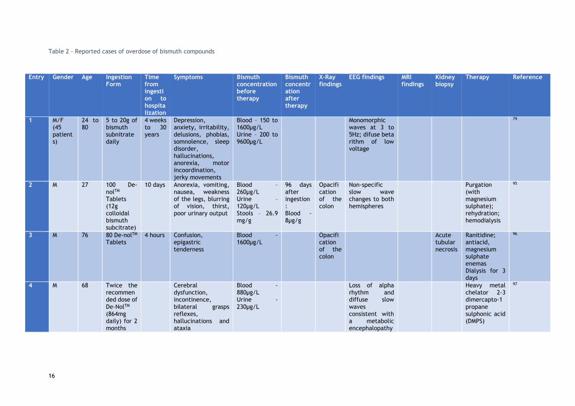
Table 1 - Bismuth compounds and its uses in medicine (adapted from 10) .......................... 3
Table 2 - Reported cases of overdose of bismuth compounds ....................................... 16
Table 3 - Reported cases of BIPP toxicity ............................................................... 19
Table 4 - Half maximal proliferation inhibitory concentration activity (IC50) values (µM) - 95%
confidence intervals, for an exposure to the compounds of 48 hours. ND – Not Defined ....... 27
Table 5 - Absorbances at 405nm of all the compounds and cysteine .............................. 34

xiv

xv
List of acronyms
BAL 2,3 – Dimercapto-1-propanol
BIPP Bismuth Iodoform Paraffin paste
CBS Colloidal Bismuth Subcitrate
CT Computerized tomography
DCF 2’,7’-dichlorofluorescein
DCFDA 2’,7’ –dichlorofluorescein diacetate
DMPS 2,3-Dimercapto-1-propanesulfonic acid
DNA Deoxyribonucleic acid
DTNB 5,5'-dithiobis-(2-nitrobenzoic acid)
EDTA Ethylenediamine tetraacetic acid
EEG Electroencephalography
FBS Fetal Bovine Serum
MBP Metal-binding protein
MRI Magnetic Ressonance Imaging
MTT 3-(4,5-dimethylthiazol-2-yl)-2,5-diphenyltetrazolium bromide
PBS Phosphate buffer saline
PI Propidium Iodide
ROS Reactive oxygen species
TBHP Tert-butyl hydroperoxide

xvi

1
1 Introduction
1.1 Bismuth It is thought that the name bismuth derives from the German word Weissmuth or Wismut, which
means white substance. Bismuth has an atomic mass of 208.980 and is the heaviest stable
element (83rd element of the periodic table, being the least abundant of the elements of the
Group 15)1. Bismuth is sometimes classified as a semi-metal or metalloid, since it has the
characteristics of a metal and possesses properties alike those of semiconductors and
insulators2. Bismuth is a relatively rare element, with an abundance comparable to that of
silver and mercury, although not quite as expensive since large amounts are recovered as a by-
product of copper and tin refining2. The world production of Bismuth in 2015 was above 13000
metric tons and the main producers were China and Vietnam3, as can be seen in figure 1.
Figure 1 - Bismuth mine production in 2015
Estimated world bismuth reserves suggest that China is the country with more abundance of
this element, followed by Vietnam, as shown in figure 2.
Bismuth is used in the most varied fields, and the most prominent use for this element is in low
melting alloys and metallurgical additives, including electronic and thermoelectric
applications4. Nonetheless bismuth is also used as a catalyst, in pharmaceuticals and industrial
chemicals and as a pearlescent pigment in cosmetics.
7 500
5 000
700
40 10 30
1 000
2 000
3 000
4 000
5 000
6 000
7 000
8 000
China Vietnam Mexico Russia Bolivia Canada

2
Figure 2 - Bismuth world reserves as of 2015 (metric tons)5
1.2 Bismuth environmental levels and exposure Concentrations of bismuth in rural air range from 0.1 to 0.6ng/m3 and in urban air this number
can be between 1 and 66ng/m3 4. The daily inhalation of bismuth is estimated to be less than
0.01-0.76µg 6.
In a recent review7 it was reported that bismuth concentrations vary from 10 to 30 ng/L in
seawater and from a few ng to a few µg/L in freshwater. As levels of bismuth in food are
relatively low, 0.1 to 1µg/kg, the exposure to bismuth through water and food is likely to be
minimal.
Bismuth levels in soil are roughly 1µg/kg, and in rocks like coal and sandstone, values range
from 0.1 to 3µg/kg8.
Exposure to bismuth can also occur through the use of cosmetics, as bismuth oxychloride is
present in some cosmetics9 mainly in those marketed as “mineral makeup”, because it presents
a distinct shimmery, pearlescent appearance and a fine white powder texture that adheres well
to the skin. Therefore, mostly pharmaceuticals but also cosmetics are a source of more
prolonged exposure to bismuth, but not to all population.
1.3 Bismuth in medicine
1.3.1 Bismuth compounds in medicine
Based in the gradually understood characteristics of this element, many bismuth compounds
have been prepared and some have clinical and health applications. Bismuth salts have been
used for over two centuries in the therapy of a large variety of clinical conditions including
dyspepsia, diarrhea, syphilis, oral and upper respiratory tract infections, verrucae and warts10.
240 000
53 000
10 000
10 000
5 00050 000
China Vietnam Bolivia Mexico Canada Other countries

3
The bismuth salts used over these two centuries have been diverse, including compounds such
as bismuth subcitrate, bismuth subsalicylate, bismuth subgallate and others that are shown in
table 1. The most relevant will be presented in the next subsections.
Table 1 - Bismuth compounds and its uses in medicine (adapted from 10)
Name Therapeutic end
Bismuth aluminate Antacid
Bismuth butylthiolaurate Antisyphilitic
Bismuth-D-camphocarboxylic acid salt basic Antisyphilitic
Bismuth chloride oxide Antisyphilitic
Bismuth ethyl camphorate Antisyphilitic
Bismuth iodide oxide Antimicrobial
Bismuth iodosubgallate Antimicrobial
Bismuth oxide Astringent
Bismuth phosphate Antacid
Bismuth potassium Antisyphilitic
Bismuth sodium iodide Antisyphilitic
Bismuth sodium tartrate Antisyphilitic
Bismuth sodium thioglycollamate Lupus erythematosus
Bismuth subcarbonate Astringent
Bismuth subcitrate Gastric and duodenal ulcers
Bismuth subgallate Astringent/Antacid
Bismuth subnitrate Antacid
Bismuth subsalicylate Lupus erythematosus/ Antidiarrhoeal
Bismuth tannate Astringent
Bismuth trobromophenate Antimicrobial
1.3.1.1 Bismuth subcitrate
Bismuth subcitrate is a mineral used in the treatment of ulcers. Other names for bismuth
subcitrate include colloidal bismuth subcitrate (CBS) and tripotassium dicitratobismuthate.
According to a report by the European Medicine Agency11 earlier in 2016, a drug containing this
compound was authorized in Portugal (and other countries), with the name PyleraTM, for the
treatment of peptic ulcers with infections by Helicobacter pylori.
1.3.1.2 Bismuth subsalicylate
This compound was initially administered as an intramuscular injection for the control of
syphilis12,13. This bismuth salt is nowadays used to treat heartburn, upset stomach, indigestion,
nausea, diarrhea or symptoms associated with excesses in eating and drinking. It’s used to
decrease the number of bowel movements and make the stool firmer. It is thought that this

4
salt may limit the secretion on the digestive tract, reduce inflammation in the stomach and
intestines, and inhibit the growth of certain bacteria and viruses that can cause intestinal tract
diseases14.
1.3.1.3 Bismuth subgallate
This compound is commonly used as an internal deodorant (for flatulence and stools).
1.3.1.4 Bismuth Iodoform Paraffin Paste
Bismuth Iodoform Paraffin Paste (BIPP) is an antiseptic agent that is widely used for packing
wounds and cavities in the ear, nose and throat, and maxillofacial surgery, since it acts as a
hemostatic agent, reduces wound colonization and promotes granulation tissue formation and
wound repair15.
Currently, the major medicinal use of bismuth compounds is focused in two fields: antimicrobial
and anticancer 16.
Bismuth can interact with nucleotides and with amino acids in peptides, enzymes and other
proteins, which are closely related to its uptake, accumulation, transport and excretion in the
human body, and to their antimicrobial and anticancer activities17,18.
There are currently 13 clinical trials with bismuth according to the Clinical Trial Registry19, the
majority of which concern its activity on Helicobacter pylori.
1.3.2 Bismuth as an antimicrobial and antiulcerous agent
For the past century bismuth compounds have been used in the treatment of various
gastrointestinal disorders and microbial infections such as syphilis, colitis, wound infection,
dyspepsia, diarrhea and peptic ulcers 20.
Bismuth subsalicylate, colloidal bismuth subcitrate and ranitidine bismuth citrate are used
worldwide to treat various gastrointestinal diseases which are related to the infection of
Helicobacter pylori 16. Helicobacter pylori can prevent ulcers from healing, so bismuth
compounds have also an anti-ulcer activity, due to the inhibition of the activity of this bacteria.
In addition, bismuth cam precipitate within the ulcer crater, leading to the formation of a
glycoprotein-bismuth complex, which acts as a protective coating and contributes to the
healing of the lesion 18.
1.3.3 Bismuth as an anticancer agent
Biocoordination studies of bismuth compounds argument that the main target are non-DNA
sites, which offers opportunities for new targeted approaches in the treatment of cancer 17,18,20–
52. Several synthetic bismuth molecules including organo- and inorgano- bismuth derivatives
have been prepared by a number of research groups and evaluated in their in vitro cytotoxic

5
or antiproliferative activities against various cancer cell lines. The bismuth derivatives include
bismuth dithiolates and dithiocarbamates, a water-soluble bismuth macrocycle complex,
heterocyclic organobismuth derivatives, triarylbismuth bis (carboxylates), tris(2-(N,N-
dimethylaminomethyl)phenyl) bismuth, and bismuth 8-quinolinethiolates 20–52. Several
compounds proved to have potent antiproliferative effects, which in some cases, are superior
to those observed with cisplatin and other classical anticancer agents 20.
A known strategy for cancer treatment is the use of targeted radiation therapy, which is an
approach mostly considered in inoperable tumors, tumors situated close to radiation sensitive
organs, metastatic disease, and diseases such as leukemia and lymphoma. This therapy involves
the use of carrier molecules, for example, antibodies (Ab) and peptides, specifically targeting
cancer cells, and a selected radionuclide that should emit controlled doses of ionizing radiation
to cancer cells without affecting healthy tissue surrounding them 53,54. The most important
variables that condition the selection of a specific radionuclide are its half-life and the
existence of viable chemistry for this use or viable supply 20. As 212Bi and 213Bi meet the baseline
parameters that define reasonable use within this context, these radionuclides are probably
the most studied ɑ-emitters in this type of therapy. These radionuclides can be stably bound
to several chelating agents that can be conjugated to monoclonal antibodies, peptides, or other
vectors without significant safety measures or shielding required. The in vivo stable
sequestration of 212Bi and 213Bi radionuclides is important to maximize their delivery of
radiation to tumors and to minimize renal toxicity and other toxic effects. Several research
groups have been developing 213Bi-based systems to make rational improvements on chelation
and/or radiolabeling chemistry, radionuclide delivery, targeting vectors, molecular targets and
therapeutic strategies and performing in vitro and in vivo studies in several different cancer
models 53,54.
Although 213Bi compounds have high interest in cancer treatment, the development of
radiotherapy involving this type of radionuclide has been limited by high costs, unresolved
chemistry, and its limited availability. Furthermore, the in vivo stability and metabolism of
these compounds is not well defined and radiologic side effects are still to be observed20.
1.4 Bismuth’s Pharmacokinetics
1.4.1 Absorption
The site of bismuth absorption in man has not yet been fully determined, but bismuth
compounds are thought to be somewhat absorbed through the respiratory and intestinal tracts,
depending on their solubility4, but there are no quantitative data. The majority of ingested
bismuth is not absorbed, but excreted mainly through the feces, and less than 1% of the
administered dose is absorbed following oral dosing with bismuth subsalicylate55, tripotassium
dicitrato bismuthate56 or ranitidine bismuth citrate57.

6
Some animal studies suggest that the absorption takes place in the small bowel, although the
rapid appearance of bismuth in blood after oral intake suggests bismuth can be absorbed in the
stomach58 Absorption through the skin is interesting, since bismuth compounds are used in
cosmetics, but, once more, there are no quantitative data. An interesting study showed the
rapid intake of bismuth into cells of the gastrointestinal tract and kidneys within hours of
exposure, and some weeks later bismuth was found on a number of organ systems59.
Bismuth is methylated by the bacterial flora in the gut, and excreted as bismuth sulfide, causing
the blackening of the feces and sometimes also of the oral mucosa60.
1.4.2 Distribution
It was demonstrated that after incubation of blood with radioactive bismuth citrate, 17% of the
radioactivity was associated with erythrocytes and the remainder underwent non-specific
binding to serum proteins61. A gel filtration study of human blood after incubation with bismuth
subgallate showed an association of bismuth with the high molecular fraction (≥200.000 daltons)
consisting of a α2-macroglobulin, IgM, β-lipoprotein and haptoglobulin62.
Regarding bismuth distribution in the tissues, the highest concentration/g wet weight was
always found in the kidney58. The retention time of bismuth in the kidney is longer than in any
other organ. In other organs, 144 hours after intravenous injection of 206bismuth citrate, 12% of
the injected dose remained in the kidneys and 0.9% in the bone61. It was also demonstrated a
retention in the kidneys of rabbits and dogs with soluble bismuth compounds63.
Lee et al. 64 found a distribution pattern after administration of colloidal bismuth subcitrate to
rats for 14 months. Bismuth concentrations were ordered from high to low in kidney (13.9µg/g
wet weight), lung, spleen, liver, brain and muscle (0.13µg/g wet weight).
In patients who died from bismuth encephalopathy the highest concentrations were found in
the thalamus and in the cerebral cortex, and additionally the concentration of bismuth in the
grey matter was twice as high as the one found in white matter65.
The knowledge that bismuth can be an effective inducer of metallothionein and that it can also
bind this protein, has been applied as a protective measure against the nephrotoxicity of
anticancer drugs such as cisplatin66–68 and doxorubicin69. These groups observed in tumor-
bearing mice and patients with renal cell carcinoma that orally administered bismuth was
transported to normal tissues and not to cancerous tissues. Bismuth induction of
metallothionein has been linked to an attenuation of the teratogenic effects of cadmium in
mice70 and the adverse effects of gamma irradiation on the bone marrows of mice71.
The formation of trimethylbismuth in humans following ingestion of bismuth subcitrate was also
reported72, and a later study by others in this group showed that HepG2 cells were capable of
methylating bismuth subcitrate and bismuth cysteine but not bismuth glutathione73.

7
After a single subcutaneous injection with BiCl3 in rats, it was demonstrated that bismuth binds
to high molecular weight proteins in the kidney, but after repeated injections nearly all bismuth
bound to a low molecular weight bismuth-metal-binding protein (Bi-MBP)58. Bismuth induces
metal-binding protein (MBP) synthesis in the kidney.
The presence of intracellular particles after in vitro incubation of macrophages with colloidal
bismuth subcitrate or bismuth subnitrate was demonstrated by light and electron
microscopy74,75. Concentrations of colloidal bismuth subcitrate above 160µmol/L inhibit the in
vitro migration of macrophages from spleen fragments; these effects indicate a possible
intracellular cytotoxic effect of subcitrate particles on macrophages after phagocytosis74.
After subcutaneous administration of high doses of bismuth subnitrate to rats, it was
demonstrated that the mitochondria in the liver and proximal renal tubules underwent
morphological changes, resembling swelling and distortion of the inner mitochondrial
membrane58.
1.4.3 Excretion
The majority of ingested bismuth is not absorbed, but excreted mainly through the feces, model
values of bismuth elimination, with a daily intake of 20µg are a fecal elimination of 18µg, and
a urinary excretion of 1.6µg76.
The main elimination ways for absorbed bismuth is renal excretion, although biliary excretion
may also be important77.
The placenta is permeable to bismuth after intramuscular injection of potassium bismuth
tartrate and sodium potassium tartro-bismuthate into pregnant rabbits and cats78.
1.5 Bismuth toxicity
Despite the many beneficial qualities of bismuth, a variety of side effects, including
neurological syndromes, have been recorded. The best documented case of its neurotoxicity
was the outbreak of bismuth encephalopathy among numerous patients in France 79. Bismuth
accumulation has been shown in several cell types, including kidney cells 80, motor neurons 81,
ganglion cells 82 and Leydig cells 83. In all this cases, bismuth was found to be located in
lysosomes, which play a vital role in heavy metal metabolism. Intralysosomal bismuth induces
lysosomal rupture and decreased numbers of intact lysosomes 84.
Bismuth toxicity may develop due to excessive ingestion, or misuse when taken in large
quantities and for a prolonged period of time 85.
The reported toxic effects caused by an overdose of bismuth compounds include
encephalopathy, nephropathy, osteoarthropathy, gingivostomatitis, and colitis 86. Bismuth
poisoning mostly affects the kidney, liver and bladder. Chronic exposure to high levels of
bismuth salts result in encephalopathy, whereas acute toxicity manifests as nephrotoxicity 87.

8
1.5.1 In vitro studies
Some studies have been made regarding the cytotoxicity of bismuth compounds, for example,
Stoltenberg et al. 84 studied the bismuth uptake in the lysosomes of a histiocytic lymphoma cell
line (J774 cells). These cells were exposed to several concentrations of bismuth citrate (5, 25,
100 and 200 µM) and different times of exposure (6, 12 and 24 hours) were evaluated. The
authors concluded that cells exposed to concentrations higher than 5µM became less attached
as a function of increasing exposure times. Damaged cells with disintegrated membranes were
seen after an exposure of 12 or more hours to 100µM of bismuth citrate, and after only 6 hours
of exposure to the concentration of 200µM 84.
The cellular uptake and the cytotoxic and genotoxic effects of bismuth compounds, namely
monomethylbismuth, bismuth citrate and bismuth glutathione has been investigated by Von
Recklinghausen et al. 60. This group used HepG2 cells, human lymphocytes and human
erythrocytes. Their results showed that the uptake of bismuth glutathione, was relatively low
(<0,3%) in all these three cell types. On the other hand, the uptake of bismuth citrate by
lymphocytes and erythrocytes was 2.6% and 6.5%, respectively, whereas the uptake of methyl
bismuth was significantly higher (up to 23% by lymphocytes and 36% by erythrocytes).
In the Trypan Blue cytotoxicity test the most significant results were found with methyl
bismuth60. In the hepatocytes, the cytotoxic effect was noteworthy after methyl bismuth
treatment for 1 hour at concentrations above 350μM and after an exposure of 24h at
concentrations above 130μM. In erythrocytes after 24 hours of exposure methyl bismuth was
highly toxic at concentrations above 3.8μM (>50% cell death). In lymphocytes, methyl bismuth
showed cytotoxicity only at high concentrations an exposure time (>430μM, 24h). After 24 hours
of exposure, bismuth citrate showed cytotoxic effects in erythrocytes at concentrations above
113μM (48% cell death). This research group also established that after exposure of lymphocytes
to methyl bismuth, chromosomal type aberrations occurred (single and double strand breaks).
The results of Von Recklinghausen et al. 60 showed that the methylated bismuth compound was
more membrane permeable and more cytotoxic than bismuth glutathione and bismuth citrate.
Dopp et al. 88 tested trimethylbismuth in Caco-2, CHO-9 and HepG2 cell lines. Their results
showed that trimethylbismuth was cytotoxic in all three tested cell lines. Caco-2 cells were the
most sensitive (LC50: 110μmol/Lgv), followed by CHO-9 (LC50: 128 μmol/Lgv) and HepG2 cells
(LC50: 194 μmol/Lgv).
The in vitro cytotoxicity of bismuth nanoparticles in HeLa and MG-63 cells was studied by Luo
et al. 89 They concluded that HeLa cells are more prone to suffer cytotoxicity effects of various
surface modified bismuth nanoparticles than MG-63 cells. Using the 3-(4,5-dimethylthiazol-2-
yl)-2,5-diphenyltetrazolium bromide (MTT) assay they observed that the viabilities of HeLa cells
decreased with the increase of the concentration of bismuth nanoparticles.
The in vitro neurotoxicity of bismuth ferrite nanoparticles on PC12 cells was evaluated by Song
et al. 90 with a MTT assay. The results showed that cytotoxicity was dose-dependent, as cell
vitality of the groups exposed for 3 hours, decreased from 95% to 73% with increasing exposure

9
concentrations from 10 to 200 μg/mL. The cell vitality further dropped to 65% when the
concentration reached 500 μg/mL. The extent and mode of cell death has been assessed by the
annexin V-FITC apoptosis detection kit, which evidenced that only a small percentage of cells
undergo apoptosis (below 2%) and necrosis (below 10%) after exposure with bismuth ferrite
nanoparticles in concentrations ranging from 50 to 200 μg/mL.
Using a human skin derived cell line, HaCaT keratinocytes, Gao et al. 91 showed that bismuth
oxybromide induced a concentration-dependent loss of cell viability. Using the cytometric
analysis of annexin-V/PI, the authors observed that bismuth oxybromide triggered mainly late
apoptosis. Bismuth oxybromide caused disturbances in plasma membrane, and lead to a loss of
membrane integrity and eventually cell death.
1.5.2 In vivo studies
Here we present a data collection of some in vivo studies concerning the toxic effects bismuth
might produce.
Bismuth pellets gained popularity when the use of lead in shotgun pellets was forbidden, thus
Pamphlett et al.81 making use of the autometallographic (AMG) technique searched for bismuth
that could be released from shotgun pellets that had been inserted into mice. The pellets were
inserted into the peritoneal cavity, through an incision in the abdominal cavity of the mice.
And this group evidenced that bismuth was present in the cerebrum (in neurons in the
supraoptic, paraventricular, suprachiasmatic and arcuate nuclei), brain stem (neurons of the
trochlear, oculomotor, mesencephalic trigeminal, abducens, facial and hypoglossal nuclei),
spinal cord (cell bodies of large motor neurons), posterior root ganglia and in cell bodies of
renal tubular cells, macrophages in the lung, and dendritic cells in the liver and spleen.
Stoltenberg et al. 83 investigated the detectable bismuth in testis of rats exposed to bismuth
subnitrate using the AMG technique. This group found traces of bismuth in the interstitial tissue
as well as in the seminiferous tubules; and an abundance of bismuth was also found in Leydig
cells. The same research group 82 also aimed to determine whether bismuth is transported in
motor and sensory axons by retrograde axonal transport. For this, bismuth subnitrate was
intramuscularly injected in Wistar rats, and 3 days after the injection, this group detected
bismuth in motor neurons of the ipsilateral spinal cord and in ganglion cells of the corresponding
dorsal root ganglia. Bismuth was found to be located in lysosome-like organelles. To the best
of our knowledge Stoltenberg et al. 82 were the first group to show that bismuth can access the
nervous system by retrograde axonal transport.
The gastrointestinal absorption and systemic uptake of bismuth citrate or ranitidine bismuth
citrate after oral exposure in female mice has been studied by Larsen et al. 59, again using the
AMG technique. This group observed that bismuth is present and absorbed in gastrointestinal
epithelial cells shortly after exposure as showed by bismuth staining in gastric, duodenal and
epithelial cells. Using electron microscopy, the authors showed that bismuth was only seen in

10
lysosomes, and that higher bismuth concentrations increased the number of cells with signs of
toxic degradation: cytoplasmic vacuolation and intracellular swelling.
Sano et al. 92 evaluated the toxicity of elemental bismuth in rats by an acute oral toxicity study
and a 28-day repeated oral administration study. This group found no abnormal clinical signs in
both of their studies. They predicted that the adverse toxic effects of bismuth as a simple metal
substance would be low, when compared with the adverse effects of lead.
A 13-week intratracheal intermittent administration of bismuth study has also been reported
by the same research group 93. Low, medium and high dose levels of bismuth were tested (0.8,
4 and 20 mg/kg) and no abnormal clinical signs attributable to bismuth administration were
found. However, hair loss was detected in 3 animals, in the medium and high dose levels, and
suppression of body weight gain from day 29 forward, in the high dose level, but without
statistical significance. A slight increase in erythrocyte count and mean cell hemoglobin
concentration was also observed as well as pathological changes in the lungs and bronchial
lymph nodes. Brown patches were observed in the lungs of animals of all dose levels. Black
patches and lung collapses were detected in all animals from the two groups with higher
concentrations of bismuth (4 and 20 mg/Kg). Enlargement of bronchial lymph nodes and a white
patch in the liver was also observed in animals of all groups. These results show that bismuth
inhalation can cause dose-dependent but not specific adverse effects.
1.5.3 Clinical evidences Exposure to bismuth can cause renal failure associated with degeneration and necrosis of the
epithelium of the renal proximal tubules, necrosis of the liver, reversible dysfunction of the
nervous system, skin eruptions, and pigmentation of the gums and intestine 4.
In order to fully comprehend the scope of bismuth toxicity it is necessary to do a summary of
the most recent clinical cases, namely those of overdoses with bismuth compounds.
A case was even reported of some side effects believed to be due to the use of bismuth in skin
creams 94. In fact, two patients presented intellectual impairment, memory loss, confusion,
tremulousness, clumsiness, difficulty walking and myoclonic jerks. Bismuth was found in
cerebral venous blood in both patients, and in the cerebrospinal fluid in one. It is proposed that
bismuth can cross the blood/brain barrier and disturb oxidative cerebral metabolism 94.
1.5.3.1 Bismuth antiulcer agents
Probably the most notorious case of bismuth toxicity in man is the French outbreak of bismuth
encephalopathy. In this context, Supino-Viterbo et al. 79 reported the case of 45 patients (8
male, 37 female) to which they had the opportunity of performing EEG studies, and study the
clinical symptomatology in depth. All 45 patients had been treated with an insoluble bismuth
salt (subnitrate) between 5 and 20 grams daily, over a period of 4 weeks going to 30 years. The
blood bismuth levels (table 2, entry 1), taken on the same day as the EEG, ranged between 150
11
to 1600 µg/L (normal being less than 20µg/L). In urine samples the levels of bismuth were from
200 to 9600µg/L.
Hudson et al. 95 reported the case of a 27 year old man admitted 4 hours after an overdose of
100 tablets of De-NolTM (12g of CBS), paracetamol and alcohol. The day after the patient was
discharged as he felt normal. After 10 days the patient was admitted once more, complaining
of anorexia, nausea, vomiting, general malaise, weakness of his legs, blurring of vision, thirst
and poor urinary output. The patient showed no signs of encephalopathy. Some biochemical
tests were made, and this group found that their patient had a blood bismuth level of 260µg/l
and a urine bismuth level of 120µg/l (table 2, entry 2). An abdominal X-ray was performed, and
it showed an opacification of the colon by ingested bismuth. An EEG showed non-specific slow
wave changes over both hemispheres. The patient was diagnosed with renal failure and
neurotoxicity induced by bismuth. The patient started hemodialysis and just five days later the
renal function had returned to normal and the neurological signs were resolved.
A case of a 76 year old man that overdosed on 80 De-NolTM tablets 4 hours prior to admission
was reported by Taylor et al. 96 They performed an X-Ray that showed opacification of the
colon; just as the case reported by Hudson et al. 95. The authors detected 1600µg/l of bismuth
in the blood (table 2, entry 3). The patient had already vomited in his home and in the
emergency department and, for that reason, the gastric lavage was not performed. They noted
that the patient was oliguric, and four hours later he began passing bloody stools. They started
the patient on ranitidine, antacid and magnesium sulphate enemas. The patient was also
dialyzed for 3 days, during which time he continued to pass bloody stools, and received a blood
transfusion. He then developed acute abdominal pain with absent bowel sounds, but he was
judged unfit for surgery and died 4 days later. Necropsy revealed a perforated duodenal ulcer
and “pale kidneys” which proved to contain bismuth (11mg/g and 16mg/g) 96.
Other case reports have been described in medical literature, all evidencing that ingestion of
high amounts of bismuth salts (> 5 grams) led to gastrointestinal, renal and neurological injury.
For instance, Playford et al. 97 reported a case of a 68 year old man (table 2, entry 4) that for
two months took double the recommended dose of De-NolTM (864mg bismuth a day). On
examination the patient evidenced cerebral dysfunction, incontinence, bilateral grasp reflexes,
visual hallucinations and ataxia. An EEG demonstrated loss of alpha rhythm and diffuse slow
waves consistent with a metabolic encephalopathy. The metal chelator 2-3 dimercapto-1-
propane sulphonic acid (DMPS) was administered for 10 days and the patient´s EEG was normal
after six weeks.
Tubular necrosis has been diagnosed in young adults after ingestion of toxic doses of bismuth
compounds. Huwez et al. 98 reported the case of a 21 year-old man admitted 3 hours after
ingesting 39 tablets of bismuth subcitrate (table 2, entry 5). The patient showed epigastric pain
and an intravenous crystalloid infusion was prescribed, but over the next 2 days the urinary
output fell and the renal function deteriorated. The renal biopsy revealed moderate acute
tubular necrosis, although no bismuth was detected in the biopsy specimen. The patient was
treated with intravenous frusemide, dopamine, mannitol and crystalloids.
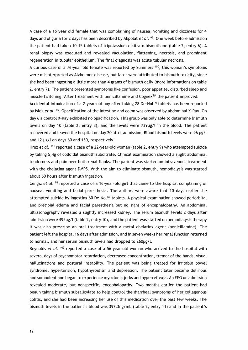
12
A case of a 16 year old female that was complaining of nausea, vomiting and dizziness for 4
days and oliguria for 2 days has been described by Akpolat et al. 99. One week before admission
the patient had taken 10-15 tablets of tripotassium dicitrato bismuthane (table 2, entry 6). A
renal biopsy was executed and revealed vacuolation, flattening, necrosis, and prominent
regeneration in tubular epithelium. The final diagnosis was acute tubular necrosis.
A curious case of a 76-year old female was reported by Summers 100; this woman’s symptoms
were misinterpreted as Alzheimer disease, but later were attributed to bismuth toxicity, since
she had been ingesting a little more than 4 grams of bismuth daily (more informations on table
2, entry 7). The patient presented symptoms like confusion, poor appetite, disturbed sleep and
muscle twitching. After treatment with penicillamine and CognexTM the patient improved.
Accidental intoxication of a 2-year-old boy after taking 28 De-NolTM tablets has been reported
by Islek et al. 85. Opacification of the intestine and colon was observed by abdominal X-Ray. On
day 6 a control X-Ray exhibited no opacification. This group was only able to determine bismuth
levels on day 10 (table 2, entry 8), and the levels were 739µg/l in the blood. The patient
recovered and leaved the hospital on day 20 after admission. Blood bismuth levels were 96 µg/l
and 12 µg/l on days 60 and 150, respectively.
Hruz et al. 101 reported a case of a 22-year-old woman (table 2, entry 9) who attempted suicide
by taking 5,4g of colloidal bismuth subcitrate. Clinical examination showed a slight abdominal
tenderness and pain over both renal flanks. The patient was started on intravenous treatment
with the chelating agent DMPS. With the aim to eliminate bismuth, hemodialysis was started
about 60 hours after bismuth ingestion.
Cengiz et al. 86 reported a case of a 16-year-old girl that came to the hospital complaining of
nausea, vomiting and facial paresthesia. The authors were aware that 10 days earlier she
attempted suicide by ingesting 60 De-NolTM tablets. A physical examination showed periorbital
and pretibial edema and facial paresthesia but no signs of encephalopathy. An abdominal
ultrasonography revealed a slightly increased kidney. The serum bismuth levels 2 days after
admission were 495µg/l (table 2, entry 10), and the patient was started on hemodialysis therapy
It was also prescribe an oral treatment with a metal chelating agent (penicillamine). The
patient left the hospital 16 days after admission, and in seven weeks her renal function returned
to normal, and her serum bismuth levels had dropped to 260µg/l.
Reynolds et al. 102 reported a case of a 56-year-old woman who arrived to the hospital with
several days of psychomotor retardation, decreased concentration, tremor of the hands, visual
hallucinations and postural instability. The patient was being treated for irritable bowel
syndrome, hypertension, hypothyroidism and depression. The patient later became delirious
and somnolent and began to experience myoclonic jerks and hyperreflexia. An EEG on admission
revealed moderate, but nonspecific, encephalopathy. Two months earlier the patient had
begun taking bismuth subsalicylate to help control the diarrheal symptoms of her collagenous
colitis, and she had been increasing her use of this medication over the past few weeks. The
bismuth levels in the patient’s blood was 397.3ng/mL (table 2, entry 11) and in the patient’s

13
urine was 292.5ng/mL. The bismuth subsalicylate was held, and in the next two days the patient
became more alert, had decreased myoclonus, and exhibited less muscular rigidity.
The case of a 21-year-old woman who was brought to the hospital 4 hours after taking 20 tablets
of (CBS)in a suicide attempt (table 2, entry 12) was reported by Erden et al. 87 A gastric lavage
was performed and the patient received intravenous fluid therapy. An abdominal
ultrasonography demonstrated slightly increased echogenicity in the renal parenchyma. The
patient became oliguric and then anuric. Blood chemistry and urine sediment showed signs of
proximal tubular dysfunction with hypophosphatemia, hypouricemia, metabolic acidosis, and
renal glycosuria despite normal plasma glucose concentration. The patient was started on a
chelating agent, sodium-2,3-dimercapto-1-propanol, and hemodialysis. After 15 days the
patient was discharged, but 8 weeks after discharge the patient’s renal function test results
remained high and the patient remained on hemodialysis for 1 year.
Akinci et al. 103 reported the case of a 16-year-old girl that came to the hospital 1 hour after
taking 19 grams of bismuth subcitrate potassium (De-NolTM) in a suicidal attempt. She had no
physical complaint and was conscious on admission. A gastric lavage was performed and the
abdominal X-Ray showed opacity, so a whole bowel irrigation was performed. On the third day
of admission the patient developed acute renal failure, metabolic acidosis and oliguria. The
patient began hemodialysis following catheterization through the jugular vein. Bicarbonated
dialysis were performed on the patient until the acute renal failure improved. Since the third
day the patient suffered from sore throat, and on examination a bilateral tonsillar ulceration
was found. On the 13th day the patient became polyuric, as a daily average of 15L of urine
were excreted. On the 15th of admission the patient developed altered mental state, and the
neurological examination revealed confusion, somnolence and cortical blindness. On the
magnetic resonance imaging (MRI) scan, hyper-intense signal alterations were observed at the
levels of bilateral parietal vertices of both cerebellar hemispheres. And intermittent rhythmic
waves were detected in the frontal region on an encephalography examination. A neurologist
diagnosed this patient with toxic metabolic encephalopathy. On the 20th day of admission the
laboratory parameter of the patient began to normalize.
1.5.3.2 Bismuth iodoform paraffin paste
Bismuth iodoform paraffin paste (BIPP) contains two active ingredients, bismuth subnitrate and
iodoform, and is used to pack cavities in ear, nose, throat, dental and neurosurgical practice.
It is believed that BIPP acts as an antiseptic and astringent.
Sharma et al. 104 reported a case of a 57-year-old-woman who in May of 1991 got a basal cell
carcinoma removed, and large areas of dura matter where exposed bilaterally with the
intervening sagittal sinus. All were packed with BIPP. On July the patient became confused and
agitated with intermittent bihemispheric signs, and eventually lapsed into a coma. A computed
tomography (CT) scan of the brain showed diffuse cerebral oedema in both parieto-occipital
lobes. In December the BIPP pack was finally removed, and the patient showed a progressive
return to full alertness, rapport, cognition and coordinated bodily activity. In later December

14
a CT scan showed complete resolution of the cerebral oedema but also showed some patchy
areas of high attenuation on the right parieto-occipital cortex subjacent to the exposed dura
matter. Later a large BIPP pack was reapplied on order to obtain a clean granular bed for later
grafting. After this reapplication of a BIPP pack the patient once again became confused,
restless, dysarthric and insomnolent. On April of the next year the patient showed rapid
deterioration in her conscious level, and became unresponsive. Only then the possibility of
bismuth toxicity was considered, and the BIPP pack was removed. At this time blood bismuth
concentration was 52ng/L (table 3, entry 1). The patient’s conscious level improved, with the
blood bismuth concentration falling to almost half by May. An MRI scan showed extensive
cerebral oedema and hyperintense areas in the dura mater, central white matter, and
periventricular ependymal lining. A more recent CT scan showed cerebral atrophy, but no
evidence of tumor.
In another case105 it was reported the situation of an 86-year-old women who was admitted to
the hospital for a partial maxillectomy (as we can see in table 3, entry 2). The patient
underwent the surgery with split skin grafting to the maxillary antrum, which was packed with
BIPP. Five days after the surgery the patient was exhausted, lightheaded and unsteady. On day
seven, the patient returned to the operating table for the replacement of the BIPP pack. The
patient became increasingly aggressive, and on day 11 she was barely eating and having various
fainting episodes. They did a CT brain scan, which was normal and electrolytes, liver function
tests and full blood scan, were also normal. On day 14 the BIPP pack was removed, the patient
was still confused and aggressive but 7 days after the removal of the BIPP pack the patient
began to improve, and being cooperative; the patient was discharged 5 days later.
Three cases of allergic contact otitis externa due to BIPP was reported by Roest et al. 106. All
three cases were women who had their external auditory meatus and concha packed with BIPP-
impregnated gauze following surgery. More information can be seen in table 3, entry 3.
Youngman et al. 107 described the case of an 81-year-old-man who suffered from epistaxis, and
after 4 days of nasal packing, hemostasis wasn’t achieved, and the patient underwent surgery.
Two days after the surgery the patient’s condition deteriorated, he became acutely confused,
he also developed dysphagia, and was becoming incontinent. The surgeon had used nasal
packing with BIPP when prolonged packing with MerocelTM failed to stop the epistaxis. The
patient’s serum bismuth level was 250µg/L (table 3, entry 4). This team stated that bismuth
toxicity was the most likely cause of his temporary, but prolonged state of confusion.
A case of a 67-year-old man with a sacral chondroma that was surgically resected and after
some troubles with the post-op the wound was irrigated with saline and packed with gauze
soaked in BIPP was reported by Ovaska et al. 108 Five days after the packing with BIPP the
patient became acutely confused, disorientated, delusional, and verbally aggressive to the
staff. He was also suffering from abdominal discomfort, nausea and tremor, even though no
cerebellar signs were present. By day 10 the patient’s condition was deteriorating and bismuth
toxicity was suspected as the patient had developed myoclonic jerks with intermittent episodes
of drowsiness and worsening confusion. The blood and urine concentrations of bismuth were
15
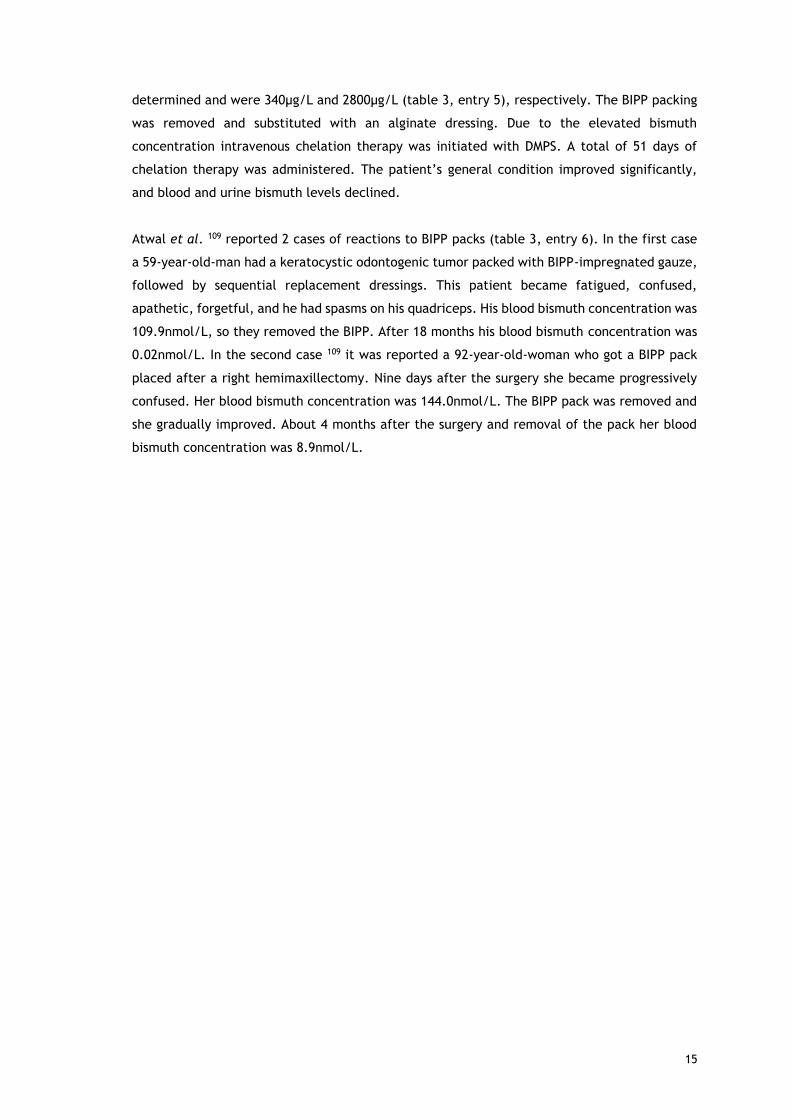
determined and were 340µg/L and 2800µg/L (table 3, entry 5), respectively. The BIPP packing
was removed and substituted with an alginate dressing. Due to the elevated bismuth
concentration intravenous chelation therapy was initiated with DMPS. A total of 51 days of
chelation therapy was administered. The patient’s general condition improved significantly,
and blood and urine bismuth levels declined.
Atwal et al. 109 reported 2 cases of reactions to BIPP packs (table 3, entry 6). In the first case
a 59-year-old-man had a keratocystic odontogenic tumor packed with BIPP-impregnated gauze,
followed by sequential replacement dressings. This patient became fatigued, confused,
apathetic, forgetful, and he had spasms on his quadriceps. His blood bismuth concentration was
109.9nmol/L, so they removed the BIPP. After 18 months his blood bismuth concentration was
0.02nmol/L. In the second case 109 it was reported a 92-year-old-woman who got a BIPP pack
placed after a right hemimaxillectomy. Nine days after the surgery she became progressively
confused. Her blood bismuth concentration was 144.0nmol/L. The BIPP pack was removed and
she gradually improved. About 4 months after the surgery and removal of the pack her blood
bismuth concentration was 8.9nmol/L.
16
Table 2 - Reported cases of overdose of bismuth compounds
Entry Gender Age Ingestion Form
Time from ingestion to hospitalization
Symptoms
Bismuth concentration before therapy
Bismuth concentration after therapy
X-Ray findings
EEG findings MRI findings
Kidney biopsy
Therapy Reference
1 M/F (45 patients)
24 to 80
5 to 20g of bismuth subnitrate daily
4 weeks to 30 years
Depression, anxiety, irritability, delusions, phobias, somnolence, sleep disorder, hallucinations, anorexia, motor incoordination, jerky movements
Blood – 150 to 1600µg/L Urine – 200 to 9600µg/L
Monomorphic waves at 3 to 5Hz; difuse beta rithm of low voltage
79
2 M 27 100 De-nolTM Tablets (12g colloidal bismuth subcitrate)
10 days Anorexia, vomiting, nausea, weakness of the legs, blurring of vision, thirst, poor urinary output
Blood – 260µg/L Urine – 120µg/L Stools – 26.9 mg/g
96 days after ingestion: Blood - 8µg/g
Opacification of the colon
Non-specific slow wave changes to both hemispheres
Purgation (with magnesium sulphate); rehydration; hemodialysis
95
3 M 76 80 De-nolTM Tablets
4 hours Confusion, epigastric tenderness
Blood - 1600µg/L
Opacification of the colon
Acute tubular necrosis
Ranitidine; antiacid, magnesium sulphate enemas Dialysis for 3 days
96
4 M 68 Twice the recommended dose of De-NolTM (864mg daily) for 2 months
Cerebral dysfunction, incontinence, bilateral grasps reflexes, hallucinations and ataxia
Blood - 880µg/L Urine - 230µg/L
Loss of alpha rhythm and diffuse slow waves consistent with a metabolic encephalopathy
Heavy metal chelator 2-3 dimercapto-1 propane sulphonic acid (DMPS)
97

17
Table 2 - (Continued)
Entry Gender Ag
e Ingestion Form
Time from ingestion to hospitalization
Symptoms
Bismuth concentration before therapy
Bismuth concentration after therapy
X-Ray findings
EEG findings MRI findings Kidney biopsy
Therapy Reference
5 M 21 39 tablets of bismuth subcitrate
3 hours Epigastric pain Blood - ~ 200µg/L Serum - ~ 1500µg/L
Blood - ~125 Serum- ~10
Acute tubular necrosis
Intravenous frusemide, dopamine, mannitol and crystalloids
98
6 F 16 10-15 tablets of tripotassium dicitrato bismuthate
1 week Nausea, vomiting, dizziness and oliguria
Acute tubular necrosis
Hemodialysis, protein restriction, metoclopramide and aluminum hydroxide
99
7 F 76 Pepto-bismolTM (4.14mg daily for 7 years)
Confusion, poor appetite, disturbed sleep, muscle twitching
On day 6: Serum - 242µg/L
After 30 days: Serum - 90µg/L After 76 days: Serum - 14µg/L
Normal Moderate atrophy, ventricular enlargement and ischemic white matter disease
Penicillamine, oral fluids, salt tablets, Cognex (Tacrine)
100
8 M 2 28 De-nolTM tablets (8.4g of colloidal bismuth subcitrate)
6 hours On day 10: Blood - 739µg/L Urine – 693µg/L
Day 105: Blood - 12µg/L
Opacification of the intestine and colon
Normal Gastric lavage, IV saline, mannitol, furosemide
85

18
Table 2 - (Continued)
Entry Gender
Age
Ingestion Form
Time from ingestion to hospitalization
Symptoms
Bismuth concentration before therapy
Bismuth concentration after therapy
X-Ray findings
EEG findings
MRI findings Kidney biopsy
Therapy Reference
9 F 22 5.4g of colloidal bismuth subcitrate
2 hours Day 3: Serum – 640µg/L
Day 11: Serum - 12µg/L
Enlarged and edematous kidneys with thinning of the cortical area
DMPS, hemodialysis, hemodiafiltrations
101
10 F 16 60 De-nolTM tablets
10 days Nausea, vomiting and facial paresthesia
Day 12: Serum- 495µg/L
Day 64: Serum - 260µg/L
Normal Hemodialysis, penicillamine
86
11 F 56 45mL (thrice per day) of bismuth subsalicylate (262mg/15mL)
Psychomotor retardation, decreased concentration, tremor of the hands, visual hallucinations and postural instability
Blood – 397.3ng/ml Urine – 292.5ng/ml
Moderate but nonspecific encephalopathy
Medication was held (bismuth subsalicylate)
102
12 F 21 20 colloidal bismuth subcitrate tablets (300mg of CBS)
4 hours Normal Normal Gastric lavage, intravenous fluids, DMPS, hemodialysis
87
13 F 16 19 grams of De-nolTM
1 hour Hyper.intense signal alterations at the level of bilateral parietal vertices of both cerebellar hemispheres
Opacities in the left side of abdomen
Intermittent rhythmic waves in the frontal region
Hyper-intense signal alterations at the levels of bilateral parietal vertices of both cerebellar hemispheres
103

19
Table 3 - Reported cases of BIPP toxicity
Entry Gender Age Operation/surgery Symptoms after packing with BIPP Bismuth levels Observations Reference
1 F 57 Removal of a basal cell carcinoma
Agitation, confusion, restlessness 52ng/L 104
2 F 86 Partial maxillectomy Exhaustion, lightheadedness, poor appetite, tremor
Day 14 – 146nmol/L Day 22 – 81nmol/L
105
3 F 16 Myringoplasty Mild erythema and swelling of the concha
Allergic contact otitis externa due to BIPP
106
F 13 Myringoplasty Allergic contact otitis externa due to BIPP
F 52 Mastoidectomy Florid eczematous reaction Allergic contact otitis externa due to BIPP
4 M 81 Epistaxis treatment with BIPP packing
Acute confusion, dysphagia 250µg/L 107
5 M 67 Resection of a sacral chondroma
Acute confusion, disorientation, delusions, aggressive, abdominal discomfort, nausea, tremor
Blood – 240 µg/L Urine – 2800 µg/L
108
6 M 59 Marsupialisationa nd packing with BIPP of a keratocystic odontogenic tumour
Fatigue, confusion, apathy, forgetfulness and spasms in the quadriceps
Blood- 109.9nmol/L After 18 months blood bismuth concentration was 0.02nmol/L
109
F 92 Right hemimaxillectomy Confusion Blood – 144.0nmol/L After 4 months blood bismuth concentration was 8.9nmol/L

20
1.6 Treatment of bismuth poisoning
The contact with bismuth preparations should be stopped promptly in case of accidental or
deliberate overdosing. According to the clinical cases reviewed above, elimination of bismuth
from the body may be improved by hemodialysis, diuresis and the use of chelating agents, such
as sodium-2,3-dimercapto-1-propanol (BAL), penicillamine and DMPS87,97,101.

21
2 Objectives Regarding all the information above, our main aim was to evaluate the toxicity of bismuth
compounds commonly used in therapy and as catalysts in organic transformations. Seeing that
so many clinical cases of side effects of bismuth compounds were reported, we set our goal to
determine if, and to what extent, bismuth compounds are indeed toxic. For that purpose, the
evaluation of the cytotoxicity of ten bismuth compounds by the colorimetric 3-(4,5-
dimethylthiazol-2-yl)-2,5-diphenyltetrazolium bromide (MTT) assay, was performed. Flow
cytometry studies using propidium iodide (PI) staining were also executed, to clarify the results
of the previous study. The 2’,7’ –dichlorofluorescein diacetate (DCFDA) study was also
implemented in order to comprehend if bismuth compounds could perhaps lead to the
production of reactive oxygen species (ROS).
Organic compounds, such as thiols, could enhance the solubility of bismuth compounds.110 In
addition, the toxicity of bismuth compounds has been partially associated to its coupling with
endogenous thiols. For this reason, the Ellman’s method was applied in order to realize if some
bismuth compounds couple to thiols more than others, and in that fashion perceive if we could
relate these results to the potential cytotoxicities of those compounds.

22
3 Materials and Methods
3.1 Compounds In this project 10 bismuth compounds were studied:
× (B1) Bismuth(III) triflate, from Sigma-Aldrich
× (B2) Bismuth(III) nitrate, from Sigma-Aldrich
× (B3) Bismuth(III) chloride, from Fluka
× (B4) Bismuth oxychloride, from Acros Organics
× (B5) Bismuth(III) oxide, from Acros Organics
× (B6) Bismuth(III) subnitrate, from Sigma-Aldrich
× (B7) Bismuth(III) subsalicylate, from Sigma-Aldrich
× (B8) Bismuth(III) citrate, from Sigma-Aldrich
× (B9) Bismuth(III) gallate basic hydrate, from Sigma-Aldrich
× (B10) Bismuth carbonate oxide, from Alfa Aesar
The solutions of the compounds were all freshly prepared, for each single experiment. All the
compounds were diluted in mili-Q water to obtain a concentration of 10mM. This solution was
then sonicated 30 to 45 minutes at a temperature of 40 to 60ºC, to favor the solubility of the
compound91. From this mother-solution appropriate dilutions of the compounds, in the different
concentrations needed, were prepared in complete culture medium, before each experience.
These prepared solutions were, once again, sonicated for 10 to 15 minutes at approximately
37ºC, so that then they could be applied to the cells.
3.2 Experimental Procedures
3.2.1 Biological Evaluation
3.2.1.1 Cell Cultures
In this study the cell cultures used were epithelial cells from a human colorectal
adenocarcinoma (Caco-2) from passages 69 to 70, epithelial cells from a mammary gland
adenocarcinoma (MCF-7) from passages 22 to 23, cells from a hepatoma of a female patient
with cirrhosis subsequent to hepatitis C virus infection (HepaRG), kindly provided by Professor
Gilberto Alves, from passages 15 to 18, non-carcinogenic human dermal fibroblasts (NHDF) from
passages 11 to 13 as well as rat dopaminergic neural cells (N27) from passages 4 to 14, all
acquired from the American Type Culture Collection (ATCC; Manassas, VA, USA). The chemicals
(of analytical grade), assay reagents, culture mediums and supplements were all obtained from
Sigma-Aldrich.
All the cell lines were cultured in 75cm3 or 175cm3 culture flasks, and maintained at 37ºC in a
humidified atmosphere incubator with 5% CO2.

23
Caco-2 cells were cultured in high-glucose Dulbecco’s modified Eagle medium (DMEM)
supplemented with 10% FBS and 1% antibiotic mixture of 10,000 U/mL penicillin G, 100mg/mL
streptomycin.
MCF-7 cells were cultured in high-glucose Dulbecco’s modified Eagle medium (DMEM)
supplemented with 10% fetal bovine serum (FBS), and 1% antibiotic/antimycotic (10,000 U/mL
penicillin G, 100 mg/mL streptomycin and 25 µg/mL anfothericin B).
HepaRG cells were cultured in Williams’ medium E, supplemented with 10% FBS, 500µL/L
insulin, 0.08mM hydrocortisone and 1% antibiotic mixture of 10,000 U/mL penicillin G,
100mg/mL streptomycin.
NHDF cells were cultured in RPMI 1640 medium supplemented with 10%FBS, 2mM L-glutamine,
10mM HEPES, 1mM sodium pyruvate and 1% antibiotic/antimycotic (10,000 U/mL penicillin G,
100 mg/mL streptomycin and 25µg/mL anfothericin B.
N27 cells were cultured in RPMI 1640 medium with 10% FBS and 1% of antibiotic mixture of
10,000 U/mL penicillin G, 100mg/mL streptomycin.
For all cell types, the medium was renewed every 2-3 days until the cells reached approximately
80-90% of confluence, at that moment the cells were detached from the culture flask by gentle
trypsinization, 125mg/L trypsin in phosphate buffer solution (PBS) and 0.02g/L ethylenediamine
tetraacetic acid (EDTA), and before the experiments, viable cells were counted with the trypan-
blue exclusion assay and adequately diluted in complete culture medium.
3.2.1.2 MTT cell proliferation assay
After the process of trypsinization, and cell counting, 96-well plates (Nunc, Apogent, Denmark)
were seeded with a cellular suspension with density of 2×10⁴ cells/mL, with 100µL per well,
and left to adhere for 48 hours. After that, the medium was replaced by the solutions of the
compounds for the concentration-response studies (0.01, 0.1, 1, 10, 100 µM) in the appropriate
medium for 3 and 48 hours. Untreated cells, to which the initial medium was replaced by fresh
medium, were used as negative controls. Each experiment was performed in quintuplicate and
independently repeated at least two times.
The in vitro antiproliferative effects were evaluated by the MTT 3-(4,5-dimethylthiazol-2-yl)-
2,5-diphenyltetrazolium bromide) assay, by measuring the extent of the MTT reduction. After
the incubation periods (3 and 48-hours) the medium was removed, 100µL per well of phosphate
buffer saline (NaCl 137mM; KCl 2.7mM, Na2HPO4 10mM and KH2PO4 1.8mM in distilled water and
pH adjusted to 7.4) were used to wash the cells and then 100µL of the MTT solution (5mg/mL),
prepared in serum free medium, were added to each well, followed by a 4-hour incubation at
37ºC. After the incubation period, the MTT containing medium was carefully removed and the
formazan crystals were dissolved in DMSO. The absorbance was measured at 570nm using Bio-
Rad xMarkTM microplate spectrophotometer. Cell viability values were expressed as relative
percentages of the absorbance in comparison with the respective controls.
24
3.2.1.3 DCFDA assay
After the process of tripsinization, and cell counting, in a 96-well black plate (Greiner) with a
clear bottom, 100µL per well were seeded with a cellular suspension of 2.5×10⁴ cells/mL, and
left to adhere for approximately 24 hours.
In one of the experiments, after the adherence time (24 hours), each well was washed with
PBS and then the cells were stained with PBS containing 20µM of DCFDA (diluted from a stock
solution in dimethyl sulfoxide)111; unstained cells serve as negative controls. The microplate
was then incubated for 45 minutes in the dark at 37ºC. After the incubation period, the DCFDA
solution was removed and the wells were washed once more with PBS. After that the previously
diluted compounds of interest were added to the plates, and left to incubate for 6 hours. For
this specific assay the compounds were diluted in PBS supplemented with 2% serum, instead of
culture medium. Tert-butyl hydroperoxide (TBHP) was also diluted to a concentration of 50µM,
as a positive control. After the incubation period, the pate was read in a fluorescence plate
reader (Spectra Max Gemini EM, Molecular devices), in the presence of the compounds, with
excitation wavelength at 485nm and emission wavelength at 535nm.
As DCFDA is not stable for more than 6 hours, in order to test the compounds for a longer period
(24 hours), some adjustments were made. The compounds were diluted in complete culture
medium, and wells with medium and without cells served as blanks. After the 24-hour
adherence period the cells were treated with the compound of interest (100µL per well) and
the positive control compound (TBHP), and left to incubate for another 24 hours. 1 hour prior
to completion of the treatment, DCFDA was diluted at two times the desired concentration (20
µM) in culture medium, and overlaid on top of the treated cells, 100µL per well. The plate was
then incubated for 30 to 45 minutes at 37ºC. At the end of the incubation period the plate was
read in the fluorescence plate reader, without washing, in the presence of compounds with
excitation wavelength at 485nm and emission wavelength at 535nm.
3.2.1.4 Flow cytometry
With the flow cytometry technique, the cell viability was analyzed after staining the dead cells
with propidium iodide (PI). The cells were seeded in a 12-well culture plate, with a density of
3×10⁴cells/mL, at 1mL per well. After 24 hours the cells were treated with compounds B1 and
B6, at a concentration of 50µM, for another 24 hours. Untreated cells were used as controls. At
the end of the incubation period and before the flow cytometry assay, the effects of the
compounds on the cell’s morphology was performed through an optic microscope (Olympus
CKX41) coupled to a digital camera (Olympus SP-500UZ) and several photographs were taken
(Zoom:100x). After that, the supernatant of each well was collected and pooled with the cells
harvested by tripsinization. The resulting cell suspension was kept on ice and pelleted by
centrifugation, the pellet was then resuspended in 400µL of complete medium.
Subsequently, 397.5µL of the cell suspension were transferred to a FACS tube containing 2.5 µL
of a solution of propidium iodide (Invitrogen) at 1mg/mL, and left to act for at least 5 minutes

25
protected from the light. A minimum of 20 000 events were acquired using a FACSCalibur flow
cytometer using FSC, SSC and FL3 channels. Both the acquisition and the analysis were
performed using the software CellQuestTMPro. In order to analyze the results a region (R4) was
created (not shown) on the SSC/FSC contour plots to exclude part of the debris. At the FSC/FL3
contour plot, gated on R4, three additional regions were created: R1, concerning viable cells;
R2, representative of dead cells; and last R3, which represents an intermediate population.
The percentage of events was calculated relating the number of events in each region with the
total number of events on R1, R2 and R3.
3.2.2 Thiols Quantification
With the Ellman’s method we tried to determine if some bismuth compounds coupled to thiols
from cysteine, more than others.
First a calibration curve of cysteine was determined. Two concentrations of each compound
were prepared from the mother-solution (10mM), at 82 and 123µM, in the reaction buffer, 0.1M
sodium phosphate, ph 8.0, containing 1mM EDTA.
Then, in each well of a clear 96-well microplate (Greiner bio-one), 100µL of reaction buffer
plus 2µL of Ellman’s Reagent solution (4mg/mL of reaction buffer) were placed, and 10µL of
each compound solution and 10 µL of the chosen cysteine solution (100 µM) were also placed in
3 wells each. The plate was mixed and incubated at room temperature for 15minutes. After
the incubation time the absorbance was read at 405nm using the Bio-Rad xMarkTM microplate
spectrophotometer.
3.2.3 Statistics In the MTT assays each experiment was performed in quintuplicate and independently repeated
at least two times. The flow cytometry studies were performed in triplicate and independently
repeated at least two times. The results of those assays were expressed as average±standard
deviation. The t-Student test was applied to determine statistical significance (p<0.05) in the
cell proliferation results. These calculations were performed using the Microsoft Excell 2010
software. The IC50 values were calculated from the concentration-response curve by a sigmoid
fitting, with a confidence interval of 95%. The cell viability in the flow cytometry studies, in
regions R1, R2 and R3 was calculated with a confidence interval of 95%.
In the DCFDA assay each experiment was performed in quadruplicate and independently
repeated at least two times. The results of this assay were expressed as average±standard
deviation. Again, the t-Student test was applied to determine statistical significance (p<0.05).
26
4 Results and discussion
4.1 MTT assay The anti-proliferative evaluation of the compounds’ cytotoxicity was executed through the MTT
assay, involving cell culture, the subsequent cell treatment with the compounds and the MTT
assay, and the posterior analysis. The biological evaluation through the MTT assay is a reliable
procedure, being widely used112.
The MTT molecule is reduced to succinate dehydrogenase in viable cells, resulting in formazan,
a purple water insoluble derivative. For this reason, the biosynthesis of formazan is considered
proportional to the cell number, therefore the more intense the purple color is, the greater
the number of living cells. The intensity of the purple color is then quantified
spectrophotometrically113.
The cell types included in the study are widely used, and are specifically pertinent in this study
since, as stated before, bismuth is thought to accumulate namely in the intestine and liver59
and can cross the blood brain barrier94.
All compounds were tested via concentration-response curve studies, done with the five cell
types mentioned, in two time frames, 3 and 48 hours of exposure to the compounds.
None of the compounds tested appeared to cause a decrease in cellular viability after 3 hours
of exposure, which demonstrated that the compounds tested do not induce acute toxicity in
the cells used. On the contrary, with a 48-hour exposure to the compounds, some effects are
noticeable. It is noteworthy that N27 cells are the most affected by the compounds, in
particular compound B1 and B6. In the Caco-2 cell line only the compound B6 had a limited
effect. In the HepaRG cell line some compounds do present a IC50, compound B6 and compound
B9, but a high one. In the MCF-7 cell line, none of the compounds was cytotoxic. At last, in the
NHDF cell line, compound B4 had a lower IC50. A group60 showed that bismuth citrate did not
induce cytotoxic effects on hepatocytes, which can be congruent with our studies since for
compound 8 (bismuth citrate) we found no IC50.
Dopp et al.88 found that Caco-2 cells were more sensitive to methylated bismuth than hepatoma
or ovary cells. In addition, Song et al.90 found that bismuth ferrite was toxic only at an exposure
of 3 hours, prolonged that time, they concluded that the cells could recover to a certain extent
with incubation time. Another study91 reported that bismuth oxybromide induced a loss of cell
viability in human keratinocyte cells.
Since compounds B1 and B6 were the most promising, in the N27 cell line, the following studies
were made with these compounds and this cell line.

27
Table 4 - Half maximal proliferation inhibitory concentration activity (IC50) values (µM) - 95% confidence intervals, for an exposure to the compounds of 48 hours. ND – Not Defined
Compound Caco-2 N27 HepaRG MCF-7 NHDF
IC50 R2 IC50 R2 IC50 R2 IC50 R2 IC50 R2
B1 >100 - 14.22 0.99 >100 - >100 - ND
B2 >100 - 21.06 0.99 >100 - >100 - ND
B3 >100 - >100 - >100 - >100 - 88.15 0.87
B4 >100 - >100 - >100 - >100 - 46.2 0.87
B5 >100 - >100 - ND >100 - >100 -
B6 50.84 0.93 14.01 0.98 89.05 0.99 >100 - >100 -
B7 >100 - >100 - ND ND >100 -
B8 >100 - 32.69 0.96 >100 - >100 - 72.05 0.95
B9 >100 - >100 - 83.19 0.86 ND >100 -
B10 >100 - >100 - >100 - >100 - >100 -
4.2 DCFDA assay
The DCFDA – cellular reactive oxygen species detection assay uses the cell permeant reagent
DCFDA, a fluorogenic dye that measures hydroxyl, peroxyl and other ROS presence within the
cell114. After diffusion into the cell, DCFDA is deacetylated by cellular esterases to a non-
fluorescent compound, which is later oxidized by ROS into 2’,7’-dichlorofluorescein (DCF)115.
This compound in highly fluorescent, and can be detected by fluorescence spectroscopy with
maximum excitation and emission spectra of 495nm and 529nm, respectively114.
When ROS production increases and overwhelms the cellular antioxidant capacity, it can induce
macromolecular damage, by reacting with DNA proteins and lipids, which can lead to apoptosis
or necrosis116. The increase of ROS production can also disrupt thiol redox circuits, which can
lead to aberrant cell signaling and dysfunctional redox control117.
In graphic 3, it can be seen the ROS detection on N27 cells after treatment during 6 hours to
the compounds B1 and B2 and to TBHP, the positive control. It can be seen that compound B1
appears to not have a significant effect in ROS production, unlike compound B6 which appears
to have a low but statistically significant increase in its lower concentration.
In graphic 4, the response of N27 cells to an exposure of 24 hours to compound B6and TBHP
(positive control) is presented, and it can be observed that this compound lead to a significant
increase in the production of ROS in its highest concentration, which almost matches the
achieved with TBHP. Gao et al. demonstrated the generation of ROS in human keratinocyte
cells for bismuth oxybromide. The production of ROS by compound B6 could be related to its
effects on cellular viability, considering that elevated ROS production could lead to apoptosis
or necrosis.

28
Figure 3 - DCFDA assay with N27 cells, exposure to the compounds of 6 hours. Results expressed in mean± standard deviation). TBHP was used as positive control. * p<0.05 in relation to a negative control (t-student test).
0
50
100
150
200
250
Cellsstained with
DCFDA
B1 10µM B1 100µM B6 10µM B6 100µM TBHP 50µM Cellswithoutstaining
*
*
v
*
v
-20
0
20
40
60
80
100
120
140
Cells stainedwith DCFDA
B6 10µM B6 100µM TBHP 50µM Cells withoutstaining
*
v
*
v
*
v
*
v
*
v
*
v
Figure 4 - DCFDA assay with N27 cells, exposure to the compounds of 24 hours. Results expressed in mean± standard deviation). TBHP was used as positive control * p<0.05 in relation to a negative control (t-student test).

29
4.3 Flow cytometry
The effects of compounds B1 and B6 were chosen to be further studied by flow cytometry
studies, as they presented pronounced effects on cell proliferation, particularly in the N27 cell
line. To analyze cell viability after treatment with these two compounds, flow cytometry assays
were performed on N27 cells. Propidium iodide (PI) was applied to identify dead cells, since it
is able to permeate comprised cell membranes. This compound intercalates DNA and emits
fluorescence proportional to the DNA content of the cell118.
Before flow cytometry analysis, to study cell morphology at 24 hours of exposure to the
compounds, photographs were taken (Figure5) . In relation to the control, it is quite evident
that both compounds induced a diminution of the cell number. Additionally, the cell
morphology is clearly altered. In the control the cells are all in their differentiated form,
elongated. With both treatments with compounds B1 and B6, the morphology of the cells is
evidently changed. With the treatment with compound B1 some cells remain “normal” but
numerous cells have an altered, rounded, morphology. With the treatment with compound B6
a few cells remain with a normal appearance, but numerous cells have a clearly altered
morphology, it is also noticeable some precipitated compound, which is explained by the low
solubility of bismuth compounds in aqueous solutions.

30
AA
BA
CA
Figure 5 - Morphology of N27 cells after 24 hours of treatment; A - cells not treated (control); B - cells treated with compound B1; C - cells treated with compound B6. Zoom: 100x

31
The results of the flow cytometry assay were analyzed. Representative contour plots of FSC
intensity versus PI intensity, a view of size of events against the fluorescence at 24h, are shown
in figure 6. The events are divided in three different regions, R1, R2 and R3. In the R1
population, in general, the events have higher size and negative fluorescence, being considered
viable cells. Usually the R2 population presents lower size and a positive fluorescence. R3
population has intermediate size and fluorescence, being considered early apoptotic or oncotic
cells, cell debris, or autoflourescent cells. This area cannot be discarded, since it has a
considerable number of events. Events out of these three main areas were not considered in
the analysis.
Compound B1 Compound B6
Control
Figure 6 - Contour plots resulting of the analysis of the non-stained/stained cells with PI (size of events versus intensity of fluorescence) 24 hours after cell treatment; R1 - living cells; R2 - dead cells; R3 - intermediate population.

32
Analyzing figure 7, it is noteworthy that with both compounds there was a statistically
significant decrease in the number of events at R1. It should also be noted that there was a
statistically significant increase of dead cells, represented by the R2 region, with the exposure
to both compounds B1 and B6. In the R3, intermediate region it can be seen a statistically
significant increase of events with the exposure to compound B6. As referred previously, this
can be due to early apoptotic or oncotic cells, cell debris, autoflourescent cells or the
precipitated compound, that can be seen in figure 5(C). This data is congruent with the MTT
assay data and the DCFDA assay data, in consideration of the diminution of the cell viability,
and with the increased production of ROS, with compound B6. A hypothesis can be made here,
perhaps the increase of ROS production lead to apoptosis, thus loss of cell viability, so in the
flow cytometry studies a decrease, in relation to the control, of viable cells exposed to
compound B6 (figure 7, R1) and an increase of the intermediate population (figure 7, R3) was
observed.
Re
lati
ve
ce
ll n
um
be
r (
%)
R1
R2
R3
0
5 0
1 0 0
1 5 0c o n tro l
B 1
B 6**
** *
Figure 7 - Effects on cell viability at 24 hours of exposure to compounds B1 and B6 - results for the three regions (R1, R2 and R3). * statistically significant in relation to the control.
4.4 Thiols quantification
The DTND-thiols assay measures sulfhydryl groups with the thiol reagent 5,5'-dithiobis-(2-
nitrobenzoic acid) (DTNB), which forms a mixed disulfide and the 5-thionitrobenzoic acid, a
measurable yellow-colored product119. Sulfhydryl groups may be estimated in a sample by
comparison to a standard curve composed of known concentrations of a sulfhydryl-containing
compound, such as cysteine. The coupling of bismuth to organic compounds such as cysteine

33
can modify bismuth solubility and therefor it can perhaps also increase the toxicity of said
compound120.
First the cysteine calibration curve was determined, as can be seen in figure 8. Testing the two
concentrations from each compound showed us that none of the compounds couple more than
others to the thiols from cysteine (see table 5), which is rather strange, because we expected
to see at least some differences between compounds. All the absorbances remain quite close
to the one from the utilized cysteine concentration, which indicates, as stated above, that the
compounds are not coupling with cysteine thiols in different degrees, in these experimental
consitions, as would be expected.
We cannot achieve a conclusion in this case, since more studies should be done. A deduction
could only be made if some bismuth compounds coupled with the cysteine thiols more than
others, and only in that case could a relation be established between that coupling and the
observed toxicities.
C is te ín e s ta n d a r d c u r v e
C is te ín e c o n c e n tra t io n (µ M )
Ab
s (
40
5n
m)
0 5 0 1 0 0 1 5 0
0 .0
0 .5
1 .0
1 .5
2 .0
2 .5
Y = 0.01503*X + 0.5123
R2= 0 .9 2 1 7
Figure 8 - Cysteine calibration curve
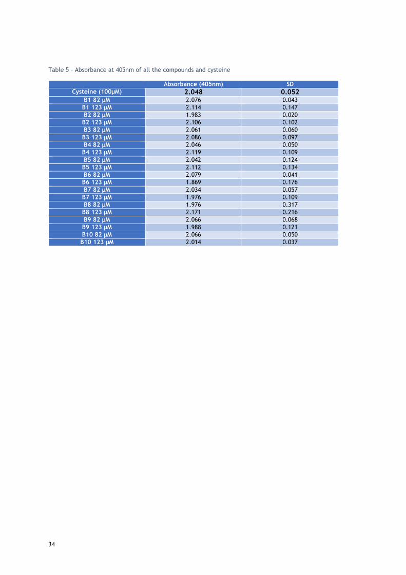
34
Table 5 - Absorbance at 405nm of all the compounds and cysteine
Absorbance (405nm) SD
Cysteine (100µM) 2.048 0.052 B1 82 µM 2.076 0.043
B1 123 µM 2.114 0.147
B2 82 µM 1.983 0.020
B2 123 µM 2.106 0.102
B3 82 µM 2.061 0.060
B3 123 µM 2.086 0.097
B4 82 µM 2.046 0.050
B4 123 µM 2.119 0.109
B5 82 µM 2.042 0.124
B5 123 µM 2.112 0.134
B6 82 µM 2.079 0.041
B6 123 µM 1.869 0.176
B7 82 µM 2.034 0.057
B7 123 µM 1.976 0.109
B8 82 µM 1.976 0.317
B8 123 µM 2.171 0.216
B9 82 µM 2.066 0.068
B9 123 µM 1.988 0.121
B10 82 µM 2.066 0.050
B10 123 µM 2.014 0.037
35
5 Conclusions and future work In summary, cellular proliferation assays, the reactive oxygen species detection assays, and
flow cytometry assays were all executed in order to evaluate the cytotoxicity of the ten bismuth
compounds tested in this project and possibly the mechanism of the cytotoxicity. It can be
concluded that most of the compounds tested do not exhibit a toxic profile in the cell cultures
used for the experiments. But, the cellular cultures used in this study are only a small sample
of the cell types that should be tested, particularly more studies should be made in the future
with renal cells. Nonetheless, two compounds revealed a toxic profile in the N27 cell line and
the Caco-2 cell line, showing that perhaps some more studies should be done, in order to fully
comprehend the extent of the toxicity of bismuth compounds, as they do not seem to be as
innocuous as they have been portrayed in the past. The two compounds with the most
antiproliferative effects were B1 and B6, Bismuth(III) triflate and Bismuth(III) subnitrate,
respectively; they were also tested by flow cytometry with propidium iodide staining, which
showed some cellular death, which is congruent with the reactive oxygen species detection
assay results, that showed some oxidative stress.
The study performed with the Ellman’s reagent did not produce relevant results. With the 15-
minute incubation period there seemed to not be any differences between the coupling of the
different bismuth compounds to the cysteine thiols. So in the future this experiment should be
repeated, perhaps with a longer incubation period or a different thiol source, to maybe
correlate the possible toxicity of bismuth compounds to their augmented solubility by the
coupling to thiols.

36
37
6 References
1. Mohan, R. Green bismuth. Nat. Chem. 2, 336–336 (2010).
2. Suzuki, H. & Ogawa, T. Organobismuth(III) Compounds. Organobismuth Chem. 21–245 (2001).
3. Bismuth production top countries 2010-2015 | Statistic. Statista (2016). Available at:
http://www.statista.com/statistics/264975/production-of-bismuth/. (Accessed: 12th September
2016)
4. Fowler, B. A., Sullivan Jr, D. W. & Sexton, M. J. Chapter 31 - Bismuth. Handbook on the Toxicology
of Metals (Fourth Edition) I, (2015).
5. World bismuth reserves top countries 2015 | Statistic. Statista (2016). Available at:
http://www.statista.com/statistics/264978/reserves-of-bismuth-by-major-countries/.
(Accessed: 12th September 2016)
6. Woolrich, P. F. Occurence of Trace Metals in the Environment An Overview. Am. Ind. Hyg. Assoc.
J. 34, 217–226 (1973).
7. Filella, M. et al. How reliable are environmental data on ‘orphan’ elements? The case of bismuth
concentrations in surface waters. J. Environ. Monit. 12, 90–109 (2010).
8. Bowen, H. J. M. Trace Elements in Biochemistry. (Academic Press, 1966).
9. U.S. Food and Drug administration. Cosmetic Ingredient Listing. (Accessed: 13th September 2016)
10. Bradley, B., Singleton, M. & Po, A. L. W. Review Article Bismuth Toxicity-a Reassessment. J. Clin.
Pharm. Ther. 423–441 (1989).
11. European Medicines Agency’s Commitee on Medicinal Produtcs for Human Use. List of nationally
authorised medicinal products Active substance : cilostazol. 44, (2016).
12. Leonard, C. S. Studies in the pharmacology of bismuth salts. Tissue distribution of bismuth. J.
Pharmacol. Exp. Ther. 333–345 (1928).
13. McCarthy, F. & Dexter, S. Oral manifestations of bismuth. N. Engl. J. Med. 345–353
14. Bismuth subsalicylate: Indications, Side Effects, Warnings - Drugs.com. Available at:
https://www.drugs.com/cdi/bismuth-subsalicylate.html. (Accessed: 13th September 2016)
15. Chevretton, E. B., McRae, R. D. & Booth, J. B. Mastoidectomy packs: Xeroform or BIPP? J.
Laryngol. Otol. 105, 916–7 (1991).
16. Yang, N. & Sun, H. Biocoordination chemistry of bismuth: Recent advances. Coord. Chem. Rev.
251, 2354–2366 (2007).
17. Ge, R. & Sun, H. Bioinorganic chemistry of bismuth and antimony: Target sites of metallodrugs.
Acc. Chem. Res. 40, 267–274 (2007).
18. N, Y. & Sun, H. Biological Chemistry of Arsenic, Antimony and Bismuth. Biological chemistry of
Arsenic, Antimony and Bismuth (John Wiley & Sons Ltd, 2011).
19. Clinical Trials register - Search for bismuth. Available at:
https://www.clinicaltrialsregister.eu/ctr-search/search?query=bismuth. (Accessed: 12th
38
September 2016)
20. Salvador, J. A., Figueiredo, S. A., Pinto, R. M. & Silvestre, S. M. Bismuth compounds in medicinal
chemistry. Future Med. Chem. 4, 1495–1523 (2012).
21. Gabbiani, C. et al. Outstanding plasmodicidal properties within a small panel of metallic
compounds: Hints for the development of new metal-based antimalarials. J. Inorg. Biochem. 103,
310–312 (2009).
22. Andrews, P. C. et al. Anti-Leishmanial activity of homo- and heteroleptic bismuth(III)
carboxylates. J. Inorg. Biochem. 105, 454–461 (2011).
23. Yang, N. et al. Bismuth complexes inhibit the SARS coronavirus. Angew. Chemie - Int. Ed. 46,
6464–6468 (2007).
24. Li, H. & Sun, H. Recent advances in bioinorganic chemistry of bismuth. Curr. Opin. Chem. Biol.
16, 74–83 (2012).
25. Silvestru, C., Breunig, H. J. & Althaus, H. Structural Chemistry of Bismuth Compounds. I.
Organobismuth Derivatives. Chem. Rev. 99, 3277–3327 (1999).
26. Li, W. et al. Structure of colloidal bismuth subcitrate (CBS) in dilute HCl: Unique assembly of
bismuth citrate dinuclear units ([Bi(cit)2Bi] 2-). J. Am. Chem. Soc. 125, 12408–12409 (2003).
27. Andrews, P. C. et al. Towards a structural understanding of the anti-ulcer and anti-gastritis drug
bismuth subsalicylate. Angew. Chemie - Int. Ed. 45, 5638–5642 (2006).
28. Chey, W. D., Wong, B. C. Y. & Practice Parameters Committee of the American College of
Gastroenterology. American College of Gastroenterology Guideline on the Management of
Helicobacter pylori Infection. Am. J. Gastroenterol. 102, 1808–1825 (2007).
29. Malfertheiner, P. et al. Current concepts in the management of Helicobacter pylori infection: The
Maastricht III Consensus Report. Gut 56, 772–81 (2007).
30. Malfertheiner, P. et al. Helicobacter pylori eradication with a capsule containing bismuth
subcitrate potassium, metronidazole, and tetracycline given with omeprazole versus
clarithromycin-based triple therapy: A randomised, open-label, non-inferiority, phase 3 trial.
Lancet 377, 905–913 (2011).
31. Tsang, C. N., Ho, K. S., Sun, H. & Chan, W. T. Tracking bismuth antiulcer drug uptake in single
helicobacter pylori cells. J. Am. Chem. Soc. 133, 7355–7357 (2011).
32. Ge, R. et al. A proteomic approach for the identification of bismuth-binding proteins in
Helicobacter pylori. J. Biol. Inorg. Chem. 12, 831–842 (2007).
33. Cun, S. & Sun, H. A zinc-binding site by negative selection induces metallodrug susceptibility in
an essential chaperonin. Proc. Natl. Acad. Sci. U. S. A. 107, 4943–4948 (2010).
34. Chen, R. et al. Fabrication of bismuth subcarbonate nanotube arrays from bismuth citrate. Chem.
Commun. (Camb). 2265–7 (2006). doi:10.1039/b601764a
35. Chen, R. et al. Bismuth subcarbonate nanoparticles fabricated by water-in-oil microemulsion-
assisted hydrothermal process exhibit anti-Helicobacter pylori properties. Mater. Res. Bull. 45,
654–658 (2010).
36. Shaikh, A. R., Giridhar, R., Megraud, F. & Yadav, M. R. Metalloantibiotics: synthesis,
39
characterization and antimicrobial evaluation of bismuth-fluoroquinolone complexes against
Helicobacter pylori. Acta Pharm. 59, 259–271 (2009).
37. Andrews, P. C. et al. Bismuth(III) complexes derived from non-steroidal anti-inflammatory drugs
and their activity against Helicobacter pylori. Dalton Trans. 39, 2861–8 (2010).
38. Andrews, P. C. et al. Bismuth(III) 5-sulfosalicylate complexes: structure, solubility and activity
against Helicobacter pylori. Dalton Trans. 28, 6377–84 (2009).
39. Andrews, P. C. et al. Structural and solution studies of phenylbismuth(III) sulfonate complexes
and their activity against Helicobacter pylori. Dalton Trans. 39, 9633–41 (2010).
40. Kotani, T. et al. Antibacterial Properties of Some Cyclic Organobismuth ( III ) Compounds.
Antimicrob. Agents Chemother. 49, 2729–2734 (2005).
41. Chauhan, H. P. S., Shaik, N. M. & Singh, U. P. Synthetic, spectroscopic and antimicrobial studies
of bis(dialkyldithiocarbamato)diorganodithiophosphatobismuth(III) complexes. Appl. Organomet.
Chem. 19, 1132–1139 (2005).
42. Chauhan, H. P. S., Bakshi, A. & Bhatiya, S. Synthetic, spectral as well as in vitro antimicrobial
studies on some bismuth(III) bis ( N , N -dialkyldithiocarbamato) alkylenedithiophosphates. Appl.
Organomet. Chem. 24, 317–325 (2010).
43. Tripathi, U. N., Siddiqui, A. & Solanki, J. S. Synthesis , Spectral Characterization , and
Antimicrobial Activity of Arsenic ( III ) and Bismuth ( III ) phenyl ) pyrazolinates ]. 33, 257–266
(2009).
44. Solanki, J. S., Thapak, T. R. & Bhardwaj, A. Synthesis , structural characterization , and in vitro
antimicrobial properties of salicylate and pyrazoline complexes of bismuth ( III ). J. Coord. Chem.
64, 369–376 (2011).
45. Dawara, L. & Singh, R. V. Microwave-assisted synthesis, characterization, antimicrobial, and
pesticidal activity of bismuth and antimony complexes with coumarin-based ligands. J. Coord.
Chem. 64, 931–941 (2011).
46. Shaikh, A. R., Giridhar, R. & Yadav, M. R. Bismuth-norfloxacin complex: Synthesis,
physicochemical and antimicrobial evaluation. Int. J. Pharm. 332, 24–30 (2007).
47. Halwani, M. et al. Liposomal bismuth-ethanedithiol formulation enhances antimicrobial activity
of tobramycin. Int. J. Pharm. 358, 278–284 (2008).
48. Alipour, M., Suntres, Z. E., Lafrenie, R. M. & Omri, A. Attenuation of Pseudomonas aeruginosa
virulence factors and biofilms by co-encapsulation of bismuth-ethanedithiol with tobramycin in
liposomes. J. Antimicrob. Chemother. 65, 684–693 (2010).
49. Halwani, M. et al. Bismuth-thiol incorporation enhances biological activities of liposomal
tobramycin against bacterial biofilm and quorum sensing molecules production by Pseudomonas
aeruginosa. Int. J. Pharm. 373, 141–146 (2009).
50. Murafuji, T. et al. Antifungal activity of organobismuth compounds against the yeast
Saccharomyces cerevisiae: Structure-activity relationship. J. Inorg. Biochem. 98, 547–552 (2004).
51. Murafuji, T. et al. Bismuth heterocycles based on a diphenyl sulfone scaffold: Synthesis and
substituent effect on the antifungal activity against Saccharomyces cerevisiae. Eur. J. Med. Chem.
46, 519–525 (2011).

40
52. Sousa, M. C. & Poiares-Da-Silva, J. Cytotoxicity induced by bismuth subcitrate in Giardia lamblia
trophozoites. Toxicol. Vitr. 13, 591–598 (1999).
53. Kim, Y. S. & Brechbiel, M. W. An overview of targeted alpha therapy. Tumor Biol. 33, 573–590
(2012).
54. Brechbiel, M. W. Targeted α-therapy: past, present, future? Dalt. Trans. 4918 (2007).
55. Bierer, D. Bismuth subsalycilate. History, chemistry, and safety. Rev Infect Dis 12, 3–8 (1990).
56. Treiber, G., Gladziwa, U. & Ittel, T. Tripotassium dicitrato bismuthate: absorption and urinary
excretion of bismuth in patients with normal and impaired renal function. Pharmacol Ther 491–
502 (1991).
57. Lacey, L., Frazer, N., Keene, O. & Smith, J. Comparative pharmacokinetics of bismuth from
ranitidine bismuth citrate (GR122311X), a novel anti-ulcerant, and tri-potassium dicitrato
bismuthate (TDB). Eur J Clin Pharmacol 177–180 (1994).
58. Slikkerveer, A. & de Wolff, F. A. Pharmacokinetics and toxicity of bismuth compounds. Med.
Toxicol. Adverse Drug Exp. 4, 303–23
59. Larsen, A., Martiny, N., Stoltenberg, M., Danscher, G. & Rungby, J. Gastrointestinal and Systemic
Uptake of Bismuth in Mice after Oral Exposure. Pharmacol. Toxicol. 93, 82–90 (2003).
60. Von Recklinghausen, U. et al. Methylated bismuth, but not bismuth citrate or bismuth glutathione,
induces cyto- and genotoxic effects in human cells in vitro. Chem. Res. Toxicol. 21, 1219–1228
(2008).
61. Russ, G., Bigler, R., Tilbury, R., Woodard, H. & Laughlin, J. Metabolic studies with radiobismuth,
I: retention and distribution of 206Bi in the normal rat. Radiat. Res. 443–454 (1975).
62. Thomas, D. W., Sobecki, S., Hartley, T. F., Coyle, P. & Alp, M. H. Chemical toxicology and clinical
chemistry of metals. (Academic Press, 1983).
63. Sollmann, T., Seifter, J., Mangun, G. H., Ramvouzec, E. S. & O’Hare, J. A. Intravenous injections
of soluble bismuth compounds: their toxicity, and their sojourn in the blood and organs. J.
Pharmacol. Exp. Ther. 74, 134–154 (1942).
64. Lee, S. P., Lim, T. H., Pybus, J. & Clarke, A. C. Tissue distribution of arally administred bismuth
in the rat. Clin. Exp. Pharmacol. Physiol. 7, 319–324 (1980).
65. Escourolle, R., Bourdon, R., Galli, A., Galle, P. & Jaudon, M. C. Étude neuropathologique et
toxicologique de douze cas d’encéphalopathie bismuthique. Rev. Neurol. (Paris). 153–163 (1977).
66. Kondo, Y., Satoh, M., Imura, N. & Akimoto, M. Effect of bismuth nitrate given in combination with
cis-diamminedichloroplatinum(II) on the antitumor activity and renal toxicity of the latter in nude
mice inoculated with human bladder tumor. Cancer Chemother. Pharmacol. 29, 19–23 (1991).
67. Kondo, Y., Satoh, M., Imura, N. & Akimoto, M. Tissue-specific induction of metallothionein by
bismuth as a promising protocol for chemotherapy with repeated administration of cis-
diamminedichloroplatinum (II) against bladder tumor. Anticancer Res. 12, 2303–7
68. Kondo, Y. et al. Citrate enhances the protective effect of orally administered bismuth subnitrate
against the nephrotoxicity of cis-diamminedichloroplatinum. Cancer Chemother. Pharmacol. 53,
33–8 (2004).
41
69. Satoh, M., Naganuma, A. & Imura, N. Modulation of adriamycin toxicity by tissue-specific induction
of metallothionein synthesis in mice. Life Sci. 67, 627–34 (2000).
70. Naruse, I. & Hayashi, Y. Amelioration of the teratogenicity of cadmium by the metallothionein
induced by bismuth nitrate. Teratology 40, 459–65 (1989).
71. Satoh, M. et al. Prevention of adverse effects of gamma-ray irradiation after metallothionein
induction by bismuth subnitrate in mice. Eur. J. Cancer Clin. Oncol. 25, 1727–31 (1989).
72. Boertz, J. et al. Determination of Trimethylbismuth in the Human Body after Ingestion of Colloidal
Bismuth Subcitrate. Drug Metab. Dispos. 37, 352–358 (2009).
73. Hollmann, M., Boertz, J., Dopp, E., Hippler, J. & Hirner, A. V. Parallel on-line detection of a
methylbismuth species by hyphenated GC/EI-MS/ICP-MS technique as evidence for bismuth
methylation by human hepatic cells. Metallomics 2, 52–6 (2010).
74. Soutar, R. L. et al. Interaction of tripotassium dicitrato bismuthate with macrophages in the rat
and in vitro. Gastroenterology 91, 84–93 (1977).
75. Boudène, C., Bouley, G., Belegaud, J. & Comoy, E. Recherches étiologiques et pathogéniques
expérimentales sur la toxicité du sousnitrate de bismuth lourd micronisé chez le rat. J. Pharmacol.
93–104 (1979).
76. Sollmann, T. & Seifter, J. The pharmacology of trimethyl bismuth. J. Pharmacol. Exp. Ther. 67,
17–49 (1939).
77. Benet, L. Safety and pharmacokinetics: colloidal bismuth subcitrate. Scand J Gastroenterol 26,
29–35 (1991).
78. Leonard, C. S. & Love, R. B. Studies in the pharmacology of bismuth salts VI. The permeability of
the placenta to bismuth. J. Pharmacol. Exp. Ther. 34, 347–354 (1928).
79. Supino-Viterbo, V., Sicard, C., Risvegliato, M., Rancurel, G. & Buge, a. Toxic encephalopathy due
to ingestion of bismuth salts: clinical and EEG studies of 45 patients. J. Neurol. Neurosurg.
Psychiatry 40, 748–752 (1977).
80. Stoltenberg, M. & Danscher, G. Histochemical differentiation of autometallographically traceable
metals (Au, Ag, Hg, Bi, Zn): Protocols for chemical removal of separate autometallographic metal
clusters in Epon sections. Histochem. J. 32, 645–652 (2000).
81. Pamphlett, R., Danscher, G., Rungby, J. & Stoltenberg, M. Tissue uptake of bismuth from shotgun
pellets. Environ. Res. 82, 258–262 (2000).
82. Stoltenberg, M., Schiønning, J. D. & Danscher, G. Retrograde axonal transport of bismuth: an
autometallographic study. Acta Neuropathol. 101, 49–52 (2001).
83. Stoltenberg, M., Danscher, G., Pamphlett, R., Christensen, M. M. & Rungby, J. Histochemical
tracing of bismuth in testis from rats exposed intraperitoneally to bismuth subnitrate. Reprod.
Toxicol. 14, 65–71 (2000).
84. Stoltenberg, M., Larsen, a, Zhao, M., Danscher, G. & Brunk, U. T. Bismuth-induced lysosomal
rupture in J774 cells. APMIS 110, 396–402 (2002).
85. Işlek, I., Uysal, S., Gök, F., Dündaröz, R. & Küçüködük, Ş. ̧. Reversible nephrotoxicity after
overdose of colloidal bismuth subcitrate. Pediatr. Nephrol. 16, 510–514 (2001).

42
86. Cengiz, N., Uslu, Y., Gök, F. & Anarat, A. Acute renal failure after overdose of colloidal bismuth
subcitrate. Pediatr. Nephrol. 20, 1355–1358 (2005).
87. Erden, A. et al. A case of bismuth intoxication with irreversible renal damage. Int. J. Nephrol.
Renovasc. Dis. 6, 241–243 (2013).
88. Dopp, E. et al. Toxicity of volatile methylated species of bismuth, arsenic, tin, and mercury in
Mammalian cells in vitro. J. Toxicol. 2011, 503576 (2011).
89. Luo, Y. et al. In vitro cytotoxicity of surface modified bismuth nanoparticles. J. Mater. Sci. Mater.
Med. 23, 2563–2573 (2012).
90. Song, Q. et al. The acute cytotoxicity of bismuth ferrite nanoparticles on PC12 cells. J.
Nanoparticle Res. 16, 2408 (2014).
91. Gao, X. et al. Comparative toxicities of bismuth oxybromide and titanium dioxide exposure on
human skin keratinocyte cells. Chemosphere 135, 83–93 (2015).
92. Ano, Y. S. et al. Oral Toxicity of Bismuth in Rat : Single and 28-Day Repeated Administration
Studies. 293–298 (2005).
93. Ano, Y. S. et al. A 13-Week Toxicity Study of Bismuth in Rats by Intratracheal Intermittent
Administration. 242–248 (2005).
94. Kruger, G., Thomas, D. J., Weinhardt, F. & Hoyer, S. Disturbed Oxidative Metabolism in Organic
Brain Syndrome Caused By Bismuth in Skin Creams. Lancet 485–487 (1976).
95. Hudson, M. & Mowat, N. a. Reversible toxicity in poisoning with colloidal bismuth subcitrate. BMJ
299, 159 (1989).
96. Taylor, E. G. & Klenerman, P. Acute renal failure after colloidal bismuth subcitrate overdose.
Lancet 670–671 (1990).
97. R.J;, P. et al. Bismuth induced encephalopathy caused by tri potassium dicitrato bismuthate in a
patient with chronic renal failure. Gut 31, 359–360 (1990).
98. Huwez, F., Pall, A., Lyons, D. & Stewart, M. J. Acute renal failure after overdose of colloidal
bismuth subcitrate. Lancet 1298 (1992).
99. Akpolat, I. et al. Acute renal failure due to overdose of colloidal bismuth. Nephrol. Dial.
ransplantationransplantation 1890,1891 (1996).
100. Summers, W. K. Bismuth Toxicity Masquerading as Alzheimer’s Dementia. J. Alzheimers. Dis. 1,
57–59 (1998).
101. Hruz, P., Mayr, M., Löw, R., Drewe, J. & Huber, G. Fanconi’s syndrome, acute renal failure, and
tonsil ulcerations after colloidal bismuth subcitrate intoxication. Am. J. Kidney Dis. 39, 1–5
(2002).
102. Reynolds, P. T. Bismuth Toxicity: A Rare Cause of Neurologic Dysfunction. Int. J. Clin. Med. 3, 46–
48 (2012).
103. Akinci, E. et al. Acute bismuth intoxication: Acute renal failure, tonsillar ulceration and posterior
reversible encephalopathy syndrome. Hong Kong J. Emerg. Med. 22, 121–125 (2015).
104. Sharma, R. R., Cast, I. P., Redfern, R. M. & O’Brien, C. Extradural application of bismuth iodoform
paraffin paste causing relapsing bismuth encephalopathy: a case report with CT and MRI studies.

43
J. Neurol. Neurosurg. Psychiatry 57, 990–3 (1994).
105. Harris, R. A. & Poole, A. Beware of bismuth: Post maxillectomy delirium. ANZ J. Surg. 72, 846–
847 (2002).
106. MA, R., S, S. & DI, O. Allergic contact otitis externa due to iodoform in BIPP cavity dressings.
Contact Dermatitis 46, 360 (2002).
107. Youngman, L. & Harris, S. BIPP madness; an iatrogenic cause of acute confusion. Age Ageing 33,
406–407 (2004).
108. Ovaska, H. et al. Severe iatrogenic bismuth poisoning with bismuth iodoform paraffin paste
treated with DMPS chelation. Clin. Toxicol. 46, 855–857 (2008).
109. Atwal, a & Cousin, G. C. S. Bismuth toxicity in patients treated with bismuth iodoform paraffin
packs. Br. J. Oral Maxillofac. Surg. 54, 111–2 (2016).
110. Thomas, F., Bialek, B. & Hensel, R. Medical Use of Bismuth : the Two Sides of the Coin. J. Clin.
Toxicol. 2012, S3-4 (2012).
111. Wu, D. & Yotnda, P. Production and detection of reactive oxygen species (ROS) in cancers. J. Vis.
Exp. (2011). doi:10.3791/3357
112. Riss, T. L. et al. Cell Viability Assays. Assay Guidance Manual (Eli Lilly & Company and the National
Center for Advancing Translational Sciences, 2004).
113. Mosmann, T. Rapid colorimetric assay for cellular growth and survival: Application to proliferation
and cytotoxicity assays. J. Immunol. Methods 65, 55–63 (1983).
114. Wojtala, A. et al. in Methods in Enzymology 542, 243–262 (2014).
115. Roesslein, M., Hirsch, C., Kaiser, J.-P., Krug, H. F. & Wick, P. Comparability of in vitro tests for
bioactive nanoparticles: a common assay to detect reactive oxygen species as an example. Int. J.
Mol. Sci. 14, 24320–37 (2013).
116. Liou, G.-Y. & Storz, P. Reactive oxygen species in cancer. Free Radic. Res. 44, 479–96 (2010).
117. Circu, M. L. & Aw, T. Y. Reactive oxygen species, cellular redox systems, and apoptosis. Free
Radic. Biol. Med. 48, 749–62 (2010).
118. Henry, C. M., Hollville, E. & Martin, S. J. Measuring apoptosis by microscopy and flow cytometry.
Methods 61, 90–7 (2013).
119. Ellman, G. L., Courtney, K. D., Andres, V. & Featherstone, R. M. A new and rapid colorimetric
determination of acetylcholinesterase activity. Biochem. Pharmacol. 7, 88–95 (1961).
120. Krari, N., Mauras, Y. & Allain. Enhancement of bismuth toxicity by l-cysteine. Res. Commun. Mol.
Pathol. Pharmacol. 89, 357–64 (1995).

44
7 Attachments
3 h o u r t re a tm e n t w ith c o m p o u n d B 1
% C
ell
Via
bil
ity
Caco
-2
N27
Hep
aR
G
MC
F-7
NH
DF
0
5 0
1 0 0
1 5 0
2 0 0
C o n tro l
0 .0 1 M
0 .1 M
1M
1 0 M
1 0 0 M*
* ***
*
4 8 h o u r t re a tm e n t w ith c o m p o u n d B 1
Caco
-2
N27
Hep
aR
G
MC
F-7
NH
DF
0
5 0
1 0 0
1 5 0
2 0 0
**
*
3 h o u r t re a tm e n t w ith c o m p o u n d B 2
% C
ell
Via
bil
ity
Caco
-2
N27
Hep
aR
G
MC
F-7
NH
DF
0
5 0
1 0 0
1 5 0
2 0 0
C o n tro l
0 .0 1 M
0 .1 M
1M
1 0 M
1 0 0 M
***
** *
4 8 h o u r t re a tm e n t w ith c o m p o u n d B 2
Caco
-2
N27
Hep
aR
G
MC
F-7
NH
DF
0
5 0
1 0 0
1 5 0
2 0 0
*
*
*
**
** *
*
*
3 h o u r t re a tm e n t w ith c o m p o u n d B 3
% C
ell
Via
bil
ity
Caco
-2
N27
Hep
aR
G
MC
F-7
NH
DF
0
5 0
1 0 0
1 5 0
2 0 0
C o n tro l
0 .0 1 M
0 .1 M
1M
1 0 M
1 0 0 M**
*
*
*
*
*
*
4 8 h o u r t re a tm e n t w ith c o m p o u n d B 3
Caco
-2
N27
Hep
aR
G
MC
F-7
NH
DF
0
5 0
1 0 0
1 5 0
*
*
** *
** * **
**
*
Attachment 1 - Relative proliferation of Caco-2, N27, HepaRG, MCF-7 and NHDF cell lines after 3 and 48h exposure to compounds 1, 2, and 3 (results expressed as mean ± SD) * p<0.05 in relation to the control (t-student test).

45
3 h o u r t re a tm e n t w ith c o m p o u n d B 4
% C
ell
Via
bil
ity
Caco
-2
N27
Hep
aR
G
MC
F-7
NH
DF
0
5 0
1 0 0
1 5 0
2 0 0
C o n tro l
0 .0 1 M
0 .1 M
1M
1 0 M
1 0 0 M*
*
***
**
4 8 h o u r t re a tm e n t w ith c o m p o u n d B 4
Caco
-2
N27
Hep
aR
G
MC
F-7
NH
DF
0
5 0
1 0 0
1 5 0
* * **
*
*
**
* * *
**
*
3 h o u r t re a tm e n t w ith c o m p o u n d B 5
% C
ell
Via
bil
ity
Caco
-2
N27
Hep
aR
G
MC
F-7
NH
DF
0
5 0
1 0 0
1 5 0
2 0 0
2 5 0
C o n tro l
0 .0 1 M
0 .1 M
1M
1 0 M
1 0 0 M
***
*
**
*
*
*
***
*
4 8 h o u r t re a tm e n t w ith c o m p o u n d B 5
Caco
-2
N27
Hep
aR
G
MC
F-7
NH
DF
0
5 0
1 0 0
1 5 0
2 0 0
*
*
**
*****
* *
*
3 h o u r t re a tm e n t w ith c o m p o u n d B 6
% C
ell
Via
bil
ity
Caco
-2
N27
Hep
aR
G
MC
F-7
NH
DF
0
5 0
1 0 0
1 5 0
2 0 0
2 5 0
C o n tro l
0 .0 1 M
0 .1 M
1M
1 0 M
1 0 0 M
*
**
*
*
** *
*
4 8 h o u r t re a tm e n t w ith c o m p o u n d B 6
Caco
-2
N27
Hep
aR
G
MC
F-7
NH
DF
0
5 0
1 0 0
1 5 0
2 0 0
*
*
* *
*
*
** * *
*
Attachment 2 - Relative proliferation of Caco-2, N27, HepaRG, MCF-7 and NHDF cell lines after 3 and 48h exposure to compounds 4,5 and 6 (results expressed as mean ± SD) * p<0.05 in relation to the control (t-student test).

46
3 h o u r t re a tm e n t w ith c o m p o u n d B 7
% C
ell
Via
bil
ity
Caco
-2
N27
Hep
aR
G
MC
F-7
NH
DF
0
5 0
1 0 0
1 5 0
C o n tro l
0 .0 1 M
0 .1 M
1M
1 0 M
1 0 0 M
* *
* ** *
**
4 8 h o u r t re a tm e n t w ith c o m p o u n d B 7
Caco
-2
N27
Hep
aR
G
MC
F-7
NH
DF
0
5 0
1 0 0
1 5 0
2 0 0
* * *
*
*
*
* ** * *
*
*
3 h o u r t re a tm e n t w ith c o m p o u n d B 8
% C
ell
Via
bil
ity
Caco
-2
N27
Hep
aR
G
MC
F-7
NH
DF
0
5 0
1 0 0
1 5 0
C o n tro l
0 .0 1 M
0 .1 M
1M
1 0 M
1 0 0 M
*
* *
*
*
4 8 h o u r t re a tm e n t w ith c o m p o u n d B 8
Caco
-2
N27
Hep
aR
G
MC
F-7
NH
DF
0
5 0
1 0 0
1 5 0
** *
*
**
*
3 h o u r t re a tm e n t w ith c o m p o u n d B 9
% C
ell
Via
bil
ity
Caco
-2
N27
Hep
aR
G
MC
F-7
NH
DF
0
5 0
1 0 0
1 5 0
2 0 0
2 5 0
C o n tro l
0 .0 1 M
0 .1 M
1M
1 0 M
1 0 0 M
**
*
4 8 h o u r t re a tm e n t w ith c o m p o u n d B 9
Caco
-2
N27
Hep
aR
G
MC
F-7
NH
DF
0
5 0
1 0 0
1 5 0
2 0 0
* *
** *
** * *
*
*
*
3 h o u r tre a tm e n t w ith c o m p o u n d B 1 0
% C
ell
Via
bil
ity
Caco
-2
N27
Hep
aR
G
MC
F-7
NH
DF
0
5 0
1 0 0
1 5 0
2 0 0
2 5 0
C o n tro l
0 .0 1 M
0 .1 M
1M
1 0 M
1 0 0 M
*
**
*
***
4 8 h o u r tr e a tm e n t w ith c o m p o u n d B 1 0
Caco
-2
N27
Hep
aR
G
MC
F-7
NH
DF
0
5 0
1 0 0
1 5 0
2 0 0
*****
*
**
Attachment 2 - Relative proliferation of Caco-2, N27, HepaRG, MCF-7 and NHDF cell lines after 3 and 48h exposure to compounds 7, 8, 9 and 10 (results expressed as mean ± SD) * p<0.05 in relation to the control (t-student test).

47


