

Inhibitory effect of Calotropis gigantea extract on Ovalbumin-induced airway inflammation and Arachidonic acid induced inflammation in a murine model of asthma
a a a a a bVipin Bulani* , Kailash Biyani , Ravindra Kale , Unmesh Joshi , Kishor Charhate , Dinesh Kumar , aRamesh Pagore
A R T I C L E I N F O A B S T R A C T
Keywords:
Original article
Asthma arachidonic acidCalotropis giganteaOvalbumin
1. Introduction
The root of Calotropis gigantea has been reported as a traditional folkloric medicine in
treatment of asthma in the Indian literature. Root contain α-and β-amyrin are reported to
possess anti-lipoxygenase activity. Present study was undertaken to investigate the effect of
methanolic extract of root of Calotropis gigantea (Linn.) R.Br. (CG) on ovalbumin induced
asthma and arachidonic acid induced paw edema in rats. In ovalbumin induced asthma, rats
were sensitized and challenged with ovalbumin (OVA). The effect of CG at 100, 200, 400 mg/kg,
p.o. on inflammatory cell count, level of nitric oxide and total protein in bronchalveolar lavage
(BAL) fluid, lung antioxidant enzymes (LPO, GSH, SOD, Catalase) and histopathological
changes were observed. Change in paw edema volume was measured in arachidonic acid
induced paw edema model. CG at 200, 400 mg/kg, p.o. showed significant inhibition of
eosinophil, neutrophil and lymphocyte and total leukocyte count in bronchalveolar lavage
(BAL) fluid (p<0.05). In BAL fluid, CG significantly reduced the nitric oxide and total protein
levels (p<0.05). CG significantly restored the levels of GSH, SOD and LPO in lungs (p<0.01). CG
at doses of 200, 400 mg/kg significantly inhibited OVA induced histological changes (p<0.01).
CG significantly reduced the arachidonic acid induced paw edema volume (p<0.05). These
results suggest that CG may prove to be potential therapeutic drug for treating asthma owing to
its anti-inflammatory, anti-lipoxygenase and antioxidant activities.
The prevalence of asthma is rapidly increasing around the
world, especially in young children, and it has become a significant
cause of morbidity and mortality in developed countries [1].
Asthma is chronic respiratory disease characterized by reversible
airway obstruction, increased mucus production, infiltration of
eosinophils and nonspecific airway hyper-responsiveness [2].
Asthma causes different phenotypes and varies with age, gender, and ethnic groups. Inhaled pollutants including allergens, viruses,
bacteria, fungi, tobacco smoke and ozone enhances the risk of
developing asthma [3, 4]. OVA-induced asthma has been
recognized as a disease that results from chronic airway
inflammation characteristically associated with infiltration of
lymphocytes, eosinophils, and neutrophils into bronchial lumen.
The levels of reactive nitrogen and oxygen species are also
increased [5].
An increasing number of clinical and experimental evidence
suggest that ROS plays important role in the pathogenesis of
airway inflammation [6, 7]. Hence by inhibiting oxidative stress we
may be able to treat asthmatic condition.
Various medicinal plants are used for treatment of asthmatic
patients. Calotropis gigantea (Linn.) R.Br. (Asclepiadaceae),
commonly known as milkweed or swallow-wort, is found chiefly in
wasteland throughout India [8]. It has been reported as a
traditional folkloric medicine in treatment of asthma in the Indian
Copyright 2011. CurrentSciDirect Publications. IJCBMS - All rights reserved.
Copyright 2011. CurrentSciDirect Publications. IJCBMS - All rights reserved.
c
c
a* Department of Pharmacology, Department of Pharmaceutical Chemistry, Anuradha College of Pharmacy, Chikhli, Buldhana 443201. India.
b Department of Pharmaceutical Science, University of Kashmir, J&K 190006. India.
* Corresponding Author : Dr. Vipin BulanDepartment of Pharmacology, Anuradha College of Pharmacy, Chikhli, Buldhana 443201. India.Email: [email protected]
Contents lists available at SciDirect PublicationsCurrent
Journal homepage: www.currentscidirect.com
International Journal of Current Biological and Medical Science
Int J Cur Bio Med Sci. 2011; 1(2): 19 – 25.
CurrentSciDirectPublication

literature [9]. Traditionally, the root of Calotropis gigantea is used
in treatment of leprosy, asthma, bronchitis, and expectorant [10].
Root of CG contains α-amyrin, β-amyrin, taraxasterol, β-sitosterol,
stigmasterol [11, 12], α-and β-amyrin are reported to possess
anti-lipoxygenase activity by inhibiting 5-HETE [13]. Calotropis
gigantea reported free radical scavenging [14, 15],
procoagulation activity [16], antidiarrheal [17], anticonvulsant
[18, 19], analgesic [20], pregnancy interceptive [21], anticancer
[22], immunomodulatory [23], wound healing activity [24], anti-
inflammatory [25, 26, 27, 28], hepatoprotective [29, 15] and Anti-
diabetic [30].
Hence, taking into consideration the traditional claims and
reported activities, the present study was planned to investigate
the effect of CG on OVA induced airway inflammation and
arachidonic acid induced inflammation in murine model of
asthma, as no work is done in this direction and to check possible
role of plant in asthma.
The inhibitory effects of CG extract on inflammation were
compared with doses of 1mg/kg, i.p. dexamethasone [31], 10
mg/kg, i.p. Indomethacin [32] and 10 mg/kg, i.p. montelukast
[33].
dexamethasone (1mg/kg, i.p.) and CG-100, CG-200,CG-400
received Calotropis gigantea 100, 200, 400 mg/kg, p.o.,
respectively, 5 hr before the antigen challenge. Bronchoalveolar
lavage (BAL) fluid was collected by lavaging the lungs with 2
aliquots of 5 ml of 0.9% sodium chloride solution. Total recovery
volume per rat was approximately 8 ml. The total cell count in the
bronchoalveolar lavage was calculated using a hemocytometer.
For the differential white cell count, BAL fluid was centrifuged at
1500 rpm for 10 mins using a Remi refrigerated centrifuge,
supernatant liquid was discarded and cellular pellets were
resuspended in 100 μl of PBS for differential count using
Leishmans stain [36].
For histological evaluation of the lung tissue, the lung was
isolated and immersed in formalin and embedded in paraffin wax.
Sections of lung tissue were cut (5 μm thickness), mounted on
glass slides and stained with hematoxylin and eosin (H & E, 400×)
to assess the lung histopathology. Asthmatic lung injury was
graded from 0 (normal) to 4 (severe) in each of the following:
Infiltration of leucocytes (infiltration of eosinophils, neutrophils),
type of inflammatory exudates (catarrhal and mucoid material
present in the bronchiolar epithelium), status of bronchi
(constriction of the secondary bronchus and some of the tertiary
bronchi.), perivascular status of lung blood vessels (infiltration of
mononuclear cells around the lung blood vessels), integrity of
alveoli (focal alveolar emphysema, and hemorrhages) and
activation of alveolar macrophages.
Whole lung samples were dissected out and washed
immediately with ice cold saline to remove as much blood as
possible. Lung homogenates (5% w/v) were prepared in cold 50
mM Tris buffer (pH 7.4) using Remi homogenizer. The unbroken
cells and cell debris were removed by centrifugation at 3000 rpm
for 10 min using a Remi refrigerated centrifuge. The supernatant
was used for the estimation of GSH [37], malondialdehyde (MDA)
[38], superoxide dismutase (SOD) [39] and catalase [40] levels.
The pulmonary production of nitric oxide in the BAL fluid
was spectrophotometrically determined by assaying BAL fluid for
nitrite using the Griess reagent (1% sulfanilic acid, 0.1% N-1
naphthylethylenediamine dihydrochloride, 5% phosphoric acid).
Absorbance was measured at 550 nm and nitrite concentration
was determined using sodium nitrite as a standard [41]. Total
protein was estimated according to the manufacturer's
instructions (Biolab diagnostic Pvt. Ltd., India).
The pulmonary edema was determined by calculating the
wet/dry weight ratio of lung tissues. The whole lung was excised
and immediately weighed using a precision balance to obtain the
“wet” weight then re-weighed after being dried at 80°C for 72 h to
obtain the “dry” weight. The wet/dry ratio was calculated by
dividing wet weight by dry weight.
Wistar rats (200 to 250 g) were divided into six groups (n=6).
Paw edema was induced by subplantar injection of 0.1 ml 0.5%
Male Wistar rats weighing 200 to 250 g were used for study
and were kept in animal house at 24 ± 2°C with relative humidity
44-56 % along with light and dark cycles of 12 h respectively.
Animals were provided with standard diet and water ad libitum.
Laboratory animal handling and experimental procedures were
performed in accordance with the guidelines of CPCSEA and
experimental protocol was approved by Institutional Animal
Ethics Committee (198/99/CPCSEA).
Standardized dry methanolic extract of root of Calotropis
gigantea was procure from Amsar Pvt. Ltd., Batch No. 6386,
Indore (M.P.), India, along with certificate of analysis.
The acute toxicity study for methanolic extract of root of
Calotropis gigantea (Linn.) R.Br. was performed using rats. The
animals were fasted overnight prior to the experiment and
maintained under standard conditions. CG was found safe up to
dose of 2000 mg/kg, p.o.
Wistar rats (200 to 250 g) were divided into six groups (n = 6)
viz. NS, S, DEXA, CG-100, CG-200 and CG-400. The animals, except
in the non-sensetized group (NS), were sensitized by an intra-
peritoneal injection of 1 ml alum precipitate antigen containing
20 µg of ovalbumin (Central Drug House Pvt. Ltd., India) and 8 mg
of alum suspended in 0.9% sodium chloride solution. A booster
injection of this alum-ovalbumin mixture was given 7 days later.
Non-sensitized (NS) animals were injected with alum only. Seven
days after (15 day) second injection, the animals were exposed to
aerosolized ovalbumin (1%) for 30 min into a closed plexiglass
chamber. The 'S' group served as a sensitized control and received
distilled water 10 ml/kg p.o., DEXA group received
2.3.5. Measurement of lung wet/dry (W/D) weight
2.3.6. Arachidonic acid induced rat paw edema [32]
Vipin Bulani et al. / Int J Cur Bio Med Sci. 2011; 1(2): 19 – 25.
2. Materials and methods
2.1. Animals
2.2. Plant material
2.3. Experimental design
2.3.1. Acute toxicity study [34]
2.3.2. Sensitization and challenge with antigen [35]
2.3.3. Lung tissue histopathology
2.3.4. Lung antioxidant enzyme assay (Estimation of MDA, GSH, SOD,
and CAT).
2.3.5. Nitric oxide and Total Protein analysis.
20

3. Statistical analysis
4.1. Effect of CG on inflammatory cell counts in BAL fluid.
arachidonic acid dissolved in carbonate buffer, pH 8.5 into the right
hind paw. Indomethacin (10 mg/kg, i.p., cycloxygenase inhibitor)
and montelukast (10 mg/kg, i.p., lipoxygenase inhibitor) as
reference standards and methanolic extract of Calotropis gigantea
(100, 200, 400 mg/kg, p.o.) was administered 30 min before
arachidonic acid injection. Percentage inhibition of paw edema
volume was measured by a plethysmograph (UGO Basile 7140,
Italy) immediately after arachidonic acid injection at 30, 60, 90 and
120 min.
The results were expressed as Mean ± SEM and statistically
analyzed by one-way analysis of variance (ANOVA) followed by
Dunnett's test, with level of significance set at p<0.05.
Fig.1. Effect of CG on the recruitment of inflammatory cells in
BAL fluid obtained from OVA-induced asthma model in rat. Fig.3. Effect of CG on OVA- induced histopathological changes in
lung tissue.
The total leukocyte, eosinophil, neutrophil, macrophage,
lymphocyte and monocyte count was significantly (p<0.001)
increased in the OVA sensitized group when compared with non-
sensitized group (Fig.1). Dexamethasone (1 mg/kg, i.p.) showed
significant (p<0.01) suppressive effect on the total leukocyte,
eosinophil, neutrophil, macrophage, lymphocyte and monocyte
count in the BAL fluid as compared to the sensitized group. CG
extract significantly inhibited (p<0.01) the total leukocytes,
eosinophilia and lymphocytes at 200, 400 mg/kg (Fig. 1), where as
it significantly reduced (p<0.05) the neutrophil count at the dose of
200, 400 mg/kg. CG extract did not produce any alteration in the
macrophage count, while at 400 mg/kg p.o., it showed significant
inhibition (p<0.05) of monocytes.
Statistical analysis done by ANOVA followed by Dunnett's test. Data are expressed as Mean ± S.E.M, n=6, Total cells (a) and Differential cells (b) in BAL fluid., NS = Non-sensitized received 8 mg alum in 1 ml, i.p., S = Sensitized group received ovalbumin 20 µg + 8 mg alum in 1 ml, i.p., DEXA = Dexamethasone 1mg/kg, i.p., CG = Methanolic extract of Calotropis gigantea 100, 200, 400 mg/kg, p.o.,
###p<0.001 compared with non-sensitized; *p<0.05 and **p<0.01 compared with sensitized.
Statistical analysis done by using Nonparametric Kruskal Wallis followed by Dunn's test. Data are expressed as Mean ± S.E.M, n=6, NS = Non-sensitized received 8 mg alum in 1 ml, i.p., S = Sensitized received ovalbumin 20 µg + 8 mg alum in 1 ml, i.p., DEXA = Dexamethasone 1mg/kg, i.p., CG = Methanolic extract of Calotropis gigantea 100, 200, 400 mg/kg, p.o.,
###p<0.001 compared with non-sensitized; *p<0.05 and **p<0.01 compared with sensitized.
4.2. Effect of CG on histopathological changes in lung tissue.
Vipin Bulani et al. / Int J Cur Bio Med Sci. 2011; 1(2): 19 – 25.21

The lung section shown that (a) non-sensitized received 8 mg alum
in 1 ml, i.p., (b) sensitized received ovalbumin 20 µg + 8 mg alum in 1
ml, i.p., (c) Dexamethasone 1 mg/kg, i.p., (d), (e), (f) received
methanolic extract of Calotropis gigantea 100, 200, 400 mg/kg p.o.
respectively.
Ovalbumin significantly (p<0.001) increased the level of LPO
and decreased the level of GSH, SOD and CAT in the OVA sensitized
group when compared with non-sensitized group (Table.1).
Dexamethasone (1 mg/kg i.p.) did not shown any effect on GSH, SOD
and CAT levels but it significantly decreased (p<0.01) the level of
LPO as compared to the sensitized group. Sensitized group showed
significant increase in LPO level of the lung tissue, while animals
treated with CG 200, 400 mg/kg significant reduced (p<0.05) the
LPO level. Treatment with CG (200, 400 mg/kg) significantly
restored (p<0.05) the level of GSH and SOD but did not show any
effect on catalase when compared with sensitized group.
Statistical analysis done by ANOVA followed by Dunnett's test. Data
are expressed in Mean ± SEM., NS = non-sensitized received 8 mg
alum in 1 ml, i.p., S = sensitized group received ovalbumin 20 µg + 8
mg alum in 1 ml, i.p., DEXA = dexamethasone 1 mg/kg, i.p., CG =
methanolic extract of Calotropis gigantea 100, 200, 400 mg/kg, p.o.,
LPO as nM of MDA/gm of tissue, SOD as units/gm of tissue, GSH is
expressed as µg of GSH/gm of tissue, Catalase as μM of H2O2
consumed/gm of tissue.
###p<0.001 compared with non-sensitized; *p<0.05 and **p<0.01
compared with sensitized.
The nitric oxide and total protein level in the BAL fluid was
significantly (p<0.001) increased in the sensitized group as
compared to the non-sensitized group (Table.2). The nitric oxide
and total protein level was significantly decreased (p<0.05) by CG at
100, 200 and 400 mg/kg when compared with the sensitized group.
Dexamethasone showed significant reduction in nitric oxide and
total protein level (p<0.01) when compared with the sensitized
group.
Statistical analysis done by ANOVA followed by Dunnett's test.
Data expressed as Mean ± S.E.M, n=6, NS = Non-sensitized received
8 mg alum in 1 ml, i.p., S = sensitized group received ovalbumin 20
µg + 8 mg alum in 1 ml, i.p., DEXA = dexamethasone 1 mg/kg, i.p., CG
= methanolic extract of Calotropis gigantea 100, 200, 400 mg/kg,
p.o.,
###p<0.001 compared with non-sensitized; *p<0.05 and **p<0.01
compared with sensitized.
Wet/dry weight ratio was higher in the sensitized group when
compared with the non-sensitized group. Pretreatment with CG
(200 and 400 mg/kg) significantly reduced (p<0.05) the wet/dry
weight ratio which was increased during the OVA induced asthma
(Table.3).
Statistical analysis done by ANOVA followed by Dunnett's test.
Data are expressed as Mean ± S.E.M, n=6, NS = Non-sensitized
received 8 mg alum in 1 ml, i.p., S = Sensitized group received
ovalbumin 20 µg + 8 mg alum in 1 ml, i.p., DEXA = Dexamethasone 1
mg/kg, i.p., CG = Methanolic extract of Calotropis gigantea 100, 200,
400 mg/kg, p.o.,
###p<0.001 compared with non-sensitized; *p<0.05 and **p<0.01
compared with sensitized.
4.5. Effect of CG on lung wet-to-dry weight ratio.
Table.3. Effect of CG on lung wet-to-dry (W/D) weight ratio in
asthma and chronic
4.4. Effect of CG on nitric oxide and total protein level in BAL fluid
4.3. Effect of CG on LPO, GSH, SOD, and CAT level in lung tissue.
Table.1. Effect of CG on lung antioxidant status
Groups Antioxidant status
NS
NS
S
DEXA
CG-100
CG-200
CG-400
LPO
8.92±0.45
###19.62 ± 0.76
13.43±0.29**
17.85 ± 0.94
16.92 ± 0.90*
14.67 ± 0.57**
SOD
95.58 ±9.23
###54.44 ± 4.46
82.32±7.37
67.42 ± 4.99
79.78 ± 4.52*
90.85 ± 7.66**
GSH
345.67 ± 31.34
###204.24 ± 19.48
215.83 ± 17.622
48.43 ± 26.54
298.67 ± 23.12
334.52 ± 28.23**
Catalase
11.45 ± 0.42
###5.98±0.46
6.4 ± 0.23
6.59 ± 0.45
7.12 ± 0.59
7.7 ± 0.78
Table.2. Effect of CG on nitric oxide and total protein level in
BAL fluid.
Groups
Groups
Nitrite(µMol/lit )
Lung (W/D) ratio
Total protein (Gms/dl)
NS
S
DEXA
CG-100
CG-200
CG-200
NS
S
DEXA
CG-100
CG-200
CG-200
2.96 ± 0.07
###4.48 ± 0.09
3.21 ± 0.08**
4.15± 0.33
3.74 ± 0.07*
3.42± 0.13**
19 ± 1.2
###36 ± 2.6
21 ± 1.8**
34 ± 2.1*
27 ± 1.7**
23±2.0**
0.98 ± 0.04
###3.46 ± 0.044
1.89 ± 0.07**
3.31 ± 0.082
3.12 ± 0.13*
2.87 ± 0.14**
Vipin Bulani et al. / Int J Cur Bio Med Sci. 2011; 1(2): 19 – 25.22
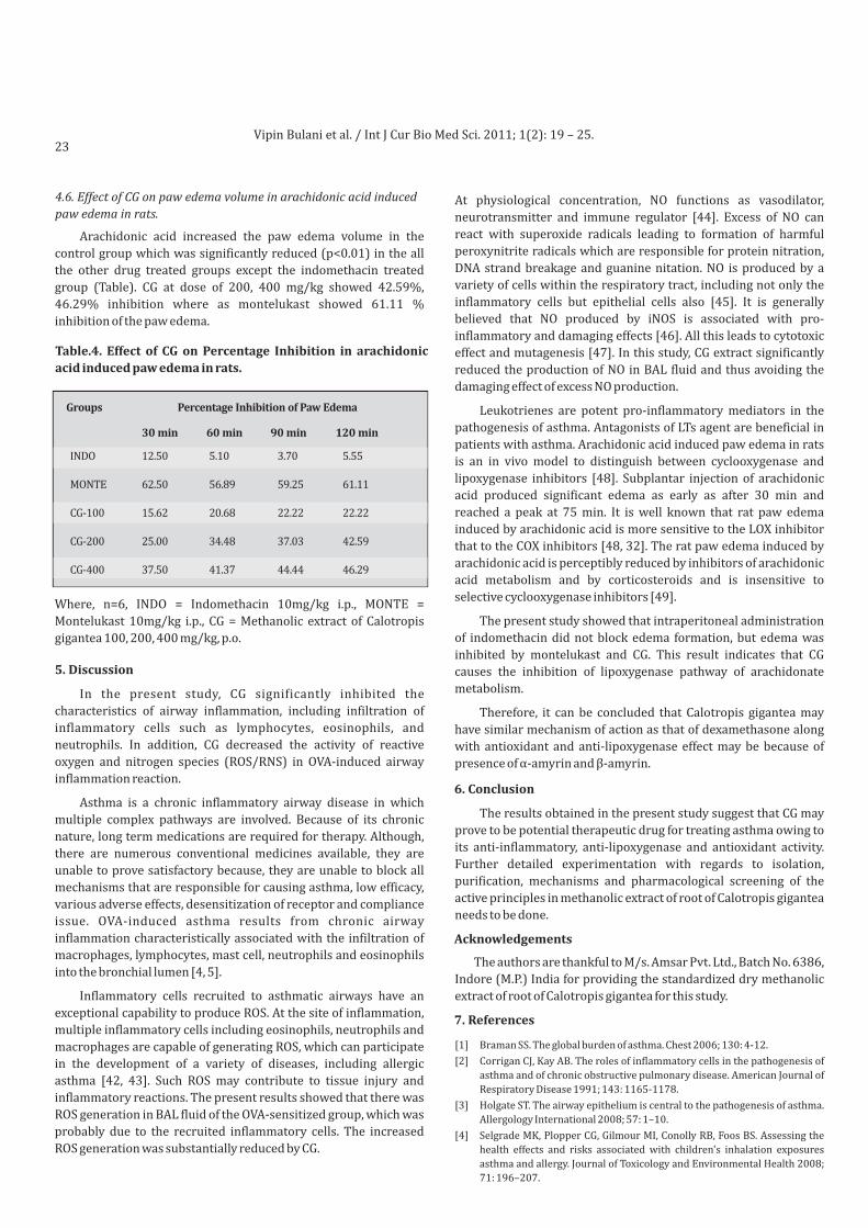
4.6. Effect of CG on paw edema volume in arachidonic acid induced
paw edema in rats.
Arachidonic acid increased the paw edema volume in the
control group which was significantly reduced (p<0.01) in the all
the other drug treated groups except the indomethacin treated
group (Table). CG at dose of 200, 400 mg/kg showed 42.59%,
46.29% inhibition where as montelukast showed 61.11 %
inhibition of the paw edema.
Where, n=6, INDO = Indomethacin 10mg/kg i.p., MONTE =
Montelukast 10mg/kg i.p., CG = Methanolic extract of Calotropis
gigantea 100, 200, 400 mg/kg, p.o.
In the present study, CG significantly inhibited the
characteristics of airway inflammation, including infiltration of
inflammatory cells such as lymphocytes, eosinophils, and
neutrophils. In addition, CG decreased the activity of reactive
oxygen and nitrogen species (ROS/RNS) in OVA-induced airway
inflammation reaction.
Asthma is a chronic inflammatory airway disease in which
multiple complex pathways are involved. Because of its chronic
nature, long term medications are required for therapy. Although,
there are numerous conventional medicines available, they are
unable to prove satisfactory because, they are unable to block all
mechanisms that are responsible for causing asthma, low efficacy,
various adverse effects, desensitization of receptor and compliance
issue. OVA-induced asthma results from chronic airway
inflammation characteristically associated with the infiltration of
macrophages, lymphocytes, mast cell, neutrophils and eosinophils
into the bronchial lumen [4, 5].
Inflammatory cells recruited to asthmatic airways have an
exceptional capability to produce ROS. At the site of inflammation,
multiple inflammatory cells including eosinophils, neutrophils and
macrophages are capable of generating ROS, which can participate
in the development of a variety of diseases, including allergic
asthma [42, 43]. Such ROS may contribute to tissue injury and
inflammatory reactions. The present results showed that there was
ROS generation in BAL fluid of the OVA-sensitized group, which was
probably due to the recruited inflammatory cells. The increased
ROS generation was substantially reduced by CG.
At physiological concentration, NO functions as vasodilator,
neurotransmitter and immune regulator [44]. Excess of NO can
react with superoxide radicals leading to formation of harmful
peroxynitrite radicals which are responsible for protein nitration,
DNA strand breakage and guanine nitation. NO is produced by a
variety of cells within the respiratory tract, including not only the
inflammatory cells but epithelial cells also [45]. It is generally
believed that NO produced by iNOS is associated with pro-
inflammatory and damaging effects [46]. All this leads to cytotoxic
effect and mutagenesis [47]. In this study, CG extract significantly
reduced the production of NO in BAL fluid and thus avoiding the
damaging effect of excess NO production.
Leukotrienes are potent pro-inflammatory mediators in the
pathogenesis of asthma. Antagonists of LTs agent are beneficial in
patients with asthma. Arachidonic acid induced paw edema in rats
is an in vivo model to distinguish between cyclooxygenase and
lipoxygenase inhibitors [48]. Subplantar injection of arachidonic
acid produced significant edema as early as after 30 min and
reached a peak at 75 min. It is well known that rat paw edema
induced by arachidonic acid is more sensitive to the LOX inhibitor
that to the COX inhibitors [48, 32]. The rat paw edema induced by
arachidonic acid is perceptibly reduced by inhibitors of arachidonic
acid metabolism and by corticosteroids and is insensitive to
selective cyclooxygenase inhibitors [49].
The present study showed that intraperitoneal administration
of indomethacin did not block edema formation, but edema was
inhibited by montelukast and CG. This result indicates that CG
causes the inhibition of lipoxygenase pathway of arachidonate
metabolism.
Therefore, it can be concluded that Calotropis gigantea may
have similar mechanism of action as that of dexamethasone along
with antioxidant and anti-lipoxygenase effect may be because of
presence of α-amyrin and β-amyrin.
The results obtained in the present study suggest that CG may
prove to be potential therapeutic drug for treating asthma owing to
its anti-inflammatory, anti-lipoxygenase and antioxidant activity.
Further detailed experimentation with regards to isolation,
purification, mechanisms and pharmacological screening of the
active principles in methanolic extract of root of Calotropis gigantea
needs to be done.
The authors are thankful to M/s. Amsar Pvt. Ltd., Batch No. 6386,
Indore (M.P.) India for providing the standardized dry methanolic
extract of root of Calotropis gigantea for this study.
[1] Braman SS. The global burden of asthma. Chest 2006; 130: 4-12.
[2] Corrigan CJ, Kay AB. The roles of inflammatory cells in the pathogenesis of
asthma and of chronic obstructive pulmonary disease. American Journal of
Respiratory Disease 1991; 143: 1165-1178.
[3] Holgate ST. The airway epithelium is central to the pathogenesis of asthma.
Allergology International 2008; 57: 1–10.
[4] Selgrade MK, Plopper CG, Gilmour MI, Conolly RB, Foos BS. Assessing the
health effects and risks associated with children's inhalation exposures
asthma and allergy. Journal of Toxicology and Environmental Health 2008;
71: 196–207.
5. Discussion
6. Conclusion
Acknowledgements
7. References
Table.4. Effect of CG on Percentage Inhibition in arachidonic
acid induced paw edema in rats.
Groups Percentage Inhibition of Paw Edema
INDO
MONTE
CG-100
CG-200
CG-400
12.50
62.50
15.62
25.00
37.50
5.10
56.89
20.68
34.48
41.37
3.70
59.25
22.22
37.03
44.44
5.55
61.11
22.22
42.59
46.29
30 min 60 min 90 min 120 min
Vipin Bulani et al. / Int J Cur Bio Med Sci. 2011; 1(2): 19 – 25.23
[1] Braman SS. The global burden of asthma. Chest 2006; 130: 4-12.
[2] Corrigan CJ, Kay AB. The roles of inflammatory cells in the pathogenesis of
asthma and of chronic obstructive pulmonary disease. American Journal of
Respiratory Disease 1991; 143: 1165-1178.
[3] Holgate ST. The airway epithelium is central to the pathogenesis of asthma.
Allergology International 2008; 57: 1–10.
[4] Selgrade MK, Plopper CG, Gilmour MI, Conolly RB, Foos BS. Assessing the
health effects and risks associated with children's inhalation exposures
asthma and allergy. Journal of Toxicology and Environmental Health 2008;
71: 196–207.
[5] Roh SS, Kim SH, Lee YC, Seo YB. Effects of radix adenophorae and
cyclosporine A on an OVA-induced murine model of asthma by suppressing
to T cells activity, eosinophilia, and bronchial hyper-responsiveness.
Mediators of Inflammation 2008; 781425: 1-11.
[6] Hamelmann E, Schwarze J, Takeda K, Oshiba A, Larsen GL, Irvin CG, Gelfand
EW. Noninvasive measurement of airway responsiveness in allergic mice
using barometric plethysmography. American Journal of Respiratory and
Critical Care Medicine 1997; 156: 766–775.
[7] Kwak YG, Song CH, Yi HK, Hwang PH, Kim JS, Lee KS, Lee YC. Involvement of
PTEN in airway hyper-responsiveness and inflammation in bronchial
asthma. The Journal of Clinical Investigation 2003; 111: 1083–1092.
[8] Singh U, Wadhwani AM, Johri BM. Dictionary of economic plants of India. In:
Jaiswal PL, editor. Indian Council of Agricultural Research New Delhi 83.
1996, pp 38–9.
[9] Varier PS. Indian Medicinal Plants. Orient Longman Pvt. Ltd. Vol.1. New
Delhi. 2003, pp 341-43.
[10] Kirtikar KR, Basu BD. Indian Medicinal Plants. Vol.3. International Book
Distributors, Dehradun. 2005, pp 1607-1609.
[11] Anjaneyulu V, Row LR. The triterpenes esters of Calotropis gigantea Linn.
Current Science 1968; 6: 156-157.
[12] Rowshanul HM, Farjana N, Matiar R, Ekramul HM, Rezaul KM. Isolation of
stigmasterol and ß-sitosterol from methanolic extract of root bark of
Calotropis gigantea. Pak J Bio Sci. 2007; 22: 4174–6.
[13] Kweifio G, Macrides TA. Antilipoxygenase activity of amyrin triterpenes.
Respiratory Communica Chem Pathol Pharmacol 1992; 78: 367-372.
[14] Mueen Ahmed KK, Rana AC, Dixit VK. Free radical scavenging activity of
Calotropis species. Indian drugs 2003; 40: 654–655.
[15] Kshirsagar A, Purnima A, Ingawale D, Vyawahare N, Ingale K, Hadambar A.
Antioxidant and hepatoprotective activity of ethanolic extract of Calotropis
gigantea against paracetamol induced liver damage in mice.
Journal of Cell and Tissue Research 2009; 9: 1859-1864.
[16] Rajesh R, Raghavendra Gowda CD, Nataraju A, Dhananjaya BL, Kemparaju K,
et al. Procoagulant activity of Calotropis gigantea latex associated with
fibrin(ogen)olytic activity. Toxicon. 2005; 46: 84–92.
[17] Chitme R, Chandra R, Kaushik S. Studies on anti-diarrheal activity of
Calotropis gigantea R.Br. in experimental animals. Journal of Pharmacy and
Pharmaceutical Science 2004; 7: 70-75.
[18] Jain SK, Sinha, BK, Saklani A. Medicinal plants known among tribal societies
of India. Ethnobotany 2001; 1: 92.
[19] Argal A, Pathak AK. CNS activity of Calotropis gigantea roots. Journal of
Ethnopharmacology 2006; 106: 142-145.
[20] Pathak AK, Argal A. Analgesic activity of Calotropis gigantea flower.
Fitoterapia 2007; 78: 142-145.
[21] Srivastava SR, Keshri G, Bhargavan B, Singh C, Singh MM. Pregnancy
interceptive activity of the roots of Calotropis gigantea Linn. in rats.
Contraception 2007; 75: 318-322
[22] Pardesi GS, Gadgoli C, Vaidya MD, Hasni HY, More BH, Bhuskat P. Preliminary
studies on antimitotic and anticancer activity of Calotropis gigantea.
Pharmacologyonline 2008a; 1: 38-47.
[23] Pardesi GS, Gadgoli C, Vaidya MD, Hasni HY, More BH, Bhuskat P.
Immunomodulatory Activity of Calotropis gigantea by cyclophosphamide
induced myelosuppression. Pharmacologyonline 2008b; 2: 164-167.
[24] Deshmukh PT, Fernandes J, Akarte A. Wound healing activity of Calotropis
gigantea root bark in rats. Journal of Ethnopharmacology 2009; 125: 178-
81.
[25] Adak M, Gupta JK. Evaluation of anti-inflammatory activity of Calotropis
gigantea (AKANDA) in various biological system. Nepal Med. Coll. J. 2006; 8:
156-61.
[26] Chitme HR, Chandra R, Kaushik S. Studies on anti-inflammatory activity of
Calotropis gigantea in experimental animals. Asia Pacific Journal of
Pharmacology 2006; 16: 163-168.
[27] Saumya D, Sanjita D, Manas KD, Saumya PB. Evaluation of anti-inflammatory
effect of Calotropis gigantea and Tridax procumbens on Wistar albino rats. J.
Pharm. Sci. & Res. 2009; 4: 123-126.
[28] Awasthi S, Irshad M, Das MK, Ganti SS, Moshahid A. Rizvi. Anti-Inflammatory
Activity of Calotropis gigantea and Tridax procumbens on Carrageenin-
Induced Paw Edema in Rats Ethnobotanical Leaflets. 2009; 13: 568-77.
Lodhi G
[29] Singh HK, Pant KK, Hussain Z. Hepatoprotective effects of Calotropis
gigantea extract against carbon tetrachloride induced liver injury in rats.
Acta Pharm. 2009; 59: 89-96.
[30] Rathod NR, Raghuveer I, Chitme HR, Chandra R. Free Radical Scavenging
Activity of Calotropis gigantea on streptozotocin-induced diabetic rats
Indian J Pharm Sci. 2009; 71: 615–621.
[31] Caramori G, Adcock I. 2005. Anti-inflammatory mechanisms of
glucocorticoids targeting granulocytes. Current drug targets. Inflammation
and Allergy 4, 455–463.
[32] De Fatima AM, Dmitrieva EG, Franzotti EM, Antoniolli AR, Antrada MR,
Marchioro M. Anti-inflammatory and analgesic activity of Peperomia
pellucida (L.) HBK (Piperaceae). Journal of Ethnopharmacology 2004; 91:
215–218.
[33] Kaan G, Fahrettin Y, Fadullah A, GuIer B, Husamettin C, Onur H. The Effects of
Montelukast on Random Pattern Skin Flap Survival: An Experimental Study
in Rats. Current Therapeutic Research 2008; 69: 459-465.
[34] Organization for Economic Co-operation Development, Guidance
Document on Acute Oral Toxicity Testing. Environment Directorate. OECD,
Paris, 2001, pp 1–24.
[35] Chapman RW, Howard AH, Richard J, Celly C. Effect of inhaled roflumilast on
the prevention and resolution of allergen-induced late phase airflow
obstruction in Brown Norway rats. European Journal of Pharmacology
2007; 571: 215-221.
[36] Benjamine NM, Maxinie M. Outline of veterinary clinical pathology, 3rd ed.
Kalyani Pub, 1997, pp 17-33.
[37] Ellaman GL. Tissue sulfhydryl group. Archives Biochemistry and
Biophysiology 1959; 82: 70-77.
[38] Slater TF, Sawyer BC. The stimulatory effect of carbon tetrachloride and
other halogenoalkanes or peroxidative reactions in rat liver fraction in vitro.
Biochemical Journal 1971; 123: 805-814.
[39] Mishra HP, Fridovich I. Role of superoxide anion in auto-oxidation of
epinephrine and a simple assay for superoxide dismutase. Journal of
Biological chemistry 1972; 247: 3170-3175.
[40] Colowick SP, Kaplan NO, Packer L. Methods in Enzymology. Academic press,
London, 1984, pp 121-125.
[41] Green LC, Wagner DA, Glogowski J, Skipper PL, Wishnok JS, Tannenbaum SR.
Analysis of nitrate, nitrite, and (15N) nitrate in biological fluids, Analytical
Biochemistry 1982; 126: 131–138.
[42] Conner EM, Grisham MB, Inflammation, free radicals, and antioxidants.
Nutrition 1996; 12: 274–277.
[43] Leusen JH, Verhoeven AJ, Roos D, Interactions between the components of
the human NADPH oxidase: intrigues in the phox family. Journal of
Laboratory and Clinical Medicine 1996; 128: 461–476.
[44] Fischer A, Folkerts G, Geppetti P, Groneberg DA. Mediators of Asthma: Nitric
oxide. Pulmonary Pharmacology & Therapeutics 2002; 15: 73-81.
[45] Vliet AV, Eiserich JP, Cross CE. Nitric oxide: a pro-inflammatory mediator in
lung disease ?. Respir Res. 2000; 1: 67–72.
[46] Laskin DL, Fakhrzadeh JD. Nitric oxide and peroxynitrite in ozone induced
lung injury. Adv. Exp. Med. Biol. 2000; 500: 183–190.
[47] Napolitano DR, Mineo JR, De Souza MA, Espinodola LS, Espinodola FS. Down
modulation of nitric oxide production in murine macrophages treated with
crude plant extracts from Brazillian cerrado. Journal of Ethnopharmacology
2005; 99: 37-41.
Vipin Bulani et al. / Int J Cur Bio Med Sci. 2011; 1(2): 19 – 25.24
[48] Tsununi K, Kyuki K, Niwa M, Mibu H, Fujimura H. Pharmacological
investigations of new anti-inflammatory agent 2-(10,11- dihydro-10-
oxodibenzo (b,f) thiepen-2-yl) propionic acid. Inhibitory effects on acute
inflammation and prostaglandin-related reactions. Arzneimitel forschung
1986; 36: 1801–1805.
[49] Di Martino MJ, Champbell GK, Wolf CE, Hanna N. The pharmacology of
arachidonic acid induced rat paw oedema. Agents and Actions 1987; 21:
303–305.
Copyright 2011. CurrentSciDirect Publications. IJCBMS - All rights reserved.c
Vipin Bulani et al. / Int J Cur Bio Med Sci. 2011; 1(2): 19 – 25.25


