
Initial assessment(ABC s)
Tissue perfusion and oxygenation
Oxygen delivery depends on CO and Hbgconcentration , Hbg oxygen concentration.

Onset of symptoms , details of events , brief identification of underlying medical problem

ABC sA:air way and neurologically injured child Protection of the cervical spineB:breathing (pulse oximeter)C:circulatory(palpitation for distal and central
pulsesBounding pulses and wide pulse pressure
are sign of vasodilatory phase of shock.D:neurologic systemE:exposure

1-respiratory failure
2-shock

Pulse oximetry and bed side measurement of glucose level

CXR : in Child with respiratory distress is important.
Culture ….when sepsis is suspected.
Electrolyte….Inadequate intravascular volume

Oxygen supplementation
Isotonic crystalloids
Packed red blood cell….If hemorrhage is highly suspected
Vasoactive substances …When respiratory and fluid are insufficient

Outcome is poor
Survival is about 6% for out of hospital arrest and 27%in hospital arrest.
Goal is optimize CO and tissue oxygen delivery , which accomplished by artificial
ventilation and chest compression.
Pediatric advanced life support and CPR:
Start chest compression immediately rather than beginning with airway and breathing.
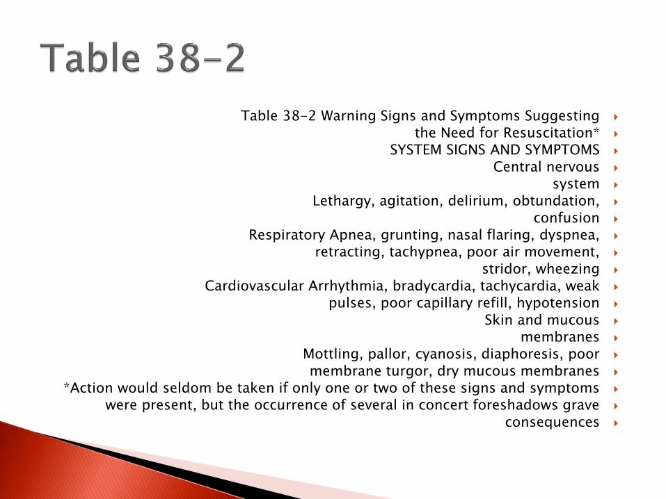
Table 38-2 Warning Signs and Symptoms Suggestingthe Need for Resuscitation*SYSTEM SIGNS AND SYMPTOMSCentral nervoussystemLethargy, agitation, delirium, obtundation,confusionRespiratory Apnea, grunting, nasal flaring, dyspnea,retracting, tachypnea, poor air movement,stridor, wheezingCardiovascular Arrhythmia, bradycardia, tachycardia, weakpulses, poor capillary refill, hypotensionSkin and mucousmembranesMottling, pallor, cyanosis, diaphoresis, poormembrane turgor, dry mucous membranes*Action would seldom be taken if only one or two of these signs and symptomswere present, but the occurrence of several in concert foreshadows graveconsequences

Chest compression : effective CPR requires compression depth of one third to one half
of the ANT-POST diameter of chest with complete chest recoil.
Chest compression Rate 100\min and breaths 8-10 times per min

Head tilt –chin lift maneuver
Jaw thrust
Cricoid pressure
Induction medication
Correct size for tube:mid-fifth phalanx
4+(age\4)
DOPE : in deteriorates patient
Displacement
or Obstruction
Pneumothorax
Equipment failure

Table 38-3 Target Organs for Hypoxic-IschemicDamageORGAN EFFECTBrain Seizures, cerebral edema, infarction,herniation, anoxic damage, SIADH, diabetesinsipidusCardiovascular Heart failure, myocardial infarctLung and pulmonaryvasculatureAcute respiratory distress syndrome,pulmonary hypertensionLiver Infarction, necrosis, cholestasisKidney Acute tubular necrosis, acute corticalnecrosisGastrointestinaltractGastric ulceration, mucosal damageHematologic Disseminated intravascular coagulationSIADH

Interosseous is recommended when iv access is not present.
Some drugs are effectively through endotrachealtube.
Epinephrine
Sodium bicarbonate is not recommended.
side effects are:
hyper Na,
hyper osmolality
Hyper K
Metabolic alkalosis

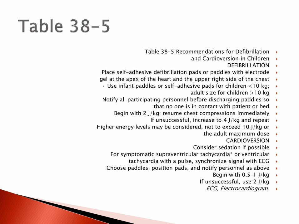
Table 38-5 Recommendations for Defibrillation
and Cardioversion in Children
DEFIBRILLATION
Place self-adhesive defibrillation pads or paddles with electrode
gel at the apex of the heart and the upper right side of the chest
• Use infant paddles or self-adhesive pads for children <10 kg;
adult size for children >10 kg
Notify all participating personnel before discharging paddles so
that no one is in contact with patient or bed
Begin with 2 J/kg; resume chest compressions immediately
If unsuccessful, increase to 4 J/kg and repeat
Higher energy levels may be considered, not to exceed 10 J/kg or
the adult maximum dose
CARDIOVERSION
Consider sedation if possible
For symptomatic supraventricular tachycardia* or ventricular
tachycardia with a pulse, synchronize signal with ECG
Choose paddles, position pads, and notify personnel as above
Begin with 0.5–1 J/kg
If unsuccessful, use 2 J/kg
ECG, Electrocardiogram.


Classified by:
Hypercarbic (pa co2 greater than 50 mmhg)
Hypoxemic(pao2 less than 60 mmhg) it cause by V\Q mismatch.
Acute Respiratory failure may occure with (ALI) or (ARDS)

Early sign of hypoxic respiratory failure:
Tachypnea and tachycardia
Late signs of inadequate oxygen delivery include cyanosis and altered mental status.

CXR
In pt with stridor or upper airway obstraction
LAT NECK or CT
Flexible bronchoscopy shows abnormalities of anatomic airway.

Bag mask ventilation is for pt with apnea.
Oxygen therapy….appropriate method
Intubation..based on pco2 alone
Noninvasive ventilation
Mechanical ventilation

Multi organ dysfunction 2 or more of following: respiratory failure - cardiac failure
Renal insufficiency or failure – GE or hepatic insufficiency – intra vascular coagulation and
hypoxic –ischemic brain injury.

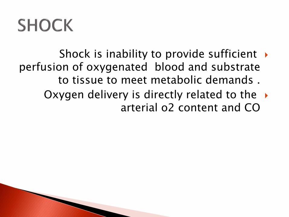
Shock is inability to provide sufficient perfusion of oxygenated blood and substrate
to tissue to meet metabolic demands .
Oxygen delivery is directly related to the arterial o2 content and CO

diabetes mellitusBurnsAdrenogenital syndromeCapillary leakDistributive Vasodilation →venous pooling →decreased preloadSepsisMaldistribution ofregional blood flowAnaphylaxisCNS/spinal injuryDrug intoxicationCardiogenic DecreasedmyocardialcontractilityCongenital heart diseaseArrhythmiaHypoxic/ischemic injuriesCardiomyopathyMetabolic derangementsMyocarditisDrug intoxicationKawasaki diseaseObstructive Mechanicalobstruction toventricular filling or

Most common cause:
Vomiting
Diarrhea
Blood loss
Capillary leak syndrome
Pathologic renal fluid losses

Septic shock is the most common type of distributive shock in children.
It present with systemic inflamatory response syndrom(SIRS):
T greater than 38 or less than 36HR}90 min or more than 2 standard deviation above
normal for ageTachypneaWBC]12000 cell\mm3 or less than 4000 cell\mm3 or
greater than 10% immature forms
Other cause of shock are anaphylaxis , neurologic injury, drug related cause

It occur in:
Congenital heart disease
In healthy children secondary to:
viral myocarditis ,
dysrhythmias
toxic metabolic
after hypoxic –ischemic injury

Causes by:
CO_ AO
Interrupted AO arch
Sever AO valvular stenosis
Acquired disease (hypertrophic cardiomyopathy)
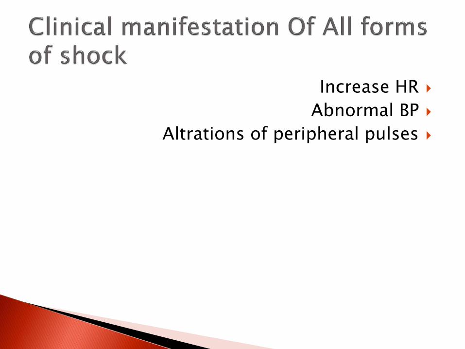
Increase HR
Abnormal BP
Altrations of peripheral pulses

Absence of sign of heart failure or sepsis
Tachycardia
Vasoconstriction
Clinical manifestation are :
Sign of dehydration such as:
Dry mucous membrane
Decrease urine out-put
pallor

Tachycardia
Alteration of peripheral perfusion

Because of :
Heart failure progressing to death may be rapid.
Tachycardia
Tachypnea
Enlarge liver
Gallop
Distension of jugular vein
Oliguria
Peripheral edema

Narrow pulse pressure
Capillary refill is delayed
Enlarged liver
Distention of jugular vein

Tachycardia
Tachypnea
Altration of mental status
Cardiovascular collaps

In all pt with shock check:Blood glucose
ABGblood lactate level
CBCElectrolyte(Na,K,Ca,P,Mg)
BUNdistributive shock require bacterial and viral
cultureCardiogenic or obstructive need ECHO

ABC resuscitation
Intubation ,combined with mechanichalventilation
Blood pressure support
Monitoring require maintaining access to the arterial and central venous circulation

Fluid resuscitation Crystalloid volume expanded are recommended20 cc\kg bolus of isotonic crystalloid over 5-15 min.Cardiovascular support:Dopamin at 3 to 15 mcg\kg\minEpinephrin or NE prefer in patient withv decompensated shockRenal salvagePrerenal azotemia is associated with :Serume BUN to Cr ratio greater than 10:1 andUrine Na level less than 20mEq\LATN:BUN\Cr 10:1 or less and urine Na level between 40-60
mEq\L


Some form of septic shock prevented by immunization(HI ; influ type b;
meningococcal, pneumococcal vaccine)
Decrease the risk of sepsis by:
Hand washing
Isolated practices
Minimizing the duration of catheters


52%of death caused by Motor car crashes Drowning 15%Poisoning 9%Burns 5%Suffocation 4%Injury occurs through interaction of the Host
and agentThe age of the child may determine the
exposure to various agent and enviroments.


Assessment and resuscitation
Pre hospital trauma care are :
Immobilization ; trasportation
Primary survey ; resuscitation ; secondary survey ; post resuscitation monitoring and
definitive care

Primary survey include…..ABCDE
Airway and breathing and cervical spine
Full stomach (risk of aspiration pneumonia)
Circulation(HR;skin collor;mental status)and control of bleeding
Disability(neurological status include pupilsize and reactivity
mental status(AVPU_Glasgow coma scale)
Secondary survey :head to toe examination
Tertiary survey include repeat primary and secondary survey along laboratory tests and
radiologic studies

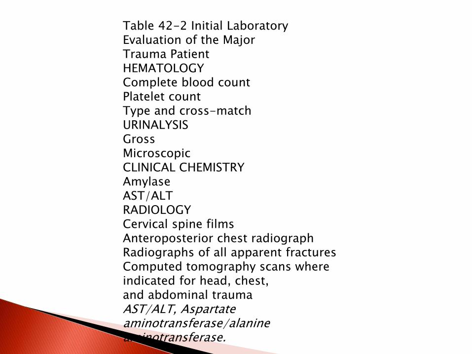
Table 42-2 Initial Laboratory Evaluation of the MajorTrauma PatientHEMATOLOGYComplete blood countPlatelet countType and cross-matchURINALYSISGrossMicroscopicCLINICAL CHEMISTRYAmylaseAST/ALTRADIOLOGYCervical spine filmsAnteroposterior chest radiographRadiographs of all apparent fracturesComputed tomography scans where indicated for head, chest,and abdominal traumaAST/ALT, Aspartateaminotransferase/alanineaminotransferase.
Radiologic studies are determined by the pattern of injury.
CT : in patient with head trauma or history of LOC
FAST
DPL
Enhanced CT in Ao injury

Spinal cord injury:
Cervical spine radiographs
SCIWORA:spinal cord injury without radiologic abnormality
Thoracic trauma:
Lung contusion
PNX
Rib fracture

Abdominal trauma:occure 8% of pediatric traumaAbdominal CT and serial PH\EXPresence of peritoneal irritation or abdominal wall
discoloration ; together with signs of intravascular volume loss ;indicates laparotomy
Spleen injury is most common (kehr sign)Liver trauma is a seriouse cause of morbidityRenal injury : IN YOUNG CHILDREN KIDNEY IS
VULNERABLE IN TRAUMAPANCREATIC INJURY : LESS COMMONELEVATION OF AMYLASE AND LIPASE SEVERAL DAYS
AFTER INJURYINTESTINALINJURY: DEODENAL HEMATOMA

RF secondary to myoglobinuria


ETIOLOGYSubmersion or immersion results in
aspiration of small amounts of fluid into the larynx ;triggering breath holding or laryngo
spasm
EPIDEMIOLOGY
Cause of injury death for 1-4 years of age and the second leading cause of injury death
for 1-18 years of age

Hypoxemia
Secondary to pulmonary endothelial injury ; increased capillary permeability ; destruction
of surfactant
The hypoxic ischemic injury lead to depressed myocardial function resulting in
tachycardia impaired perfusion and cardiovascular collapse.

Laboratory and imaging studies
ABG
Elevated liver Enz
Electrolytes

ABC s
Victim of unwitnessed drowning required:
Stabilized cervical spine
Optimizing oxygenation
Patient with evidence lung injury ;cardiovascular compromise should be
monitored in ICU.
Prophylactic AB not beneficial.
Unfavorable prognostic markers include:
the need for CPR for more than 25 min ,
continued CPR at the hospital ;
glosgow coma scale of 5 or less
fixed and dilated pupils seizure and coma for more than 72 H


ETIOLOGYPathophysiology of burn injury is caused by
disruption of the 3 key function of the skin:Regulation of heat lossPreservation of body fluidBarrier to infection BURN CLASSIFIED: on the basis of 4 criteria:Depth of injuryPercent of body surface area involvedLocation of the burnAssossiation with other injury


SUPERFICIAL(first degree):Red;painful;drySeen with sun exposureHeal in 2-5 days without scarring and not included in
burn surface area calculations.SECOND degree: entire epidermis and superficial
dermis….deep partial thickness is also second degreeThird and four degree:full thickness
Inhalation injuries

Estimate :
Each upper extremity 9%
Each lower extremity 18%
Ant trunk 18%
Post trunk 18%
Head 9%
Perineum 1%

CBC
Type and crossmatch
Coagulation studies
ABG
CXR
Cyanid level(smoke inhalation and loc)

American burn association criteria :
Partial and full thickness burns greater than 10%total body surface area(TBSA)
in patient less than 10 years old
Or
more than 50 years old
or
greater than 20% TBSA in other age groups
p artial and full thickness burns involving the face; hand; feet; genitalia ; perineum
or
major joints ;
electerical burns
chemichal burns
inhalation injury
in patient with preexisting medical condition that could complevated management prolong recovery ; increase mortality ; any burn with concomitant trauma in
wich the burn inury poses the greatest risk.

Significant burn :
Rapid bolus 20 cc\kg of lactate ringer solution
Total fluids are 2-4 cc\kg\percent burn\24 h
Half in 8 hours
Colloid therapy in extensive burns
Hypermetabolic response:require nutritional support
Factors that may modify the hypermetabolic state such as beta blocker ;androgenic steroids,and
other are being investigated

Wound care :Cleaning and debridingSilver sulfadiazineIf the burn is shallow; poly myxin
B\bacitracin\neomycinSulfamylon has the benefit of penetrating
eschar;but it is pailfull and can cause methabolic acidosis
Silver cause electrolyte abnormality

Table 44-1 Complications of Burns
PROBLEM TREATMENT
Sepsis Monitor for infection, avoid
prophylactic antibiotics
Hypovolemia Fluid replacement
Hypothermia Adjust ambient temperature: dry
blankets in field
Laryngeal edema Endotracheal intubation, tracheostomy
Carbon monoxide
poisoning
100% oxygen, hyperbaric oxygen
Cyanide poisoning 100% O2 plus amyl nitrate, sodium
nitrate, and sodium thiosulfate
Cardiac dysfunction Inotropic agents, diuretics
Gastric ulcers H2-receptor antagonist, antacids
Compartment syndrome Escharotomy incision
Contractures Physical therapy
Hypermetabolic state Enteral and parenteral nutritional
support
Renal failure Supportive care, dialysis
Transient antidiuresis Expectant management
Anemia Transfusions as indicated
Psychological trauma Psychological rehabilitation
Pulmonary infiltrates PEEP, ventilation, oxygen
Pulmonary edema Avoid overhydration, give diuretics
Pneumonia Antibiotics
Bronchospasm β-Agonist aerosols
PEEP, Positive


Etiology and epidemiology in children include cosmetics
person care product
analgesics
cleaning solution
Fetal childhood poisoning are commonly caused by:
analgesics
anti histamin
sedative hypnotics ;fumes ; gases;vapors

Any child who presents with unexplained symptoms include altered mental
status;seizure;cardiovascular compromise or metabolic abnormality should be considered

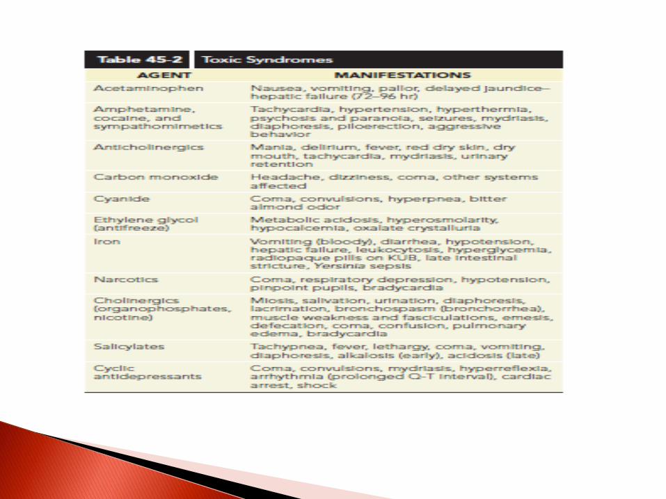
A poisoned child can exhibit any one of six basic clinical patterns:
Coma;toxicity;metabolic acidosis;heartrhythm abberantion ;gasterointestinal
symptms;seizure

Emesis or lavage should not be initated in a child who has ingested volatile hydrocarbons.
Caustic ingestion may cause dysphasia ;epigastric pain ; oral mocusal burns and
low grade fever
Bateries: remain in the esophagus and cause esophageal burns and erosion and should
remove with endoscope.
Acid agents can injured the lungs


Metabolic acidosis
Metabolic acidosis with high AG(mnemonic MODPILES)


Dysrhythmias:
Prolonged QT (phenothiazine-anti histamin)
QRS widening(TCA-quinidine)
Brady cardia suggest:
digoxin;cyanid;cholinergic agent or BB ingestion.

ABG
Electrolyte
Osmolel
Glucose
AG and Osmolar gap
ECG
Urine screen

Four foci of treatment are :Supportive careDecontaminationEnhanced eliminationSpecific antidotesIf the level of consciousness is depressed and a
toxic substance is suspected Glucose 1gr\kg 100% o2naloxone

IPECAC should not be administered Gastric lavage not be used
Single dose activated charcoal within 1 hour of ingestion.
Charcoal is ineffective against…….. Caustic
corrosive agentshydrocarbons
;heavy metals(arsenic,lead;iron;Li;glycols and water insoluble compounds)
A cathartic (sorbitol or Mg citrate)alone has no role in the management of the poisoned patient.
Whole bowel irrigation using PEG effective for toxic ingestion or sustained –release or enteric –coated drugs.

Multi dose activected charcoal should be considered only if patient has ingested a
life-threatening amount of carbamazepin;dapson;phenobarbital;quinine;
theophylline.
Alkalinization of urine :salicylate or MTX ingestion
Dialysis : methanol ; ethylen glycol;salicylate ; theophylline ; bromide; li

Table 45-5 Drugs Amenable to Therapeutic
Monitoring for Drug Toxicity
ANTIBIOTICS
Aminoglycosides—gentamicin, tobramycin, and amikacin
Chloramphenicol
Vancomycin
IMMUNOSUPPRESSION
Methotrexate
Cyclosporine
ANTIPYRETICS
Acetaminophen
Salicylate
OTHER
Digoxin
Lithium
Theophylline
Anticonvulsant drugs
Serotonin uptake inhibitor agent

Nonprocedural sedation:
Many ventilated pediatric pt require sedation and some analgesia while intubated.
Most common choice is a combination of a longer acting BNZ and an opioid.
Local anesthetics such as lidocaine use for minor procedures.
Use of EMLA a cream containing lidocaine and prilocaine is less effective than interadermal
lidocaine .

Table 46-1 Agents that Produce SedationSEDATIVES EFFECT CONCERNSMidazolam Anxiolysis, sedation, muscle relaxation, amnesia Tolerance is
possible; apnea, hypotension, depressed myocardialfunction; short actionLorazepam Anxiolysis, sedation, muscle relaxation, amnesia Same as
midazolam; long actionDexmedetomidine Sedation without respiratory depression May cause
bradycardiaKetamine Anesthesia, analgesia, amnesia Dissociative reactions,
tachycardia, hypertension, increasedbronchial secretions, emergent delirium, hallucinations; increasesintracranial pressureChloral hydrate Sedative Emesis, hypotension, arrhythmias, hepatic
dysfunctionPropofol Rapid-onset sedative for induction andmaintenance of anesthesiaMetabolic acidosis in children; may depress cardiac function

Table 46-2 Agents that Produce AnalgesiaANALGESIC EFFECT COMPLICATIONSAcetaminophen andNSAIDsModerate analgesia, antipyresis Ceiling effect, requires PO administrationNSAIDs—gastrointestinal bleed, ulcerationOpioids No ceiling effect; respiratory depression, sedation, pruritus,
nausea/vomiting,decreased gastric motility, urinary retention, tolerance with abuse potentialMorphine Analgesia May cause myocardial depressionCodeine Analgesia Nausea/vomitingFentanyl, alfentanil,sufentanilAnalgesia, sedation No adverse effects on cardiovascular system; stiff chest
syndromeMethadone AnalgesiaNSAIDs, Nonsteroidal anti-inflammatory drugs; PO, oral