
International Journal of PharmTech Research
CODEN (USA): IJPRIF, ISSN: 0974-4304 Vol.8, No.4, pp 750-769, 2015
Biosynthesis, Characterization, Antimicrobial Activity Of Copper Nanoparticles Using Fresh Aqueous Ananas Comosus
L. (Pineapple) Extract
A.Mercy Ranjitham, G.Selva Ranjani, Dr. G.Caroling.
Department of Chemistry, Ethiraj College for Women(Autonomous)Chennai-08, Tamilnadu, India.
Abstract: The synthesis of metal nanoparticles is a growing area for research due to its
potentiality in the application and development of advanced technologies. In general,
nanoparticles are synthesized by using chemical methods which are not eco-friendly. In this
study we used the fast, convenient, eco-friendly method for synthesis of copper nano particles
by using thefresh aqueous pineapple extract.
The fresh aqueous pineapple extract was mixed with copper sulphate solution by heating to a
temperature of 60-80˚C and under optimum condition at ph 11. The reduction reaction was
studied by observing the color change from blue to reddish brown. Water soluble antioxidant
constituents present in the fresh aqueous pineapple extract were mainly responsible for the
reduction of copper ions to nano sized Cu particles.
The resulting copper nanoparticles were characterized by UV Visible Absorption
Spectrometer, powder X-Ray Diffraction (PXRD), Fourier Transform Infrared (FTIR),
Scanning Electron Microscopy (SEM) and EDAX analysis. UV-Vis spectral analysis showed
Copper nanoparticles exhibit an absorption peak at around 576 nm. X-ray diffraction
analysis shows that the crystalline morphology of nanoparticles are FCC in nature. The FTIR
spectrum analysis has confirmed the presence of functional groups and active proteins in the
biomass before and after reduction. SEM results display the formation of copper
nanoparticles with an average size of 30-50 nm. The presence of elemental copper was
characterized by EDS. The antibacterial and antifungal activity of these nanoparticles was
studied against E-Coil, Staphylococcus aureus, Aspergillus niger and Trichoderma viride .
The biosynthesized nanoparticles are quite stable in aqueous solution for a month without any
sign of precipitation.
Keywords: Eco-friendly, copper nanoparticles, fresh aqueous pineapple extract, UV-Vis, FT-
IR, EDS, SEM, Antimicrobial activity, Antioxidant.
Introduction
The term Nanotechnology comes from the combination of two words: the Greek numerical prefix nano
referring to a billionth and the word technology. As an outcome, Nanotechnology or Nanoscaled Technology is
generally considered to be at a size below 0:1 m or 100 nm (a nanometer is one billionth of a meter, 109 m).
Nanoscale science (or nanoscience) studies the phenomena, properties, and responses of materials at atomic,
molecular, and macromolecular scales, and in general at sizes between 1 and 100 nm. In this scale, and
especially below 5 nm, the properties of matter differ significantly (i.e., quantum-scale effects play an important
role) from that at a larger particulate scale. Nanotechnology is considered an emerging technology due to the
possibility to advance well-established products and to create new products with totally new characteristics and
functions with enormous potential in a wide range of applications. In addition to various industrial uses, great
G.Selva Ranjani et al /Int.J. PharmTech Res. 2015,8(4),pp 750-769. 751
innovations are foreseen in information and communication technology, in biology and biotechnology, in
medicine and medical technology, in metrology, etc. Significant applications of nanosciences and
nanoengineering lie in the fields of pharmaceutics, cosmetics, sports, therapy, automotives, environmental
monitoring, fuel cells and energy devices, water purification, food and beverage industry, chemical engineering,
high-performance materials, electronics, precision mechanics, optics, and environmental sciences.
Among the various nanoparticles, metal nanoparticles assume special importance because they are
easier and cheaper to synthesis and are most promising in application. Synthesis of copper nanoparticles is of
great interest because of many advantages. Copper is highly conductive and also less expensive than silver and
gold. Copper nanoparticles have wide applications as heat transfer system, antimicrobial materials, super strong
materials, sensors and catalysts. Copper nanoparticles are very reactive because of their high surface-to-volume
ratio and can easily interact with other particles and increase their antimicrobial efficiency.
However, aggregation appears immediately due to surface oxidation are the main problems concerned
with copper nanoparticles. But, we can overcome these problems by selecting suitable stabilizer for capping of
copper nanoparticles [1].
Copper is most widely used material in the world due to their electrical, optical, catalytic, biomedical
and antifungal/antibacterial applications among various metal particles such as gold, silver, iron, palladium,
zinc and quantum dots.
Copper can give more yields and reaction rate in mild reaction conditions when compared to other
traditional catalysts [2]. Green synthesis of colloidal copper nanoparticles is used for treating the rubber tree
which is affected by pink disease [3].
The need for biosynthesis of nanoparticles rises because the physical and chemical processes are costly,
hazardous, longer time, tedious process to isolate nanoparticles. This green synthesis method have several
advantages over other methods namely cost effectiveness, simplicity, use of less temperature, the usage of
less toxic materials, moreover it is compatible for medical and food applications. Therefore, in the search
for cheaper pathways for Nano particle synthesis, scientists used microorganisms and then plant extracts for
synthesis.
Many researchers used green synthesis methods for different metal nanoparticles due to their
growing need of eco-friendly properties. Green synthesis method was found to be the best method when
compared to the other method such as chemical reduction, photochemical reduction, electrochemical
reduction, heat evaporation etc., In this method, the plant extract has been used as capping and reducing
agent for the synthesis of copper nanoparticles due to their reducing properties present in the leaf and
fruit extract.
Ananas comosus L. belongs to Bromeliaceae family, a large, diverse family of about 2000 species and
is a subtropical fruit native to Thailand, Phillipines, China, Brazil and India.
Pineapple has several beneficial properties including antioxidant activity. It contains mixture of
protease enzymes which acts as a nutritional supplement to “promote digestive health” and used as an anti-
inflammatory medication.
Phytochemicals, especially phenolics, in fruits and vegetables are suggested to be the major bioactive
compounds for the health benefits [4]. Pineapple contains phytochemicals which include antioxidant substances
that fight against free radical cell damage and have been commonly used in traditional Chinese medicine [5].
Polyphenols possess outstanding antioxidant and free radical scavenging properties, suggesting a possible
protective role in humans [6,7]. Their antioxidant potential is closely related to the number of hydroxyls, the
higher the number, the more potent the chain breaking antioxidant action of the compound. Plant phenolics are
the largest class of plant secondary metabolites, which serve in plant defense mechanism to counteract reactive
oxygen species (ROS) in order to survive and prevent molecular damage.
G.Selva Ranjani et al /Int.J. PharmTech Res. 2015,8(4),pp 750-769. 752
Antimicrobial Applications
Copper as an antimicrobial agent which is able to reduce specific harmful bacteria linked to potentially
deadly microbial infections (European Copper Institute, 2008). In addition, no research has discovered any
bacteria are able to develop immunity to copper as they often do with antibiotics. Copper nanoparticles act as
antimicrobial agent in various fields. The copper is highly toxic to microorganism such as bacteria (E-Coli,
Staphylococcus Aureus, Pseudomonas aeruginosa) and non-toxic to animal cells, due to these phenomena it is
considered to be an effective bactericidal metal. It is consider safe for human beings in food package and in
water treatment applications.CuNps with bactericidal properties can be immobilized and coated on to the
surface find application in water treatment and in medical instrument and devices as sterilizing agent
respectively.
Keeping this in view the present work is focused for the first time to carry out the biosynthesis of
copper nanoparticle using fresh aqueous pineapple extract and to evaluate the antifungal and antibacterial
activities against human pathogens.
Materials and Methods
Pineapple, the sample for the biosynthesis of the copper nanoparticles was procured from the local
supermarket (Fig.1). The bacterial and fungal strains employed in this work were procured from Microbial
Type Culture Collection Centre (MTCCC) located at the institute of microbial technology, Chandigarh, India.
Fig.1: Photograph of fresh pineapple fruit
Scientific classification of pineapple
Table-1:- Scientific Classification of pineapple
Kingdom Plantae – Plants
Subkingdom Tracheobionta – Vascular plants
Superdivision Spermatophyta – Seed plants
Division Magnoliophyta – Flowering plants
Class Liliopsida – Monocotyledons
Subclass Zingiberidae
Order Bromeliales
Family Bromeliaceae – Bromeliad family
Genus Ananas Mill. – pineapple
Species Ananas comosus (L.) Merr. – pineapple
G.Selva Ranjani et al /Int.J. PharmTech Res. 2015,8(4),pp 750-769. 753
Preparation of sample extract
50g of pineapple fruit was accurately weighed, thoroughly washed under running tap water followed
by washing it with double deionized water to remove surface impurities. They were crushed using a blender
and finely macerated. After homogenization 100ml of double deionized water was added and heated over
a water bath maintained at 80 degree Celsius for 15 minutes. The extract obtained was filtered through muslin
cloth and then through whatmann no: 1filter paper and used immediately for the biosynthesis of copper
nanoparticles.
Biosynthesis of copper nanoparticles
The four step preparation scheme for copper nanoparticles starts with dissolving copper sulphate
(0.02M) in deionised water to obtain a blue solution. Next, poly ethylene glycol 6000 (PEG 6000) 0.1 M was
dissolved in water and added to the aqueous solution containing the copper salt with vigorous stirring. In this
step, solution colour changes from blue to white. In the third step, fresh pineapple extract was added to the
CuSO4 solution containing PEG 6000. The colour change occurred in the aqueous phase from white to blue.
Finally 0.1 M sodium hydroxide were added to the solution under continuous rapid stirring and instant colour
change started to occur in the aqueous phase from blue to green in cold condition. The appearance of this colour
indicates that the reduction has started. The formation of copper nanoparticle is confirmed by the colour change
from green to reddish brown when it is kept on the waterbath at 80°C. The formation of copper nanoparticles is
strongly inferred by visual observation followed by UV-Visible spectrum, FTIR, SEM, XRD and EDAX
studies.
Fixation of parameters for biosynthesis of copper nanoparticles
Biosynthesis of copper nanoparticles using different composition
( volume ratio of CuSO4 : Extract ):-
The biosynthesis of copper nanoparticles was carried out at different volume ratio of copper sulphate
and extract (1:1, 1:2, 1:3, 1:4) and vice versa at pH 11. The time taken for the colour change in the reaction
mixture as well as the formation of nanoparticle was monitored by visual inspection and also by UV-Visible
spectrometer.
Biosynthesis of copper nanoparticles at different temperatures:-
The biosynthesis of copper nanoparticles was done at different temperatures namely; room temperature
and at 80°C over water bath and the time taken for visual colour change from green to reddish brown was
recorded followed by recording the UV-Visible spectrum.
Biosynthesis of copper nanoparticles at different pH:-
The biosynthesis of copper nanoparticles was carried out at different pH viz 8, 9, 10, 11, 12. The time
taken for colour changes as well as the UV-Visible spectrum for the reaction mixture was monitored.
Biosynthesis of copper nanoparticles at different intervals of time:-
The synthesis was carried out at pH 11 in the ratio 1:4 (CuSO4: Extract) and the time taken for the
formation was noted at the interval of every 30 minutes. The completion of the reaction was monitored by the
colour change as well as recording the UV-Visible spectrum.
Biosynthesis of copper nanoparticles in presence of peg 6000:-
The biosynthesis of copper nanoparticles was carried at pH 11 in the ratio 1:4 with and without PEG
6000 and completion of the reaction was monitored by the colour change as well as the UV-Visible spectrum.
Biosynthesis of copper nanoparticles in the presence of starch:-
The biosynthesis of copper nanoparticles was carried out at pH 11 in the ratio 1:4 using three different
concentration of freshly prepared starch viz (1 %, 2%, 3%). Completion of the reaction was monitored by the
colour change as well as the UV-Visible spectrum.
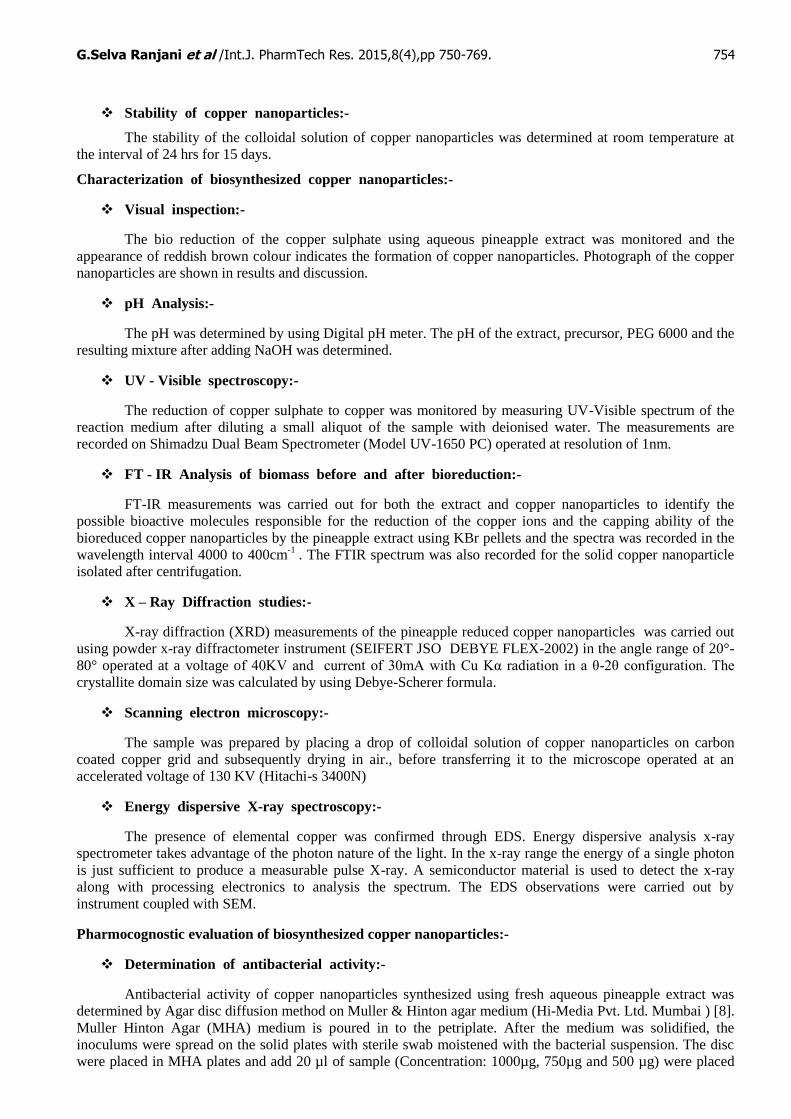
G.Selva Ranjani et al /Int.J. PharmTech Res. 2015,8(4),pp 750-769. 754
Stability of copper nanoparticles:-
The stability of the colloidal solution of copper nanoparticles was determined at room temperature at
the interval of 24 hrs for 15 days.
Characterization of biosynthesized copper nanoparticles:-
Visual inspection:-
The bio reduction of the copper sulphate using aqueous pineapple extract was monitored and the
appearance of reddish brown colour indicates the formation of copper nanoparticles. Photograph of the copper
nanoparticles are shown in results and discussion.
pH Analysis:-
The pH was determined by using Digital pH meter. The pH of the extract, precursor, PEG 6000 and the
resulting mixture after adding NaOH was determined.
UV - Visible spectroscopy:-
The reduction of copper sulphate to copper was monitored by measuring UV-Visible spectrum of the
reaction medium after diluting a small aliquot of the sample with deionised water. The measurements are
recorded on Shimadzu Dual Beam Spectrometer (Model UV-1650 PC) operated at resolution of 1nm.
FT - IR Analysis of biomass before and after bioreduction:-
FT-IR measurements was carried out for both the extract and copper nanoparticles to identify the
possible bioactive molecules responsible for the reduction of the copper ions and the capping ability of the
bioreduced copper nanoparticles by the pineapple extract using KBr pellets and the spectra was recorded in the
wavelength interval 4000 to 400cm-1
. The FTIR spectrum was also recorded for the solid copper nanoparticle
isolated after centrifugation.
X – Ray Diffraction studies:-
X-ray diffraction (XRD) measurements of the pineapple reduced copper nanoparticles was carried out
using powder x-ray diffractometer instrument (SEIFERT JSO DEBYE FLEX-2002) in the angle range of 20°-
80° operated at a voltage of 40KV and current of 30mA with Cu Kα radiation in a θ-2θ configuration. The
crystallite domain size was calculated by using Debye-Scherer formula.
Scanning electron microscopy:-
The sample was prepared by placing a drop of colloidal solution of copper nanoparticles on carbon
coated copper grid and subsequently drying in air., before transferring it to the microscope operated at an
accelerated voltage of 130 KV (Hitachi-s 3400N)
Energy dispersive X-ray spectroscopy:-
The presence of elemental copper was confirmed through EDS. Energy dispersive analysis x-ray
spectrometer takes advantage of the photon nature of the light. In the x-ray range the energy of a single photon
is just sufficient to produce a measurable pulse X-ray. A semiconductor material is used to detect the x-ray
along with processing electronics to analysis the spectrum. The EDS observations were carried out by
instrument coupled with SEM.
Pharmocognostic evaluation of biosynthesized copper nanoparticles:-
Determination of antibacterial activity:-
Antibacterial activity of copper nanoparticles synthesized using fresh aqueous pineapple extract was
determined by Agar disc diffusion method on Muller & Hinton agar medium (Hi-Media Pvt. Ltd. Mumbai ) [8].
Muller Hinton Agar (MHA) medium is poured in to the petriplate. After the medium was solidified, the
inoculums were spread on the solid plates with sterile swab moistened with the bacterial suspension. The disc
were placed in MHA plates and add 20 µl of sample (Concentration: 1000µg, 750µg and 500 µg) were placed
G.Selva Ranjani et al /Int.J. PharmTech Res. 2015,8(4),pp 750-769. 755
in the disc .The plates were incubated at 37ºC for 24 hrs. Then the antimicrobial activity was determined by
measuring the diameter of zone of inhibition.
Determination of antifungal activity:-
Antifungal activity of copper nanoparticles synthesized using fresh aqueous pineapple of extracts was
determined by disc diffusion method on Sabouraud Dextrose agar (SDA) medium [8]. SDA medium is poured
in to the petriplate. After the medium was solidified, the inoculums were spread on the solid plates with sterile
swab moistened with the fungal suspension. The disc were placed in SDA plates and add 20 µl of sample
(Concentration: 1000µg, 750µg and 500 µg) were placed in the disc .The plates were incubated at 37ºC for 24
hrs. Then the antifungal activity was determined by measuring the diameter of zone of inhibition.
Results and Discussion
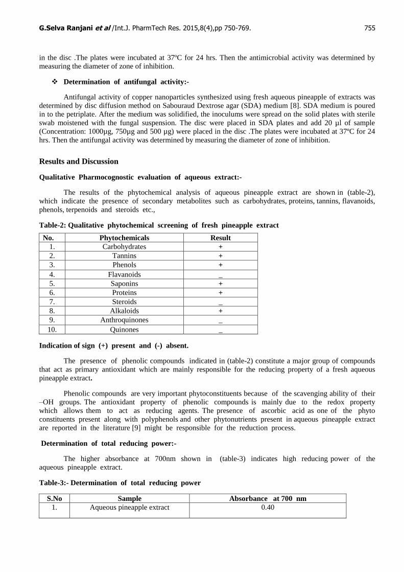
Qualitative Pharmocognostic evaluation of aqueous extract:-
The results of the phytochemical analysis of aqueous pineapple extract are shown in (table-2),
which indicate the presence of secondary metabolites such as carbohydrates, proteins, tannins, flavanoids,
phenols, terpenoids and steroids etc.,
Table-2: Qualitative phytochemical screening of fresh pineapple extract
Indication of sign (+) present and (-) absent.
The presence of phenolic compounds indicated in (table-2) constitute a major group of compounds
that act as primary antioxidant which are mainly responsible for the reducing property of a fresh aqueous
pineapple extract.
Phenolic compounds are very important phytoconstituents because of the scavenging ability of their
–OH groups. The antioxidant property of phenolic compounds is mainly due to the redox property
which allows them to act as reducing agents. The presence of ascorbic acid as one of the phyto
constituents present along with polyphenols and other phytonutrients present in aqueous pineapple extract
are reported in the literature [9] might be responsible for the reduction process.
Determination of total reducing power:-
The higher absorbance at 700nm shown in (table-3) indicates high reducing power of the
aqueous pineapple extract.
Table-3:- Determination of total reducing power
S.No Sample Absorbance at 700 nm
1. Aqueous pineapple extract 0.40
No. Phytochemicals Result
1. Carbohydrates +
2. Tannins +
3. Phenols +
4. Flavanoids _
5. Saponins +
6. Proteins +
7. Steroids _
8. Alkaloids +
9. Anthroquinones _
10. Quinones _
G.Selva Ranjani et al /Int.J. PharmTech Res. 2015,8(4),pp 750-769. 756
Determination of total antioxidant capacity:-
Pineapple contain a complex network of antioxidant metabolites and enzymes that work together
to prevent oxidative damage to cellular components. Pineapple contains a number of essential nutrients,
including vitamin C, manganese, and fiber. It also contains beneficial phytochemicals (Ferulic acid and
chlorogenic acid) which have antioxidant and anti-cancer activities. These antioxidative compounds delay
or inhibit the oxidation of molecules by inhibiting the initiation or propagation of oxidative chain
reaction. The antioxidative activity of phenolic compounds is mainly due to their redox property, which
plays an important role in absorbing and neutralizing free radicals, quenching singlet and triplet oxygen
or decomposing peroxides [10].
Another phenol, with high radical scavenging capacity present in the pineapple extract is the
chlorogenic acid. Hydrocinnamic acids, caeffic acids, rosemarinic acid and chlorogenic acids are more
efficient scavengers of free radicals than benzoic acid derivatives [11]. Thus the different types of
antioxidants presented in the pineapple extract synergistically reduce the Cu metal ions as each
antioxidant is unique in terms of its structure and antioxidant function of trapping the different free
radicals. In addition different antioxidants react with one another to regenerate each other forming an
antioxidant network.
Therefore it is necessary to determine the free radical scavenging activity by DPPH method. The
DPPH activity of fresh aqueous pineapple extract was found to be 82.8% shown in (table-4) indicates
that fresh aqueous pineapple extract has potent antioxidant property.
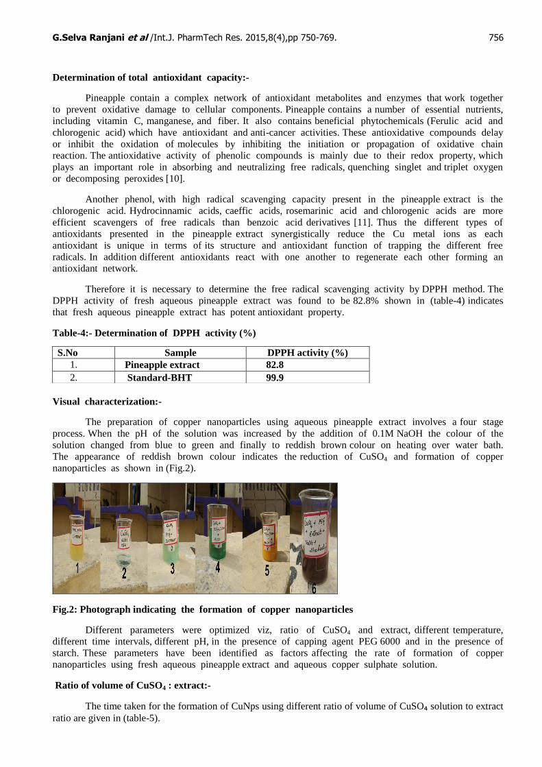
Table-4:- Determination of DPPH activity (%)
Visual characterization:-
The preparation of copper nanoparticles using aqueous pineapple extract involves a four stage
process. When the pH of the solution was increased by the addition of 0.1M NaOH the colour of the
solution changed from blue to green and finally to reddish brown colour on heating over water bath.
The appearance of reddish brown colour indicates the reduction of CuSO4 and formation of copper
nanoparticles as shown in (Fig.2).
Fig.2: Photograph indicating the formation of copper nanoparticles
Different parameters were optimized viz, ratio of CuSO4 and extract, different temperature,
different time intervals, different pH, in the presence of capping agent PEG 6000 and in the presence of
starch. These parameters have been identified as factors affecting the rate of formation of copper
nanoparticles using fresh aqueous pineapple extract and aqueous copper sulphate solution.
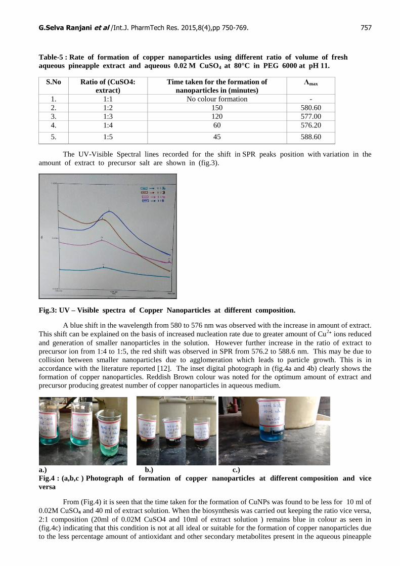
Ratio of volume of CuSO4 : extract:-
The time taken for the formation of CuNps using different ratio of volume of CuSO₄ solution to extract
ratio are given in (table-5).
S.No Sample DPPH activity (%)
1. Pineapple extract 82.8
2. Standard-BHT 99.9
G.Selva Ranjani et al /Int.J. PharmTech Res. 2015,8(4),pp 750-769. 757
Table-5 : Rate of formation of copper nanoparticles using different ratio of volume of fresh
aqueous pineapple extract and aqueous 0.02 M CuSO4 at 80°C in PEG 6000 at pH 11.
The UV-Visible Spectral lines recorded for the shift in SPR peaks position with variation in the
amount of extract to precursor salt are shown in (fig.3).
Fig.3: UV – Visible spectra of Copper Nanoparticles at different composition.
A blue shift in the wavelength from 580 to 576 nm was observed with the increase in amount of extract.
This shift can be explained on the basis of increased nucleation rate due to greater amount of Cu2⁺ ions reduced
and generation of smaller nanoparticles in the solution. However further increase in the ratio of extract to
precursor ion from 1:4 to 1:5, the red shift was observed in SPR from 576.2 to 588.6 nm. This may be due to
collision between smaller nanoparticles due to agglomeration which leads to particle growth. This is in
accordance with the literature reported [12]. The inset digital photograph in (fig.4a and 4b) clearly shows the
formation of copper nanoparticles. Reddish Brown colour was noted for the optimum amount of extract and
precursor producing greatest number of copper nanoparticles in aqueous medium.
a.) b.) c.)
Fig.4 : (a,b,c ) Photograph of formation of copper nanoparticles at different composition and vice
versa
From (Fig.4) it is seen that the time taken for the formation of CuNPs was found to be less for 10 ml of
0.02M CuSO₄ and 40 ml of extract solution. When the biosynthesis was carried out keeping the ratio vice versa,
2:1 composition (20ml of 0.02M CuSO4 and 10ml of extract solution ) remains blue in colour as seen in
(fig.4c) indicating that this condition is not at all ideal or suitable for the formation of copper nanoparticles due
to the less percentage amount of antioxidant and other secondary metabolites present in the aqueous pineapple
S.No Ratio of (CuSO4:
extract)
Time taken for the formation of
nanoparticles in (minutes)
Λmax
1. 1:1 No colour formation -
2. 1:2 150 580.60
3. 1:3 120 577.00
4. 1:4 60 576.20
5. 1:5 45 588.60
G.Selva Ranjani et al /Int.J. PharmTech Res. 2015,8(4),pp 750-769. 758
extract. Hence it needed a large amount of aqueous pineapple extract to reduce a Cu2+
ions. It is also inferred
that the amount of antioxidant and other secondary metabolites present in the aqueous pineapple extract plays
an important role in the reducing reaction as indicated by the formation of nanoparticles.
Effect of temperature on biosynthesis of copper nanoparticles:-
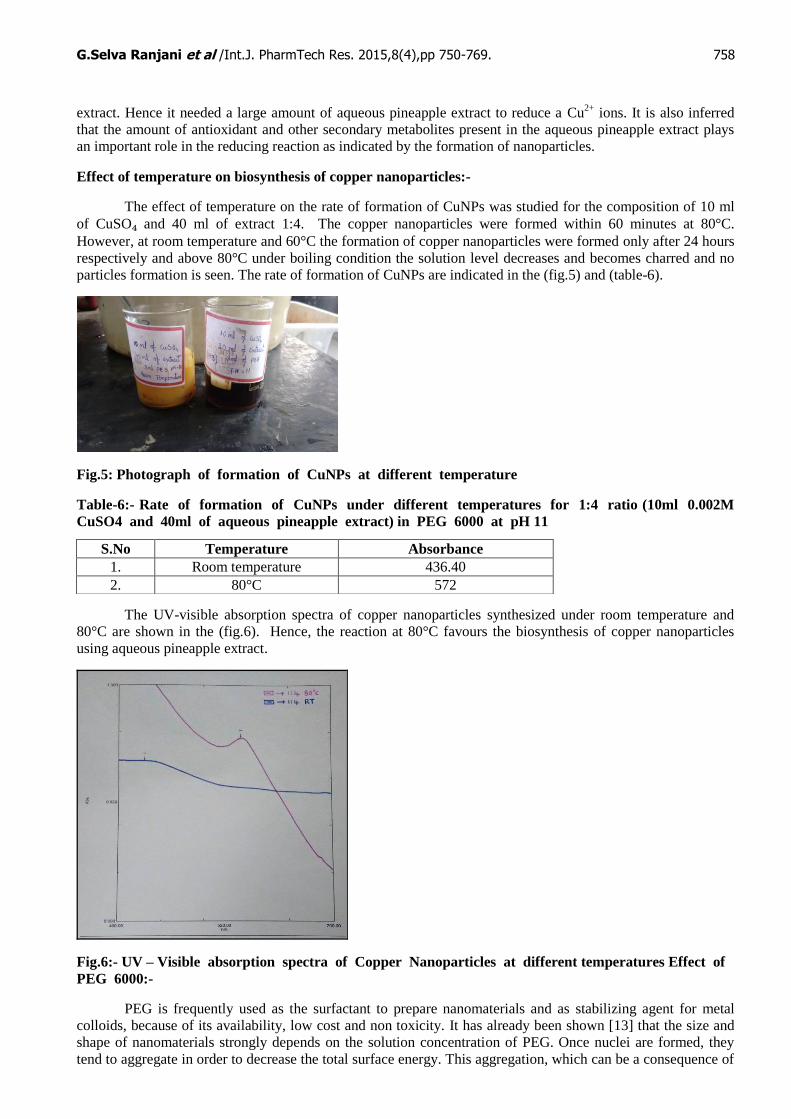
The effect of temperature on the rate of formation of CuNPs was studied for the composition of 10 ml
of CuSO₄ and 40 ml of extract 1:4. The copper nanoparticles were formed within 60 minutes at 80°C.
However, at room temperature and 60°C the formation of copper nanoparticles were formed only after 24 hours
respectively and above 80°C under boiling condition the solution level decreases and becomes charred and no
particles formation is seen. The rate of formation of CuNPs are indicated in the (fig.5) and (table-6).
Fig.5: Photograph of formation of CuNPs at different temperature
Table-6:- Rate of formation of CuNPs under different temperatures for 1:4 ratio (10ml 0.002M
CuSO4 and 40ml of aqueous pineapple extract) in PEG 6000 at pH 11
The UV-visible absorption spectra of copper nanoparticles synthesized under room temperature and
80°C are shown in the (fig.6). Hence, the reaction at 80°C favours the biosynthesis of copper nanoparticles
using aqueous pineapple extract.
Fig.6:- UV – Visible absorption spectra of Copper Nanoparticles at different temperatures Effect of
PEG 6000:-
PEG is frequently used as the surfactant to prepare nanomaterials and as stabilizing agent for metal
colloids, because of its availability, low cost and non toxicity. It has already been shown [13] that the size and
shape of nanomaterials strongly depends on the solution concentration of PEG. Once nuclei are formed, they
tend to aggregate in order to decrease the total surface energy. This aggregation, which can be a consequence of
S.No Temperature Absorbance
1. Room temperature 436.40
2. 80°C 572
G.Selva Ranjani et al /Int.J. PharmTech Res. 2015,8(4),pp 750-769. 759
attractive Van der Waals forces between crystals, should be inhibited or limited to restrict the final particle size
at the nanometric scale. One way to prevent nanoparticles from aggregation is the use of substances that lead to
steric repulsion between individuals. PEG inhibits this type of growth and aggregation among biosynthesized
nanoparticles.
In the present work PEG 6000 was used, which act as size controller and polymeric capping agent, as it
hinders the nuclei from aggregation through the polar groups which are strongly adsorbed at the surface of the
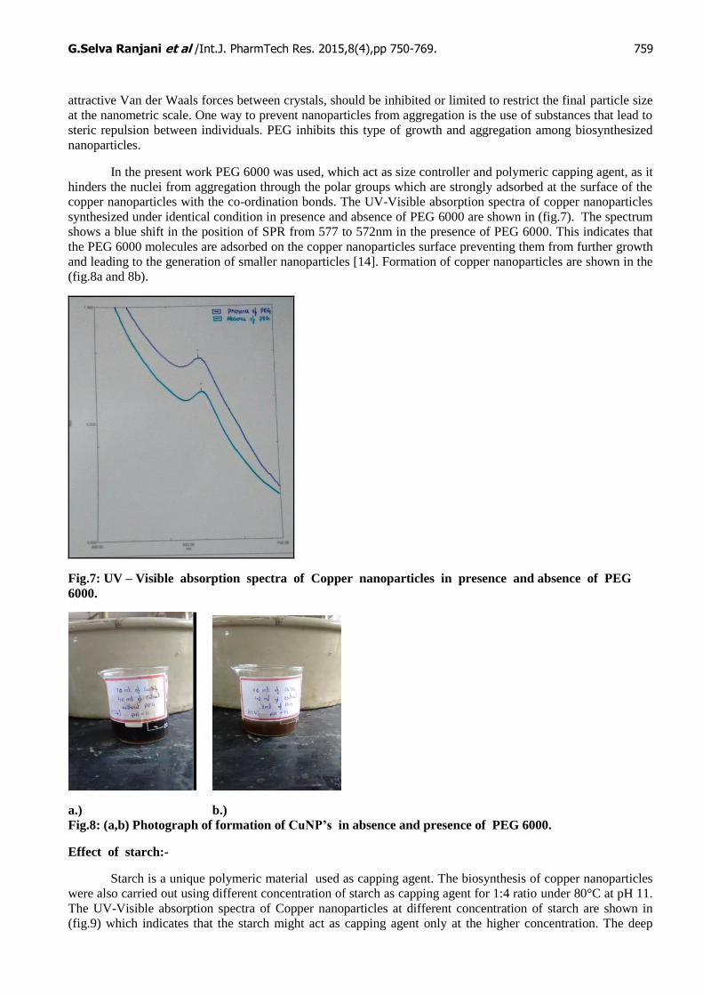
copper nanoparticles with the co-ordination bonds. The UV-Visible absorption spectra of copper nanoparticles
synthesized under identical condition in presence and absence of PEG 6000 are shown in (fig.7). The spectrum
shows a blue shift in the position of SPR from 577 to 572nm in the presence of PEG 6000. This indicates that
the PEG 6000 molecules are adsorbed on the copper nanoparticles surface preventing them from further growth
and leading to the generation of smaller nanoparticles [14]. Formation of copper nanoparticles are shown in the
(fig.8a and 8b).
Fig.7: UV – Visible absorption spectra of Copper nanoparticles in presence and absence of PEG
6000.
a.) b.)
Fig.8: (a,b) Photograph of formation of CuNP’s in absence and presence of PEG 6000.
Effect of starch:-
Starch is a unique polymeric material used as capping agent. The biosynthesis of copper nanoparticles
were also carried out using different concentration of starch as capping agent for 1:4 ratio under 80°C at pH 11.
The UV-Visible absorption spectra of Copper nanoparticles at different concentration of starch are shown in
(fig.9) which indicates that the starch might act as capping agent only at the higher concentration. The deep

G.Selva Ranjani et al /Int.J. PharmTech Res. 2015,8(4),pp 750-769. 760
investigation with regard to starch will be carried out in future. As far as the stability is concerned, it is stable
only for 4 days where as biosynthesized copper nanoparticle using PEG6000 was stable for more than a week.
Fig. 9:- UV – Visible Absorption spectra of formation of Copper Nanoparticles at different
concentration of starch.
Effect of pH:-
The work reported by Thi My Dung Dang et al [15] showed that the pH of the solution has an influence
on the progress of copper sulphate reduction reaction. The pH of the pineapple extract, copper sulphate and
PEG 6000 on mixing was found to be 5. The probable kinetic enhancement could be conducive to a reduction in
crystallite size because of the enhancement of the nucleation rate. The antioxidant present in the extract induces
a reduction in the solution pH which was adjusted back in the range from 7 to 12 with addition of 0.1 M NaOH
solution and it also accelerates the reduction reaction in water. The UV-Visible absorption spectra for the pH
ranging from 7 to 12 are shown in (fig.10). The Surface Plasmon absorbance of copper solution nanoparticles
was obtained for all pH except at pH 7. This probably indicates the formation of very small particles at such
low pH. The Plasmon Resonance is clearly visible for pH 9 to 11. At pH 12 the peak is still detectable but
shows red shift when compared with other pH values.
The maximum blue shift in SPR peak position at pH 11 could be attributed due to the decrease in
particle size [16]. The exact position of the Plasmon absorption may depend on several factors (including
particle size, shape, solvent type and capping agent) and in this case, there might be some variation in the
arrangement of capping molecules around the copper nanoparticles as a consequence of variation in pH. The pH
11 is found to be ideal due to the appearance of the reddish brown colour within 60 minutes because
biosynthesized copper nanoparticles showed maximum absorption at 576.20 nm which is in agreement with the
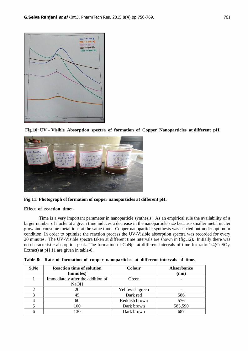
reported values in literature [17]. The formation of copper nanoparticle at different pH are shown in (fig.11).
The rate of formation of CuNPs at different pH for ratio 1:4 (CuSO4: Extract) are given in table-7.
Table-7: Rate of formation of copper nanoparticles at different pH
S.N
o
pH Time Taken For The Formation Of Copper
Nanoparticles in (minutes)
Absorbance
1. 7 360 -
2. 8 240 593.00
3. 9 120 580.60
4. 10 90 577.00
5. 11 60 576.20
6. 12 45 588.60
G.Selva Ranjani et al /Int.J. PharmTech Res. 2015,8(4),pp 750-769. 761
Fig.10: UV – Visible Absorption spectra of formation of Copper Nanoparticles at different pH.
Fig.11: Photograph of formation of copper nanoparticles at different pH.
Effect of reaction time:-
Time is a very important parameter in nanoparticle synthesis. As an empirical rule the availability of a
larger number of nuclei at a given time induces a decrease in the nanoparticle size because smaller metal nuclei
grow and consume metal ions at the same time. Copper nanoparticle synthesis was carried out under optimum
condition. In order to optimize the reaction process the UV-Visible absorption spectra was recorded for every
20 minutes. The UV-Visible spectra taken at different time intervals are shown in (fig.12). Initially there was
no characteristic absorption peak. The formation of CuNps at different intervals of time for ratio 1:4(CuSO₄: Extract) at pH 11 are given in table-8.
Table-8:- Rate of formation of copper nanoparticles at different intervals of time.
S.No Reaction time of solution
(minutes)
Colour Absorbance
(nm)
1 Immediately after the addition of
NaOH
Green -
2 20 Yellowish green -
3 45 Dark red 586
4 60 Reddish brown 576
5 100 Dark brown 583,590
6 130 Dark brown 687
G.Selva Ranjani et al /Int.J. PharmTech Res. 2015,8(4),pp 750-769. 762
Fig.12: UV-Visible Absorption spectra of CuNPs at different intervals of time
From (table-7) and the (fig.12) it is seen that the CuNps were formed within 60 minutes exhibited
Plasmon Resonance at 576nm. It suggests a homogenization mechanism, which provides a larger number of
nuclei with time. For 100 and 130 minutes no clear Plasmon Resonance was observed at higher range of
wavelength 590, 687nm. This could indicate an formation of copper oxide on heating for longer time interval.
At this moment the mechanism associated with this phenomenon is not clearly understood. Ascorbic acid
present in the pineapple extract as reported in the literature [18] is well known to scavenge free radicals. This
provides the right condition for subsequent rapid reduction by phytonutrients, polyphenols along with ascorbic
acid and hence copper nanoparticles formation [14].
Stability:-
The stability of nanoparticles dispersion is a key factor in their application. In order to prevent
agglomeration of nanoparticles, several capping agent is added in this media. In this present work, antioxidant
present in the pineapple extract acts as both reducing and capping agent along with the protecting agent viz
PEG 6000. The antioxidant and PEG 6000 stabilized copper nanoparticles dispersion was centrifuged at 8000
rpm for 15 minutes. It is seen that the residue gets settled at the bottom of the centrifuge tube. The supernant
liquid was decanted under ambient conditions and no sign of sedimentation was observed even after 10 days of
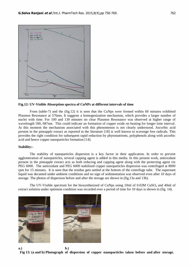
storage. The photos of dispersion before and after the storage are shown in (fig.13a and 13b).
The UV-Visible spectrum for the biosynthesized of CuNps using 10ml of 0.02M CuSO4 and 40ml of
extract solution under optimum condition was recorded over a period of time for 10 days is shown in (fig. 14).
a.) b.)
Fig 13: (a and b) Photograph of dispersion of copper nanoparticles taken before and after storage.
G.Selva Ranjani et al /Int.J. PharmTech Res. 2015,8(4),pp 750-769. 763
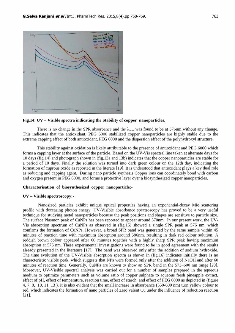
Fig.14: UV – Visible spectra indicating the Stability of copper nanoparticles.
There is no change in the SPR absorbance and the λmax was found to be at 576nm without any change.
This indicates that the antioxidant, PEG 6000 stabilized copper nanoparticles are highly stable due to the
extreme capping effect of both antioxidant, PEG 6000 and the dispersion effect of the polyhydroxyl structure.
This stability against oxidation is likely attributable to the presence of antioxidant and PEG 6000 which
forms a capping layer at the surface of the particle. Based on the UV-Vis spectral line taken at alternate days for
10 days (fig.14) and photograph shown in (fig.13a and 13b) indicates that the copper nanoparticles are stable for
a period of 10 days. Finally the solution was turned into dark green colour on the 12th day, indicating the
formation of cuprous oxide as reported in the literate [19]. It is understood that antioxidant plays a key dual role
as reducing and capping agent. During nano particle synthesis Copper ions can coordinately bond with carbon
and oxygen present in PEG 6000, and forms a protective layer over a biosynthesized copper nanoparticles.
Characterisation of biosynthesized copper nanoparticle:-
UV – Visible spectroscopy:-
Nanosized particles exhibit unique optical properties having an exponential-decay Mie scattering
profile with deceasing photon energy. UV-Visible absorbance spectroscopy has proved to be a very useful
technique for studying metal nanoparticles because the peak positions and shapes are sensitive to particle size.
The surface Plasmon peak of CuNPs has been reported to appear around 570nm. In our present work, the UV-
Vis absorption spectrum of CuNPs as observed in (fig.15) showed a single SPR peak at 576 nm, which
confirms the formation of CuNPs. However, a broad SPR band was generated by the same sample within 45
minutes of reaction time with maximum absorption around 586nm, resulting in dark red colour solution. A
reddish brown colour appeared after 60 minutes together with a highly sharp SPR peak having maximum
absorption at 576 nm. These experimental investigations were found to be in good agreement with the results
already presented in the literature [17]. The band was observed only after the addition of sodium hydroxide.
The time evolution of the UV-Visible absorption spectra as shown in (fig.16) indicates initially there is no
characteristic visible peak, which suggests that NPs were formed only after the addition of NaOH and after 60
minutes of reaction time. Generally, CuNPs are known to show an SPR band in the 573–600 nm range [20].
Moreover, UV-Visible spectral analysis was carried out for a number of samples prepared in the aqueous
medium to optimize parameters such as volume ratio of copper sulphate to aqueous fresh pineapple extract,
effect of pH, effect of temperature, reaction time, effect of starch and effect of PEG 6000 as depicted in (figure
4, 7, 8, 10, 11, 13 ). It is also evident that the small increase in absorbance (550-600 nm) turn yellow colour to
red, which indicates the formation of nano particles of Zero valent Cu under the influence of reduction reaction
[21].
G.Selva Ranjani et al /Int.J. PharmTech Res. 2015,8(4),pp 750-769. 764
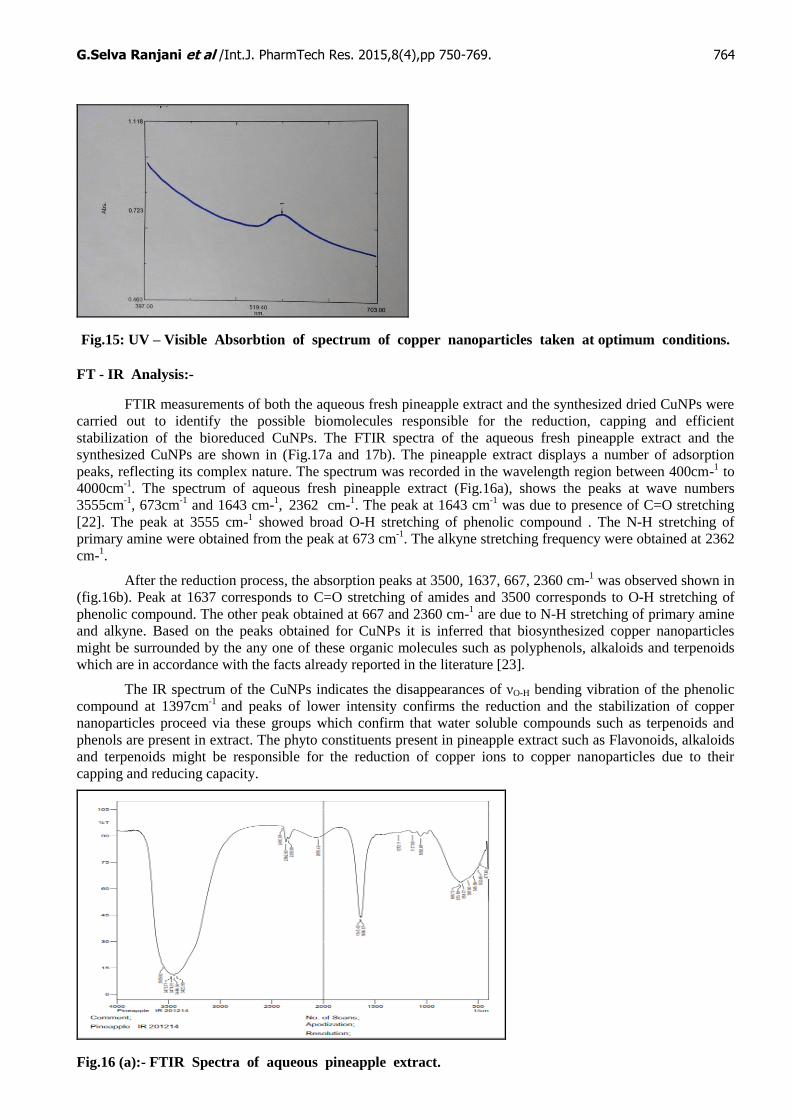
Fig.15: UV – Visible Absorbtion of spectrum of copper nanoparticles taken at optimum conditions.
FT - IR Analysis:-
FTIR measurements of both the aqueous fresh pineapple extract and the synthesized dried CuNPs were
carried out to identify the possible biomolecules responsible for the reduction, capping and efficient
stabilization of the bioreduced CuNPs. The FTIR spectra of the aqueous fresh pineapple extract and the
synthesized CuNPs are shown in (Fig.17a and 17b). The pineapple extract displays a number of adsorption
peaks, reflecting its complex nature. The spectrum was recorded in the wavelength region between 400cm-1 to
4000cm-1
. The spectrum of aqueous fresh pineapple extract (Fig.16a), shows the peaks at wave numbers
3555cm-1
, 673cm-1
and 1643 cm-1, 2362 cm-
1. The peak at 1643 cm
-1 was due to presence of C=O stretching
[22]. The peak at 3555 cm-1 showed broad O-H stretching of phenolic compound . The N-H stretching of
primary amine were obtained from the peak at 673 cm-1
. The alkyne stretching frequency were obtained at 2362
cm-1.
After the reduction process, the absorption peaks at 3500, 1637, 667, 2360 cm-1 was observed shown in
(fig.16b). Peak at 1637 corresponds to C=O stretching of amides and 3500 corresponds to O-H stretching of
phenolic compound. The other peak obtained at 667 and 2360 cm-1 are due to N-H stretching of primary amine
and alkyne. Based on the peaks obtained for CuNPs it is inferred that biosynthesized copper nanoparticles
might be surrounded by the any one of these organic molecules such as polyphenols, alkaloids and terpenoids
which are in accordance with the facts already reported in the literature [23].
The IR spectrum of the CuNPs indicates the disappearances of νO-H bending vibration of the phenolic
compound at 1397cm-1
and peaks of lower intensity confirms the reduction and the stabilization of copper
nanoparticles proceed via these groups which confirm that water soluble compounds such as terpenoids and
phenols are present in extract. The phyto constituents present in pineapple extract such as Flavonoids, alkaloids
and terpenoids might be responsible for the reduction of copper ions to copper nanoparticles due to their
capping and reducing capacity.
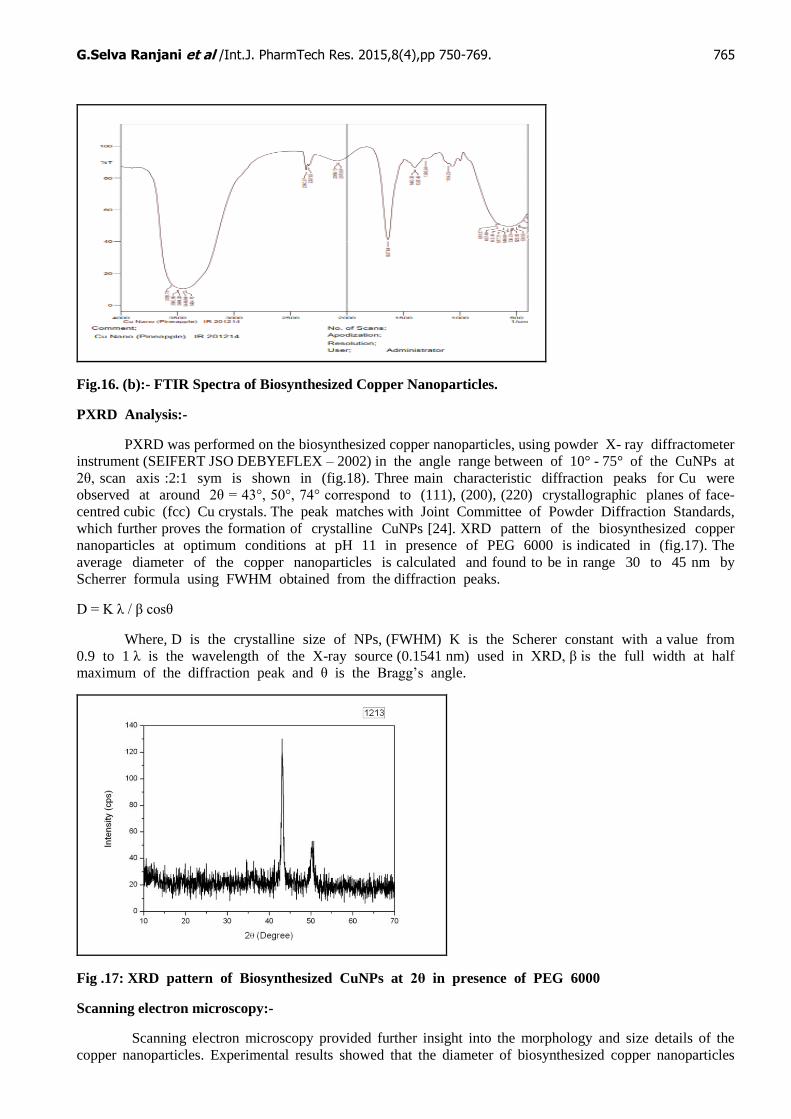
Fig.16 (a):- FTIR Spectra of aqueous pineapple extract.
G.Selva Ranjani et al /Int.J. PharmTech Res. 2015,8(4),pp 750-769. 765
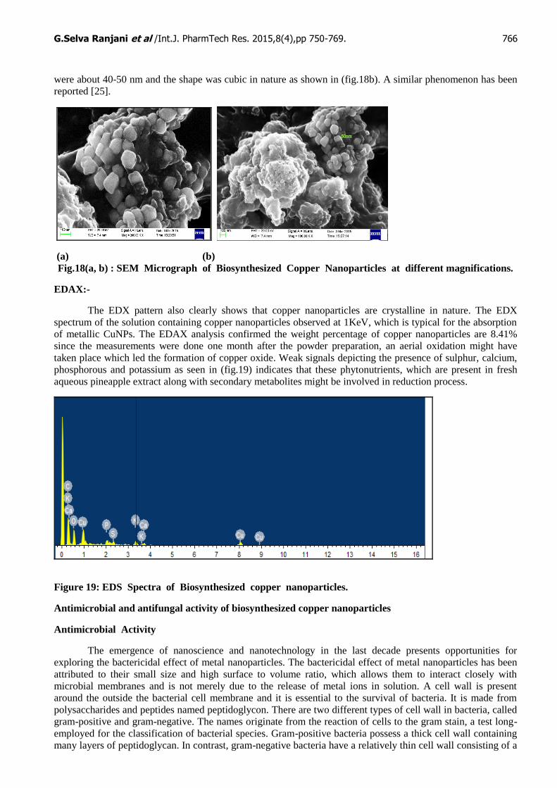
Fig.16. (b):- FTIR Spectra of Biosynthesized Copper Nanoparticles.
PXRD Analysis:-
PXRD was performed on the biosynthesized copper nanoparticles, using powder X- ray diffractometer
instrument (SEIFERT JSO DEBYEFLEX – 2002) in the angle range between of 10° - 75° of the CuNPs at
2θ, scan axis :2:1 sym is shown in (fig.18). Three main characteristic diffraction peaks for Cu were
observed at around 2θ = 43°, 50°, 74° correspond to (111), (200), (220) crystallographic planes of face-
centred cubic (fcc) Cu crystals. The peak matches with Joint Committee of Powder Diffraction Standards,
which further proves the formation of crystalline CuNPs [24]. XRD pattern of the biosynthesized copper
nanoparticles at optimum conditions at pH 11 in presence of PEG 6000 is indicated in (fig.17). The
average diameter of the copper nanoparticles is calculated and found to be in range 30 to 45 nm by
Scherrer formula using FWHM obtained from the diffraction peaks.
D = K λ / β cosθ
Where, D is the crystalline size of NPs, (FWHM) K is the Scherer constant with a value from
0.9 to 1 λ is the wavelength of the X-ray source (0.1541 nm) used in XRD, β is the full width at half
maximum of the diffraction peak and θ is the Bragg’s angle.
Fig .17: XRD pattern of Biosynthesized CuNPs at 2θ in presence of PEG 6000
Scanning electron microscopy:-
Scanning electron microscopy provided further insight into the morphology and size details of the
copper nanoparticles. Experimental results showed that the diameter of biosynthesized copper nanoparticles
G.Selva Ranjani et al /Int.J. PharmTech Res. 2015,8(4),pp 750-769. 766
were about 40-50 nm and the shape was cubic in nature as shown in (fig.18b). A similar phenomenon has been
reported [25].
(a) (b)
Fig.18(a, b) : SEM Micrograph of Biosynthesized Copper Nanoparticles at different magnifications.
EDAX:-
The EDX pattern also clearly shows that copper nanoparticles are crystalline in nature. The EDX
spectrum of the solution containing copper nanoparticles observed at 1KeV, which is typical for the absorption
of metallic CuNPs. The EDAX analysis confirmed the weight percentage of copper nanoparticles are 8.41%
since the measurements were done one month after the powder preparation, an aerial oxidation might have
taken place which led the formation of copper oxide. Weak signals depicting the presence of sulphur, calcium,
phosphorous and potassium as seen in (fig.19) indicates that these phytonutrients, which are present in fresh
aqueous pineapple extract along with secondary metabolites might be involved in reduction process.
Figure 19: EDS Spectra of Biosynthesized copper nanoparticles.
Antimicrobial and antifungal activity of biosynthesized copper nanoparticles
Antimicrobial Activity
The emergence of nanoscience and nanotechnology in the last decade presents opportunities for
exploring the bactericidal effect of metal nanoparticles. The bactericidal effect of metal nanoparticles has been
attributed to their small size and high surface to volume ratio, which allows them to interact closely with
microbial membranes and is not merely due to the release of metal ions in solution. A cell wall is present
around the outside the bacterial cell membrane and it is essential to the survival of bacteria. It is made from
polysaccharides and peptides named peptidoglycon. There are two different types of cell wall in bacteria, called
gram-positive and gram-negative. The names originate from the reaction of cells to the gram stain, a test long-
employed for the classification of bacterial species. Gram-positive bacteria possess a thick cell wall containing
many layers of peptidoglycan. In contrast, gram-negative bacteria have a relatively thin cell wall consisting of a
G.Selva Ranjani et al /Int.J. PharmTech Res. 2015,8(4),pp 750-769. 767
few layers of peptidoglycan. Surfaces of copper nanoparticles affect / interact directly with the bacterial outer
membrane, causing the membrane to rupture and killing bacteria.
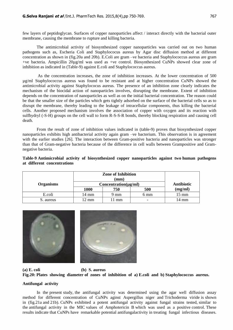
The antimicrobial activity of biosynthesized copper nanoparticles was carried out on two human
pathogens such as, Escheria Coli and Staphylococus aureus by Agar disc diffusion method at different
concentration as shown in (fig.20a and 20b). E.Coli are gram –ve bacteria and Staphylococcus aureus are gram
+ve bacteria. Ampicillin 20µg/ml was used as +ve control. Biosynthesized CuNPs showed clear zone of
inhibition as indicated in (Table-9) against E.coli and Staphylococcus aureus.
As the concentration increases, the zone of inhibition increases. At the lower concentration of 500
µg/ml Staphylococcus aureus was found to be resistant and at higher concentration CuNPs showed the
antimicrobial activity against Staphylococus aureus. The presence of an inhibition zone clearly indicates the
mechanism of the biocidal action of nanoparticles involves, disrupting the membrane. Extent of inhibition
depends on the concentration of nanoparticles as well as on the initial bacterial concentration. The reason could
be that the smaller size of the particles which gets tightly adsorbed on the surface of the bacterial cells so as to
disrupt the membrane, thereby leading to the leakage of intracellular components, thus killing the bacterial
cells. Another proposed mechanism involves the association of copper with oxygen and its reaction with
sulfhydryl (-S-H) groups on the cell wall to form R-S-S-R bonds, thereby blocking respiration and causing cell
death.
From the result of zone of inhibition values indicated in (table-9) proves that biosynthesized copper
nanoparticles exhibits high antibacterial activity again gram –ve bacterium. This observation is in agreement
with the earlier studies [26]. The interaction between Gram-positive bacteria and nanoparticles was stronger
than that of Gram-negative bacteria because of the difference in cell walls between Grampositive and Gram-
negative bacteria.
Table-9 Antimicrobial activity of biosynthesized copper nanoparticles against two human pathogens
at different concentrations
(a) E. coli (b) S. aureus
Fig.20: Plates showing diameter of zones of inhibition of a) E.coli and b) Staphylococcus aureus.
Antifungal activity
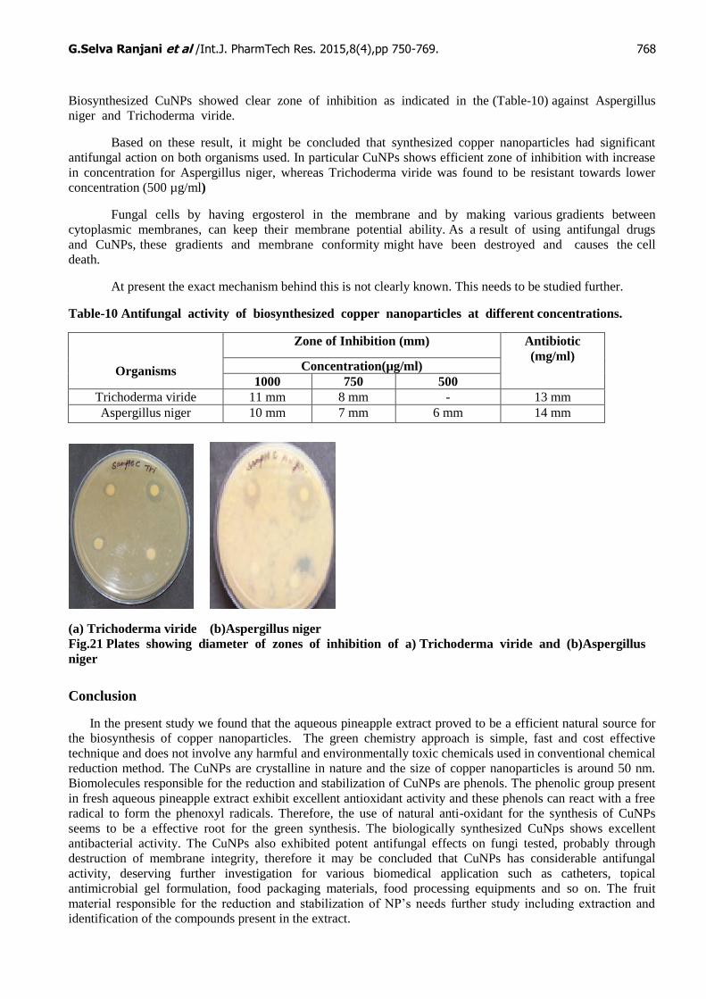
In the present study, the antifungal activity was determined using the agar well diffusion assay
method for different concentration of CuNPs aginst Aspergillus niger and Trichoderma viride is shown
in (fig.21a and 21b). CuNPs exhibited a potent antifungal activity against fungal strains tested, similar to
the antifungal activity in the MIC values of Amphotericin B which was used as a positive control. These
results indicate that CuNPs have remarkable potential antifungalactivity in treating fungal infectious diseases.
Organisms
Zone of Inhibition
(mm)
Antibiotic
(mg/ml) Concentration(µg/ml)
1000 750 500
E.coli 14 mm 9 mm 6 mm 15 mm
S. aureus 12 mm 11 mm - 14 mm
G.Selva Ranjani et al /Int.J. PharmTech Res. 2015,8(4),pp 750-769. 768
Biosynthesized CuNPs showed clear zone of inhibition as indicated in the (Table-10) against Aspergillus
niger and Trichoderma viride.
Based on these result, it might be concluded that synthesized copper nanoparticles had significant
antifungal action on both organisms used. In particular CuNPs shows efficient zone of inhibition with increase
in concentration for Aspergillus niger, whereas Trichoderma viride was found to be resistant towards lower
concentration (500 µg/ml)
Fungal cells by having ergosterol in the membrane and by making various gradients between
cytoplasmic membranes, can keep their membrane potential ability. As a result of using antifungal drugs
and CuNPs, these gradients and membrane conformity might have been destroyed and causes the cell
death.
At present the exact mechanism behind this is not clearly known. This needs to be studied further.
Table-10 Antifungal activity of biosynthesized copper nanoparticles at different concentrations.
Organisms
Zone of Inhibition (mm) Antibiotic
(mg/ml) Concentration(µg/ml)
1000 750 500
Trichoderma viride 11 mm 8 mm - 13 mm
Aspergillus niger 10 mm 7 mm 6 mm 14 mm
(a) Trichoderma viride (b)Aspergillus niger
Fig.21 Plates showing diameter of zones of inhibition of a) Trichoderma viride and (b)Aspergillus
niger
Conclusion
In the present study we found that the aqueous pineapple extract proved to be a efficient natural source for
the biosynthesis of copper nanoparticles. The green chemistry approach is simple, fast and cost effective
technique and does not involve any harmful and environmentally toxic chemicals used in conventional chemical
reduction method. The CuNPs are crystalline in nature and the size of copper nanoparticles is around 50 nm.
Biomolecules responsible for the reduction and stabilization of CuNPs are phenols. The phenolic group present
in fresh aqueous pineapple extract exhibit excellent antioxidant activity and these phenols can react with a free
radical to form the phenoxyl radicals. Therefore, the use of natural anti-oxidant for the synthesis of CuNPs
seems to be a effective root for the green synthesis. The biologically synthesized CuNps shows excellent
antibacterial activity. The CuNPs also exhibited potent antifungal effects on fungi tested, probably through
destruction of membrane integrity, therefore it may be concluded that CuNPs has considerable antifungal
activity, deserving further investigation for various biomedical application such as catheters, topical
antimicrobial gel formulation, food packaging materials, food processing equipments and so on. The fruit
material responsible for the reduction and stabilization of NP’s needs further study including extraction and
identification of the compounds present in the extract.
G.Selva Ranjani et al /Int.J. PharmTech Res. 2015,8(4),pp 750-769. 769
References:
1. Narayanan,R. and EI-Sayed,2003.M.A.J.Am.Chem.Soc., 125:8340.
2. Nguyen thi Phuong Phong, Vo Quoc Khuong, Tran Duc Tho, Cao Van Du, Ngo Hoang Minh.
3. Proceedings of IWNA 2011; November 10-12.
4. J. Sun, Y. Chu, X. Wu and R. Liu, “Antioxidant and An- tiproliferative Activities of Common Fruits,”
Journal of Agricultural and Food Chemistry, Vol. 50, No. 25, 2002, pp. 7449-7454.
doi:10.1021/jf0207530
5. M. Mhatre, J. Tilak-Jain, S. De and T. P. Devasagayam, “Evaluation of the Antioxidant Activity of
Non-Trans- formed and Transformed Pineapple: A Comparative Study,” Food and Chemical
Toxicology, Vol. 47, No. 11, 2009, pp. 2696-2702. doi:10.1016/j.fct.2009.06.031
6. B. C. Scott, J. B. Healliwll and O. B. Aruoma, “Evalua- tion of the Antioxidant Actions of Ferulic Acid
and Catechins,” Free Radical Research Communications, Vol. 19, No. 4, 1993, pp. 241-253.
doi:10.3109/10715769309056512
7. R. Hussain, S. J. Cillard and P. Cillard, “Hydroxyl Radi- cal Scavenging Activity of Flavonoids,”
Phytochemistry, Vol. 26, No. 9, 1989, pp. 2489-2491. doi:10.1016/S0031-9422(00)83860-1
8. Baur AW, Kirby WMM, Sherris JC, Turck M. Antibiotic Susceptibility testing by a standardized single
disk method. Am J ClinPathol. 1996;45:493-496.
9. Y. Z. Caia, Q. Luob, M. Sunc and H. Corke, “Antioxidant Activity and Phenolic Compounds of 112
Traditional Chinese Medicinal Plants Associated with Anticancer,” Life Sciences, Vol. 74, No. 17,
2004, pp. 2157-2184. doi:10.1016/j.lfs.2003.09.047
10. S. Panchawat and S. S. Sisodia, “In Vitro Antioxidant activity of Saraca Asoca Roxb. De Wilde Stem
Bark Ex-tracts from Various Extraction Processes,” Asian Journal of Pharmaceutical and Clinical
Research, Vol. 3, No. 3, 2010, pp. 231-233.
11. J. H. Chen and C. T. Ho, “Antioxidant Activities of Caf- feic Acid and Its Related Hydroxycinnamic
Acid Com- pounds,” Journal of Agricultural and Food Chemistry, Vol. 45, No. 7, 1997, pp. 2374-2378.
12. Kantabathini Venkata Pavani, Nandigam Srujana, Guntur Preethi, Tandale Swati. Synthesis of Copper
Nanoparticles by Aspergillus species. Open Access Journal. 2013; vol-2: 110-113
13. Zhang X, Yin H, Cheng X, Hu H, Yu Q and Wang A 2006 Mater. Res. Bull. 41 2041
14. Thi My Dung Dang, Thi Tuyet Thu Le, Eric Fribourg-Blanc and Mau Chien Dang. Synthesis and
optical properties of copper nanoparticles prepared by a chemical reduction method. Advances in
Natural Sciences, Nanoscience and Nanotechnology. 2011; (http://iobscience.iop.org/2043-6262/2/1/)
15. Zhang H X, Siegert U, Liu R and Cai W B 2009 Nanoscale Res. Lett. 4 705
16. Jing Xiong, Ye Wang, et al., Synthesis of highly stable dispersions of nanosized Copper Nanoparticles
using L- ascorbic acid. Green chem. 2011; 13: 900.
17. Pacheco M J G, Sánchez J E M, Hernández G, Ruiz F. Mater. Lett. 2010; 64: 1361. DOI: 10.
1016/j.matlet.2010.03.029.
18. A. Haripyarie, K. Guneshwor and M. Damayanti, “Evaluation of Antioxidant Properties of Phenolics
Ex- tracted from Ananas comosus L.,” Notulae Scientia Biologicae, Vol. 2, No. 2, 2010, pp. 68-71
19. Kooti, M.; Matouri, L. Transac. F: Nanotech. 2010, 17, 73.
20. Dang T M D, T Le T T, Fribourg-Blanc E and Dang M C. Adv. Nat. Sci: Nanosci. Nanotechnol. 2011;
2: 015009. DOI:10.1088/2043-6362/2/1/015009.
21. Liu Q, Yu R L, Qiu G Z, Fang Z, Chen A L and Zhao Z W. Transac. Nonfer. Metal. Soc.China. 2008;
18: 1258.DOI: 10.1016/S1003-6326(08)60213-7.
22. Vasudev D. Kulkarni, Pramod S. Kulkarni Green Synthesis of Copper Nanoparticles Using Ocimum
Sanctum Leaf Extract International Journal of Chemical Studies ISSN: 2321-4902 Volume 1 Issue 3.
23. K. saranyaadevi, V.subha et al. synthesis of copper nanoparicles using Capparis Zeylanicaleaf extract.
International Journal of Chem Tech Research.2014;6(10).
24. Theivasanthi and Alagar M, X-ray Diffraction studies of Copper nanopowder.
25. Sathishkumar M, Sneha K et al.,Cinnamon zeylanicum bark extract and powder mediated green
synthesis of nano-crystalline silver particles and its bactericidal activity. Colloids Surf B Biointerfaces.
2009; Oct 15;73(2):332-8.doi: 10.1016/j.colsurfb.2009.06.005.
26. Y. Abboud. T. Saffaj et al., Biosynthesis, characterization and antimicrobial activity of copper oxide
nanoparticles (CONPs) produced using brown alga extract Appl Nanosci (2014) 4:571–576
*****
G.Selva Ranjani et al /Int.J. PharmTech Res. 2015,8(4),pp 750-769. 770
International Journal of PharmTech Research
log on to - www.sphinxsai.com
International Journal of
PharmTech Research is an open
access Bimonthly Journal, 7.5 Years old. It
contains more than 2200 published papers
since 2009.
Subject areas: This journal publishes the
Research and Review papers of the following subject/areas. Pharmaceutics, Pharmaceutical
Chemistry, Biopharma, Pharmacology, Pharmacy Practice, Pharmacognosy, Analytical Chemistry,
Biotechnology, Microbiology, Biochemistry, , Medicinal Science, Clinical Pharmacy, Medichem, and
applied related subject areas.
[1] RANKING:
It has been ranked from India (subject: Pharma Sciences) from India at International platform,
by SCOPUS- scimagojr. It has topped in total number of CITES AND CITABLE DOCUMENTS.
Find more by clicking on SCOPUS-scimagojr SITE....AS BELOW.....
http://www.scimagojr.com/journalrank.php?area=3000&category=0&country=IN&year=2013&o
rder=tc&min=0&min_type=tc
Please log on to - www.sphinxsai.com
*****
Indexed/Abstracted/ Ranked by
Else vier SCOPUS- scimagojr.


