

18/04/2013
1
Mohammed A. Al-Muharraqi MBChB (Dnd.), BDS (Dnd.), MDSc (Dnd.), MRCS (Glas.), FFD RCS (Irel.), MFDS RCS (Eng.)
Consultant Maxill Facial Surgeon
Royal Medical Services, Bahrain Defence Force,
KINGDOM OF BAHRAIN
1 2
Introduction Ortho (orthos – straight) gnathic (gnathos – jaw)
Facial disproportion (dento-facial deformity)
20% of population
MDT approach: orthodontist, OMFS, restorative dentist,
hygienists, psychologist/psychiatrist, technician,
anaesthetist
Aetiology anomalous facial development is complex &
multi-factorial Extremes of variation in normal development
Associated with recognisable syndromes
3
Orthognathic Surgery
The correction of functional and aesthetic
consequences of severe dentofacial deformity
through a combination of orthodontic, surgical
and possible, restorative dentistry
Simply its a surgical procedure that changes the
position of the jaws
4
5 6
Severe Class III skeletal pattern
Severe Class II skeletal pattern
Long face syndrome / Anterior open bite
Facial Asymmetries
Craniofacial anomalies, e.g. Cleft lip and palate
Retruded Mandible Protruded Mandible
18/04/2013
2
7
Orthodontic Indications
1. Aesthetics
2. Function
3. Stability
8
Orthodontic Indications
Patient Factors
1. Age and Sex – influence the amount of growth
remaining
2. Race – influence profile considerations
3. Medical History – contraindications for surgery
4. Psychological – patients perception of the
problem
9
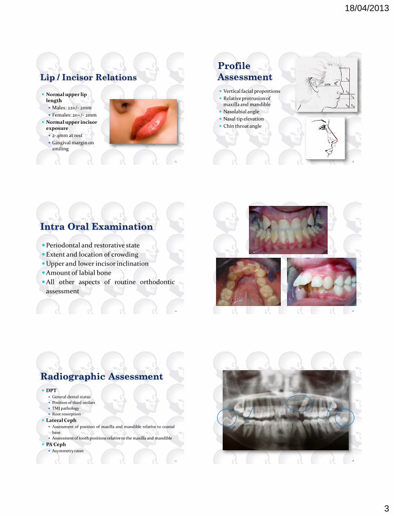
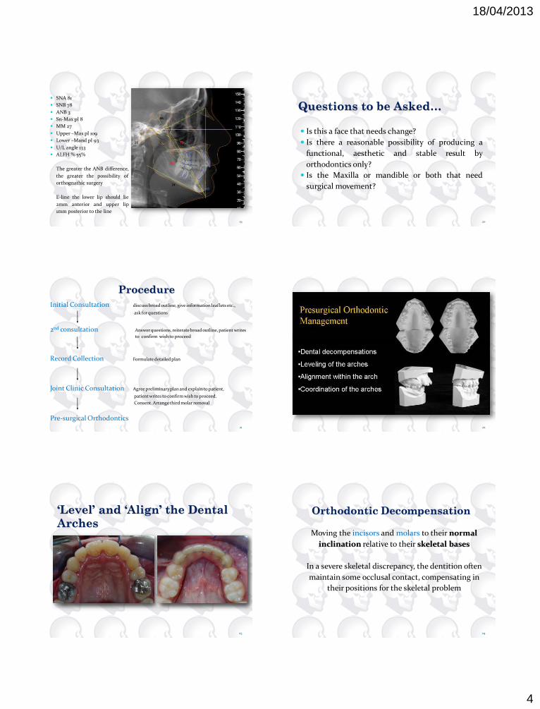
Frontal
Assessment Assessment of Facial Thirds
Symmetry
Vertical proportions i.e. Facial 1/3’s
Mid line in relation to maxilla,
mandible, nose and chin
Chin
Scleral Exposure
Normally the lower border of the iris
should lie behind the upper border
of the lower lid
Scleral exposure indicates maxillary
hypoplasia
Alar Base Width
Upper Lip / Incisor Relationship
10
11 12
18/04/2013
3
Lip / Incisor Relations
Normal upper lip length
Males: 22+/- 2mm
Females: 20+/- 2mm
Normal upper incisor exposure
2-4mm at rest
Gingival margin on smiling
13
Profile
Assessment
Vertical facial proportions
Relative protrusion of maxilla and mandible
Nasolabial angle
Nasal tip elevation
Chin throat angle
14
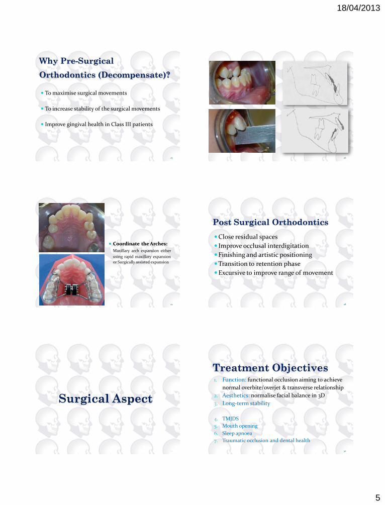
Intra Oral Examination
Periodontal and restorative state
Extent and location of crowding
Upper and lower incisor inclination
Amount of labial bone
All other aspects of routine orthodontic
assessment
15 16
Radiographic Assessment
DPT General dental status
Position of third molars
TMJ pathology
Root resorption
Lateral Ceph Assessment of position of maxilla and mandible relative to cranial
base
Assessment of tooth positions relative to the maxilla and mandible
PA Ceph Asymmetry cases
17 18
18/04/2013
4
19
SNA 81
SNB 78
ANB 3
Sn-Max pl 8
MM 27
Upper –Max pl 109
Lower –Mand pl 93
U/L angle 133
ALFH % 55%
The greater the ANB difference,
the greater the possibility of
orthognathic surgery
E-line the lower lip should lie
2mm anterior and upper lip
1mm posterior to the line
Questions to be Asked…
Is this a face that needs change?
Is there a reasonable possibility of producing a
functional, aesthetic and stable result by
orthodontics only?
Is the Maxilla or mandible or both that need
surgical movement?
20
Procedure
Initial Consultation discuss broad outline, give information leaf lets etc.,
ask for questions
2nd consultation Answer questions, reiterate broad outline, patient writes
to confirm wish to proceed
Record Collection Formulate detailed plan
Joint Clinic Consultation Agree preliminary plan and explain to patient,
patient writes to confirm wish to proceed.
Consent. Arrange third molar removal
Pre-surgical Orthodontics 21 22
23
‘Level’ and ‘Align’ the Dental
Arches
Orthodontic Decompensation
Moving the incisors and molars to their normal
inclination relative to their skeletal bases
In a severe skeletal discrepancy, the dentition often
maintain some occlusal contact, compensating in
their positions for the skeletal problem
24
18/04/2013
5
Why Pre-Surgical
Orthodontics (Decompensate)?
To maximise surgical movements
To increase stability of the surgical movements
Improve gingival health in Class III patients
25 26
27
Coordinate the Arches:
Maxillary arch expansion either
using rapid maxillary expansion
or Surgically assisted expansion
Post Surgical Orthodontics
Close residual spaces
Improve occlusal interdigitation
Finishing and artistic positioning
Transition to retention phase
Excursive to improve range of movement
28
29
Treatment Objectives 1. Function: functional occlusion aiming to achieve
normal overbite/overjet & transverse relationship
2. Aesthetics: normalise facial balance in 3D
3. Long-term stability
4. TMJDS
5. Mouth opening
6. Sleep apnoea
7. Traumatic occlusion and dental health
30
18/04/2013
6
Surgical Treatment
Planning Check list: essential information for planning
Class I/II/II
Skeletal base relationship
Maxilla AP – hypoplastic/normal
Vertical Maxillary Excess (VME)
Upper incisor show at rest & smiling
Centre lines – upper dental/lower dental/chin point
Overjet/overbite
Occlusal cant
Naso-labial angle
Upper lip length
Alar base width
31 32
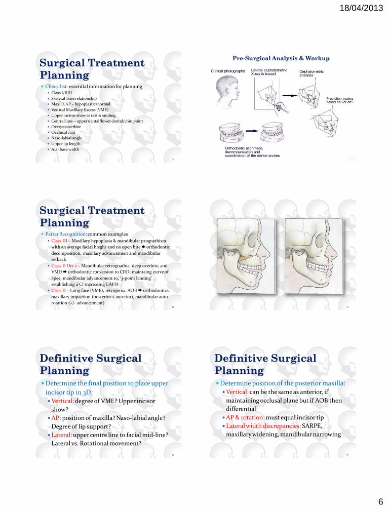
Pre-Surgical Analysis & Workup
Surgical Treatment
Planning Patter Recognition: common examples
Class III – Maxillary hypoplasia & mandibular prognathism
with an average facial hieght and no open bite orthodontic
decomposition, maxillary advancement and mandibular
setback
Class II Div 2 – Mandibular retrognathia, deep overbite, and
VMD orthodontic conversion to CIID1 maintaing curve of
Spee, mandibular advancement to, ‘3-point landing’
establishing a CI increasing LAFH
Class II – Long face (VME), retrogenia, AOB orthodontics,
maxillary impaction (posterior > anterior), mandibular auto-
rotation (+/- advancement) 33 34
Definitive Surgical
Planning Determine the final position to place upper
incisor tip in 3D:
Vertical: degree of VME? Upper incisor
show?
AP: position of maxilla? Naso-labial angle?
Degree of lip support?
Lateral: upper centre line to facial mid-line?
Lateral vs. Rotational movement?
35
Definitive Surgical
Planning Determine position of the posterior maxilla:
Vertical: can be the same as anterior, if
maintaining occlusal plane but if AOB then
differential
AP & rotation: must equal incisor tip
Lateral width discrepancies: SARPE,
maxillary widening, mandibular narrowing
36

18/04/2013
7
Definitive Surgical
Planning Mandibular positioning:
Deliver to class I
Consider lower centre line to facial
midline/dental midline
Chin:
Consider AP, vertical, lateral need for
genioplasty
It will be influenced by changing occlusion
37
Soft Tissue
Considerations Nasal tip relative to A point
1:3 in LeFort I (1:2 LFII, 1:1 LFIII)
Upper lip
80% of advancement, 50% of setback, 10-40% of
impaction, 50% of down grafting
Lower lip
85% of advancement, 60% of set-back
Chin:
Pogonion moves consistently in a 1:1
38
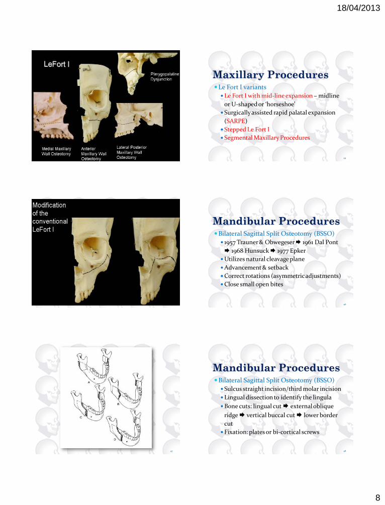
Maxillary Procedures Based on Le Fort (Rene, French surgeon 1869 –
1951) fracture lines
Le Fort I most popular
Total maxillary osteotomy through lateral wall of
maxilla, lateral wall of nose & nasal septum
Once mobilised can be moved in any dimension
Segmentalization to correct width, occlusal plane,
dento-alveolar discrepancies
39 40
Maxillary Procedures Le Fort I
Sulcus incision
Zygomatic buttress, IO nerve, piriform aperture & pterygo-
palatine fissure
Floor of the nose
Bone cuts: from posterior aspect of zygomatic buttress (5mm
above teeth) lateral wall of sinus base of piriform
aperture
Division of lateral nasal walls, nasal septum from crest,
pterygo-maxillary dysjunction
Down #, mobilisation, trimming of interferences
41 42
18/04/2013
8
43
Maxillary Procedures Le Fort I variants
Le Fort I with mid-line expansion – midline
or U-shaped or ‘horseshoe’
Surgically assisted rapid palatal expansion
(SARPE)
Stepped Le Fort I
Segmental Maxillary Procedures
44
45
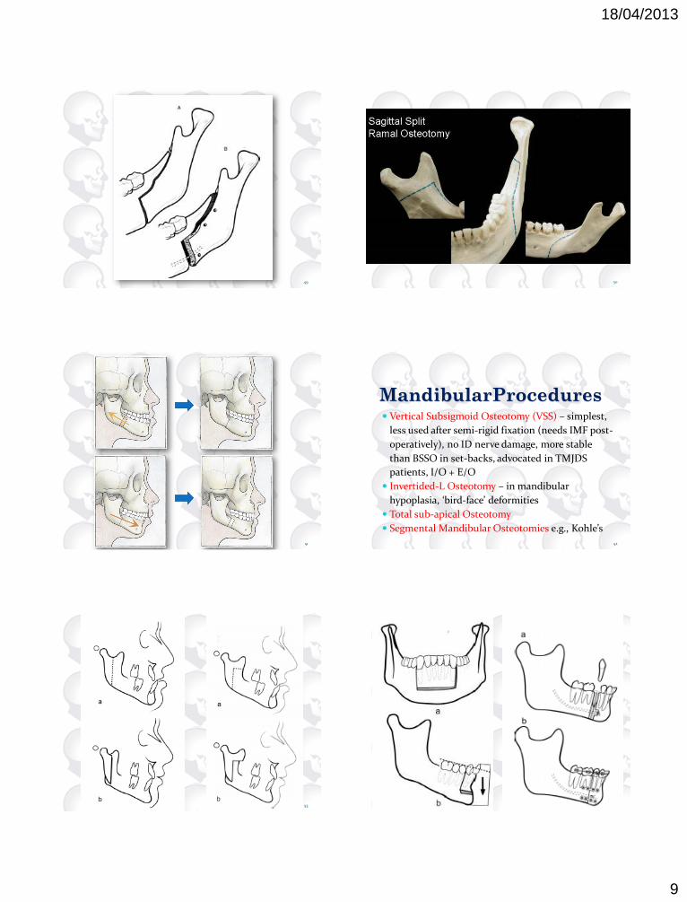
Mandibular Procedures Bilateral Sagittal Split Osteotomy (BSSO)
1957 Trauner & Obwegeser 1961 Dal Pont
1968 Hunsuck 1977 Epker
Utilizes natural cleavage plane
Advancement & setback
Correct rotations (asymmetric adjustments)
Close small open bites
46
47
Mandibular Procedures Bilateral Sagittal Split Osteotomy (BSSO)
Sulcus straight incision/third molar incision
Lingual dissection to identify the lingula
Bone cuts: lingual cut external oblique
ridge vertical buccal cut lower border
cut
Fixation: plates or bi-cortical screws
48
18/04/2013
9
49 50
51
MandibularProcedures Vertical Subsigmoid Osteotomy (VSS) – simplest,
less used after semi-rigid fixation (needs IMF post-
operatively), no ID nerve damage, more stable
than BSSO in set-backs, advocated in TMJDS
patients, I/O + E/O
Invertided-L Osteotomy – in mandibular
hypoplasia, ‘bird-face’ deformities
Total sub-apical Osteotomy
Segmental Mandibular Osteotomies e.g., Kohle’s
52
53 54
18/04/2013
10
Complications Haemorrhage – pterygoid venous plexus, greater
palatine nerve, naso-palatine vessels, maxillary
artery
Reduced by: LA, diathermy, hypotensive GA, table
position, efficient surgery, anti-fibrinolytics, care
in pterygo-maxillary dysjunction
Management: diathermy/ligation, packing,
haemostatic gauze, fixation, ligation of external
carotid, angiography and embolization
55
Complications Unfavorable Osteotomy – unwanted patterns
(most common in BSSO), ≤ 23%
Predisposed by: thin ascending ramus, unfavorable
bone texture, wisdom teeth, failure to divide lower
border at buccal cut
Undesirable fractures in maxillary osteotomies
may propagate to base of the skull
56
Complications Nerve Damage –
ID nerve during BSSO, long-term varies from 3% -
25% (up to 85%)
Risk factors: age, lag-screws, BSS + genioplasty
Facial nerve VII damage rare (0.5% - 1%)
Labial gingival and anterior palatal mucosa
CN II, III, IV, VI, X and XII due to unfavorable
fractures propagating up to the base of the skull
57
Complications Condyle Positioning –
Mal-occlusion will result
Upright vs. supine patient, paralyzed vs.
unparalyzed patient
Notorious in: Mandibular rotations: rotation of the condyle, winging
of the proximal fragment, inability to get fragments to
sit passively (excessive torquing)
Le Fort I impaction with interferences distracting the
condyle causing an AOB on releasing IMF 58
Complications Tooth Damage –
Direct damage (bone cuts, screws) or due to
ischemic changes
Soft Tissue Changes –
Nasal changes – alar base widens, nasal tip
upturned, naso-labial angle decreases
(advancement)
Reduction of nasal septum (to prevent buckling),
cinch sutures, V-Y closure
59
Stability Relapse/Migration
Surgical/Orthodontic
Careful planning building in relapse
Hierarchy of stability (Proffit, Yurvey & Phillips) Maxillary impaction mandibular advancement
maxillary advancement maxillary impaction with
mandibular advancement maxillary advancement
with mandibular setback mandibular setback
increasing maxillary width inferior positioning of
maxilla 60

18/04/2013
11
Stability Maxillary Surgery
Impaction is the most stable, but stability decreases if
combined with advancement
Larger moves (>8mm) are potentially unstable
Short period of IMF may aid stability
Inferior positioning of maxilla: relapse of 30-50%, mal or
non-union common, must be accompanied by grafting
Surgical widening: very unstable, 6/52 support (acrylic
plate or accessory bucco-labial arch wire), accompanied by
grafting
61
Stability Mandibular Surgery
Degree of advancement/setback & fixation
BSSO relapses forward , VSS migrates posteriorly
Plates vs. screws – same results
For large advancement (>10mm), suspension wires
1/52
Larger moves in setback (>8mm) are also potentially
unstable – pterygo-masseteric sling impingement
(mitigated by Hunsuck modification)
62
Stability Condylar Resorption
6 – 18 months post-surgery
2-8% incidence
Clinically: Horizontal relapse with AOB,
radiologically: flatting of condylar head with
posterior shortening/angulation of neck
Risks: ♀ patients, CII, high FP-M plane angle,
TMJDS, large mandibular advances (<10mm),
counter clockwise rotations for AOB
63
Stability Condylar Resorption
Aetiology: increased pressure on posterior surface
of the condylar head thereby increasing load on
the TMJ stimulating resorption
Management: ‘self burn out’ then stabilize, an
unloading splint, extra-capsular approach
recommended, further corrective surgery confined
to maxilla?
64
65


