

CHAPTER- 1
INTRODUCTION

CHAPTER -1 INTRODUCTION
1.1 INT)RODUCTI'ON
Shilajit, also known as salajit, shilajatu, murnie or ixiummiyo is a pale-brown to blackish
-brown exudation, of variable consistency, coming out from layer of rocks in many
mountain ranges of the world, especially the Himalayan ranges of the Indian subcontinent
(Chopra et al, 1958; Ghosal, 1992a; Agarwal et ctL, 2007a). It is also found in many
other mountain ranges of the world, e.g. Afghanistan (Hindukush), Australia (Northern
Pollock Ranges), and in the former USSR (Tieii-Shan, Pamir, Caucasus, Ural), where it is
collected in small quantities from steep rock faces at altitudes between 1000 and 5000m
(Ghosal, 2002a). We have recently reported the physico-chemical, spectral and thermal
properties of Shilajit which further confirm its humic nature (Agarwal et ciL, 2007 b).
wShilajit has been reported to contain a number of components including resins, fatty
acids, sterols, triterpenes, aromatic carboxylic acids, 3,4-beu/.coumarins and a-
aminoacids (Ghosal et.al, 1976). The biological effects of shilajit have been ascribed to
two distinct classes of compounds (Ghosal et.al., 1991).
❖ The low molecular weight bioactive organic compounds such as oxygenated
dibenzo-a-pyrones, and
❖ The medium molecuhii- weight Fulvic acids (FA) and Humic acids (HA),
Wliile the benzopyrones act as the active principles, fulvic and humic acids acts act as
carrier molecules for in-vivo trmisportation of these bioactive substances (Anwer et.al.,
2007).The interior of this complexing agents are thus capable of forming inclusion
complexes with non-polar solutes and drug molecules with low bioavailability. These
drug molecules can be entrapped in the hydrophobic interior so as to increase their
solubility, dissolution and stability, thereby enhancing their bioavailability (Kliamia,
2006),
Such entrapment is also capable of enhancing the stability of the drug molecules. In fact,
it has been reported that the bioactive principles of shilajit owe their stability in the
natural habitat due to their entrapment in the voids (micropores) of the fulvic acids of
shilajit humus (Ghosal, 1992b, Agarwal et al, 2008a, c).). A purified fulvic acid carrier
Dept, o f Pharmaceutics, Jamia Uamdard, New Delhi-62 1
having a sponge like structure punctured by voids of about 200-1000A in diameter and an
average molecular weight of about 700-2500 to which a water insoluble and unstable
active ingredient added to fill the voids (Ghosal, 1992c).
So far, except Jamia tiamdard (Saltija, 2001; Khanna, 2006; Karmarkar, 2007; Anwer,
2005, Ahmad, 2006; Vashisht, 2006; Mirza, 2007 and Tyagi, 2007) there is no report in
the literature on the use of fulvic and humic acids in enhancing the bioavailabilty of any
drug. There are some scattered reports of their use as bioenhancers of trace elements and
vitamins.
Aspirin (ASA) is an old drug but still possesses high medical value, and its health
protection function such as antipyretic, anti-inflammatory, analgesic and anti-aggregatory
activity has received more and more attention. The acetylsalicylic acid molecule has a
carboxyl group and an ester group. The ester group can be easily hydrolyzed, which
reduces the medical value and has side effects on humans. A need exists to learn how to
inliibit the hydrolysis of acetylsalicylic acid and to reduce its toxicity.
In the present project we use humic and fulvic acids extracted from shilajit as complexing
agents for such moisture sensitive drug can be a potential approach to Inhibit the
degradation, such an interaction or association between the drug molecule either with
humic or fulvic acid can lead to an increase in the drug bioavailability, decreasing
toxicity and a better pharmacodynamic profile. A com parative study has been done
between complexes of humic acid/fulvic acid and hydroxy propyl-(3-
cyclodextrin (HP-P-CD) - aspirin complex.
1.2 L IT E R A T U R E R EV IEW
12,1 SMlaJIt
Shilajit is a pale-brown to blackish-brown exudation, of variable consistency, oozes from
the rocks of the Himalayas, as they become warm in the summer months. It is said to
cm-y the healing power of these great mountains (Chopra et a l, 1958; Ghosal, 1993;
Ghoaal et al, 2000; Frawley, 2001). It is also found in Russia, Tibet, Norway and other
countries, where it is collected in small quantities from steep rock faces at altitudes
CHAPTER ______________________ INTRODUCTION
Dept, o f Pharmaceutics, Jamia Harndard, New Delhi-62 2

CHAPTER -1 INTRODUCTION
between 1000 and 5000m. Shilajit samples from different region of the world however
vary in their physiological properties (Chopra al., 1958; Ghosal, 1992c).
Shilajit is an important drug of the ancient liiiidu materia medica and is to tliis day used
extensively by the Hindu physicians for a variety of diseases. Early ayurvedic writings
from the Charaka Samhita and Susruta Samhita describe shilajit as a cure for all disease
as well as a rasayana (rejuvenative) able to increasing longevity from 100 to 1000 years
of age. Shilajit is one such remedy, which has been in use as a folk medicine for over
3,000 years as a rejuvenator and adaptogen (Sharma et.al., 2000). It has been used by
Vaidyas and Hakims for ages and has a unique place in the ancicnt texts. It has been said
that there is hardly any curable disease, which cannot be controlled or cured with the aid
of shilajit. Although this is a tall order, scientific studies over the last 20-25 years have
shown that it is indeed a panacea of traditional medicine, effective in a number of
ailments. We present here a brief review of the ancient claims for this panacea and the
modern scientific findings, which have validated these claims.
1.2.1.1 Synonyms of .slillajlt
Languages ■ : Name S.<?ferences' .
Sanskrit Shilajit, Silajit, Silaras (Chopra 1958)
Hindi, Gujarati and Marathi Silajita (Chopra, et a l, 1958)
Hindi Ral-yahtidi (Nadkarni, 1954)
Bengali Silajatu (Chopra er a/., 1958)
Arabic Hajar-ul-musa (Chopra et aL, 1958)
English Vegetable Asphalt (Tirtha, 1998)
Botanical description Bitumen mineral (Puri, 2003)
Russian Mummio, Mumie (Bucci, 2000)
Persian Momiai Faciurual Yahud (Nadkarni, 1954)
Tamil Perangyum, Uerangyum (Nadkarni 1954)
Latin Asphaltum (Tirtha, 1998)
Dept, o f Pharmaceutics, Jamia Hamdard, New Delhi-62

CHAPTER-1 INTRODUCTION
Fig. 1.1: Rock Shilajit, Dabur
1.2.1.2 Shilajit in ancient texts
Tribal villagers of Himalaya, who were observing white monkeys moving to the higher mountains in summer months, made the discovery of shilajit. The monkeys were observed to lick the semi-solid substance exuding out the rock crevices. Since observing the animal behaviors was an important part of healthcare research in ancient times, those villagers attributed the great strength, longevity and wisdom of those monkeys to this
substance. Curious by the thought, they themselves started taking the substance and reported a broad spectrum of improvement in their health and stamina. It gave them more energy, relieve digestive problems, increase sex drive, improve to memory etc., with the passage of the time traditional health practitioners established the methods to purify the substance (Dabur, 2003; Tewari e t c ii, 1973).
1.2.1.3 Source of Shilajit
The statement o f Charaka Samhita
“Stones of metal like gold, silver, black iron etc in the mountains get heated up by the
sun and form exudates that comes out of them and results in the formation of smooth and clean gum called ^ilajatu” . Sharma adds that metals like gold do not produces exudate
and what was actually intended was that stones containing gold would produce shilajit (Sharma e/.rt/., 2000).
Dept, of Pharmaceutics, Jarnia Hamdard, New Delhi-62
The statement ofSushruta Samhita
“A gelatinous substance that is secreted from the side of the mountains when they have
become heated by the rays of the sun in the months of Jyaishta and Ashadha. This
substance is what is know as ^"ilajatu and it cures all distempers of the body.”
(Bhishagratna, 1998). It is found in abundance in the lower Himalayan hills near
Haridwar, Simla and also in Nepal. (Chopra et.al., 1958).
L2.1.4 V arieties o f S h ila jit
According to Charaka Samhita four types of shilajit were found based upon four types of
metals on stone from which it exudes; gold, silver, copper and black iron. The shilajit
obtained from the stone containing gold is the best. If administered according to proper
procedure, it produces rejuvenating and aphrodisiac effects and cures diseases (Sharma
e/.a/., 2000).
The Sushruta Sa,mhita states that there are six types based on their origins, hi addition to
the four types of metal associated with shilajit listed above he explained presence of tin
and lead. Each type has the same taste (I'asa) and potency (virya) as the metal to whose
essence it owes its origin. He goes on to note that tin, lead, iron, copper, silver and gold
are progressively more efficacious, so the different types of shilajit that derive from these
metals £ue also progressively more efficacious in their application (Bhishagratna, 1998).
The Astanga Hardayam also noted the six types of shilajit but they mentioned that the
shilajit coming out of iron is the best (Murthy, 2001).
The description of six types in wSushruta relates to both the rejuvenation therapy and
treatment of diseases. Charaka describes only the rejuvenating effects of shilajit, and this
effect is available in all four types of shilajit that he lists. (Sharma et.al, 2000).
Chopra (1958) states there are four types each with its own unique color; gold (red),
silver (white), copper (blue), iron (blackish brown).
There m-e several vai'ieties of the substance, of which the black color has the main
therapeutic properties (Frawley, 2001).
CHAPTER-1 . _ _ INTROD UCTION
Dept, o f Pharmaceutics, Jamia Hamdard, New Delhi-62 5
CHAPTER -1 INTRODUCTION
i:’he blcick form of shilajit is the most commonly used mediciiuU form (Halperm, 2003).
1.2.1.5 Chemical Coiislituenls of Shilajit
Fixtensive research has been carried out to know the exact chemical iiature ot shilajit.
Earlier work on sliiiajit slrowed ttiat its major organic constitueiits included benzoic acid,
hippuric acid, fatty acids, resin and waxy materials, gums, albuminoids and vegetable
matter with benzoic acid being the active ingredient (Kong e! al, 1987; Ghosal et at..,
1976). Extensive research in the eighties show'ed that the major organic mass of shilajit
comprised of humus (60-80%) along with other components such as benzoic acid,
hippuric acid, fatty acid, ichthyol, eliagic acid, resin, triterpenes, sterol, aromatic
carboxylic acid, 3,4-benzocoimiarins, amino acids and phenolic lipids (Gliosal et al.,
1988a & b). The major physiological action of shilajit was found to be due to the
presence of the bioactive dibenzo-alpha-pyrones along with hurnic and ixdvic acids which
acted as a carrier molecules for the active ingredients (Ghosal, 1990; Ghosal, 1980).
The composition of shilajit is influenced by factors such as the plant-species involved, the
geological nature of the rock, local temperature profiles, humidity and altitude, etc. For
example, it was fbund that shilajit obtained from hidia in the region of Kumoaii contains
higher percentage of fulvic acids (21.4%) as compared to shilajit obtained from Nepal
(15.4%), Pakistan (15.5%) and Russia (19.0%). On the other hand the bioactive low
molecular weight compound found to be high in shilajit obtained from Nepal. Similarly
pH of the 1% aqueous solution of shilajit is different obtained from different countries,
viz., 6.2 for India (Ktimoan), 7.5 for Nepal (Dolpa), 6.8 for Pakistan (Peshawar) and 8.2
for Russia (Tien-Shan). Similarly, humic constituents in shilajit samples obtained fxom
these countries also varied. (Ghosal et al, 1991).
1.2.1.6 Purification and Fornmlation of Shilajit
Modern research has shown that shilajit in its natural form is often contaminated with
varied amount of impurities such as mycotoxins, heavy metal ions, polymeric quinones,
reactive free radical etc. Mycotoxins ai-e produced by mold or limgi and can cause illness
or death in man. Free radicals can be harmful to cells and are believed to be a causativeifactor in aging. Polymeric quinones ai'e an oxidation product of quinic acid which is
Dept, o f Pharmaceutics, Jamia Hamdard, New Delhi-62
found in some plants. Hence, it is necessary to purify the shilajit before it is consumed.
Tiie findings are consistent, with the ancient texts which recommend purification of
shilajit before consumption (Ghosal et al., 1996).
1.2.1.7 Biological effects of shilajit
The biological effects of shilajit were evaluated by pharmacological and inmiunological
screening of pure shilajit and its n:iajor components. The biological effects of shilajit are
attributed to a combination of two broad groups of compounds:
(1) DCPs (DBP-chromoproteins), comprising several low and medium molecular weight
compounds, as prosthetic group, and intercalated entities, and low and medium molecular
weight conjugated proteins (e.g protamines and histones)., and
(2) Low and medium molecular weight fulvic acids (FAs) and ixisoms obtained ixom
shilajit humus. FAs and fusoms act as systemic caiiier of tiie bioactive molecules.
Appropriate combination of these two groups of compounds has exhibited the biological
effects enumerated and detailed below;
(a) Anti-iilcerogenic activity
Shilajit possesses both anti-inflammatory and anti-ulcerogenic activity and can be safely
utilized in clinical practice (Goal et. al., 1990). Shilajit increases the thickness of
protective layer of mucous secreted by the mucus secreting cells in the lining of the
stomach. This protects the wall qf the stomach from the acid preventing and
allowing ulcers to heal, and allows proper digestion and assimilation of food
(Fortan, 1978).
FAs containing DBFs and 4 ’-methoxy-6-carbomethoxy-biphenyl (MCB), isolated from
shilajit significantly reduced the resistant-stress-induced ulcer index in pylorus ligated
albino rats, compared to the control and the aspirin treated group (Ghosal et.ai, 1989;
Ghosal e ta l, 1988b).
CHA PTER - / INTROD UCTION
Dept, o f Pharmaceutics, Jamia Hamdard, New Delhi-62

(b) Aiiti-Dilabetic activity
Subcutaneous administration of shilajit alone and in combination with insulin on plasma
glucose level were determined in streptozotocin-induced diabetic rats. Shilajit alone did
not alter the glucose level But same dose of shilajit with insulin significantly potentiated
and prolonged the hypoglycemic action of insulin (Kanikkannan et. at., 1995).
Purified shilajit was found to attentuate streptozotocin induced Diabetes mellitus and
decrease in pancreatic islet superoxide dismutase activity in albino rats (Bhattacharya
et.al., 1995a). Shilajit produced a significant reduction in blood glucose level and
also produced beneficial effects on lipid profile. (Trivedi.e/' ai, 2004).
(c) Imriiunomodiilatory activity
Purified shilajit was found to augment the lytic potential of activated lymphocyte. When
treated according to methods reported in literature (Zarling and Bach, 1976), shilajit
produced T-cell mediated cytotoxicity. This was evident from the ability of the shilajit
treated lymphocyte to lyse "‘' ’Cr labeled tumor cells. Shilajit produced significant
morphometric and functional changes in macrophages (Bhaumik et.al, 1993; Ghosal
et.al, 1995a). Effect of shilajit was determined on the level of brain monoamine in rats. It
was observed that shilajit administered at a dose of 25 and 50 mg/kg i.p., for five days,
has significantly reduced the level of 5-hydroxy tryptamine and 5-hydroxy indole acetic
acid and enhanced tlie level of dopamine, noradrenaline and their metabolites in rat brain
(Ghosal, 1992b). These changes in neurotransmitter levels showed an increase in humoral
activity (immune activity).
(d) Aiitioxidaiit activity
The antioxidant property of processed shilajit was compared to unprocessed shilajit and
vitamin C (ascorbic acid). Processed shilajit exhibited significant antioxidant activity of
itself and also had the ability to regenerate (recycle) ascorbic acid after it had neutralized
free radicals. The dihydroxybenzo-alpha-pyrones in shilajit caused recycling
(regeneration) of ascorbic acid. Unprocessed shilajit did not consistently exhibit the
antioxidant activity (Salil et.al, 1995).
CHAPTER -1 _ _ _ INTRODUCTION
Dept, o f Pharmaceutics, Jamia Hamdard, New Delhi-62

111 another experiment, processed shilajit was tested for its ability to neutralize sulphite
anion, hydroxy and nitric oxide free radicals. Chemical polymerization by tree radicals
was measured with and without processed shilajit. Processed shilajit provided almost
complete protection of methyl methacrylate against hydroxyl radical-induced
polymerization and significantly inhibited the polymerization of methylmethacrylate by
the sulphite free radical. Processed shilajit efficiently trapped nitric oxide free radicals.
The study showed concentration dependent antioxidant effects. Higher concentrations of
processed shilajit provided greater free radical protection (Ghosal et a/., 1995b;
Bhattacharya et al, 1995a & b).
In a separate experiment, the effect of shilajit on lipid peroxidation and glutahione
content in rat liver honiogenates was also investigated. It was found that shilajit inhibited
lipid peroxidation induced by cumene hydroperoxide and ADP/Fe^"*' complex in a dose
dependent manner (Ghosal, 2000). Shilajit also decreased the rate of oxidation of reduced
glutathione content and inhibited the ongoing lipid peroxidation which was induced by
these agents immediately after its addition to the incubation system (Tripathi et
al. 1996).
(e) Lesiriiiiig augmentation
The study was carried out to test the validity of use of shilajit as an Ayurvedic medha
rasayana (enhancer of memory and learning) in albino rats. Processed shilajit, native
shilajit and a preparation consisting of a mixture of ethyl acetate extractive and fulvic
acids obtained from processed shilajit were evaluated in an active avoidance, elevated
plus-maze and open field behavior pai'adigms. It was found that processed shilajit and its
active constituents (total ethyl acetate fraction and fulvic acids) significantly increased
the leai'ning acquisition and memory retention in old albino rats (Ghosal et al., 1993),
However native shilajit produced erratic response (both augmentive and retendative) in
the above parameters.
(f) Anxiolytic activity
The effect of shilajit was investigated for putative nanotropic and anxiolytic activity in
Charles foster sti’ain albino rats. The nanotropic activity was assessed by passive
CHAPTER - . / _ ^
Dept, o f Pharmaceutics, Jarnia Hamdard, New Delhi-62 9

CHAPTER - / INTRODUCTION
avoidance learning acquisition and retention while the anxiolytic activity was studied and
evaluated by the elevated plus-raaze technique. The results of these studies indicated that
shilajit had significant nanotropic and anxiolytic activity. The biochemical studies carried
out for level of monoamines indicated that acute treatment with shilajit had insignificant
effect on rat brain monoamines and monoamine metabolite levels. However, it was
observed that subacute (5 days) dose treatment caused a decrease in 5-hydroxy indole
acetic acid concentration and an increase in the level of dopamine, homovallanic acid and
3,4-dihydroxyphenyl acetic acid concentration with insignificant effect on noradrenaline
and 3-methoxy-4- hydrophenylethylene glycol levels. The observed neurochemical
studies on shilajit indicate a decrease in rat brain 5-hydroxytryptamine turnover,
associated with an increase in dopaminergic activity leading to an increase in memory
and anxiolytic acdvity in albino rats (.Taiswal et ciL, 1992).
(g) Aiitistress activity
Shilajit collected from hidia, Nepal, Pakistan and Russia and organic constituents isolated
fi'om them were studied for their antistress effect in albino mice. It was found that shilajit
from Kumoan (India), Dolpa (Nepal) and a combination of the total ethyl acetate extract
and fulvic acids extracted from Kumoan shilajit produced statistically significant
improvement in forced swimming induced immobility in albino mice (Ghosal et a l,
1991).
(Ii) Antiallergic activity
The effect of shilajit and its main active constituents fulvic acids, 4’-methoxy-6-
carbomethoxybiphenyl and 3,8-dihydroxy-dibenzo-alpha-pyrone were studied in relation
to the degranulation and disruption of mast cell against noxious stimuli. Shilajit and its
active constituents provided satisfactory significant protection to antigen-induced
degranulation of sensitized mast cells, markedly inhibited the antigen induced spasm of
sensitized guinea-pig ileum and prevented mast cell disruption (Ghosal et al., 1989).
These findings ai’e consistent with the therapeutic use of shilajit in the treatment of
allergic disorders.
Dept, o f Pharmaceutics, Jamici Hamdard, New Delhi-62 10
(i) Anti AIDS activity
Shilajit is endowed with both immunopotentiating (Ghosai, 1990; 1992 a &b; Ghosal,
1998; Ghosal et al 1995c & d; Bhauniik et ciL, 1993) and viral load reducing properties
(Ghosal, 2000; Ghosal, 2002a), Clinical studies were conducted in AIDS patients with a
rnulti-component natural product-formulation, comprising three essential and three
supportive ingredients, in which shilajit was one of the essential constituent. Out of 36
patients enrolled, 22 who received the treatment with the formulation containing shilajit,
for 6 months showed positive sign of improvement, Their CD4 and CDS ccll counts wore
increased from 259 ± 119 (CD4) and 733 ± 483 (CDS) to 356 ± 203 and 984 ± 356,
respectively. Ten patients who received the treatment for one year, showed distinct
improvement in the symptoms and augmentation in the CD4, 526 ± 272; CDS 1157 ± 428
ccll counts.
(j) Spermatogeiik and Ovogenic activily
In the shilajit treated male rats, the number of sperms in the testes and epididymides was
significantly higher than in the control. A histological examination revealed an apparent
increase in the number of seminiferous tubular cell layers in the testes of the treated rats.
However, there were no significant differences in the weights of heart, spleen, liver,
kidney, brain, testes and epididymides. In the female rats, the effect of Shilajit was
estimated by the ovulation inducing activity. Over a 5-day, ovulation was induced in
seven out of nine rats in the Shilajit administration group and in three out of nine rats in
the control. It was estimated that Shilajit had both a spermiogenic and ovogenic effect in
mature rats (Park et.al, 2006).
1 .2 .1J PatCBts on Shilajit
There are several patents filed with United States Patents and Indian patents &
Intellectual Property Rights office on Shilajit. Extensive proof has been submitted on
the healing, anti-aging and restorative properties of shilajit. This has been verified,
approved.
CHAPTER - 7 _ _ _ _ _ ____________ __ INTRODUCTION
Dept, o f Pharmaceutics, Jamia Harndard, New Delhi-62 11
i;2.1,8.1 United Stales Patents on Shllajit
(i) Patent N um ber 6 , 440, 436 (Ghosal, 2002b)
“Process fo r preparing purified shilajit composition form native shilqjit”
This invention relates to shilajit coniposi.tions, and particularly to purified shilajit
compositions obtained' from native shilajit, which compositions have an abundance
of defined bio-active constituents and are devoid of toxic components, and tlieir
application to personal care, pharmaceutical and nutritional use formulations
thereof.1
Aging and its associated problems are < degenerative diseases. The aging process
involves the action of highly reactive ixee radicals, produced systemically, which
interact with other cellular compounds and produce oxidative damages and
eventually kills cells and tissues and impairs the immune fiinction of the
organisms. Such free radical damage accumulates and increases w ith age, creating
degenerative diseases, such as Alzheimer's, cardiovascular, arthritis, cancer and
over a hundred other diseases.
(ii) Patent N um ber 6, 558, 712 (Ghosal, 2003)
‘Delivery system fo r pharmaceutical, nutritional and cosmetic ingredient”
This invention relates to delivery systems for active ingredients, and more
particularly, to a water soluble delivery system for pharmaceutical, nutritional and
cosmetic active ingredients, which includes a purified shilajit composition obtained
by extraction fron\ native shilajit containing a carrier which is a purified fulvic
acid and wherein the active ingredient is added to and present in voids of the
carrier.
A feature of the invention is the provision of a stable delivery system including a
purified fulvic acid carrier having a predetermined molecular weight and void
sizes which can accept different active ingredients advantageously to deliver and
release them smoothly at cell-receptor sites.
CHAPTER -1 INTRODUCTION
Dept, o f Pharmaceutics, Jamia Hamdard, New Delhi-62 12
The invention will be described hereinafter with refcrcocc to the following
examples
(1) Purified ftilvic acid - glibenclamide drug deliverry system
(2) Pentazocin (Ptz) - Purified fulvic acid carrier compositions
(3) Potentiation of Anti-diabetic effect of insulin (p.o) by purified fulvic
acid-Insulin compositions
(iii) Patent Num ber 5, 405, 613 (Rowland, 1995)
‘‘Vitamin/mmeral composition”
The present inventor has found that shilajit over and above its nutritional and herbal
content has novel energetic properties. Measurement of subtle energy changes indicate
that shilajit has a vibratory field that is substantially stronger than any vitamin, mineral,
food substance or herb.
The present inventor has also surprisingly found that when a small amount of shilajit is
added to a vitamin or mineral preparation, the energetic properties of the vitamin or
mineral preparation are enhanced. In particular, the present inventor has found that the
addition of a small amount of shilajit to a vitamin or mineral preparation increases the
energy field of the entire prepai'ation to at or near the vibratory level of pure shilajit.
(Iv) Patent Number 10/128, 832
“Herbo-Mineral compositions ”
This invention relates to herbo-mineral cmpositions for treating mineral-deficient
conditions, and more particularly, to compositions which include a bioactive metal-
complexing agent which is purified Shilajit containing purified dimeric and
oligomeric d-pyrones (DBFs), obtained from native shilajit, and, optionally, in
synergistic combination with gallo/ellagi tamioids (GET) extracted from the
Ernblica officinalis plant, and added mineral supplement, such as iron, copper,
CHAPTER-1 _ _ i n t r o d u c t i o n
Dept, o f Pharmaceutics, Jamia Hamdard, New Delhi~62 13
chromium and the like, which coinpositioiis can be readily al^sorbed in the body
without gastric upset or side effects (Ghosai, 2002a)
l,2.1iL.2 Indian Patents on Shilajit
(i) Patent application N um ber 531/.Del/200S
(Kl'uinna, R, Agarwal, SP and Khar RK, 2005a)
“A novel complexing agent”
The invention relates to isolation and characterization of humic and fulvic acids
from shilajit. The physiocheinicai properties of HA and FA obtained have been
determined. These complexing agents could increase the solubility, wettability,
dissolution characteristics, permeability and hence bioavailability of poorly water
soluble drugs.
(il) Patent application N um ber S32/JDel/2005
(Khanna, R, Agarwal, SP and Khar RK, 2005b)
“A novel complexes*’
Complexes of itraconazle, ketoconazole, artirnesinin and acyclovir were prepared
with fulvic and humic acids Irom shilajit. Complexation resulted in a significant
increase in the solubility, dissolution rate, permeability and bioavailability of
drugs. Complexes were prepared in rnolar ratio 1:1 by solvent evaporation or
freeze drying methods. The complexes were characterized by differential scanning
calorimetry and powder X-ray diffractometry and fourier transform infra red
spectroscopy.
(iii) Patent application N um ber 814/I>el/2001 (Saiuja, A and Agarwal SP, 2001)
“A new non-steoidal anti-inflammatory and analgesic drug composition o f
piroxicam and humic acid extracted from shilajit.”
C H A P T E R ___ __ __________________ ___ ^
Dept, q f Pharmaceutics, Jamia Hamdard, New Delhi-62 14

FMroxicain a non-steroidal aiiti-inflaminatory drug was coinplexed with huniic acid
extracted IVoni slulajit. Tiie complex prepared by freeze drying technique was
characterized by differential scanning calorimetry and powder X-ray Diffractornetry.
Improved solubility, bioavailability and reduced gastrointestinal side effects were
obtained. The anti-iuflamrnatory activity of the complex was improved when
evaluated by carrageenan induced edema test.
L3 PMOPEMTIES OF BiUMIC SUBSl’ANCES
I J . l CoiTipoiieats of H uink substaiaces
Humic substances along with other colloida! organic materials are fascinating
substances that can have profound environmental consequences. Researchers have
recognized their ability to complex metals and radio nuclides for sometime. The
micellar properties of humic and fulvic acids also give them the ability to play
important role in the solubilization and transport of hydrophobic chemical entities
by acting as surfectant like agents (Stevenson, 1982).
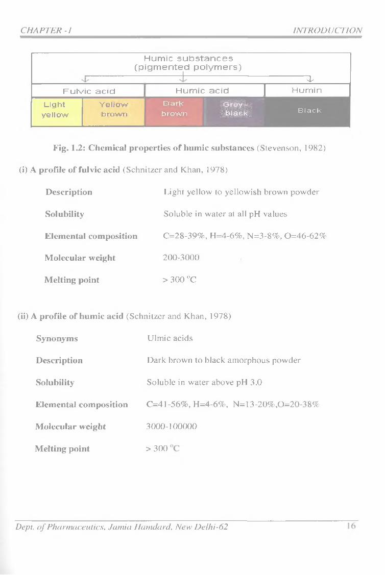
These materials are usually divided into the following tlu'ee main fractions.
1) liuiiiic acids; The fraction of humic substances that is not soluble in water
under acidic conditions (pH < 2) but is soluble at higher pH values. They are
dark brown to black in color.
2) Fulvic acids: The fraction of humic substances that is soluble in water under
all pH conditions. They remain in solution after removal of humic acid by
acidification. They are light yellow to yellow-brown in color.
3) Huiniin: The fraction of humic substances that is not soluble in water at any
pH value. Humin is black in color.
CHAPTER-1
Dept, o f Pharmaceutics, Jarnia Hamdarcl, New Delhi-62 15
CHAPTER-} INTRODUCTION
Hum ic s u b s ta n c e s (p ig m e n te d p o ly m e rs )
4Fulvic acid Hum ic acid | Humin
Lightyellow
YeKowbrown
Darkbrown ■■‘-Jalacfcii Black
Fig. 1.2: Chemical properties o f humic substances (Stevenson, 1982)
(i) A profile of fulvic acid (Schnitzer and Khan, 1978)
Description
Solubility
Elemental composition
Molecular weight
Melting point
Light yellow to yellowish brown powder
Soluble in water at all pH values
C=28-39%, H=4-6%, N=3-8%, 0=46-62%
200-3000
> 300 ”C
(ii) A profile of humic acid (Schnitzer and Khan, 1978)
Synonyms
Description
Solubility
Ulmic acids
Dark brown to black amorphous powder
Soluble in water above pH 3.0
Elemental composition C=41-56%, H=4-6%, N=13-20%,0=20-38%
Molecular weight
Melting point
3000-100000
> 300
Dept, o f Phanmiceulics, Jamia Hamdard, New Delhi-62

CHAPTER ~1 INTRODUCTION
1.3.2 Colloidal charact:eristics
The colloidal state represent a phase intermediate between true solution, where
species are of ionic or molecular dimensions, and suspended particulates, where
species are sufficiently large to settle under the force of gravity. Chemical and
physical reactions are generally enhanced in colloidal systems due to large surface
area of colloidal particles. At the same time, mobility through water or
groundwater is also enhanced, approaching that for true solutions. The range of
molecular size for humic acids places them in the colloidal range when in solution
i.e. from 0.001 to l|jm(Gaffiney et cil, 1996).
Humic colloidal material is thought to consist of coiled, long chained or three-
dimensional cross-linked macromolecules with electrical charges variously
distributed on the particle. The presence of charged sites arising fi'om ionized
groups, results in mutual repulsion and causes maximum expansion of the
macromolecule. Trapping of biologicals (peptides, carbohydrates) and anthropogenic
substances such as pesticides and plasticizers in the voids of these macromolecular
substances has been investigated.(Gaffney et ciL, 1996)
1.3.3 M icellar iiatare
It was shown that humic acids have surfactant properties. Humic acids are
predominantly hydrophilic (except at lower pH) but they also contain a substantial
concentration of aromatic rings, fatty acid esters, aliphatic hydrocarbon and other
hydrophobic substances, which together with the hydrophilic groups account for
the surface activity of these materials. The hydrophilic oxygen containing
functional groups (COOH, C=0, OH) are thought to play a significant role in
lowering the surface tension of water and so increasing aqueous wettability of
hydrophobic materials.
It has been recognized that the presence of even a small amount of humic acid
in aqueous solution can significantly enhance the water solubility of hydrophobic
organic compounds (Schnitzer and Khan, 1978). This solubilization in solution is
often attributed to the presence of micelle (Guetzloff et al, 1996).
Dept, o f Pharmaceutics, Jamia Hamdard, New Delhi~62 17
CHAPTER ~1 INTRODUCTION
Huniic acids being highly aromatic as compared to fulvic acids become insohible
at low pH values when the carboxylate groups become prot;onated that may also
lead to formation of intraniolccular pseudomicelles.
The structure of humic acids is such that it allows them to function as surfactants
with the ability to bind both hydrophilic and hydrophobic materials. This function
in combination with their colloidal properties makes humic acids effective agents
in transporting both organic and inorganic materials in the environment
1.4 METHODS FOR BIO AVAILABILITY ENHANCEMENT
A review of monographs in the European Pharmacopoeia has revealed that more
thtin 40 per cent of the drug substances have aqueous solubilities below 1 mg/ml,
and that 32 per cerit have an aqueous solubility below 0.1 mg/ml (Philip e ra i;
1986) The implementation of dissolution and absorption enhancing methods is,
therefore, a major field in the formulation of drug dosage forms, in particular for
the oral route of administration. A wide range of principles and methods for
enhancing dissolution rate of low-solubility substances, are available which include:
❖ Selection of salt form for weak acids and bases,
❖ Reduction of particle size,
❖ Preparation of solid dispersions,
❖ Change of crystal form by precipitation with hydrophilic polymers,
❖ Lipophilic formulations, i.e. emulsions, rnicroernulsions etc.,
❖ Use of surfactants for increased wettability,
❖ Complexation with cyclodextrins
Dept, o f Pharmaceutics, Jamia Hamdard, New Delhi-62 18

(i) Salt formation
Selection of an appropriate salt form or in case of liquid preparations, adjustment
of pH is first choice for weakly acidic and basic drug substances. Substances
having aqueous solubilities above 10 mg/ml are formulated as hydrochlorides,
sulphates, rnaleates, citrates etc. of basic drugs or for acidic drugs potassium,
sodium, calcium or other salts. But it is limited to drug with ionizable groups
only. Moreover poor crystallinity and hygroscopicity are major problems.
(ii) Micronization
In principle, the lower the aqueous solubility, the lower particle size is required to
achieve a satisfactory dissolution rale. For the practically insoluble substances (<
0.1 mg/ml), the required particle' size is so low that it may be technically difficult
to prepare the desired size range. On the other hand, physical instability of the
drug as well as the size distribution is introduced when the size range is reduced
to micrometer and sub-micrometer scale (Dalmora 2001).
(Mi) Solid dispersions
Several solid dispersions and coprecipitates with hydrophilic polymers have been
prepared where the drug substance typically is present in an amorphou.s state but
these are highly energetic systems (Chiou et al; 1971). Therefore physical
instability is a major problem and that is why only a few products based on solid
dispersions have been marketed (Arias et.al, 1994).
(iv) Microeiimlsioii
The newest trend in the formulation of low-solnbility drugs is accordingly the use
of so-called Upidic formulations, in particular microemnlsions and other self-
emulgating systems which can dissolve a sufficient amount of the drug substance.
But physical and chemical instability are again major problems.
CHAPTER -1 • INTRODUCTION
Dept, o f Pharmaceutics, Jarnia Eamdard, New Delhi-62 19
CHAPTER -] INTRODUCTION
l.S CYCLODEXTRIN COMPLEXATION
Physical and chemically stable compounds can be made by the preparation of
inclusion complexes of drugs with cyclodextrias. The potentials of the Vcu'ious
types of cyclodextrins for solubility and absorption enhancement are well
documented in the literature (Baboota et.ai, 2000, 2001, 2002a & b, 2003 a & b, 2004
a & b) Comjjlexation with cj/clodextrins has been used as a novel approach for
designing drug delivery system because of numerous advantages provided by these
carrier systems like:
❖ Cyclodextrins being natural carriers have low toxicity and are almost inert,
❖ They are usually safe and can be used almost through every possible route
of administration like oral, ocular, nasal, buccal, parenteral and rectal.
❖ They are insignificantly absorbed through intestinal mucosa.
❖ A large number of cyclodextrins with different cavity sizes are available
❖ They have a well defined chemical structure which provides a number of
potential sites for chemical modification or conjugation
❖ They ai-e versatile complexing agent and can accommodate almost every
type of organic molecule
❖ They are equally applicable for both ionizable and non-ionizable drugs
❖ They are stable enough to withstand high temperature (upto 280 °C) during
manufacturing processes like preparation and sterilization
❖ They protect the included drugs from biodegradation.
Dept, o f Pharmaceutics, Jamia Hamdard, New Delhi-62 20
1.5.1 Coniplexatiors phenom enoia '■
A “complex” is a species formed by interaction of two or more molecules or ions
with a definite substrate to ligand stoichiometry (Eccleston et.al; 1994).
mS -I- nL SmLn
Substrate Ligand Complex
A “substrate” S is the interactant whose physical or chemical properties are
obs erV ed e xperi mental 1 y.
A “ligand” L is the second interactant whose concentration may be varied
independently in an experimental study.
1.5.1.1 Types of complexes
The definition of a complex leads to a classification into two groups (Higuchi and
Connors, 1995).
CHAPTER -I , INTRODUCTION
Chemical Bonding
1) Co-ordinate complexes These are formed by co-ordinate bonds in which
transfer of a pair of electrons takes place e.g metal and ammonium ion co
ordination complexes between metal ions and bases (Amiji et.al, 2003).
o Inorganic complexes eg. [Ag (NH3)2j' , [Co CF etc.
o Chelates: Ligand have more than one donor groups eg. EDTA
2) Molecular compIexe.s These are mainly formed by non covalent interaction
between the substrate and the ligand such as electrostatic induction and
dispersion interactions with the exception of charge transfer or electron
donor acceptor complexes which may have some covalent character hence
appearance of new UV absorption bands (Amiji et.al, 2003).
Dept, o f Pharmaceutics, Jamia Hcimdard, New Delhi-62 21
B. Type of Bonding or Interaction
1) Charge transfer e.g. nitrobenzene complex
2) Hydrogen bonding e.g. Caffeine complexes
3) Hydrophobic interaction
4) Stacking intei'action
Type or struclare olf tnteractaiits
1) Sinali molecule - Small molecule intraction
2) Small molecule - macromolecule binding
3) Drug-protien binding
4) Drug-receptor binding
5) Enzyme-substrate complex
1.5.1.2 Type or structure of complex
1) Self association; It is a complexation of molecule with others of its own
species for example benzene forms dimer,
2) Micelle: A special form of self aggregated complex in which interactant is a
surfactant.
3) The inclusion complex: One interactant (guest) is entrapped within the cavity
formed by other macrocyclic interactant called as host (Szejtli, 1998 and
Eastburn eta l, 1994).
1.5.2 Methods of preparing isicliislon complexes
Several methods have been described in literature for preparing complexes of
drugs. Since cyclodextrins has been studied in gi'eat details as a complexing
agents and the following methods have been used to prepare cyclodextrin
CHAPTER ~1 INTRODUCTION
Dept, o f Pharmaceutics, Jamia Hamdard, New Delhi-62 22

complexes, these are given here as these methods can be modified to Form
complexes with iuirnic and fuivic acids as well. By only trial and error one can
find a method which will give the best result for a given drug. Complexes can he
inarmed by a variety of techniques that depend on the properties of drug, the
equilibrium kinetics, the formulation ingredients and processes and final dosage
form desired. However, each of these process depends on a small amount of
water to help drive the thermodynamics. Among the methods used are simple dry
mixing, mixing in solutions and suspensions followed by a suitable separation, tiie
preparation of pastes and several thermo-mechanical techniques.
1) GriEcliiig
Incltision complexes can be prepared by simply grinding the guest with a
complexing agent such as cyclodextrin. This is a very slow' process for making
inclusion complex and degree of complexation achieved is very low (Szejtli, 1988).
2 ) Solid clLspersion/Co-evaporatecI dispersion
The drug is dissolved in ethanol and cyclodextrin is either dissolved in a
alcoholic solution or dissolved separately in water or other suitable medium. The
cyclodextrin solution is then added to the drug solution or vice- versa and stirred
to attain equilibrium. The resulting solution is evaporated to dryness preferably under
vacuum.
3) Neutralization method
Martin and Udupa 1995, reported this method for various fluoroquinolones. In this
method equimolar concentration of drug and cyclodextrin are separately dissolved
in 0.1 N NaOH, mixed and stirred for about half an hour, pH is recorded and
0.1 N HCl is added dropwise with stirring until pH reaches 7.5, upon when the
complex precipitates. The residue is filtered and washed until free from Cl". It is dried
at 25'* C for 24 hours and stored in a dessicator.
CHAPTER -1 ^ ____________________________ ____ INTRODUCTION
Dept, o f Pharmaceutics, Jamia Haindard, New Delhi-62 23

4) Kneading :
In this method cyclodextrin is not dissolved but iaieaded like a paste, cither with
small amount of water to which the guest component has been added. Guest
component can be added without a solvent or in small amount of ethanol in
which guest has been suspended. Several hours of grinding of paste in mortar
results in evaporation of solvent and formation of powder like complex (Otero-
Espiner er fl/., 1992).
5) Precipitation
The guest which shows Bs type phase solubility curve are suitable for this
method of complex formation. In this method the drug (guest) mid cyclodextrin
are dispersed in water and the solution is heated to obtain concentrated, viscous
and translucent liquid. The solution is left to give a precipitate of inclusion
complex. Precipitate obtained is separated and dried to get solid inclusion
complex.
6 ) Spray drying
In this method first a rnonophasic solution of drug and cyclodextrin is prepared
using a suitable solvent (generally hydroalcoholic solution is used). The solution is
then stirred to attain equilibrium following which the solvent is removed by spray
drying (Bietti et aL, 1992)'
7) Freeze-drying
Freeze drying method is similar to spray drying method except that in this
method, the solvent is removed by freeze-drying after attaining the equilibrium
(Becirevic-Lacen e/a/., 1996)’
8) P reparation of ,su.si3ension
Cyclodextrin need not be dissolved. Simply stirring the guest in an aqueous
suspension of CD can achieve • complexation within 2-24 hrs at ambient
temperature. This is recommended method for industrial application (Szetli, 1988)'
CHAPTER-I _ _ _ _ _ INTRODUCTION
Dept, o f Pharmaceutias, Jamia Harndard, New Delhi-62 24
9) Melting
Complexes can be prepared by simply melting the guest, mixed with finely
powdered cyclodextrin. In this procedure there has to be a large excess of guest,
and after cooling this excess is removed by careful washing with weak complex
forming solvent or by vacuum sublimation. This latter is preferred method and is
used to sublimate guests such as menthol (Szetli, 198K)
1.5.3 Cliaracterizalion of inclusion complexes
Several methods have been proposed for the analytical characterization of
drug/cyclodextrin complexes, according to the physical state considered, i.e. solution or
solid. The formation of inclusion complex can be studied and characterized in two
ways (Szetjli, 1988)
1) In solid state (by DSC, XRD, FTIR, SEM)
2) In solution (by solubility studies, dissolution tests; UV spectral studies, ‘H-
NMR studies, TLC)
1) Characterization in solid state
(i) Differentia! scanning calorimetry (DSC)
DSC is the measurement of rate of beat evolved or absorbed by the sample,
during a temperature program, It is extensively employed to check any variation
of crystalline properties due to the interaction with the CD. The reduction in the
degree of crystallinity of the drug is often taken as an indication of complexation.
The DSC curve of cyclodextrin generally show an endotherm neai' IOO°C which
signifies removal of water. The DSC curve of the guest molecule shows a sharp
intense peak (endotherm) at its melting temperature (m.p.) and then it starts
decomposing. In DSC curve of cyclodextrin-guest inclusion complex, these peaks
are either diminished or absent. Partial complex formation may be fihowii by varying
patterns e.g. small exotherrn adjacent to the melting endotherm of guest molecule.
CHAPTER-I _ _____ INTROLyUCnON
Dept, o f Pharmaceutics, Janiia Harndard, New Delhi-62 25
CHAPTER -1 INTRODUCTION
The D5iC curve of simple mixtiu'e would resemble the combination of curves _ of
pure substance i.e. guest and cyclodextrin.
(il) Powder X-ray diffraction (XMD)
This is an important technique for the determination of three dimensional structure
of molecule and distinguishes between amorphous and crystalline forms. The
diffraction pattern is characteristic of a substance. The crystalline substance has sharp
intense peaks in its powder diffraction pattern whereas amorphous substance shows
only undefined, broad, diffused peaks of low intensity. Generally the complex has
an amorphous nature i.e. broad, undefined peaks with low intensities.
(lit) Fourier transfontis liitra-recl spectroscopy (FT-IR)
It is another useful technique to verify formation of inclusion complexes. The
guest molecule within the cavity show shift in its peaks or shows peaks of less
intensity. Basically, peaks which lie in the fingerprint region and peaks due to C-
O or O-Ii stretching are affected (shifted or intensity is changed). FO R technique
is known to have superior sensitivity and resolution, absolute wavelength accuracy
and higher precision of measurement than conventional IR technique.
(iv) Scanning electron Biicroscopy (SEM)
SEM is done to observe tlie crystalline structure of the sample. SEM studies help
us to observe the changes that occur in the crystal structure during or after the
preparation procedure. Generally a change from crystalline to amorphous nature
(of the drug) can be seen upon compiexation with fulvic and humic acids.
2) Characterization of iiiclE,sioii complexes In solution
(i) Phase solubility studies and dissolution tests
The most common and widely used method to evaluate the ability of CD to
complex a drug is by phase solubility studies. Phase solubility analysis allows the
determination of both the stability constant and the stoichiometry of the complex
formed in solution. Higu.chi and Connors, 1965 have classified the various
Dept, o f Pharmaceutics, Jamia Hamdard, New Delhi-62 26
CHAPTER -1 INTRODUCTION
solubility behavior seen during complex fonnation as A-type (a soluble inclusion
complex is formed) or B-type (an inclusion compound of finite solubility is
formed).
(ii) TLC (Thin layer chromatography)
TLC may also be useful for verification of complex formation, since the Rf
values are altered considerably. Rf values are usually diminished provided the
complex is sufficiently stable in the solvent mixture used.
(iii) Proton nuclear magnetic resonance (^H-NMR)
It is useful not only for verification of complex formation but also to guess how
the guest is geometrically aligned in the cyclodextrin cavity. The inclusion of
guest molecule into the cyclodextrin cavity clearly induces some changes in the
chemical shift values. The chemical shift values are also indicative of the
interactions, if any between protons of cyclodextrin and guest (Djedani 1991).
1,5,4 Studies Carried on Complexation of drugs with Hiiiiiic Snbstaiices
K arniarkar, R.M (2007) developed ketoconazole complexes with lidvic and humic acid
isolated from shilajit. Solubility, dissolution and antimicrobial activity were improved as
compared to ketoconazole alone. A successful bioequivalence study was performed on
healthy human volunteers with significant increase in bioavailabilty of ketoconazole
complex as compared to uncomplexed dosage form of ketoconazole (Agarwal et.ai,
2008b),
Tyagi, B (2007) prepared complexes of paclitaxel, an anticancer drug with humic and
fulvic acid of shilajit. Complexes were evaluated for solubility, dissolution and
characterized by FT-IR, DSC, XRD and SEM.
M irza, A (2007) investigated the effect of fulvic acid on the solubility, dissolution and
bioavailability of Carbamazepine. The complexes were prepared using different
techniques like freeze drying, solvent evaporation, kneading and physical mixture and
evaluated for solubility, dissolution, differential scanning calorimetry (DSC), fourier
Dept, o f Pharmaceutics, Jmnici Hamdard, New Delhi-62 27
transform infrared spectroscopy (FT-IR), X-Ray diffraction. A marked increase in
aqueoLLs solubilily and dissolution profile was observed.
Ahinadl, 1) (2{I06) investigated the influence of sliilajit extracted fulvic acids on
complcxation with melatonin and to develop an oral dosage form of melatonin in order to
increase the solubility, dissolution rate. The complexes were characterized by using
differential Scanning calorimetry (DSC), X-Ray diffraction (XRD), Foinier transform
Infra - red spectroscopy (FT-IR) and Scanning Electron Microscopy (SEM) and it was
concluded that maximum coniplexation was achieved by lyophilized complex.
Aiiwer, M .K (2005) prepared complexes of furosemide with humic and fulvic acid using
different techniques, Freeze drying, solvent evaporation and grinding and chacterized
them by FT-IR, DSC, XRD and SEM. A significant enhancement in solubility,
dissolution profile and diuretic activity were observed in comparison to the uncomplexed
furosemide dosage form (Agarwal et.al, 2008a).
Klianiia, R (200S) developed and validated a tiew' method lo extract humic and fulvic
acid using ion exchange resins. Method gives better yield of humic and fulvic acids and
even the solubility of fulvic acids obtained by this method is better than obtained by
established Ghosal method. Complexes of itraconazole and acyclovir were prepared with
humic and ixilvic acid and these complexes were found to have improved solubility,
dissolution and therapeutic efficticy than the uncomplexcd drugs, Permeability of tiie
drugs was also found to be improved in comparison to the uncomplexed form.
Bioequivalence study was conducted on healthy human volunteer, it was found that
bioavailability of itraconozole increased significantly.
Saliija, A (2001) proved that hunric acid-piroxicam complex has better solubility than
pure piroxicam powder, it also, found that dissolution profile of the tablets of these
complexes are better thaii the marketed preparation containing piroxicam in uncomplexed
form. Gastric ulceration was significantly reduced as compared to uncomplexed
piroxicam.
CHAPTER -1 _ _ _ ^ ^ _ _ _ _ _ Z^ON
Dept, o f Pharmaceutics, Jamia Hanulard, New Delhi~62 28

1.6 STEATRGirF:S TO IMPROVE ASPIRIN STABILITY
Mroso et.al (2006) identified salicyl,salicylic acid and acetylsalicylsalicylic acid as
decomposition products of aspirin when mixtures of the drug wilh magnesium stearate
were stored in the solid state at 60° and 15% relative humidity. The effect of increasing
tlie concentration of inagnesium stearate and the addition of other alkali stearates on. the
rale of decomposition of aspirin were studied. The validity of the theory that pH changes
indticed by ihe alkali stearates account for the catalytic effect of the lubricants on the
decornpo.sition was tested. The changes observed were modeled and the mechanism
involved elucidated. The potential use of the melting points of aspirin mixtures in
predicting the stability of the drug in such drug-excipient mixtures is demonstrated.
Wlllianii.s et. al (1999) investigated the effect of formulation technique for 2-
hydroxypropyl-P-cyclodextrin (HPpCD) on the stability of aspirin in a suspension based
pressurized metered dose--iiilialer (pMDI) formulation containing a hydrofluoroalkane
(HFA) propellant. The chemical stability of aspirin in pMDI formulation was determined
over 6 month storage at 5, 25 and 40 °C. Aspirin in the iyophilized inclusion complex
exhibited the most significant degree. of degradation during 6 month storage, while
aspirin alone in the pMDl demonstrated a moderate degree of degradation.
M ario et.al (1965) studied the ell'ect of ultrasound on the hydrolysis of aspirin solutions
at various temperature and pH values. The reaction kinetics followed a pseudo first-order
rate, both with and without the influence of ultrasound. The rate of hydrolysis was
increased in all cases by applying sound energy.
Gore et.al (1968) investigated the significance of salicylic acid sublimation in stability
testing of aspirin- containing sohds. Under conditions of accelerated stability testing, the
loss of salicylic acid from the system by sublimation can incur appreciable eiTors in the
detection of overestimating aspirin stability. Since aspirin was not detected to sublime
under these same conditions, its residual content is an improved indication of its stability.
A method for its simultaiieons determination with salicylic acid is presented.
Chang et.al (1984) conducted study on 0.2% w/v aspirin liquid formulation in a wide
range of water-propylene glycol mixture and water-trietliylene glycol diacetate mixture at
CHAPTER -1 INTRODUCTION
Dept, o f Pharmaceutics, Jamia Hamdard, New Delhi-62 29
CHAPTER -1 INTRODUCTION
fouf temperatures. The effect of surfactant, polyoxyethyleue (20) sorbitan rnoaoIai,u-ate,
on aspirin stability was investigated. There was a linear relationship between water
content and degradation rate cotistants. Formulation containing the higher concentration
of the surfactant showed the greater aspirin degradation.
M ihranyaii et.al (2005) studied the effect of cellulose powder structure on moisture
induced degradation of acetylsalicylic acid. Different cellulose powders were
manufactured and characterized by X-ray diffraction and N2 BET gas adsorption.
Cellulose with lower crystallinity index exhibited lower degradation rate than the sample
with the higher crystallinity index. It should be noted that higher ASA degradation rate
were observed in the sample with comparably higher moisture content.
Mlzobiichl ei.fil (2001) prepared the external preparation containing aspirin which were
stored for a long term and was superior in dermal absorbability. Formulation were
prepared by mixing aspirin together with at least one substance selected from an ester of
an organic acid ester having 2 to 20 carbon atoms, a glycerol fatty acid ester, silicon oil,
hydrocarbon oil and crotamiton.
Sriavely et.al (1993) coducted the study on the stability of a direct compression tablet
formulation containing aspirin as a model hydrolabile drug with Erndex (a mixed sugar
diluent containing approximately 8 percent moisture) and steaiic acid. Compressed tablet
and uncompressed powder blend were packaged in storage container and placed on
stability at different temperatures. Analysis of the aspirin data showed that the rate of
aspirin decomposition increased with temperature. The formulation showed good stability
with less than one percent decomposition occurring after 1.75 year of storage at room
temperature.
Choi, H,S (1989) investigated the molecular nature of aspirin hydrolysis as biometric
model for esterase. The structural specificity and the chemical dynamics of these
inclusion complexes in the solid state and in the solution state were detemined by FT-IR,
UV, FAB-MS, 'HNMR and '"’CNMR spectroscopy. Dissociation constants were obtained
by the kinetic method under alkaline condition (Choi, 1992).
Dept o f Pharmaceutics, Jcmiia Hamdard, New Delhi-62 30

1.7 OBJECTIVE OF THE STUDY
Non-steroidal anti-inflammatory drags (NS AIDs) are among the most widely used of all
therapeutic agents. They are drugs of choice for the management of a variety of acute and
chronic infhimmation., They are frequently prescribed for long-term treatment of
rheumatic musculo-skeletal complaints. The major drawback to anti-inflammatory drug
use is the occurrence of gastrointestinal side effects with majority of agents. Aspirin is
very old drug but still having a very high market value. It possesses antipyretic, anti-
inflanimatory, analgesic and anti-aggregatory activity (Chang et.al., 1984). The
acetylsalicylic acid molecule has a carboxyl group and an ester group. The ester group
can be easily hydrolyzed, which reduces the medical value and causes side effects on
humans (Gore et.al, 1968 and Mario et.al, 1965). A strategy was designed how' to inhibit
the hydrolytic decomposition and enhatice solubility and dissolution of aspirin inside the
void of humic and fulvic acid of shilajit. We propose to investigate the effects of humic
and ftdvic acid as carrier on aspirin in enhancing the solubility, dissolution rate,
bioavailability, stability, decreasing the toxicity and obtaining a better pharmacodynamic
profile of aspirin through complexation and compare the prepared complex with
hydroxy-propy 1-p-eyclodextrin.
The main objectives of study were.,
1. To isolate pure fulvic acid and humic acid from shilajit and to characterize them.
2. To prepare complexes of drugs with fulvic, hiirnic acids and HP-f3-CD and study
their stoichiometi'y and nature of complexation through techniques like FT-IR, •
DSC, SEM and X-ray diffraction.
3. To caiTy out saturation solubility studies of prepared complexes and compm'ed
with HP'P-CD complexes
4. To carry out in-vitro dissolution behaviour of the solid complexes of humic/fulvic
acid and conipai'e with HP-P-CD complex and drug alone
5. Selection of preparative technique and optimization of complexation method
CHAPJ:ER^-1 _ _ _ _ _ _ _ _ _ _ INTRODUCTION
Dept, o f Pharmaceutics, Jamia Hamdard, New Delhi-62 31
6. Effcct of fulvic/humic acid on solubility/dissolution of aspirin and comparison
with HP-P-CD
7. Effect of humic/lxilvic acid on stability of aspirin and comparison with HP-P-CD
8. Effect of fulvic acid on permeability of aspirin and comparison with HP-P~CD
9. Effect of fulvic/humic acid on anti-inflammatory activity and comparison with
HP-f3-Cr3
10. Effect of fulvic/humic acid on gastric ulceration and comparision w'ith HP-p-CD
11. Preparation of dosage forms and in-vitro release of the drug from tlie prepared
dosage form
12. Stability studies of the prepared dosage forms of fulvic acid complex and compare
with dosage form of HP-p-CD comple?c and innovator product
13. To determine the stability of complexes by using computational method
1.8 HUMIC SUBSTANCES AS CARRIER
From the literature the following properties of the humic substances were observed. This
encouraged us to test their bioavailabiiity and stability enhancing potential
1. They have a sponge like structure punctured by voids of about 200-1000A in
diameter. A water insoluble and unstable active ingredient can be added to fill the
voids.
2. They are naturally occurring and are toxicologically safe.
3. They have surfactant properties which gives them an advantage over cyclodextrin
as a bioenliancer.
4. Their ubiquitous occurrence in natttre can provide the pharmaceutical industry a
large amount of ready to use bioenhancers.
CHAPTER -I _ INTRODUCTION
Dept, o f Phannaceutics, Jamia Harndard, New Delhi-62 3 2

5. Established pathways of the formation of humic substances provides a scientific
basis for exploring the possibilities of in house production of humic substances.
6. As the traditional literature boasts of various pharmacological activities of hiunic
substances, establishment of their pharmacological and safety profile could give
us magic molecules which will not only have their own pharmevcological activity
but will also help in enhancing the bioavailability of various poorly bioavailable
and unstable drugs, thus reducing the amount required to produce their
pharmacological effect. ,
L9 SELECTION OF MODEL DRUG CANDIDATE
Aspiriti is a very old drug but still having excellent medicinal value, and its health
protection function such as analgesic, anti-inflammatory, antithrombotic, and antipyretic,
has received more and more attention (Choi, 1992). The aspirin molecule has a carbo.xyl
group and an ester group. The ester group can be easily hydrolyzed, which reduces the
medicinal usefulness and has gastrointestinal side effects on humans (Connors et.al
1986), A need exists to learn how to inhibit the hydrolysis of aspirin. A number of papers
are available describing decomposition of aspirin, Aspirin is degraded into salicylic acid
and acetic acid by influence of moisture. (Connors eial., 1986). The decomposition of
aspirin complexes with cyclodextrin has been studied and found significant degradation
during 6 month of storage as compared to aspirin (Williams et.al., 1999), In another
study, Influence of cellulose powder with lower crystallinity index exhibited lower
degradation rate of aspirin than the sample with the higher crystallinity index (Mihranyan
et a l, 2005), The degradation of aspirin increased by increasing the specific surface area
of excipient (dicalcium phosphate dihydrate powders),
Fulvic acids are the major constituent of shilajit, having relatively open, flexible
structure punctured by voids (micropores) of different diameters (Agarwal et.al.,
2007a & b). These compounds were, presumably, loosely held in the core structure
of shilajit (Agarwal et.al.,200&a). The plant secondary metabolites which are trapped
in the internal voids of fulvic acids are spared from and become resistant to
conmron chemical and biological decomposition (Agarwal et a l, 2007c), Taking a clue
CHAPTER 4 _ ______ INTRODUCTION
Dept, o f Pharmaceutics, Jainia Hamdard, New Delhi-62 33
CHAPTER -I INTRODUCTION
on this point we started to investigate ttie potential of fulvic and humic acid as a novel
coinplexing agent in order to increase the stability of aspirin .Theses fulvic and humic
acid provide tiie protective layer around aspirin in wiiich water is excluded as inucli as
possible and reduce the decomposition.
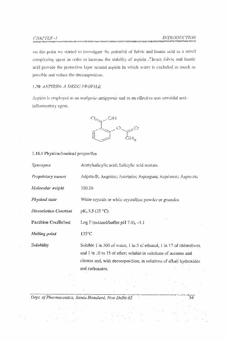
1,10 ASPIRIN- A DRUG PROFILE
Aspirin is employed as an analgesic-antipyretic and as an effective non-steroidal anti
inflammatory agent.
..O H
C hL.
1.10.1 Physicocheniica'l properties
Synonyms
Proprietary nam es
Molecular weight
Physical state
Dissociation Constant
Partition Coefficient
Melting point
Solubility
Acetylsalicylic acid; Salicylic acid acetate.
Adprin-B; Angettes; Ascriptin; Aspergum; Asprimox; Aspro etc
180.16
White crystals or white cryLStalline powder or granules
pKa3.5 (25 °C).
Log P (octanol/bufTer pH 7,4), -1.1
135°C
Soluble 1 in 300 of water, 1 in 5 of ethanol, 1 in 17 of chloroform,
and 1 in 10 to 15 of ether; soluble in solutions of acetates and
citrates and, with decomposition, in solutions of alkali hydroxides
and carbonates.
Dept, o f Pharmaceutics, Jamia Hamdard, New Delhi-62 34

1.10.2 Pharmacology ,
Aspirin is analgesic, anti-inflammatory, antipyretic and an inhibitor of platelet
aggregation. It inhibits prostaglandin G/H synthase (Roth et al; 1975). This enzyme
catalyses the first step in the synthesis of prostaglandins and thromboxanes from
ai-achidonate. Aspirin is relatively specific for type-I isoenzyme which is constitutively
expressed in platelets and other tissues and arc involved in platelet/endothelial cell
interaction (Smith et.al, 1992). Aspirin acetylates the hydroxyl group of a serine residue
at a position 529 of the polypeptide chain, thereby preventing access of substrate to the
active site by steric hindrance and causing irreversible loss of cyclooxygenase activity
(DeWitt et.ai, 1988 and Funk et al., 1991). Aspirin also inhibits type-II prostaglandin
G/H synthase which is not expressed constitutively brrt is induced by cytokines during the
inflammatory response (Xie, et. a l, 1991 and Kujubu et.al, 1991). Most of the
pharmacological effects of aspiiin are caused by inliibition of formation of prostaglandins
and thromboxanes.
Aspirin has an active metabolite (salicylate) which in addition to possessing some anti
inflammatory properties in its own right also has important effects on respiration acid-
base balance, and the stomach. Salicylates stimulate respiration by a direct effect on
medulla, and at high concentration, uncouple oxidative phosphorylation in muscle,
increasing oxygen consumption and carbon dioxide production. Salicylate have a direct
irritant effect on the gastric mucosa and further predispose to ulceration by inhibiting
synthesis of vasodilator and cytoprotective prostaglandins. Large doses of salicylates
(greater than 5 g per day) are uricosuric, but such doses are poorly tolerated and
salicylates are no longer used to treat gout.
1.10.3 Toxicology
An in~viiro study (Joshlco et.al,, 1993) looked at rat embryos cidtured for 48 h (days 9.5-
11.5 of gestation) in 100-300 pg/ml salicylic acid, a metabolite of aspirin. When
compared with growth in control embryo, a significant dose-dependant decrease in crown
rump lengths, somite members, and yolk sac diameters was observed in the rat embryos
cultured with salicylic acid. There was also a significant increase in overall
CHAPTER __________ _ _ _ _ INTRODUCTION
Dept, o f Pharmaceutics, Jamia Harndard, New Delhi-62 3 5

dysmorpliology, including eye, bronciiial arcli and anomalies, and an absence of; foreiimb
buds. The neural tube was especially vulnerable and had frequently failed to close.
Clinical and experimental evidence indicates that exposure to relatively large doses of
aspirin prolongs parturition. A study of the dose-response relationship for salicylic acid
on labor and gestation times in rat used, as a positive control pregnant rat exposed to 260
mg/kg per day of aspirin fi'om day 15 to day 21. The aspirin-treated rats had both
prolonged labor and gestation times, as well as increased maternal peripartum death.
1.10.4 Clinical Pharmacology
Aspirin is generally well tolerated at doses up to 2 g daily. Higher doses are associated
with numerous side effects, including tinnitus, abdominal discomfort, nausea, vomiting
and gastrointestinal bleeding. Increasingly toxic concenti'ations cause deafness, vertigo,
headache, hyperpnea, acid-base disturbance, fever, sweating, tachycardia, hallucination,
delirium, loss of conciousness, circulatory collapse, respiratory failure and death.
The analgesic effect of aspirin is a peripheral effect owing to its inhibition of the
cyclooxygenase enzyme. In areas of inflammation, increased amounts of PGE2 and PGFac
are produced. These lower die threshold for triggering pain fibers. This can be
demonstrated by injecting prostanoids into the skin, which causes an area hyperesthesia.
PGE2 also act as a vasodailator in areas of inflammation and this combined with other
substances which increase vascular permeability, contributes to the vascularity and
swelling in areas of inflammation. The cmti-inflamraatory effects of aspirin and salicylate
related to the decreased vascularity which results from inhibition of PGE2 synthesis.
The inhibition of platelet prostaglandin G/FI synthase has been investigated through
measurement of serum thromboxane B2 (Patrono et.al, 1980; Patrignani etal, 1982;
Patrono et.al, 1985) and urinary thromboxane metabolite (FitzGerald et.al., 1983 and
Ritter et.al., 1989). Single oral dose of 5-100 mg aspirin cause dose- dependant inhibition
of serum thromboxane B2 generation, with 100 mg causing near' maximal inlaibition.
Aspirin irreversibly acetylates prostaglandin G/H synthase, its duration of action on
platelets substantially outlives its presence in the body. Its effect on platelet
thrpmboxanae biosynthesis and on bleeding time persist for many days after dosing is
CHAPTER -I INTRODUCTION
Dept, o f Pharmaceutics, Jarnia Hamdard, New Delhi-62 3 6

discontinued, recovery being determined by the entry of new platelets into the circulation
(Patrono et.ciL, 1980). When aspirin is used as an analgesic in postoperative dental pain,
large doses are required (around 1200 mg), analgesia only lasts for few hours and there is
a significant correlation between analgesia and plasma salicylate concentration (Seymour
et.ai, 1982). However, the correlation between analgesia and plasma salicylate
concentration is fortuitous, and sodium salicylate does not cause significant analgesia
(Seymour et.ai, 1984). It therefore seems likely that the analgesic action of aspirin
depend on acetylation of cyclooxygenase and transience of the analgesia is probably
explained by biosynthesis of new prostaglandin G/H synthase.
1,10.5 Pliarniaceiitics
Aspirin is available froms many manufacturers. Preparations are available for oral and
rectal administrations. In some countries lysine aspirin is available for intramuscular or
intravenous administration.
Oral dosage form are available as capsule, plain uncoated tablets, dispersible tablets,
effervescent tablets, soluble tablets, enteric-coated tablets, enteric-coated capsules,
buffered tablets and modified -release tablets. The usual strengths available include 300
mg, 75 mg and 500 mg. Plain tablets or capsules and dispersible or soluble tablets should
be taken with or after food. Dispersible or soluble tablets should be dispersed or
dissolved in water. Enteric-coated tablets or enteric-coated capsules should be swallowed
whole, not chewed and should not be taken at the same time as indigestion remedies.
Modified-release tablets should be swallowed whole, not chewed.
Suppositories are available containing 60 mg to 1.2 g aspirin in USA and 150 or 300 mg
in the UK. Generally aspirin preparations should be stored at room temperature, protected
from moisture, in air-tight container.
Aspirin is available in combination with many other drugs - for example, analgesic such
as aloxiprin and acetaminophen; opoid analgesic such as codeine-dihydrocoedine,
propoxyphene napsylate and ethohepazine citrate; muscle relaxants such as meprobamate
and methocarbamol; histamine Hp receptor antagonist such as chlorpheniramine and
CHAPTER -1 INTRODUCTION
Dept, o f Pharmaceutics, Jamia Hamdard, New Delhi~62 37

CHAPTER -1 INTRODUCTION
cj'clizine. Other drugs iuclude aluminium or niagnesium hydroxide, ascorbic acid and
plienylephrine.
1,10.6 Mechanism of pH dependent hydrolysis of aspirin
Esters group of aspirin are susceptible to catalytic hydrolysis by both aqueous acids and
bases. The possible mechanisms are given below:
1.10.6,1 Acidic hydrolysis
0
R"' "OCH3
■ H"
fast OCH3.
H2O
slow
OH
'OCH-,
ff*’ transfer fast
R'' fast
0 '"H
R'"' ""oh
+ CH3 OHslov/ R'
OH
"(yCHgOH
Fig. 1.3: Scheme for acidic hydrolysis of aspirin
If the proton is hydronium ion (H.iO' ) the catalysis is known as specific acid catalysis.
Source of proton is from dissociated acid and the ester of aspirin is protonated in the
transition state of the reaction. For specific acid catalysis, the observed rate constant, tobs
is described by equation 1.
'obs = ko + /chIH.iO'" ] 1
Wliere ko is the rate constant for the uiicataiyzed process and kn is the rate constant for
the acid catalyzed process. Note that there is no term in the equation for any
undissociated acid present in the reaction mixture.
Dept, o f Pharmaceutics, Jamia Harndard, New Delhi-62 38
CHAPTER - i INTRODUCTION
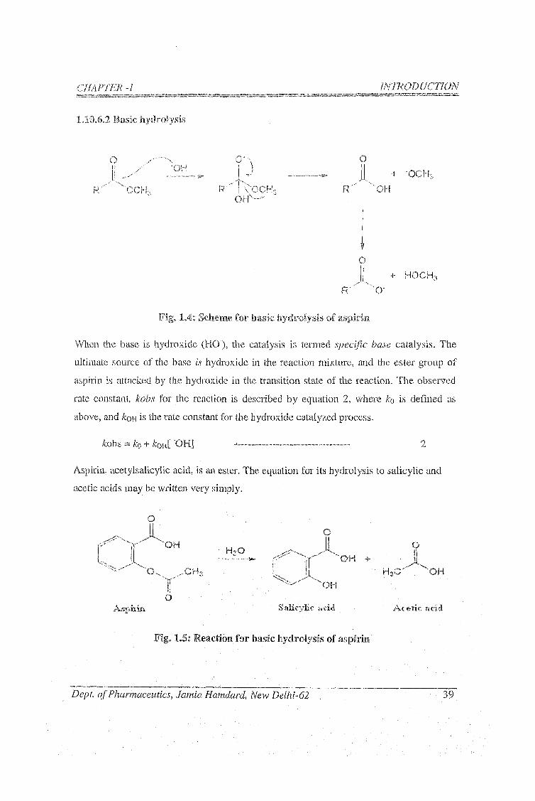
1,10.6.2 Basic hydrolysis
O
R"'"..... '" OCHa
•OH
R
O" \
VO CH 3 OH''...
+ -O C H 3
R'" '■"'OH
0j + HOCH3
R""'....'"■■O-
Fig. 1.4; Scheme for basic hydrolysis of aspirin
When the base is hydroxide (HO"), the catalysis is termed specific base catalysis. The
ultimate source of the base is hydroxide in the reaction mixture, and the ester group of
aspirin is attacked by the hydroxide in the transition state of the reaction. The observed
rate constant, kohs for the reaction is described by equation 2, where /co is defined as
above, and /cqh is the rate constant for the hydroxide catalyzed process.
/cobs = /co + /cohI OFI]
Aspirin, acetylsalicylic acid, is an ester. The equation for its hydrolysis to salicylic and
acetic acids may be written very simply.
H20 Ir'-" ''O H +
"'■OHo
Aspkia Salicylic acid A c e tic acid
Fig. 1.5: Reaction for basic hycIroly.sis of aspiriB
Dept, o f Pharmaceutics, Jarnia Hamdard, New Delhl-62 39

The exact mechanism of hydrolysis is a bit more dilficult to describe, since the hydrolysis
of aspirin may occur by one or more of the mechanisms described above.
1;ICI.7 Method of Analysis
The preferred analytical method is lugh performance Liquid chromatography (Lo et.al,
1980). The sensitivity is 0.5 mg/L for both aspirin and salicylate. Blood samples are
collected in the presence of potassium fluoride to inliibit plasma esterases and must; be
analyzed immediately if reliable information on aspirin itself is needed. For routine
toxieological purposes, determination of salicylate suffices, and efforts to prevent
hydrolysis of acetylsalicylic acid are unnecessary. Absorption after oral administration of
a solution of aspirin is usually complete, while enteric-coated tablet are less reliably
absorbed (Levy et.al, 1960).
However, GC-M5] is also used to dctcct aspirin in plasma. (Tsikas et al, 1998). Limit of
detection of aspirin, salicylic acid, and salicyluric acid is 50 pg/L in plasma (Buskin et
al., 1982). Llimits of detection in plasma are 100 pg/L for aspirin and salicyluric acid,
500 pg/L for salicylic acid, and 200 pg/L for gentisic acid (Rumble et a/., 1981). In
plasma, LOD for aspirin and salicylic acid is 100 pg/L (Kees et al., 1996). In plasma or
skin, LOD for aspirin and salicylic acid is 0.1 pg/mL and 0.1 pg/cm^ respectively (Pirola
e ta l, 1998).
CHAPTim^-1 ____________ __________ ______ ___ INTRODUCTION
Degt o f Pharmaceutics, Jarnia Hamdard, New Delhi-62 40

CHAPTER -1 INTRODUCTION
Table 1,1: Reported HPLC methods of aspirin
S.
NoSam ple
S.P
(coliHiin)M .P F .R D etection- Meference.s
1
Forrmilation/H\iman
serum
c , «
150 nun x
4.6 iriiii
acel.onitrile-0.1 %
aqiieou.s
orthophosphoric acid
2.0
m l / m i n
fl uorescence
detection
(Ibrahim.
«/; 2007)
2 Tablet
3.0 (11 n
non-
porous
silica
ODS
98:2 v/v 50 mM
phosphate buffer pH
3.0-acetonitrile
1.5
ml/minUV-220 run
(Xu X et.al,
2000)
3 Tal5let
C |8
column
(150x4,60
mm, 5 |,0
0.1% v/v
triethylamine (pH
4.0):ucetonitrile in
the ratio 25:75%
(v/v)
1 nil/ininUV 225
n m
(Grand himatlii
et,al,2007)
4 Dosage form
Bondapak
CIS
rever.se
phase
' C o l u m n
methanol; water
(35:65; v/v)1.8nil/min UV-235 nm
(Akay et.al.,
2008)
5
Capsule of
combination dosage
form
C -18 ,5
m m
c o l u m n
having
250 X 4.6
m m
0.02 M
pota.ss iumd ihydro gen
phosphate: methanol
(20:80) adjusted to
pH 4
1.0
m l/m inU V-240 nin
(Shahet.al,,
2007)
S.P- Stationary phase; M.P- Mobile phase; F.R - Flow rate
Dept, o f Pharmaceutics, Jamia Hamdard, New Delhi-62 4 1
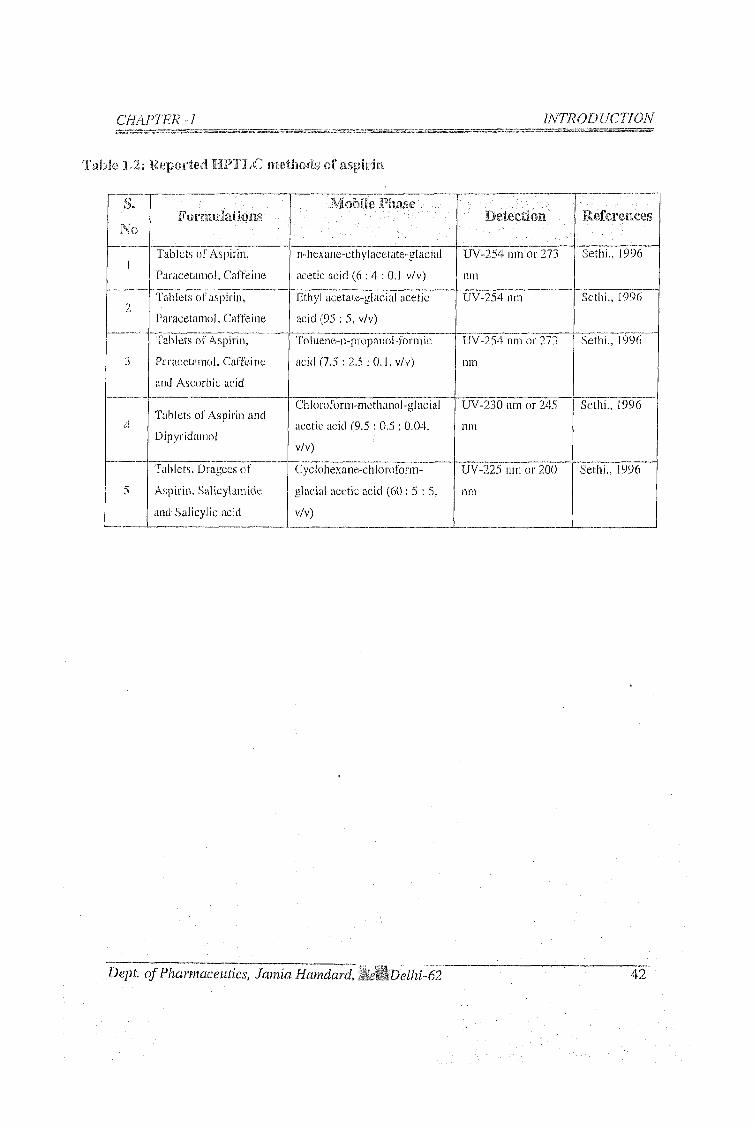
CHAPTER -1 INTRODUCTION
Table 1,2: Reported HPTLC methods of aspirm
S.
N oF o n m ila t io i is
J?hase .D e te c tio n ReferenccvS
1Tablets of Aspirin,
Paracetam ol, Caffeine
n-liexanc-ethylacetate-glacial
acetic acid (6 : 4 : 0.1 v/v)
UV-254 nin or 273
11 tn
Sethi., 1996
2Tablets o f aspirin.
Paracetam ol, Caffeine
Etliyl acetate-glacial acetic
acid (95 : 5, v/v)
UV-254 nm Sethi., 1996
3
Tablets of Aspirin,
Paracetam ol, Caffei ne
and Ascorbic acid
Toluene-n-propanol-form ic
acid (7.5 : 2.5 : 0 .1, v/v)
UV-254 nm or 273
nin
Sethi., 1996
4Tablets of Aspirin and
Dipyridamol
Chloroforin-methanol'glacial
acetic acid (9.5 : 0.5 ; 0,04,
v/v)
UV-230 nm or 245
nm
Sethi., 1996
5
Tablets, Dragees o f
Aspirin, Salicylamide
and Salicylic acid
Cyclohexane-chloroforni-
glacial acetic acid (60 : 5 : 5,
v/v)
UV-225 iim or 200
nm
Sethi., 1996
Dept, o f Pharm.aceutics, Jamia Hamdard, MiM'Delhi-62 4 2

CHAPTER -i INTRODUCTION
1,11 PLAN OF ¥FORK
1 J 1 ,1. l i te ra tu re search
1.11.2 Prociirenieiit of sliilajit and model drug cancllclate, aspirin
1 . 1 L3 Method development a n d standardization for aspirin analysis
1.1 i . 3.1 Analytical methods for routine analysis (HPLC and HFFLC)
1.11.4 Characterization anti identification of aspirin
1.11.4.1 Physico-chemical characterization
1.11.4.2 Spectra! characterization.
l.lloS Ctiaracterization and autlientification of sliilajit of different -sources
1.11.6 Extraciion of Imsiiiic and flilvic ad d from sfailajit
1.11.6.1 Standardization of reported method
1.11.6.2 Development of an improved method.
1.11.7 Characterizati oil of iiiiiniic and fiilvic acid
1.7.7.1 S pectral characterization
1.11.7.2 Elemental composition analysis by FT-ICR mass spectrometry
1.11.8 Phase solubility studies
1.11.9 Preparation of complexes using following methods
1.11.9.1 Solvent evaporation
1.11.9.2 Freeze drying.
1.11.9.3 Spray drying
1.11.10 Characterization of complexes
1. .11.10.1 Differential scanning calorimetry
1.11.10.2 FT-IR spectroscopy
1.11.10.3 X~ray diffraction
1.11.10.4 Scanning electron microscopy.
Dept o f Pharmaceutics, Jamia Hamdard, New Delhi-62 43

CHAPTER -7 IN T R O D ir i l t IS
1.11.10.5 Nuclear magnetic resonance spectrometry
1.11.10.6 Saturation solubility of prepared complexes
i. 11.10.6.1 iiiO.lNHCl
1.11.10.6.2 in acetate buffer pH 4.5
1.1 1. i 0.6.3 in phosphate buffer of pH 6.8
1.11.10.7 Dissolution studies of solid complexes
1.11.10.8 Stability studies of aspirin and their complexes as per iCH Guideline
1.11.10.9 Accelerated stability studies according to WHO for shelf life
determination
1.11.10.10 Drug permeation study across rat everted gut sac
1.11.10.11 pH stability profile of aspirin and their optimized complexes
1.11.10.12 Forced degradation of aspirin and their complexes
l .H . l l Pliarmacoclynamic studies of the optiinKed conipiexes using
established animal iiKwlel
1.11.11.1 Anti-inflammatory studies: The rat paw edema method
J .11.11.2 Pylorus ligated gastric ulceration
1.11. i 1.3 Histopathological study of stomach
1.11.12 Formulation of tablets u.sing fiilvic acid and HP-p-CD complexes
1.11.12.1 Release and in-vitro equivalence study
1.11.12.2 Stability studies of the optimized formulation
1.11.13 Molecular method; Computational methods
Dept, o f Pharmaceutics, Jcimia Hamdard, New Delhi-62 44


