

4
Journal of Kurdistan Board of Medical Specialties (2019) Vol. 5, No. 2
Kurdistan Board of Medical Specialties
Risk factors, presentations, associated anomalies, and outcomes of patients with encephaloceleNawzhin Jange Jalyzada*, Ari Sami Hussain Nadhim**, Nabaz Muhamad Mustafa***
*MBChB, KBMS (Neurosurgery) trainee
**MBChB, FRCS, FIBMS, Consultant Neurosurgeon
***MBChB, FIBMS (Neurosurgery)
***, **, *Iraq, Kurdistan Region, Sulaimani City, Shar Hospital, Department of Neurosurgery
Corresponding author: Nawzhin Jange Jalyzada, [email protected]
Abstract Background and objectives: Encephalocele is a rare neural tube birth defect. Aim of this study was to find the risk
factors and associated anomalies of patients with encephalocele, and its outcomes. Methods: We used a case-series
study for 30 patients with encephalocele who were admitted to Shahid Dr. Aso Hospital from January 2017 to July
2018. The patients’ clinical features and computed tomography findings were recorded and their outcomes classified
into death, cerebral palsy, delayed mile stones, and good. Results: The genders of the patients were 11 (36.7%) male
and 19 (63.3%) female with a female: male ratio of 1.7:1. There were significant associations between the site of the
skull defect with microcephalus, seizure, and outcome and statistically significant association between the content of
the sac and family history. The content of the sac was mostly mixed brain and cerebrospinal fluid (56.7%). The size
of the sac was significantly associated with family history and hydrocephalus and all of encephaloceles were located
in the midline mostly at occipital and occipito-cervical region. The outcomes were death (10%), poor (10%), delayed
milestones (23.3%), and 56.7% good. There was also statistically significant association between outcome and other
brain abnormality and microcephalus. Conclusions: Encephalocele was located in midline mostly at occipito-cervical
region, the size of its sac is associated with hydrocephalus, the content was mixed brain and CSF, and its site was
associated with seizure. Moreover, content and size of the sac was significantly associated with family history of neural
tube defect.
Keywords: Congenital brain anomaly; Congenital diseases; Encephalocele; Folic acid; Hydrocephalus; Microcephalus;
Neural tube defect.
Introduction The neural tube is a congenital narrow channel which
closes during the third and fourth weeks of pregnancy to
form the brain and spinal cord. Encephalocele, sometimes
known as cranium bifidum, is a rare congenital neural tube
birth defect and it occurs as a result of a defect in the
skull1-9. It is a sac-like protrusion of brain tissue with its
covering meninges through the defect1-3, 5-10. Moreover, it
results from failure of closure of the neural tube; therefore,
the defect is in the midline from nasion (Figures 1) to oc-
ciput (Figures 2)1, 3, 8, 11. The encephalocele can be pedun-
culated (Figure 2) or sessile (Figure 3) cystic lesion and it
may contain herniated meninges and brain tissue which is
called encephalocele or meningoencephalocele (Figure 3)
or only meninges which is called meningocele (Figure 2)
or meninges, brain tissue and part of ventricular system
which is called encephalomeningocystocele or encepha-
lomeningohydrocele and it is called encephalomyelocele if
it contained both the brain and spinal cord tissues12.
Figure (1): The images are for a 3-month-old female in-

5
Journal of Kurdistan Board of Medical Specialties (2019) Vol. 5, No. 2
Kurdistan Board of Medical Specialties
fant. (A) large encephalocele in nasion. (B) and (C) are
polydactyly (left hand) and handicap (right hand). (D) Plain
radiograph of head. (E) Intrauterine CT scan of the baby.
(F) Post-operative image of the baby after resection of the
encephalocele.
Figure (2): The images of 2-day-old male infant. (A) and
(B) large pedunculated occipital encephalocele.
Figure (3): The images of 16-day-old male infant. (A)
occipito-parietal encephalocele with scar. (B) CT scan of
head (bone window) shows the bony defect (arrowhead).
(C) Sessile encephalocele which contains brain tissue.
The incidence of encephalocele is 1:3000-100001-2, 7, 10, 13
and its prevalence is 0.08-0.5 per 1000 live birth14.
The size of encephalocele is variable; it may be very small
and just seen by naked eyes (Figure 4) or as large as the
size of the skull or even larger (Figure 2). Furthermore, it
usually associated with anomalies of cerebrum, cerebel-
lum, and midbrain1, 15.
Figure (4): CT scan of a 2-month-old female infant which
shows atretic encephalocele (arrows).
The cause of encephalocele is unknown, but many risk
factors were postulated to be associated with neural tube
defects; including encephalocele, such as genetic factor,
environmental factors like exposure to radiation, viral in-
fection, and some drugs like salicylic acid treatment dur-
ing early period of pregnancy, hypervitaminosis, hypoxia,
maternal nutrition deficiencies such as folic acid, aflatoxin
exposure, advanced paternal age, and long intervals be-
tween pregnancies1, 16.
Clinical features of encephalocele can vary from individual
to another because they depend on many factors such as
location, size of the encephalocele and the amount and
type of neural tissue protruded17.
The diagnosis is usually prenatally by ultrasonography
and magnetic resonance imaging (MRI), but after birth it
is usually apparent from the clinical features and imaging
techniques such as MRI and computed tomography (CT)
scan are used to find the associated anomalies12. Usually,
CT scan is used because it can be performed rapidly and
it’s superior to MRI in showing details of bone defect, but
MRI needs longer duration for its performance12.
Generally, the sac is covered by healthy skin, but urgent
surgical intervention is needed if cerebrospinal fluid (CSF)
leak occured1.
The treatment of choice is surgery; usually separating the
sac and finding the bony defect edges, followed by cut-
ting the sac and removal of the sequestrated brain tissues.
Furthermore, the patients require long period follow up1.
In our study, we wanted to know risk factors for patients
with encephalocele, its associated anomalies and out-
comes.
Patients and methods We used a case-series research design for our study and
we collected 30 patients with encephalocele who were

6
Journal of Kurdistan Board of Medical Specialties (2019) Vol. 5, No. 2
Kurdistan Board of Medical Specialties
admitted to Shahid Dr. Aso Hospital during January 2017
to July 2018. They were 11 (36.7%) male and 19 (63.3%)
female with a female: male ratio of 1.7:1. The patients
were questioned and examined at one time. In addition,
informed consents had been taken from the patients’ par-
ents for the inclusion of their children in this study and the
study was accepted by the ethical committee of Kurdistan
Board for Medical Specialties (KBMS).
The inclusion criterion was patients who presented with
encephalocele.
The patients’ age, gender, parental consanguinity, family
history of neural tube defect, folic acid intake during the
pregnancy, and seizure were asked. Clinical examinations,
including trans-illumination test, were performed to speci-
fy the site, size (the size roughly was classified into atretic
which is a small encephalocele that just can be seen by
eyes (Figure 4), large about the size of the head of the
patients (Figure 1 and 2), and medium in between these
two sizes (Figure 3) and the content of the sac. Due to the
administrative and technical issues, we could not perform
MRI for the patients and therefore, we solely depended on
cranial CT scan to identify the exact content of the sac, hy-
drocephalus and other brain anomalies. The patients’ out-
comes classified into death, cerebral palsy (CP), delayed
mile stones, and good.
The “IBM SPSS Statistics version 25” was used for the
analysis of the data and both descriptive and inferential
statistics were used. Furthermore, a p-values of ≤0.05
were considered as statistically significant, and highly
significant associations, consecutively. In addition, Pear-
son Chi-Square was used to find out the significancy of
association between independent and dependent variable
pairs, and Pearson’s R Correlation was used to calculate
the direction of the correlation between the two variables
Results There was zero percent history of radiation during the
pregnancies. In addition, there were other associated con-
genital anomalies such as micrognathia (3.3%), cleft pal-
ate (3.3%), and 6.7% of the patients presented with cleft
lip and palate and syndactyly.
There were statistically insignificant relationship between
site of the defect and gender, parent consanguinity, family
history, folic acid intake, drug history, hydrocephalus, but
a statistically significant association of site of the defect
with microcephaly, seizure, and outcome, Table 1.
Table 1 shows that microcephaly and seizure is more
common in patients with frontal encephalocele while
worse outcome and death is more common in patients
with occipital and occipito-cervical encephalocele.
Table (1): The statistical relationships between variables with the site of the defect.
7
Journal of Kurdistan Board of Medical Specialties (2019) Vol. 5, No. 2
Kurdistan Board of Medical Specialties
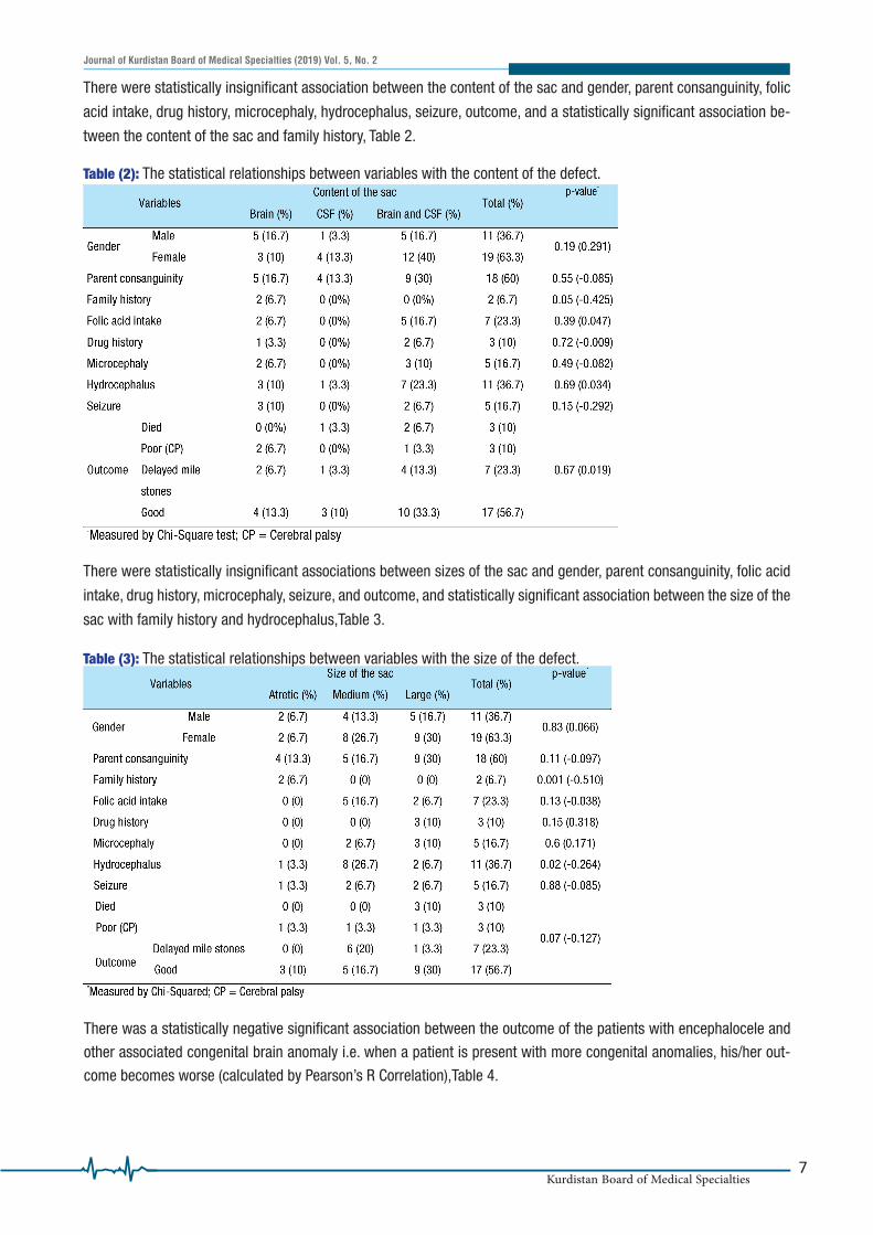
There were statistically insignificant association between the content of the sac and gender, parent consanguinity, folic
acid intake, drug history, microcephaly, hydrocephalus, seizure, outcome, and a statistically significant association be-
tween the content of the sac and family history, Table 2.
Table (2): The statistical relationships between variables with the content of the defect.
There were statistically insignificant associations between sizes of the sac and gender, parent consanguinity, folic acid
intake, drug history, microcephaly, seizure, and outcome, and statistically significant association between the size of the
sac with family history and hydrocephalus,Table 3.
Table (3): The statistical relationships between variables with the size of the defect.
There was a statistically negative significant association between the outcome of the patients with encephalocele and
other associated congenital brain anomaly i.e. when a patient is present with more congenital anomalies, his/her out-
come becomes worse (calculated by Pearson’s R Correlation),Table 4.

8
Journal of Kurdistan Board of Medical Specialties (2019) Vol. 5, No. 2
Kurdistan Board of Medical Specialties
Table (4): Association of outcome with other brain abnormality.
There were statistically insignificant association between outcome and hydrocephalus and seizure, and statistically
significant association between the outcome and microcephaly,Table 5.
Table (5): Association of some clinical features of encephalocele patients with their outcome.
Discussion Neural tube develops in the midline during embryonic
stage. Therefore, almost all of the neural tube defects oc-
cur in the midline and mostly at lumbosacral and occipital
regions1, 2, 7-9, 13, 18. But, there are some sporadic cases in
which encephalocele occurred in other than midline such
as anterioinferior region of temporal lobe5, 7, 19.
The Matson and Ingraham classification of encephalocele
is widely accepted and it is based on the location of the
encephalocele and it includes: basal, sincipital, convexi-
ty (frontal, parietal, occipital, and cervico-occipital), and
atretic7. According to this classification, the most common
location is cervico-occipital region7. Out findings are com-
patible with the Matson and Ingraham classification; all
of our patients presented with midline encephalocele and
occipital area was predominant; 40% occipital, 26.7% oc-
cipito-cervical, 20% vertex, 10% frontal, and 3.3% nasal
,Table 1.
Most of encephaloceles are congenital in origin i.e. prima-
ry1-4, 7, 10-11, 15, 18-20, but acquired or secondary encephalocele
can occur as a result of increased intracranial pressure
e.g. due to tumor, surgery, trauma, and infection5-7. En-
cephalocele can also be acquired from congenital menin-
gocele14. Furthermore, all of our patients had congenital
encephalocele and this may be the cause of the occur-
rence of encephalocele at midline especially occipital re-
gion in our patients.
The size of the encephalocele is not important predictor of
outcome because the prognosis depends on the location
and amount of neural tissue inside the sac and associated
anomalies2, 5. Therefore, the size of the defect is not as
important as the content of the sac; thence, a small sac
may contain large amount of neural tissue and associat-
ed more with microcephaly than larger defect. In addition,
hydrocephalus and microcephalus are most critical risk
factors2. Contrary to that, our results showed a statistically
significant association between the site of the enceph-
alocele and its outcome; the sites were mostly occipital
and occipito-cervical followed by vertex and this may have
been caused more pressure on vital structures in posterior

9
Journal of Kurdistan Board of Medical Specialties (2019) Vol. 5, No. 2
Kurdistan Board of Medical Specialties
fossa, Table 1, and a statistically insignificant association
between the content of the sac and outcome. This may be
because of the small sample size of our study due to rarity
of the condition, Table 2. Furthermore, the size of the sac
had no statistically significant association with the out-
come because a larger size may contain only CSF, Table 3.
Encephalocele can contain CSF, brain tissue and CSF, and
rarely brain tissue only3, 8. Furthermore, the content of the
encephalocele sac in our patients were as follows: 56.7%
brain tissue and CSF, 26.7% brain tissue only, and 16.6%
CSF only, Table 2. This congenital neural tube defect; en-
cephalocele, may coexist with other congenital anomalies
such as transposition of great arteries, atrial defect, ocu-
lar malformation, and craniofacial defects2, 5, in addition
to other congenital brain anomalies like: corpus callosum
and cerebellar agenesis, cortical dysplasia and agene-
sis, ventricular anomaly, Arnold Chiari and Dandy Walker
malformation, microcephaly and hydrocephalus2, 5. In our
study, we found a very significant association between the
outcome and other brain anomalies; we found 13.3% of
patients with corpus callosal agenesis, 6.7% with Dan-
dy Walker, 3.3% with Dandy Walker and corpus callosal
agenesis, 3.3% with right cerebellar agenesis, 3.3% with
arachnoid cyst, and 3.3% with hydrocephalus, Table 4.
About one third of the patients with encephalocele die and
half of the patients who live beyond the first day of birth
have various degree of developmental delay2. Our study
showed a 10% death, 10% poor outcome, 23.3% delay
in milestones, and 56.7% good outcome, Tables 1-4. The
cause of death in our study may be due to that, most of
the died-babies were presented with large occipito-cer-
vical encephalocele and they were associated with cere-
bellar and corpus callosum agenesis. In the literature we
searched, 32% of the patients had hydrocephalus10. Hy-
drocephalus and microcephalus were considered as the
two important risk factors for worst outcome2. Our study
showed half agreement with these findings; we found that
microcephalus is statistically associated significantly with
the worst outcome but hydrocephalus did not have signif-
icant association with the outcome although it accounted
for 36.7% of the patients, Table 5. It may be due to that;
hydrocephalus per se has better outcome if managed well.
ConclusionsEncephalocele is located mostly at occipito-cervical re-
gion. The size of its sac is associated with hydrocephalus,
the content is mixed brain and CSF, and its site is associ-
ated with seizure. Moreover, the content and size of the
sac is significantly associated with family history of neural
tube defect. In addition, the outcome of encephalocele is
not so good and if associated with other congenital and
brain anomalies, occipital encephalocele, and microceph-
aly, it has worst outcome.
We do recommend doing more researches on the assess-
ment of risk factors/causes and preventive methods of
neural tube defects.
Acknowledgement
We are thankful for “Tropk for Computer and Scientific Re-
search (Tropk.org)” for their help in analysis of our data
and edition and proofreading of this manuscript.
Conflict of interest
Nothing to declare.
References1. Yucetas SC, Uçler N. A Retrospective Analysis of Neonatal Enceph-
alocele Predisposing Factors and Outcomes. Pediatr Neurosurg 2017;
52:73-6.
2. Inan C, Sayin NC, Gurkan H, et al. A large posterior encephalocele
associated with severe ventriculomegaly, cerebellar atrophy and trans-
position of the great arteries. J Clin Ultrasound 2018;46(9):588-90.
3. Jeyaraj P. Management of the frontoethmoidal encephalomenin-
gocele. Ann Maxillofac Surg 2018; 8:56-60.
4. Arifin M, Suryaningtyas W, Bajamal AH. Frontoethmoidal encepha-
locele: clinical presentation, diagnosis, treatment, and complications in
400 cases. Childs Nerv Syst. 2018; 34(6):1161-8.
5. Bannout F, Harder S, Lee M, et al. Epilepsy Surgery for Skull-Base
Temporal Lobe Encephaloceles: Should We Spare the Hippocampus
from Resection? Brain Sci. 2018; 8(3):42.
6. Baro V, D’Errico I, d’Avella D, Denaro L. Transethmoidal Encephalocele
and High Intracranial Pressure. Pediatr Neurosurg. 2018; 53(4):286-7.
7. Shi C, Flores B, Fisher S, Barnett SL. Symptomatic Parietal Intradiplo-
ic Encephalocele—A Case Report and Literature Review. J Neurol Surg
Rep. 2017; 78:e40–e45.
8. Satyarthee GD, Moscote-Salazar LR, Escobar-Hernandez N, et al.
A Giant Occipital Encephalocele in Neonate with Spontaneous Hem-
orrhage into the Encephalocele Sac: Surgical Management. J Pediatr
Neurosci. 2017; 12(3):268–70.
9. Yhoshu E, Dash V, Bawa M. Double encephalocele: An unusual pres-

10
Journal of Kurdistan Board of Medical Specialties (2019) Vol. 5, No. 2
Kurdistan Board of Medical Specialties
entation. J Pediatr Neurosci 2018; 13:264-6.
10. Alshamrani A, Habalrih F, Altweijri I, Alsaleh S, Ajlan A. Endoscop-
ic trans-nasal repair of basal encephalocele associated with morning
glory syndrome. Br J Neurosurg. 2018; 10:1-3. Available from: https://
www.tandfonline.com/doi/full/10.1080/02688697.2018.1494264
11. Horcajadas A, Palma A, Khalon BM. Frontoethmoidal encephalocele.
Report of a case. Neurocirugia (Astur). Forthcoming 2018;30. Available
from: http://dx.doi.org/10.1016/j.neucir.2018.02.006
12. Cortazar AZ, Martinez CM, Feliubadalo CD, Cueto MR, Serra L. Mag-
netic resonance imaging in the prenatal diagnosis of neural tube de-
fects. Insights Imaging. 2013; 4(2):225–37.
13. Richardson S, Khandeparker R, Raghuvaram A, Mohan R. Modified
two flap palatoplasty in asymptomatic transsphenoidal encephalocele:
a case report. J Korean Assoc Oral Maxillofac Surg. 2018; 44(2):86-90.
14. Gandhoke G, Goldschmidt E, Kellogg R, Greene S. Encephalocele
development from a congenital meningocele: case report. J Neurosurg
Pediatr. 2017; 20(5):419-22.
15. Lau W, Yung W, Leung W, et al. A case of “familial atretic encepha-
locele”. Ultrasound Obstet Gynecol. 2018; 27. doi: 10.1002/uog.20109.
16. Bear K, Solomon B, Roessler E, et al. Evidence for SHH as a can-
didate gene for encephalocele. Clin Dysmorphol. 2012; 21(3):148–51.
17. Greene ND, Copp AJ. Neural Tube Defects. Annu Rev Neurosci.
2014;37: 221–42.
18. Raeiq A. Posterior Fontanelle Encephalomeningocele in a Neonate:
A Case Report. Cureus. 2018;10(3): e2315.
19. Wang ZI, McBride A, Grinenko O, et al. Utility of CISS sequence in
detecting anteroinferior
temporal encephalocele. J Neurol Sci. 2017; 381:59–61.
20. Rehman L, Farooq G, Bukhari I. Neurosurgical interventions for oc-
cipital encephalocele. Asian J Neurosurg. 2018; 13:233-7.


