

Lymphedema
James Laredo, MD, PhDByung Boong Lee, MD, PhD

PHLEBOLOGY & VENOUS ULTRASOUND
431
Lymphedem
a
1. IntroductionLymphedema is the result of impaired lymphat-
ic function. The lymphatic system is an essential
component of the human circulatory system. It
is comprised of a complex network of vessels.
The major function of the lymphatic system is
the maintenance of interstitial fluid homeosta-
sis and prevention of edema.1,2 Other impor-
tant functions include transportation of white
blood cells and antigen-presenting cells to the
lymphoid organs, and lipid absorption from the
gastrointestinal tract.1,2
Lymphedema is characterized by swelling of
tissues, most commonly involving the lower
extremities in 80% of cases.3,4 It can also occur
in the arms, face, trunk, and external genitalia.
Leg edema is a common condition that is seen
by all practicing clinicians. The differential di-
agnosis of lower extremity edema is extensive
and includes systemic causes such as congestive
heart failure, renal insufficiency, hepatic insuf-
ficiency, hypoalbuminemia, medications, and
local causes such as deep vein thrombosis, ve-
nous insufficiency, lymphedema, lipedema, and
cellulitis.2-5
A detailed understanding of the anatomy and
physiology of the lymphatic system and the
pathophysiology of lymphedema will contrib-
ute to the proper diagnosis and treatment of this
complex and important clinical condition.
2. Anatomy and PhysiologyThe lymphatic system is found throughout the
body and is composed of four components:
lymphatic vessels, lymph fluid, lymph nodes,
and lymphocytes.1,2 Lymphatic vessels gener-
ally accompany the venous system throughout
the body except in the central nervous system,
hepatic sinusoids, and cortical bony skeleton,
where these perivascular spaces serve the func-
tion of the lymphatic vessels.1-3 Lymphatic fluid
from the lower extremities, pelvis, abdominal
viscera, thorax, left arm, and left head and neck,
drains into the central venous system via the
thoracic duct. Lymphatic fluid from the right
arm, right head and neck, and parts of the tho-
rax, drains into the central venous system via
the right lymphatic duct.1-3 Numerous intercon-
nections exist as well as significant variants.1
In addition, an extensive system of superficial
lymphatic vessels extend over the surface of the
entire body draining into communication wa-
tersheds, regional lymph nodes, and ultimately
into the deep lymphatic system.
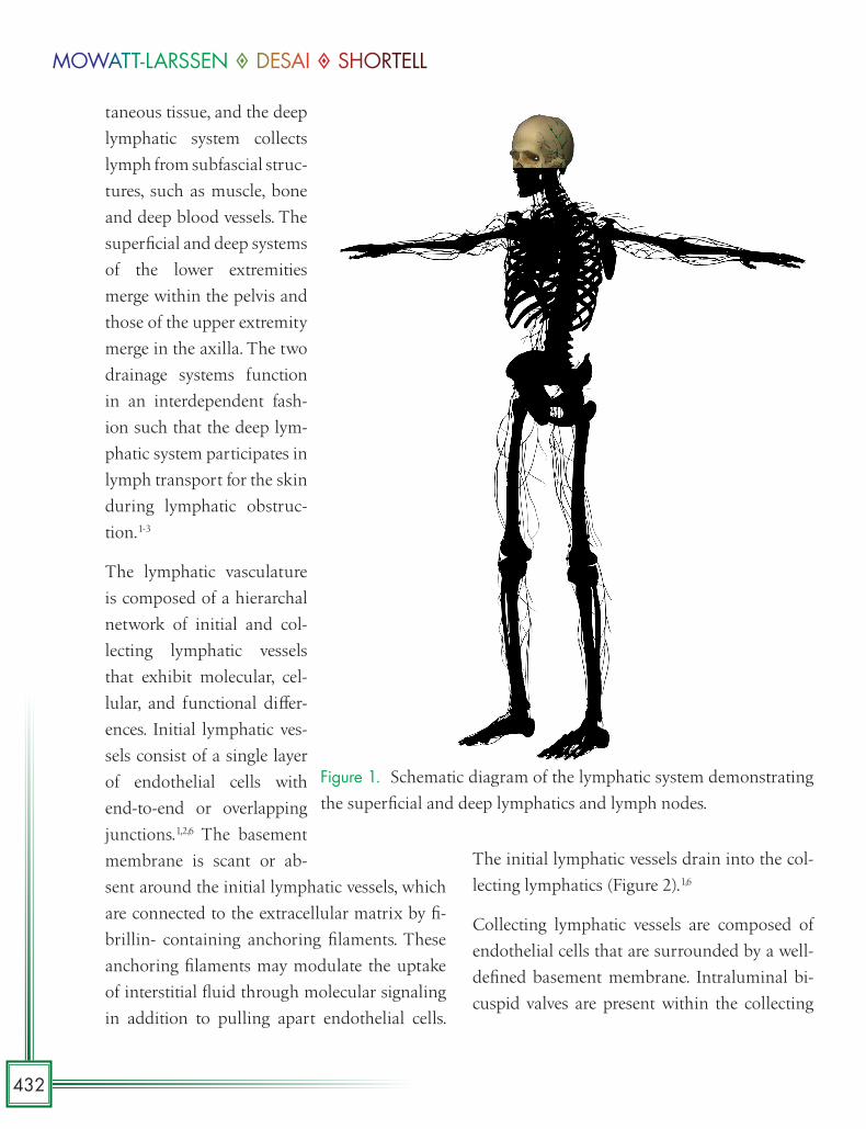
Analogous to the venous system, the lymphatic
system has both a superficial and deep system
in the extremities that is separated by the mus-
cle fascia (Figure 1).1 The superficial lymphatic
system collects lymph from the skin and subcu-
MOWATT-LARSSEN ¹ DESAI ¹ SHORTELL
432
taneous tissue, and the deep
lymphatic system collects
lymph from subfascial struc-
tures, such as muscle, bone
and deep blood vessels. The
superficial and deep systems
of the lower extremities
merge within the pelvis and
those of the upper extremity
merge in the axilla. The two
drainage systems function
in an interdependent fash-
ion such that the deep lym-
phatic system participates in
lymph transport for the skin
during lymphatic obstruc-
tion.1-3
The lymphatic vasculature
is composed of a hierarchal
network of initial and col-
lecting lymphatic vessels
that exhibit molecular, cel-
lular, and functional differ-
ences. Initial lymphatic ves-
sels consist of a single layer
of endothelial cells with
end-to-end or overlapping
junctions.1,2,6 The basement
membrane is scant or ab-
sent around the initial lymphatic vessels, which
are connected to the extracellular matrix by fi-
brillin- containing anchoring filaments. These
anchoring filaments may modulate the uptake
of interstitial fluid through molecular signaling
in addition to pulling apart endothelial cells.
The initial lymphatic vessels drain into the col-
lecting lymphatics (Figure 2).1,6
Collecting lymphatic vessels are composed of
endothelial cells that are surrounded by a well-
defined basement membrane. Intraluminal bi-
cuspid valves are present within the collecting
Figure 1. Schematic diagram of the lymphatic system demonstrating
the superficial and deep lymphatics and lymph nodes.

PHLEBOLOGY & VENOUS ULTRASOUND
433
Lymphedem
a
lymphatic vessels. Valves partition collecting
lymphatic vessels into discrete contractile seg-
ments, termed “lymphangions,” which are sur-
rounded by smooth muscle and contract to ac-
tively transport lymphatic fluid through lymph
nodes and throughout the lymphatic system
(Figure 2).1,2,6
Since the lymphatics lack a central pump, lym-
phatic fluid is propelled through the lymphat-
ics through the concerted effects of respiratory
motions, skeletal muscle contractions, and the
autocontractility of the mural smooth muscle
of the vasculature itself. In skeletal muscle, lym-
phatics are usually paired with arterioles, so that
arterial pulsations can also contribute to the pe-
riodic expansion and compression of the initial
lymphatic vessels to enhance fluid uptake.1-3,7
3. PathophysiologyLymphedema is an imbalance between lym-
phatic fluid formation and lymphatic fluid
absorption, representing a high output or low
output failure of the lymphatic system or a com-
bination of both. The increase of interstitial flu-
id leads to a cascade of remodeling that leads to
permanent changes in the tissues of the affected
limb.1-3,7
Figure 2. Schematic diagram of the lymphatic vessels. Note the initial lymphatic vessels that are
represented as solid green, smaller diameter vessels that drain into the larger diameter collecting
lymphatic vessels. Note the smooth muscle cells around the collecting lymphatics and the pres-
ence of valves. Segments of collecting lymphatic vessels located between two valves are known as
“lymphangions.”

MOWATT-LARSSEN ¹ DESAI ¹ SHORTELL
434
High output failure (also known as dynamic
insufficiency) occurs when excessive lymphatic
fluid formation exceeds the transport capac-
ity of the intact lymphatic system. Increases
in lymphatic fluid production may arise when
Starling forces shift net pressure to favor flow of
fluid into the interstitium. Increases in venous
pressure result in increased hydrostatic pressure
within the venules and capillaries increase the
driving force for ultrafiltration.1-3 Loss of oncotic
pressure, as seen in hypoproteinemic states such
as malnutrition, has a similar effect. Elevated ve-
nous pressure occurs in patients with right heart
failure, deep vein thrombosis, and venous insuf-
ficiency. Local inflammation increases capillary
permeability, accelerating the loss of fluid and
plasma proteins into the interstitium.1-3
In contrast, low out-
put failure (also
known as mechanical
insufficiency) occurs
when there is injury
or impairment of the
lymphatic system due
to paralysis, obstruc-
tion, or inadequacy of
the lymphatics (e.g.,
lymphedema from
filarial lymphatic ob-
struction or congeni-
tal hypoplasia).1-3 As
lymphatic obstruc-
tion progresses, tortu-
osity, dilatation, and
pooling of lymphatic
fluid gives way to massive ectasia, valvular de-
struction, retrograde lymph flow, and lymph co-
agulation. Intrinsic truncal contractions fail; in-
traluminal valves give way, hydrostatic pressure
increases in the superficial valveless lymphatic
watersheds. Chronic inflammation results in
mast cell infiltration, disruption of the intersti-
tial elastin fiber network, intense lymphangio-
genesis and hemangiogenesis, fibrosis, progres-
sive fat deposits, and skin thickening.1-3
Dermal edema is the hallmark of lymphedema
and represents the earliest clinical manifesta-
tion of lymphatic impairment.1,2 The presence of
dilated lymphatic vessels may also be evident.
With prolonged lymphatic impairment, tissue
changes include fibroplasia, hyperkeratosis, and
increases in stromal cells. In addition, elastic tis-
Figure 3. Primary and secondary lymphedema. Lymphedema is the result of decreased transport capacity of the lymphatic system. Lymphedema can be primary or secondary. Primary lymphedema is due to a defect in the lymph conducting pathways. Secondary lymphedema is due to an acquired cause.

PHLEBOLOGY & VENOUS ULTRASOUND
435
Lymphedem
a
sue fragmentation, clumping, and loss of ma-
ture elastic fibers also occur. Abundant subcu-
taneous fat becomes a predominant component
of the swelling seen in the affected limb.3,7
The inflammatory cells present in the edema-
tous tissue contribute to the ongoing fibrosis. It
is believed that the inflammatory cells fail to mi-
grate to the lymph nodes due to impaired lym-
phatic transport and dysfunctional lymphan-
giogenesis, leading to worsening edema and
further inflammation. This ultimately results
in impaired immune trafficking and decreased
clearance of pathogens.1,2
Lymphedema is a progressive and usually pain-
less swelling of the limbs or genitals that is the
result of decreased transport capacity of the
lymphatic system. Lymphedema can be prima-
ry or secondary. Primary lymphedema is due
to a defect in the lymph conducting pathways.
Secondary lymphedema is due to an acquired
cause (such as filariasis, previous surgery, radia-
tion therapy, malignancy, infection and inflam-
mation) that results in injury and impairment
of the lymphatic system (Figure 3).2-4,7,8
4. Stages of LymphedemaRegardless of the etiology, lymphedema is clini-
cally staged by the extent of visible tissue degra-
dation (Table 1).9-12 In the early stages of lymph-
edema (Stage I), the associated limb swelling
resembles other types of edema such as that seen
with congestive heart failure, renal insufficiency,
and venous disease. At this stage, the swelling
is completely relieved with
elevation and/or overnight
rest. The tissues are soft
and usually pitting with
no evidence of fibrosis. As
the condition progresses
(Stage II), the edema is no
longer relieved with el-
evation or rest, and skin
changes begin to appear
such as induration of the
skin and progressive hardening. The edema also
becomes non-pitting. In the late, chronic stage
of lymphedema (Stage III), the edema is severe
and the skin is fibrotic with numerous skin
changes that include hyperkeratosis, warty pro-
jections, cobblestoning, and lichenification (Fig-
ure 4). Stage III lymphedema is also known as
Table 1. Stages of lymphedema
Latency Risk for lymphedema present. No clinical change evident.
Stage IPitting, reduces overnight with simple measures (eleva-
tion). No fibrosis.
Stage IINo longer pitting, no full reduction with elevation, evi-
dent fibrosis.
Stage IIINon-reversible, hardened fibrosis and sclerosis of cutane-
ous and subcutaneous tissues.

MOWATT-LARSSEN ¹ DESAI ¹ SHORTELL
436
“elephantiasis” because the affected limb begins
to resemble the leg of an elephant.9,10,13
4.1. Primary LymphedemaIn patients with primary lymphedema, the cause
of decreased lymphatic transport can be an in-
trinsic ”defect” or a malfunction of the lymph
conducting elements, which is believed to be a
genetically determined abnormality of lymph
drainage.2 The majority of lymphedemas classi-
fied as primary lymphedema has inborn abnor-
malities of the lymphatic system that manifest
mostly with irregular or abnormal structural
development caused by abnormal (mutant)
genes.2,11 These abnormalities result in lymphat-
ic hypoplasia, aplasia, numerical hyperplasia,
or dilation (lymphangiectasia) with valvular in-
competence.2,11
Primary lymphedemas have been classified into
three groups, depending on the age of onset:
Congenital (before age 2), Praecox (onset be-
tween ages 2 and 35), and Tarda (after age 35).
Lymphedema praecox is the most common
form of primary lymphedema with a female to
male ratio of 10:1. It is usually unilateral and of-
ten limited to the foot and calf in most patients
(Figure 3).2,8,11
4.2. Secondary LymphedemaSecondary lymphedema is far more common
than primary lymphedema and represents 90%
of cases of lymphedema. The most common
causes of lower extremity lymphedema are tu-
mor (e.g., lymphoma, prostate cancer, ovarian
cancer), surgery involving the lymphatics, ra-
diation therapy, obesity, trauma, and infection.
Worldwide, infection with the parasitic nema-
tode Wuscheria bancrofti (also known as filaria-
sis), is the most common cause of lymphedema
(Figure 3).2,4,8
5. Diagnosis5.1. Clinical EvaluationEvaluation of patients with lymphedema must
include a detailed, careful history, and thorough
physical examination.3,4,7,11,12 The history should
include age at onset, travel to tropical countries
and history of all causes that could result in sec-
ondary lymphedema such as surgery, malignan-
cy, venous insufficiency, trauma, and cellulitis. A
history of temporary edema of the affected limb
or other areas must be noted and a detailed

PHLEBOLOGY & VENOUS ULTRASOUND
437
Lymphedem
a
family history of limb swelling should
also be recorded.
Signs and symptoms of lymphedema
should be documented. These include
non-pitting edema, skin changes such
as “peau d’orange,” pinkish-red skin
discoloration, hyperkeratosis, der-
matitis, eczema, ulceration, varicos-
ity, lymph vesicles, warty projections,
drainage of fluid (clear or milky), or
yellow discoloration or other abnor-
malities of the nails (Figure 4). The
presence of the Stemmer sign (inabil-
ity to pinch a fold of skin at the base of
the second toe) or puffiness of the fore-
foot (buffalo hump) should be noted
(Figure 5).3,7,11,12 The presence of venous,
arteriovenous, or capillary malforma-
tions, and any limb length discrepancy,
should be recorded. Finally, any com-
plications, such as cellulitis, lymphan-
giitis, malnutrition, immunodeficiency
or, rarely, suspicion for malignancies
(lymphangiosarcoma) must be docu-
mented.3,7,11
5.2. Non-Invasive Radiologic Studies
Plain film X-rays will identify limb length dis-
crepancies, bone abnormalities or phleboliths
in patients with combined lymphatic malforma-
tions and venous malformations.7,11
Venous duplex studies will confirm any associ-
ated venous anomalies (valvular incompetence,
obstruction, ectasia or aneurysms) and assess
Figure 4. Bilateral lower extremity lymphedema in a 56 year old man before and after complex decongestive therapy. Top: Before treatment. Note the significant limb swelling and chronic skin changes (lichenification, warty projections, and cobblestone appearance) associated with lymphedema.
Bottom: After treatment. Note the significant improvement in
limb swelling and chronic skin changes.

MOWATT-LARSSEN ¹ DESAI ¹ SHORTELL
438
for venous obstruction as an etiology or con-
tributing factor to lymphedema.7,11
5.3. Minimally Invasive Radiologic Studies5.3.1. Radionuclide Lymphoscintigraphy
This study is performed with a subcutaneous
injection of 99mTc-labeled human serum al-
bumin (HAS) or 99mTc-labeled sulphur col-
loid (SC), into the first and second web-space
of the toes (fingers), followed by radionuclide
scanning at various time intervals.7,11 It is the
test of choice to confirm or exclude lymph-
edema as the cause of chronic limb swell-
ing. Removal of the colloid from the injection
site, appearance time of activity at the knee,
groins or axilla, absence or presence of major
lymphatic collectors, number and size of ves-
sels and nodes, the presence of collaterals and
reflux, symmetric activity with the opposite
side are recorded and used for interpretation.
• An appropriate combination of non- to
minimally invasive tests normally should
provide all the information necessary to
insure an adequate diagnosis and lead to
the correct multidisciplinary, specifically
targeted and sequenced treatment strategy.
The tests and the information they provide
are indicated here.11
Basic/Essential tests:
• Radionuclide Lymphoscintigraphy
• MRI with/without contrast for the differ-
ential diagnosis
Figure 5. Clinical signs of lymphedema. A. Positive Stemmer’s sign (a failure by the examiner to pick up or pinch a fold of skin at the base of the second toe). B. Buffalo hump on the dorsum of the left foot in a patient with lymphedema.

PHLEBOLOGY & VENOUS ULTRASOUND
439
Lymphedem
a
• CT scan to exclude underlying pathology
• Duplex ultrasonography
Optional tests:
• Whole body blood pool scintigraphy
(WBBPS)
• Magnetic resonance (MR) and/or Ultra-
sound lymphography
• Volumetry
• Bio-impedance Spectrometry
• Air plethysmography
• Ultrasonographic lymphangiography: in-
vestigational for the reconstructive surgery
candidate patient
• MR lymphangiography: investigational for
the reconstructive surgery candidate pa-
tient
• Microscopic fluorescent lymphangiogra-
phy: investigational for phlebolymphede-
ma
Radionuclide lymphoscintigraphy is the most
essential part of the diagnosis of lymphedema
in addition to clinical evaluation. This study
is extremely useful for delineating the specific
lymphatic abnormality and has largely replaced
conventional oil contrast lymphography for
visualizing the lymphatic network. Lymphos-
cintigraphy remains the gold standard for the
lymphatic function evaluation, which is recom-
mended for proper clinical management.3,7,11
On some occasions an invasive study is required
for an accurate diagnosis. These tests and the in-
formation they provide are listed below:
Direct puncture percutaneous lymphangiogra-
phy
• Standard (ascending) lymphangiography
• Indirect lymphography using water-solu-
ble contrast media
• Fine needle aspiration biopsy of lymph
node
• Skin biopsy in cases of suspected sarcoma,
skin cancer or differential diagnosis of
warty lesions
Invasive tests are seldom required for diagnosis
but are occasionally needed for confirming the
diagnosis or planning surgical therapy.
Conventional oil contrast lymphangiography,
especially if coupled with computed tomog-
raphy (CT) scanning, is still advantageous in
selected patients with chylous dysplasia and
gravitational reflux disorders in order to define
more clearly the extension of the pathologic
alterations and sites of lymphatic and chylous
leakage.11 It is the only diagnostic study that can
clearly demonstrate pathologies of chylous ves-
sels, chylous cyst and thoracic duct in cases of
chylothorax, chylous ascites, protein-losing en-
teropathy, etc.
As a part of the diagnostic procedure, the sys-
temic causes of edema (e.g., heart failure, hypo-
proteinemia, pulmonary hypertension, hypo-

MOWATT-LARSSEN ¹ DESAI ¹ SHORTELL
440
thyroidism, cyclic edema) should be ruled out.
Duplex ultrasonography should be performed
initially in all forms of lymphedema to assess for
concomitant venous disease.
Diagnostic evaluation should also include ap-
propriate assessment of the patient’s under-
standing of the disease process and ability to be
compliant with the treatment regimen, since the
outcome of successful management is totally
dependent on the patient’s active participation
in the care of his or her lymphedema.
6. Treatment of Lymphedema6.1. General ConsiderationsThe importance of patient education and com-
pliance cannot be over-emphasized when treat-
ing patients with both primary and secondary
lymphedema. The patient must first understand
that lymphedema is a chronic condition and
will never be completely cured. In addition they
must also understand that there is no “quick
fix” operation, medication, or therapy that will
completely reverse the clinical condition. Treat-
ment of lymphedema is essentially management
of the medical condition and prevention of pro-
gression of the disease process. Lymphedema
can be successfully managed.
The goals of lymphedema therapy are to arrest
progression, reduce swelling, maintain that re-
duction, prevent infection, restore mobility and
range of motion, and train patients for self-man-
agement.4,10,14
The treatment of lymphedema requires dili-
gence and motivation on the part of the patient.
The patient must be an active, compliant partic-
ipant for successful management. The mainstay
of lymphedema treatment is through physical
therapeutic measures occurring in the setting of
a specific lymphedema therapy program, per-
formed by specially trained lymphedema thera-
pists.3,10,14,15
6.2. Physical Treatments Physical treatment to reduce swelling is aimed
at controlling lymph formation and improving
lymph drainage through existing lymphatic ves-
sels and collateral routes by applying normal
physical processes which stimulate lymph flow
(Table 2).3,10,12,14-16 Manual therapies in multiple
forms remain the most widely used interven-
tions for the therapeutic management of lymph-
edema, regardless of etiology.
Manual lymphatic drainage (MLD) is a highly
specialized form of massage therapy that em-
ploys very light and gentle cutaneous distension

PHLEBOLOGY & VENOUS ULTRASOUND
441
Lymphedem
a
to enhance lymph transport. MLD is believed
to stimulate and increase the intrinsic contrac-
tility of lymph collecting vessels and encourage
increased protein molecule sequestration and
subsequent transport.10,14-16 MLD is often com-
bined with other manual therapies including
compression bandaging, exercise regimens, skin
care techniques, pressure gradient garments,
and pneumatic compression devices.15-20
Complex decongestive therapy (CDT) is a com-
bined approach to lymphedema therapy that
has been standardized by multiple international
lymphatic treatment organizations and special-
ized lymphedema treatment programs.10,14-16 The
treatment regimen is composed of two phases:
intensive reduction therapy followed by mainte-
nance therapy (Table 3). This treatment regimen
utilizes MLD, compression wrapping, exercise
therapy and skin care. This highly successful
treatment regimen has become the standard of
care for lymphedema management.10,14-16 Signifi-
cant improvement and reduction of swelling is
often readily apparent after treatment (Figure 4).
Compression wrapping in various forms has
been a longstanding treatment of both venous
and lymphatic edema.10,17-20 Lymphatic wrapping
Table 2. Physical Treatments for Lymphedema
Treatment Effect
Exercise
Dynamic muscle contractions encourage movement of lymph along tis-
sue planes and non-contractile, initial lymph vessels (passive drainage)
and increased contractility of collecting lymph vessels (active drainage).
Compression Gar-
ments
Opposes capillary filtration.
Acts as a counterforce to muscle contractions generating greater inter-
stitial pressure changes.
Manual Lymphatic
Drainage
Form of massage therapy that stimulates lymph flow in more proximal,
normally draining lymphatics to “siphon” lymph from congested areas.
Compression Bandag-
ing
Used as an intensive treatment in combination with exercise to reduce
large, misshapen lower limbs and permit subsequent maintenance
treatment with compression stockings.
Pneumatic Compres-
sion
Softens and reduces limb volume but can forcibly displace fluid into
trunk and genitalia. Compression garments must be worn after treat-
ment.
ElevationDoes not stimulate lymph drainage, but lowers venous pressure and
therefore capillary filtration, allowing lymph drainage to catch up.

MOWATT-LARSSEN ¹ DESAI ¹ SHORTELL
442
techniques are complex and utilize low stretch
bandages instead of the more traditional high
stretch elastic bandages. High stretch wrapping
produces high pressures at rest that decrease
with limb muscle contraction and movement.
This decreases the ability of the wrap to raise
the tissue pressure during exercise, reducing the
hydrostatic pressure gradient and resulting in a
reduction in stimulation of lymphatic flow.10,17-20
In contrast, low stretch wrapping provides resis-
tance during muscle pump action that results in
an increase in pressure gradient and stimulates
increased fluid flow.10,17-20 Patients with signifi-
cant obesity, pain problems, or advanced disease
may not be able to comply with the complexi-
ties of wrapping. For these patients, static gradi-
ent compression devices are available.
The use of elastic compression garments is the
mainstay of the maintenance portion of any
lymphedema management program.10,17-21 Com-
pliance with daily use of compression stockings
or sleeves is critical for maintenance of limb
size and volume. Compression garments should
have graduated compression where pressure is
highest distally and decreases proximally where
the pressure is lowest at the highest level (Figure
6). For an upper extremity graduated compres-
sion sleeve, pressure is highest at the hand/wrist
and is lowest at the shoulder. For a lower ex-
tremity graduated compression stocking, pres-
sure is highest at the ankle and is lowest at the
knee, thigh, or waist, depending on the length
of the stocking. Recommended graduated com-
pression is 30-40 mmHg for the lower extrem-
ity.10,18 Upper extremity lymphedema sleeves are
available with a graduated compression of 20-
30 mmHg, which is usually adequate.10,18
In addition to providing graduated compres-
sion, compression stockings and sleeves also
assist with venous return, help preserve skin
integrity, and protect the skin from trauma.10
Currently, there are many manufacturers who
produce graduated compression stockings and
sleeves with different pressure strengths, differ-
ent fabrics, and multiple color options. The abil-
ity to independently don the compression gar-
ment is critical. Numerous devices are available
to assist with stocking and sleeve donning.
Table 3. Complex decongestive therapy.
Phase I: Intensive reduction therapy
Manual lymphatic drainage massage
Multilayered low-stretch wrapping techniques
Specific exercise regimen
Skin care education and techniques
Phase II: Maintenance therapy
Daily wear of pressure garment
Continued nightly multilayered wrapping
Self-manual lymphatic drainage massage
Exercise
Continued meticulous skin management

PHLEBOLOGY & VENOUS ULTRASOUND
443
Lymphedem
a
For decades prior to the introduction of CDT,
pneumatic compression pumps were the main-
stay of lymphedema therapy. Since the mid-
1990s, when CDT became
more widely available, use
of pneumatic compression
pump therapy has largely
become an adjunct to CDT
in both the reductive and
maintenance phases.3,10,14,22
The majority of pneumatic
compression pumps perform
sequential pumping of the
affected lymphedematous
limb from distal to proxi-
mal. These devices augment
the beneficial effects of the
standard modalities of CDT.
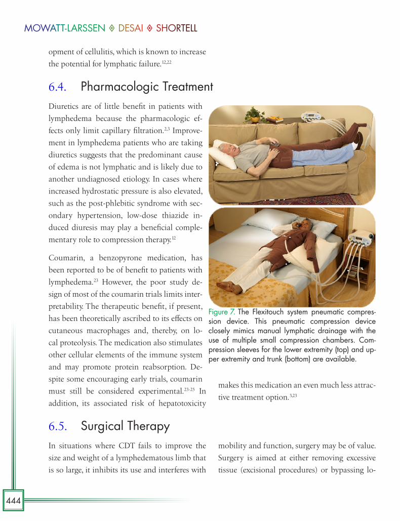
There is a new class of se-
quential pneumatic com-
pression device, known as
the Flexitouch system. This
pneumatic compression de-
vice closely mimics MLD with the use of mul-
tiple small compression chambers (Figure 7).
6.3. Prevention of InfectionPrevention of acute episodes of cellulitis or lym-
phangiitis is critical because they cause severe
deterioration in swelling and result in further
injury to the lymphatic system.12,21,22 Care of the
skin, good hygiene, control of skin diseases such
as tinea pedis, and careful antiseptic dressing
application after minor wounds are all impor-
tant. Antibiotics must be administered prompt-
ly when an acute inflammatory episode occurs.
There are no definitive studies addressing anti-
biotic prophylaxis for patients at risk for lymph-
edema, but evidence has shown the relationship
between chronic fungal infection and the devel-
Figure 6. Graduated compression stockings. Graduated compression stockings have the highest pressure at the ankle level. The pressure de-creases up the leg where the pressure is the lowest at the highest level. The ideal compression for lymphedema treatment is 30-40 mmHg.
MOWATT-LARSSEN ¹ DESAI ¹ SHORTELL
444
opment of cellulitis, which is known to increase
the potential for lymphatic failure.12,22
6.4. Pharmacologic Treatment Diuretics are of little benefit in patients with
lymphedema because the pharmacologic ef-
fects only limit capillary filtration.2,3 Improve-
ment in lymphedema patients who are taking
diuretics suggests that the predominant cause
of edema is not lymphatic and is likely due to
another undiagnosed etiology. In cases where
increased hydrostatic pressure is also elevated,
such as the post-phlebitic syndrome with sec-
ondary hypertension, low-dose thiazide in-
duced diuresis may play a beneficial comple-
mentary role to compression therapy.12
Coumarin, a benzopyrone medication, has
been reported to be of benefit to patients with
lymphedema.23 However, the poor study de-
sign of most of the coumarin trials limits inter-
pretability. The therapeutic benefit, if present,
has been theoretically ascribed to its effects on
cutaneous macrophages and, thereby, on lo-
cal proteolysis. The medication also stimulates
other cellular elements of the immune system
and may promote protein reabsorption. De-
spite some encouraging early trials, coumarin
must still be considered experimental.23-25 In
addition, its associated risk of hepatotoxicity
makes this medication an even much less attrac-
tive treatment option.3,23
6.5. Surgical TherapyIn situations where CDT fails to improve the
size and weight of a lymphedematous limb that
is so large, it inhibits its use and interferes with
mobility and function, surgery may be of value.
Surgery is aimed at either removing excessive
tissue (excisional procedures) or bypassing lo-
Figure 7. The Flexitouch system pneumatic compres-sion device. This pneumatic compression device closely mimics manual lymphatic drainage with the use of multiple small compression chambers. Com-pression sleeves for the lower extremity (top) and up-per extremity and trunk (bottom) are available.

PHLEBOLOGY & VENOUS ULTRASOUND
445
Lymphedem
a
cal lymphatic defects (lymphatic reconstruction
procedures).4,26 CDT is still required after surgi-
cal excision and reconstruction.
Excisional procedures usually involve staged
removal of the lymphedematous subcutaneous
tissue of the leg.26-29 The most radical excisional
operation, the Charles procedure, involves total
skin and subcutaneous tissue excision of the
lower extremity from the tibial tuberosity to the
malleoli, followed by skin grafting. The main
complication associated with this procedure
and other excisional procedures, is infection and
necrosis of the skin graft.26
Chronic lymphedematous tissue transforms
with time into adipose tissue, which cannot be
reduced by massage or compression treatment.
Liposuction aimed at removing this adipose tis-
sue, has been reported to be beneficial in treat-
ing lymphedematous limbs.28,29 This procedure
is not routinely performed for treatment of
lymphedema.
Developments in microvascular techniques have
allowed surgical attempts at direct lymphatic
reconstructions, performance of lymphatic-ve-
nous anastomoses or lymphatic grafting.26,30,31
These reconstructions are usually indicated in
only a small subset of patients who have proxi-
mal obstruction with preserved lymphatic ves-
sels distally.
The best outcomes are seen in patients with sec-
ondary lymphedema, with well-defined trauma
to the lymphatics, seen on lymphatic imaging,
who underwent lymphatic to venous anastomo-
ses.30,31
Lymphatic bypass procedures are only per-
formed in a few selected cases and in only a few
specialized medical centers. This is reflected in
the literature by small patient numbers in most
series reported.26,32,33 Results are variable and
lymphatic bypass procedures are generally not
routinely performed except at these few special-
ized medical centers.
7. References1. Dellinger MT, Bernas MJ, Witte MH. Lymphatic bi-
ology and pathobiology. In: Dieter RS, Dieter RA Jr, Dieter RA, eds. Venous and lymphatic diseases. New York: McGraw Hill, 2011:17-36.
2. Thanaporn PK, Rockson SG. Disease of the lymphat-ic vasculature. In: Dieter RS, Dieter RA Jr, Dieter RA, eds. Venous and lymphatic diseases. New York: Mc-Graw Hill, 2011:569-594.
3. Rockson SG. Diagnosis and management of lymphatic vascular disease. J Am Coll Cardiol. 2008;52(10):799-806.
4. Tiwari A, Cheng KS, Button M, Myint F, Hamilton G. Differential diagnosis, investigation, and current
treatment of lower limb lymphedema. Arch Surg. 2003;138(2):152-61.
5. Ely JW, Osheroff JA, Chambliss ML, Ebell MH. Ap-proach to leg edema of unclear etiology. J Am Board Fam Med. 2006;19(2):148-160.
6. Alitalo K, Tammela T, and Petrova TV. Lymphangio-genesis in development and human disease. Nature 2005;438:946-953.
7. Rooke TW, Felty C. Lymphedema: pathophysiology, classification, and clinical evaluation. In: Gloviczki P, ed. Handbook of venous disorders 3rd edition. Lon-don: Hodder Arnold, 2009:629-634.

MOWATT-LARSSEN ¹ DESAI ¹ SHORTELL
446
8. Kerchner K, Fleischer A, Yosipovitch G. Lower ex-tremity lymphedema update: pathophysiology, diag-nosis, and treatment guidelines. J Am Acad Dermatol. 2008;59(2):324-31.
9. International Society of Lymphology.The diagno-sis and treatment of peripheral lymphedema. 2009 Concensus Document of the International Society of Lymphology. Lymphology. 2009;42(2):51-60.
10. Gamble GL, Cheville A, Strick D. Lymphedema: medical and physical therapy. In: Gloviczki P, ed. Handbook of venous disorders 3rd edition. London: Hodder Arnold, 2009:649-657.
11. Lee B, Andrade M, Bergan J, Boccardo F, Campisi C, Damstra R, et al. Diagnosis and treatment of primary lymphedema. Consensus document of the Interna-tional Union of Phlebology (IUP)-2009. Int Angiol. 2010;29(5):454-470.
12. Mortimer PS. ABC of arterial and venous dis-ease swollen lower limb – 2: Lymphoedema. BMJ 2000;320:1527-1529.
13. Dean SM, Zirwas MJ, Horst AV. Elephantiasis nostras verrucosa: an institutional analysis of 21 cases. J Am Acad Dermatol. 2011 Jun;64(6):1104-10
14. Cheville AL, McGarvey CL, Petrek JA, Russo SA, Tay-lor ME, Thiadens SR. Lymphedema management. Se-min Radiat Oncol. 2003 Jul;13(3):290-301.
15. Mayrovitz HN. The standard of care for lymphede-ma: current concepts and physiological consider-ations. Lymphat Res Biol. 2009;7(2):101-8
16. Badger C, Preston N, Seers K, Mortimer P. Physical therapies for reducing and controlling lymphoedema of the limbs. Cochrane Database Syst Rev. 2004 Oct 18;(4):CD003141.
17. Partsch H, Mosti G. Thigh compression. Phlebology. 2008;23(6):252-8.
18. Partsch H, Flour M, Smith PC; International Com-pression Club. Indications for compression therapy in venous and lymphatic disease consensus based on experimental data and scientific evidence. Under the auspices of the IUP. Int Angiol. 2008 Jun;27(3):193-219.
19. Pappas CJ, O’Donnell TF Jr. Long-term results of compression treatment for lymphedema. J Vasc Surg. 1992 Oct;16(4):555-62.
20. Damstra RJ, Brouwer ER, Partsch H. Controlled, com-parative study of relation between volume changes and interface pressure under short-stretch bandages in leg lymphedema patients. Dermatol Surg. 2008 Jun;34(6):773-8.
21. Mortimer PS. Therapy approaches for lymphedema. Angiology. 1997 Jan;48(1):87-91.
22. Keeley VL. Lymphoedema and cellulitis: chicken or egg? Br J Dermatol. 2008 Jun;158(6):1175-6.
23. Badger C, Preston N, Seers K, Mortimer P. Benzo-pyrones for reducing and controlling lymphoe-dema of the limbs. Cochrane Database Syst Rev. 2004;(2):CD003140.
24. Casley-Smith JR. Benzo-pyrones in the treatment of lymphoedema. Int Angiol. 1999 Mar;18(1):31-41.
25. Loprinzi CL, Kugler JW, Sloan JA, Rooke TW, Quella SK, Novotny P, et al. Lack of effect of coumarin in women with lymphedema after treatment for breast cancer. N Engl J Med. 1999 Feb 4;340(5):346-50.
26. Gloviczki P. Principles of surgical treatment of chron-ic lymphedema. In: Gloviczki P, ed. Handbook of ve-nous disorders 3rd edition. London: Hodder Arnold, 2009:658-664.
27. Miller TA, Wyatt LE, Rudkin GH. Staged skin and subcutaneous excision for lymphedema: a favorable report of long-term results. Plast Reconstr Surg. 1998 Oct;102(5):1486-98
28. Brorson H. From lymph to fat: complete reduction of lymphoedema. Phlebology. 2010;25 Suppl 1:52-63.
29. Brorson H, Ohlin K, Olsson G, Svensson B, Svensson H. Controlled compression and liposuction treat-ment for lower extremity lymphedema. Lymphology. 2008;41(2):52-63.
30. Campisi C, Bellini C, Campisi C, Accogli S, Bonioli E, Boccardo F. Microsurgery for lymphedema: clini-cal research and long-term results. Microsurgery. 2010;30(4):256-60.
31. Campisi C, Eretta C, Pertile D, Da Rin E, Campisi C, Macciò A, et al. Microsurgery for treatment of pe-ripheral lymphedema: long-term outcome and fu-ture perspectives. Microsurgery. 2007;27(4):333-8.

PHLEBOLOGY & VENOUS ULTRASOUND
447
Lymphedem
a


