

MAHARASHTRA STATE BOARD OF TECHNICAL EDUCATION (Autonomous)
(ISO/IEC - 27001 - 2005 Certified)
SUMMER - 2017 EXAMINATION Subject Code:17666 Model Answer _____________________________________________________________________________________
Page 1 of 29
Important Instructions to examiners:
1) The answers should be examined by keywords and not as word-to-word as given in the model
answer scheme.
2) The model answer and the answer written by candidate may vary but the examiner may try to
assess the understanding level of the candidate.
3) The language errors such as grammatical, spelling errors should not be given more Importance.
(Not applicable for subject English and Communication Skills.)
4) While assessing figures, examiner may give credit for principal components indicated in the
figure. The figures drawn by candidate and model answer may vary. The examiner may give
credit for any equivalent figure drawn.
5) Credits may be given step wise for numerical problems. In some cases, the assumed constant
values may vary and there may be some difference in the candidate’s answers and model answer.
6) In case of some questions credit may be given by judgement on part of examiner of relevant
answer based on candidate’s understanding. 7) For programming language papers, credit may be given to any other program based on
equivalent concept.
Q.
No.
Question & its Answer Remark Total
Marks
Q.1
A)
Attempt ant THREE of the following 12
i) Draw a neat labeled diagram of MAN-INSTRUMENT system.
State the function of any 2 blocks.
04
Ans. Diagram:
Fig: Block diagram of Man – Instrument system
Function of Man – Instrument system : (Any two block) The basic components of the man instrument system are:
Subject: The subject is the human being on whom the measurements
02 marks
for
Diagram

MAHARASHTRA STATE BOARD OF TECHNICAL EDUCATION (Autonomous)
(ISO/IEC - 27001 - 2005 Certified)
SUMMER - 2017 EXAMINATION Subject Code:17666 Model Answer _____________________________________________________________________________________
Page 2 of 29
are made.
Stimulus: Stimulus generates response. The instrumentation used to
generate and present this stimulus to the subject is the vital part of man-
instrument system whenever responses are measure. E.g. visual (flash
of light), auditory (a tone), etc.
Transducer: A transducer is device used to produce an electrical signal
that is an analog of the phenomenon being measured.
Signal conditioning equipment: This part of the system amplifies,
modifies, or in any other ways changes the electric output of the
transducer to satisfy the functions of the system and to prepare signals
suitable for operating the display or recording equipment that follows.
Display equipment: The input to the display device is the modified
electric signal from the signal conditioning equipment which is
converted into a form that can be perceived by one o the human’s
senses in a meaningful way. E.g. graphic pen recorder for recoding
ECG signal.
Recording, Data processing, and Transmission: Recording
instruments are required to record the desirable information that can be
used to transmit or for possible later use. E.g. on line digital computer,
recording equipment etc.
Control devices: Where it is necessary or desirable to have automatic
control of the stimulus, transducers, or any other part of the man
instrument system, a control system is incorporated which uses control
devices.
02 marks
for
function of
any two
block
ii) Name any 2 electrodes each used for measurement of:
1) ECG 2) EEG
04
Ans. 1)Electrodes for ECG:
i) Limb Electrodes
ii) Floating Electrodes
iii) Pasteless Electrodes
iv) Pre-jelled Disposable Electrodes
v) Air-Jet ECG Electrodes
2) Electrodes for EEG
i) Surface Electrodes
ii) Chlorided Silver discs Electrodes
iii)Skin surface Electrodes
02 marks
for Any
two
electrodes
02 marks
for Any
two
relevant
electrodes

MAHARASHTRA STATE BOARD OF TECHNICAL EDUCATION (Autonomous)
(ISO/IEC - 27001 - 2005 Certified)
SUMMER - 2017 EXAMINATION Subject Code:17666 Model Answer _____________________________________________________________________________________
Page 3 of 29
iv)Needle Electrodes
v)Scalp Electrodes
iii) Explain the need of dialysis machine. 04
Ans. Need of dialysis machine
There is need when the original kidneys of patient are:
Unable to form urine.
Unable to removal of waste products from blood plasma.
Unable for the regulation of the composition of blood
plasma.
Unable to regulates volume, osmotic pressure in the blood
vessels .
Unable to Balance pH and electrolyte composition of the
body fluids.
04 marks
(Any four
points)
iv) State one application each of the following.
1) Centrifuge
2) Autoclave
3) Deionizer
4) Incubator
04
Ans. 1)Centrifuge
1) laboratory-scale centrifuges are used in chemistry, biology,
biochemistry and clinical medicine for isolating and separating
suspensions and immiscible liquids.
2)Centrifuges are use in nuclear power and nuclear
weapon programs to separate isotopes.
3) Centrifuges are use in the chemical industry for Synthesis of
materials.
4) In washing Machines.
5) Used to separate cream (remove fat) from milk.
2)Autoclave
It is used to sterilize medical equipment such as
1. Surgical instruments (scissors, needles etc )
2. Glass ware
3. Pathogenic hospital waste etc
4. Used in food industry
5. Used in dentistry
3)Deionizer 1. Medical
2. Laboratory
3. Pharmaceutical
01 mark
for stating
any one
applicatio
n each
MAHARASHTRA STATE BOARD OF TECHNICAL EDUCATION (Autonomous)
(ISO/IEC - 27001 - 2005 Certified)
SUMMER - 2017 EXAMINATION Subject Code:17666 Model Answer _____________________________________________________________________________________
Page 4 of 29
4. Cosmetics
5. Electronic manufacturing
6. Food processing
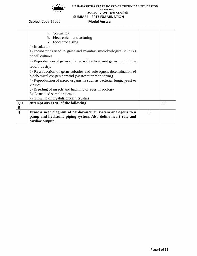
4) Incubator
1) Incubator is used to grow and maintain microbiological cultures
or cell cultures.
2) Reproduction of germ colonies with subsequent germ count in the
food industry.
3) Reproduction of germ colonies and subsequent determination of
biochemical oxygen demand (wastewater monitoring)
4) Reproduction of micro organisms such as bacteria, fungi, yeast or
viruses
5) Breeding of insects and hatching of eggs in zoology
6) Controlled sample storage
7) Growing of crystals/protein crystals
Q.1
B)
Attempt any ONE of the following 06
i) Draw a neat diagram of cardiovascular system analogous to a
pump and hydraulic piping system. Also define heart rate and
cardiac output.
06

MAHARASHTRA STATE BOARD OF TECHNICAL EDUCATION (Autonomous)
(ISO/IEC - 27001 - 2005 Certified)
SUMMER - 2017 EXAMINATION Subject Code:17666 Model Answer _____________________________________________________________________________________
Page 5 of 29
Ans.
Heart rate: The number of heartbeats per unit of time, usually per
minute. The heart rate is based on the number of contractions of
the ventricles (the lower chambers of the heart). The heart rate may
be too fast (tachycardia) or too slow (bradycardia). Normal heart
rate in healthy human is 72 beats per minute.
Cardiac output:- Cardiac output is the volume of blood pumped by
the heart per minute (mL blood/min). Cardiac output is a function of
heart rate and stroke volume. The heart rate is simply the number of
heart beats per minute. The stroke volume is the volume of blood, in
milliliters (ml), pumped out of the heart with each beat. Increasing
either heart rate or stroke volume increases cardiac output.
Cardiac Output in ml/min = heart rate (beats/min) X stroke
volume (ml/beat)
An average person has a resting heart rate of 70 beats/minute and a
04 marks
for
diagram
01 mark
01 mark

MAHARASHTRA STATE BOARD OF TECHNICAL EDUCATION (Autonomous)
(ISO/IEC - 27001 - 2005 Certified)
SUMMER - 2017 EXAMINATION Subject Code:17666 Model Answer _____________________________________________________________________________________
Page 6 of 29
resting stroke volume of 70 ml/beat. The cardiac output for this
person at rest is:
Cardiac Output = 70 (beats/min) X 70 (ml/beat) = 4900
ml/minute.
The total volume of blood in the circulatory system of an average
person is about 5 liters (5000 ml). According to our calculations, the
entire volume of blood within the circulatory system is pumped by
the heart each minute (at rest). During vigorous exercise, the cardiac
output can increase up to 5 fold (25 liters/minute).
ii) Explain the operation of an X-ray machine with a neat block
diagram.
06
Ans.
OR
03 marks
diagram

MAHARASHTRA STATE BOARD OF TECHNICAL EDUCATION (Autonomous)
(ISO/IEC - 27001 - 2005 Certified)
SUMMER - 2017 EXAMINATION Subject Code:17666 Model Answer _____________________________________________________________________________________
Page 7 of 29
Explanation: X ray machine has two parts of the circuit.
i) One of them is to produce high voltage which is applied to tubes
anode and cathode and comprises high voltage step up
transformer followed by rectification. The current through the
tube follows the high tension path way and is measured by mA
meter.
A kV selector switch facilitates change in voltage between
the exposures. The voltage is measured with the help of kV meter.
The exposure switch controls the timer and thus the duration
of application of kV. To compensate mains supply voltage
variation a voltage compensator is included in the circuit.
ii) Second part concerned the X-Ray tube filament; the filament is
heated with 6-12 volts of AC Supply at current of 3-5 A.
The filament temperature determines the tube current and therefore
the filament temp control is attached with millimeter selector.
The filament current is controlled by using in the primary side of the
filament transformer, a variable choke or rheostat. The rheostat
provides a step wise control of mA and is most commonly
used in modern machine. A preferred method of providing high
voltage dc to the anode of X-Ray tube is by use a bridge rectifier
using 4 valve tube or solid state rectifiers, which provide more
efficient system than the half wave self rectification method.
03 marks
for
explanatio
n
Q.2 Attempt ant TWO of the following 16
a) Describe resting potential and action potential with neat
diagrams and waveform.
08
Ans. i) Resting Potential:

MAHARASHTRA STATE BOARD OF TECHNICAL EDUCATION (Autonomous)
(ISO/IEC - 27001 - 2005 Certified)
SUMMER - 2017 EXAMINATION Subject Code:17666 Model Answer _____________________________________________________________________________________
Page 8 of 29
Surrounding the cell of the body or body fluids. These fluids of
conductive solutions containing charged atoms known as ions. The
principle ions are sodium (Na+), potassium (K
+) and chloride(Cl
-).
The membrane of excitable cell readily permit entry of K+ and Cl
-
restricts flow of NaCl. The inability of sodium to penetrate the
membrane results in two conditions. First, the concentration of
sodium ion inside the cell much lower than in the intercellular fluid
outside. Since the sodium ions are positive, these would tend to
make the outside of the cell more positive than the inside. Second, in
an attempt to balance the electric charge, additional potassium ions,
which are also positive, enters the cell, causing a higher
concentration of K+ ions on the inside than on the outside. These
charge balance can not be achieved, however because of the
concentration imbalance of K+ ions. Equilibrium is reached with the
potential difference across the membrane, negative on the inside and
positive on the outside.
This membrane potential is called the resting potential of the cell
and is maintained until some kind of disturbance upset the
equilibrium
Diagram of resting potential:
ii)Action Potential:
When cell is excited by any external excitation or stimulus then
property of cell membrane changes, which allows entry of Na+ ions.
The large number of Na+ ions tries to enter inside the cell than the
number of Cl- ions leaving the cell body. So after some time inside
the cell body potential is more +ve than outside. This developed
potential in the cell is called as “action potential”. A decrease in
resting membrane potential difference is called Depolarization.
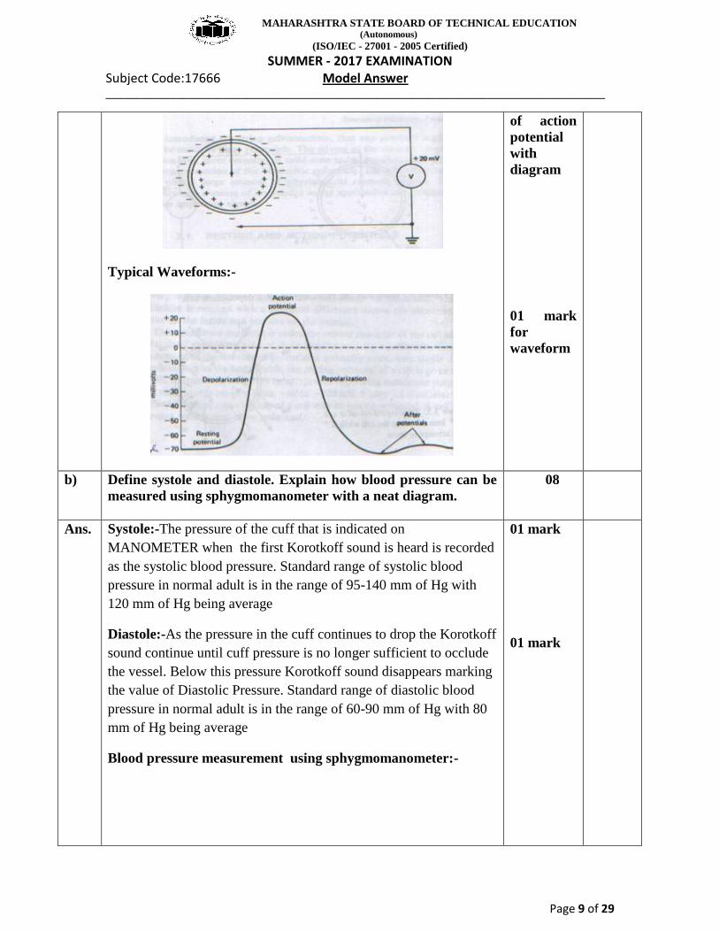
Diagram of action potential :
3 ½
marks for
definition
of resting
potential
with
diagram
3 ½ marks
for
definition
MAHARASHTRA STATE BOARD OF TECHNICAL EDUCATION (Autonomous)
(ISO/IEC - 27001 - 2005 Certified)
SUMMER - 2017 EXAMINATION Subject Code:17666 Model Answer _____________________________________________________________________________________
Page 9 of 29
Typical Waveforms:-
of action
potential
with
diagram
01 mark
for
waveform
b) Define systole and diastole. Explain how blood pressure can be
measured using sphygmomanometer with a neat diagram.
08
Ans. Systole:-The pressure of the cuff that is indicated on
MANOMETER when the first Korotkoff sound is heard is recorded
as the systolic blood pressure. Standard range of systolic blood
pressure in normal adult is in the range of 95-140 mm of Hg with
120 mm of Hg being average
Diastole:-As the pressure in the cuff continues to drop the Korotkoff
sound continue until cuff pressure is no longer sufficient to occlude
the vessel. Below this pressure Korotkoff sound disappears marking
the value of Diastolic Pressure. Standard range of diastolic blood
pressure in normal adult is in the range of 60-90 mm of Hg with 80
mm of Hg being average
Blood pressure measurement using sphygmomanometer:-
01 mark
01 mark

MAHARASHTRA STATE BOARD OF TECHNICAL EDUCATION (Autonomous)
(ISO/IEC - 27001 - 2005 Certified)
SUMMER - 2017 EXAMINATION Subject Code:17666 Model Answer _____________________________________________________________________________________
Page 10 of 29
(Note: any other relevant diagram may considered)
Description :
• The familiar indirect method of measuring blood pressure involves
use of Sphygmomanometer and a stethoscope. Sphygmomanometer
consists of an inflatable pressure cuff and mercury manometer to
measure the pressure in the cuff.
• The cuff consists of a rubber bladder inside an inelastic fabric
covering that can be wrapped around the upper arm and fastened
with either hook or a Velcro fastener. The cuff is normally inflated
manually with rubber bladder and deflated slowly through a needle
valve.
• The Sphygmomanometer works on the principle of that when the
cuff is placed on the upper arm and inflated (filled with air
pressure), arterial blood can flow past the cuff only when the arterial
pressure exceeds the pressure in the cuff.
• So first pressure in cuff is increased by inflating cuff with the help
of rubber bladder pumping manually above systolic pressure at this
point no sound is heard through the stethoscope which is placed over
the brachial artery. For that artery has been collapse by the pressure
of the cuff.
• The pressure in the artery gradually reduced by opening needle
vale slowly.
• As soon as cuff pressure falls below systolic pressure, small
amount of blood Spurt past the cuff and KOROTKOFF sounds
begin to be heard through stethoscope.
02 marks
for
diagram
04 marks
for
explanatio
n
c) Explain the working of an internal pacemaker with a neat block
diagram.
08
MAHARASHTRA STATE BOARD OF TECHNICAL EDUCATION (Autonomous)
(ISO/IEC - 27001 - 2005 Certified)
SUMMER - 2017 EXAMINATION Subject Code:17666 Model Answer _____________________________________________________________________________________
Page 11 of 29
Ans. Diagram
Explanation:
In the given block diagram the timing circuit consists of an RC
network, reference voltage source and a comparator that determines
the basic pacing rate of the pulse generator. Its output signal is given
to second RC network, the pulse width circuit which determines the
stimulation pulse duration. A third RC network and the rate limiting
circuit disable the comparator for a preset interval and thus limit the
pacing rate to a maximum of 120 pulses per minute. The output
circuit provides a voltage pulse to stimulate the heart. The voltage
monitoring circuit senses cell depletion and then signals the rate
slow down circuit and the energy compensation circuit of this event.
The rate slow down circuit shuts off some of the current to the basic
timing network to cause the rate slow down to 8+ 3 beats per
minutes. When cell depletion has occurred the energy compensation
circuit causes the pulse duration to increase as the battery voltage
decreases to maintain nearly constant stimulation to the heart. There
is a feedback loop from output circuitry to the refractory circuit
which provides a period of time following an output pulse on sensed
R wave during which the amplifier will not respond to the outside
signals. The sensing circuit detects a spontaneous R wave and resets
the oscillation timing capacitor. The reversion circuit allows the
amplifier to detect a spontaneous R wave. In the absence of R wave
this circuit allows the oscillator to pace at its present rate + beat per
minute
04 marks
for
diagram
04 marks
for
explanatio
n
Q.3 Attempt ant FOUR of the following 16
a) State functions of the following 04

MAHARASHTRA STATE BOARD OF TECHNICAL EDUCATION (Autonomous)
(ISO/IEC - 27001 - 2005 Certified)
SUMMER - 2017 EXAMINATION Subject Code:17666 Model Answer _____________________________________________________________________________________
Page 12 of 29
(i) Medulla Oblongata
(ii) Cerebellum
(iii)Cerebrum
(iv) Frontal Lobe
Ans. i)Medulla Oblongata:
It is part of brain stem and its functions are as follows
1) It control blood pressure
2) Regulate breathing, heart and blood vessel function
3) reflex center of vomiting,
4) coughing, sneezing
5) Swallowing.
OR
It contain nuclei for regulating
1) Blood pressure
2) breathing
3) Also responsible for relaying information from sense organs that
comes from cranial nerves.
ii)Cerebellum:
1) The cerebellum receives information from the sensory systems,
the spinal cord, and other parts of the brain and then regulates
motor movements.
2) The cerebellum coordinates voluntary movements such as
posture, balance, coordination, and speech, resulting in smooth
and balanced muscular activity.
iii)Cerebrum: The cerebrum or cortex is the largest part of the
human brain, associated with higher brain function such as thought
and action.
iv)Frontal Lobe: The frontal lobes are essential for intelligence,
constructive imagination & thought. Large quantities of information
stored temporarily & correlated thus making basis of higher mental
functions.
01 mark
01 mark
01 mark
01 mark
b) List the effects of leakage current on human body with
increasing current intensity.
04
Ans. Effect of leakage current on human
i) Threshold of perception: It is at approximately 500 mA or 1 mA.
ii) Accepted safe level: it is up to 5 mA. It is not considered harmful.
iii) Maximum let go current: It is in excess of 10mA or 20mA. It can
tentize the arm muscle.
iv) Danger of ventricular Fibrillation : It is above 75 mA
v) Contraction of heart (Sustained myocardial contraction): it is at
excess of 1A or 2A current.
vi) Severe burns and physical injury: It is at excess above 10A current.
01 mark
each (any
four
points)

MAHARASHTRA STATE BOARD OF TECHNICAL EDUCATION (Autonomous)
(ISO/IEC - 27001 - 2005 Certified)
SUMMER - 2017 EXAMINATION Subject Code:17666 Model Answer _____________________________________________________________________________________
Page 13 of 29
vii) Danger of respiratory paralysis: It is current excess at 100mA
onwards.
viii) Sustained Myocardial contraction: entire heart muscle contract at
current in the range of 1A- 6A.
c) With a neat diagram, explain the ultrasonic method for
measurement of blood flow.
04
Ans. Ultrasonic blood flow measurement
Ultrasonic blood flow measurement works on two principles
1. Transit type ultrasonic flow meter.
2. Doppler shift type ultrasonic blood flow meter.
In the transit time ultrasonic flow meter, a pulsed beam of
ultrasonic energy is used to measure the velocity of flowing
blood. A pulsed beam is directed through the blood vessel at a
shallow angle and its transit time is measured. When blood flows
in the direction of the energy transmission, the transit time is
shortened. If flows in the opposite direction, the transit time is
lengthened. The transit time is proportional to the velocity of
blood flow.
In Doppler principle an oscillator, operating at a frequency of
several megahertz, excites a piezoelectric transducer. This
transducer is coupled to the wall of an exposed blood vessel and
sends an ultrasonic beam with a frequency F into the flowing
blood. A small part of the transmitted energy is scattered back and
is received by a second transducer arranged opposite the first one.
Because of the scattering, due to moving blood cells the received
frequency is either F+Fd or F–Fd depending on direction of flow.
The Doppler frequency component (Fd) is proportional to
velocity of blood.
02 marks
for
diagram
02 marks
for
relevant
explanatio
n of any
one
method
d) Describe the working of a d.c. defibrillator with a diagram and
waveform.
04
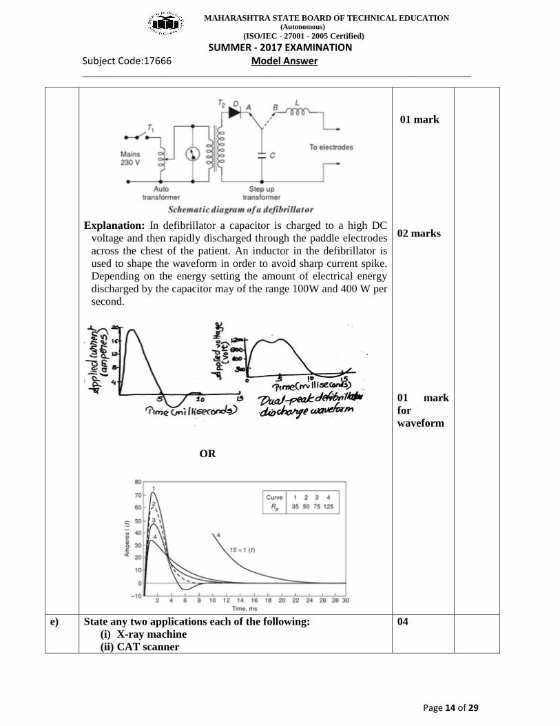
Ans. DC Defibrillator:
MAHARASHTRA STATE BOARD OF TECHNICAL EDUCATION (Autonomous)
(ISO/IEC - 27001 - 2005 Certified)
SUMMER - 2017 EXAMINATION Subject Code:17666 Model Answer _____________________________________________________________________________________
Page 14 of 29
Explanation: In defibrillator a capacitor is charged to a high DC
voltage and then rapidly discharged through the paddle electrodes
across the chest of the patient. An inductor in the defibrillator is
used to shape the waveform in order to avoid sharp current spike.
Depending on the energy setting the amount of electrical energy
discharged by the capacitor may of the range 100W and 400 W per
second.
OR
01 mark
02 marks
01 mark
for
waveform
e) State any two applications each of the following:
(i) X-ray machine
(ii) CAT scanner
04

MAHARASHTRA STATE BOARD OF TECHNICAL EDUCATION (Autonomous)
(ISO/IEC - 27001 - 2005 Certified)
SUMMER - 2017 EXAMINATION Subject Code:17666 Model Answer _____________________________________________________________________________________
Page 15 of 29
Ans. X-ray machine:
1. Used in medicine to detect fractures in bones or presence of
foreign body.
2. Used in diagnosis of tuberculosis, ulcers, cancer etc.
3. In industry they are used to test metal castings and moulds and
also to detect cracks in them.
4. They are used to test the genuineness of the diamonds and
pearls.
5. They are used to study the crystal structure.
CAT scanner:
1. It is ideally suited for studying structures in the chest and
abdomen.
2. It can be used in the diagnosis of infectious conditions, heart
disease, lung disease, diseases involving the bones and muscle.
3. It can be used to diagnose disorders related to the central
nervous system, more specifically the brain.
4. It can also be used to confirm the presence of lesions such as
cysts, solid tumors in different areas of the body.
02 marks
for (any
two
applicatio
ns)
02 marks
for (any
two
applicatio
ns)
Q.4
A)
Attempt ant THREE of the following
12
i) Describe the working of an image intensifier used with a X-ray
machine with the help of a neat diagram.
04
Ans. Image intensifier:
• This is mostly used as instead of fluoroscopic screen as image is
faint , viewed only in dark room
• In Image Intensifier faint image of fluoroscopic screen can
be made brighter with the help of electronic image intensifier
• X-ray image intensifier is used for visual observation &
reading of picture with movie camera or video recorder
• Intensifier tube contain fluorescence screen act as photo
cathode
• The electron image thus obtained is projected on a photo
phosphor screen at the end of tube by means of electronic system
• There is increase in brightness due to acceleration of electron in
the lens system.
• But o/p image is smaller than primary fluorescent image
• X ray image can be observed in normally illuminated room
• This tube is heavy so require special type of suspension
• Image can be seen from right side mirror
• Or can be seen by video placed at suitable placed monitor
• Moving camera can be used to record the image while
examining the patient
02 marks
for
explanatio
n

MAHARASHTRA STATE BOARD OF TECHNICAL EDUCATION (Autonomous)
(ISO/IEC - 27001 - 2005 Certified)
SUMMER - 2017 EXAMINATION Subject Code:17666 Model Answer _____________________________________________________________________________________
Page 16 of 29
02 marks
for
diagram
ii) Draw a neat labeled diagram of the cut section of a kidney. 04
Ans.
Cut section of a kidney:
OR
02 marks
for
diagram
02 marks
for
labeling

MAHARASHTRA STATE BOARD OF TECHNICAL EDUCATION (Autonomous)
(ISO/IEC - 27001 - 2005 Certified)
SUMMER - 2017 EXAMINATION Subject Code:17666 Model Answer _____________________________________________________________________________________
Page 17 of 29
iii)
Define the following
(a) TV (b) RV
(c) VC (d) IRV
04
Ans.
(a) Tidal Volume (TV): The volume of gas inspired or expired
(exchanged with each breath) during normal quiet breathing.
OR
The volume of air breathed in and out without conscious effort.
(b) Residual Volume (RV):
The volume of air remaining in the lungs after maximum exhalation
or forced expiration.
(c) Vital Capacity (VC):
The greatest volume of gas that can be inspired by voluntary effort
after maximum expiration irrespective of time.
OR
The total volume of air that can be exhaled after a maximum
inhalation: VC = TV + IRV + ERV
(d) Inspiratory Reserve Volume (IRV):
The volume of gas which can be inspired from a normal end.
OR
The additional volume of air that can be inhaled with maximum
effort after a normal inspiration.
01 mark
01 mark
01 mark
01 mark
iv) Explain A Scan and M Scan display modes of ultrasound
imaging.
04
Ans.
A scan: This mode is the simplest among other methods. The
transmitted signals and echo signals are applied to the Y
plates of CRT so that they are displayed as vertical deflections on
02 marks

MAHARASHTRA STATE BOARD OF TECHNICAL EDUCATION (Autonomous)
(ISO/IEC - 27001 - 2005 Certified)
SUMMER - 2017 EXAMINATION Subject Code:17666 Model Answer _____________________________________________________________________________________
Page 18 of 29
the CRT screen. The vertical sweep is calibrated in units of
distance and provides vertical deflections in various ranges
depending upon the distance of the interface. Echoencephalogram
is typical example of A scan display.
M scan: M scan is very useful in monitoring moving structure
inside the body. M scan is basically a combination of A scan and B
scan. In this system intensity or brightness of the beam is
modulated using received echoes and displayed on horizontal axis
with the help of horizontal timing information, that is horizontal
sweep.
for
explanatio
n
02 marks
for
diagram
Q.4.
B)
Attempt any one of the following 06
i)
List the precautions to be taken to minimize electric shock
hazards.
06
Ans.
Precautions to minimize electric shock hazards:
1. In the vicinity of the patient, appliances with three wire power
cords should be used.
2. Provide isolated input circuits on monitoring equipment.
3. Have periodic checks of ground wire continuity of all equipment.
4. Connectors for probes and leads should be standardized so that
current intended for powering transducers are not given to the leads
applied to pick up physiologic electric impulses.
5. Ground fault circuit interrupters should be used to disconnect the
source.
6. Reducing leakage current inside the chasis of instruments by
using layout.
7. The solid state electronic diagnostic equipment to be so selected
that they work on low voltage.
06 marks
for (any
six points)

MAHARASHTRA STATE BOARD OF TECHNICAL EDUCATION (Autonomous)
(ISO/IEC - 27001 - 2005 Certified)
SUMMER - 2017 EXAMINATION Subject Code:17666 Model Answer _____________________________________________________________________________________
Page 19 of 29
8. A separate (double) secondary layer of insulation between the
chasis and the outer case is provided to protect personnel from
ground fault.
9. Double insulation reduces leakage current and also protects
against both Macroshock and Microshock.
ii) Explain the working of dialysis machine with a neat block
diagram.
06
Block diagram of dialysis machine:
OR
Explanation : Dialysis machine works as artificial kidney which has following parts,
1. Dialyzer: This is the part in which blood filtration actually takes
place and urine is formed.
2. Proportionating Pump: It produces steady flow of quality dialysate
by having proper proportion of water and concentrated chemical.
3. Dialysate temp Control: To achieve dialysis at body temperature
the control of temperature is essential.
03 marks
for labeled
diagram
03 marks
for
explanatio
n

MAHARASHTRA STATE BOARD OF TECHNICAL EDUCATION (Autonomous)
(ISO/IEC - 27001 - 2005 Certified)
SUMMER - 2017 EXAMINATION Subject Code:17666 Model Answer _____________________________________________________________________________________
Page 20 of 29
4. Heparin infusion: It is done in order to avoid coagulation or clotting
of blood, which is taken from the patient.
5. Venous pressure gauge: It monitors the pressure of blood which is
given back to the patient.
6. Air/Foam Detector: It detects the presence of air / Foam in the
blood to avoid danger.
7. Blood leak detector: It detects the leakage of blood from the
dialyzer
8. Bypass circuit and line in clamp: It is used to bypass the dialysate
flow, for replacement, maintenance or repair of dialyzer.
Q.5 Attempt any TWO of the following 16
a) What is respiration? Describe the mechanism of breathing with
a neat diagram.
08
Ans. Respiration: Exchange of gases in any biological process is called
respiration.
OR
The exchange of gases between blood and the external environment
takes place in the lungs is termed as respiration
Diagram
OR
01 mark
02 marks
for
diagram

MAHARASHTRA STATE BOARD OF TECHNICAL EDUCATION (Autonomous)
(ISO/IEC - 27001 - 2005 Certified)
SUMMER - 2017 EXAMINATION Subject Code:17666 Model Answer _____________________________________________________________________________________
Page 21 of 29
OR
Diagram of Respiratory system
Mechanism of Breathing:
There are two main steps in breathing: inspiration and expiration:
Inspiration:
Inspiration (inhalation) is the process of breathing in, by which air is
brought into the lungs.
Inspiration involves the following steps:
i. The muscles attached to the ribs on their outer side contract. This
causes the ribs to be pulled out, expanding the chest cavity.
ii. The muscle wall between the chest cavity and the abdominal
cavity, called diaphragm, contracts and moves downwards to further
expand the chest cavity.
iii. The abdominal muscles contract.
The expansion of the chest cavity creates a partial vacuum in the
chest cavity. This sucks in air into the lungs, and fills the expanded
alveoli.
Expiration:
After the exchange of gases in the lungs, the air has to be expelled.
Expulsion of the air from the lungs is called expiration. In this
process, muscles attached to the ribs on their inner side contract, and
the diaphragm and the abdominal muscles relax. This leads to a
05 marks
for
descriptio
n

MAHARASHTRA STATE BOARD OF TECHNICAL EDUCATION (Autonomous)
(ISO/IEC - 27001 - 2005 Certified)
SUMMER - 2017 EXAMINATION Subject Code:17666 Model Answer _____________________________________________________________________________________
Page 22 of 29
decrease in the volume of the chest cavity, which increases the
pressure on the lungs. The air in the lungs is pushed out and it passes
out through the nose.
When we breathe out, not all of the air in the lungs gets expelled.
Some of it remains in the lungs. This keeps the lungs from
collapsing and allows more time for the exchange of gases.
b) Explain the working of an ECG machine with a neat block
diagram.
08
Ans.
OR
Explanation :
04 marks
for
diagram

MAHARASHTRA STATE BOARD OF TECHNICAL EDUCATION (Autonomous)
(ISO/IEC - 27001 - 2005 Certified)
SUMMER - 2017 EXAMINATION Subject Code:17666 Model Answer _____________________________________________________________________________________
Page 23 of 29
The potential picked up by the electrodes are taken to the lead
selector where lead whose output is required are selected as per lead
wire configuration. By means of capacitive coupling the signal is
connected to the differential pre amplifier.
The preamplifier is usually a 3 or 4 stage differential amplifiers
having sufficiently large negative current feedback.
The amplified output signal is given to the power amplifier.
The power amplifier is a push-pull type. The base of one input
transistor is driven by pre amplified signal and the base of other is
driven by feedback network.
The output of power amplifier deflects the writing arm.
Paper recording speed is 25 mm/s.
Amplitude measurements are made vertically made in mV.
Sensitivity of electrocardiograph is typically set to 10mm/mV.
It includes speed control circuit for a chart drive motor.
04 marks
for
explanatio
n
c) Describe the working of CAT scan machine with a neat block
diagram.
08
Ans.
04 marks
for
diagram
MAHARASHTRA STATE BOARD OF TECHNICAL EDUCATION (Autonomous)
(ISO/IEC - 27001 - 2005 Certified)
SUMMER - 2017 EXAMINATION Subject Code:17666 Model Answer _____________________________________________________________________________________
Page 24 of 29
OR
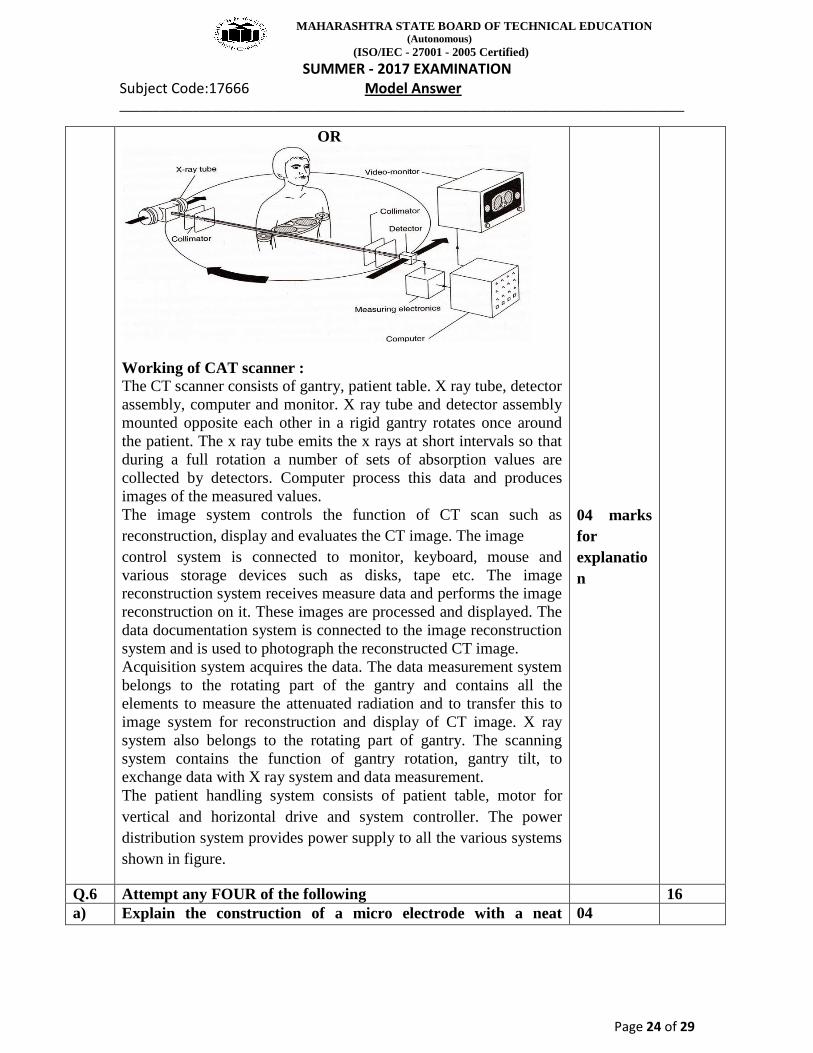
Working of CAT scanner : The CT scanner consists of gantry, patient table. X ray tube, detector
assembly, computer and monitor. X ray tube and detector assembly
mounted opposite each other in a rigid gantry rotates once around
the patient. The x ray tube emits the x rays at short intervals so that
during a full rotation a number of sets of absorption values are
collected by detectors. Computer process this data and produces
images of the measured values.
The image system controls the function of CT scan such as
reconstruction, display and evaluates the CT image. The image
control system is connected to monitor, keyboard, mouse and
various storage devices such as disks, tape etc. The image
reconstruction system receives measure data and performs the image
reconstruction on it. These images are processed and displayed. The
data documentation system is connected to the image reconstruction
system and is used to photograph the reconstructed CT image.
Acquisition system acquires the data. The data measurement system
belongs to the rotating part of the gantry and contains all the
elements to measure the attenuated radiation and to transfer this to
image system for reconstruction and display of CT image. X ray
system also belongs to the rotating part of gantry. The scanning
system contains the function of gantry rotation, gantry tilt, to
exchange data with X ray system and data measurement.
The patient handling system consists of patient table, motor for
vertical and horizontal drive and system controller. The power
distribution system provides power supply to all the various systems
shown in figure.
04 marks
for
explanatio
n
Q.6 Attempt any FOUR of the following 16
a) Explain the construction of a micro electrode with a neat 04
MAHARASHTRA STATE BOARD OF TECHNICAL EDUCATION (Autonomous)
(ISO/IEC - 27001 - 2005 Certified)
SUMMER - 2017 EXAMINATION Subject Code:17666 Model Answer _____________________________________________________________________________________
Page 25 of 29
diagram.
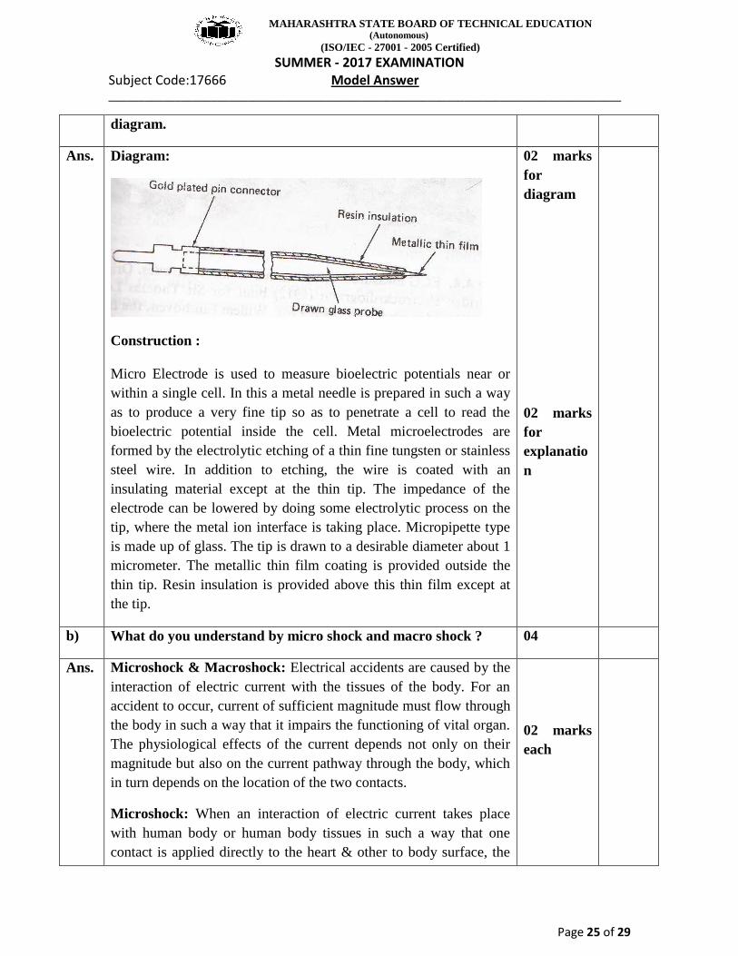
Ans. Diagram:
Construction :
Micro Electrode is used to measure bioelectric potentials near or
within a single cell. In this a metal needle is prepared in such a way
as to produce a very fine tip so as to penetrate a cell to read the
bioelectric potential inside the cell. Metal microelectrodes are
formed by the electrolytic etching of a thin fine tungsten or stainless
steel wire. In addition to etching, the wire is coated with an
insulating material except at the thin tip. The impedance of the
electrode can be lowered by doing some electrolytic process on the
tip, where the metal ion interface is taking place. Micropipette type
is made up of glass. The tip is drawn to a desirable diameter about 1
micrometer. The metallic thin film coating is provided outside the
thin tip. Resin insulation is provided above this thin film except at
the tip.
02 marks
for
diagram
02 marks
for
explanatio
n
b) What do you understand by micro shock and macro shock ? 04
Ans. Microshock & Macroshock: Electrical accidents are caused by the
interaction of electric current with the tissues of the body. For an
accident to occur, current of sufficient magnitude must flow through
the body in such a way that it impairs the functioning of vital organ.
The physiological effects of the current depends not only on their
magnitude but also on the current pathway through the body, which
in turn depends on the location of the two contacts.
Microshock: When an interaction of electric current takes place
with human body or human body tissues in such a way that one
contact is applied directly to the heart & other to body surface, the
02 marks
each

MAHARASHTRA STATE BOARD OF TECHNICAL EDUCATION (Autonomous)
(ISO/IEC - 27001 - 2005 Certified)
SUMMER - 2017 EXAMINATION Subject Code:17666 Model Answer _____________________________________________________________________________________
Page 26 of 29
effect of current applied to the heart is often referred to as
microshock.
Macroshock:
When an interaction of electric current takes place with human body
or human body tissues in such a way that current applied to the
surface contacts, the effect of current applied to the heart is called as
macroshock.
Diagram
optional
c) Explain the working of spirometer with a neat diagram. 04
Ans.
OR
02 marks
for
Diagram

MAHARASHTRA STATE BOARD OF TECHNICAL EDUCATION (Autonomous)
(ISO/IEC - 27001 - 2005 Certified)
SUMMER - 2017 EXAMINATION Subject Code:17666 Model Answer _____________________________________________________________________________________
Page 27 of 29
( Note: Any other relevant diagram should be considered )
Working of Spirometer :
Figure shows the diagram for Spirometer. Spirometer is a device
which is used to determine all lung volumes and capacities.
The standard Spirometer consists of a movable bell inverted over a
chamber of water. Inside the bell is the gas that is to be breathed.
The bell is counterbalanced by a weight to maintain the gas inside
the atmospheric pressure so that its height above the water is
proportional to the amount of gas in the bell.
A breathing tube connects the mouth of the patient to the
Spirometer. Thus as the patient breathe gas from the tube there are
changes in internal volume of Spirometer which causes proportional
displacement of bell downwards.
Similarly, as the patient breaths back into the tube, the bell moves
up proportional to the change in internal volume.
The motion is recorded on a rotating drum i.e. kymogram through a
pen that is attached to a counter balancing mechanism.
The change in bell pressure changes the volume inside the bell,
which also causes the position of the counter weight to change. We
may record the volume changes on the piece of graph paper by
attaching a pen to the counter weight or a tension string.
Some spirometer also offers an electrical output that is the electrical
analog of the respiration waveform. Most frequently the electrical
output is generated by connecting the pen and weight assembly to a
linear potential if precise and negative potentials are connected to
02 marks
for
explanatio
n

MAHARASHTRA STATE BOARD OF TECHNICAL EDUCATION (Autonomous)
(ISO/IEC - 27001 - 2005 Certified)
SUMMER - 2017 EXAMINATION Subject Code:17666 Model Answer _____________________________________________________________________________________
Page 28 of 29
the end of the potentiometer, then the electrical signal will present
the same data as the pen when no one is breathing in to the
mouthpiece, E0 will be zero, but when the patient is breathing in to
the tube, E0 will take the value proportional to the volume and a
polarity that indicates in inspiration or expiration.
Thus all lung volumes and capacities can be determined by
measuring the amount of gas inspired or expired under a given set of
condition or during a specified time interval can obtained by the use
of spirometer.
d) List the four heart sounds. How do they originate? 04
Ans. i) 1st Heart sound(lub sound): caused due to closure of the
Atrioventricular valves
ii) 2nd
Heart sound (dub sound) : caused due to the closing of
the semilunar valves.
iii) 3rd Heart sound: Occurs due to rush of blood from the atria
into the ventricles, which causes turbulence & some
vibrations of ventricular walls.
iv) Atrial Heart sound: Occurs when the atria actually do
contract, squeezing the remainder of the blood into the
ventricles.
v) Murmur: Abnormal heart sound due to improper opening of
heart valves.
The heart sounds are originating due to flow of blood through heart
valves in heart chamber.
02 marks
for listing
heart
sound
(Any four)
02 mark
for heart
sound
origination
(Any four)
e) State the functions of :
i) SA node
ii) Hypothalamus
iii) Nephron
iv) Tricuspid valve
04
Ans. i)SA node: It is natural pacemaker of human heart. It controls the
heart rate by generating electrical impulses at regular interval and
then sending electrical signals through the heart muscle, causing the
heart to contract and pump blood throughout the body.
ii)Hypothalamus
The hypothalamus acts as the 'head ganglion' of the autonomic
nervous system. The basic drives of life - hunger, thirst and sex,
originate in the hypothalamus. The hypothalamus is central to the
maintenance of homeostasis.
The functions of the hypothalamus include:(1) controls the release
01 mark
01 mark

MAHARASHTRA STATE BOARD OF TECHNICAL EDUCATION (Autonomous)
(ISO/IEC - 27001 - 2005 Certified)
SUMMER - 2017 EXAMINATION Subject Code:17666 Model Answer _____________________________________________________________________________________
Page 29 of 29
of 8 major hormones by the hypophysis, and is involved in (2)
temperature regulation, (3) control of food and water intake, (4)
sexual behaviour and reproduction, (5) control of daily cycles in
physiological state and behaviour, and (6) mediation of emotional
responses.
iii) Nephron: Its chief function is to regulate the concentration of
water and soluble substances like sodium salts by filtering the blood,
reabsorbing what is needed and excreting the rest as urine. It is
smaller unit of kidney. They are available in millions number
responsible for filtering.
iv) Tricuspid valve: The function of the valve is to prevent back
flow of blood from to right atrium to right ventricles.
01 mark
01 mark


