

i
MATERNAL AND FETAL OUTCOMES OF WOMEN
REFERRED TO CHARLOTTE MAXEKE JOHANNESBURG
ACADEMIC HOSPITAL WITH PROLONGED LABOUR
DARIN ALI OTHMAN ASHNAF
A research report submitted to the Faculty of Health Sciences, University of the Witwatersrand,
Johannesburg, in partial fulfilment of the requirements for the degree of Master of Medicine in
Obstetrics and Gynaecology
Johannesburg, February 2018

ii
DECLARATION
I Darin Ali Othman Ashnaf declare that this Research Report is my own, unaided work. It is
being submitted for the Degree of Master of Medicine at the University of the Witwatersrand,
Johannesburg. It has not been submitted before for any degree or examination at any other
University.
(Signature of candidate)
21/January /2019

iii
DEDICATION
This research dedicated to:
My husband (Dr. Hashem Burcan) and my children (Anas and Salma) who they have been after
God the source of my strength and support throughout this work. Soul of my father, my mother,
my sisters and my brothers for their support despite of the distance.
D. Ashnaf
iv
ABSTRACT
Introduction
Prolonged labour or labour dystocia is a common obstetric complication and constitutes the
most frequent indication for instrumental and caesarean delivery. Approximately eight percent
of all women giving birth are affected by prolonged labour.1 The correct diagnosis of labour is
an important part of preventing prolonged labour. It is important to have an objective diagnosis
of labour in order to avoid over diagnosing labour which can result in inappropriate
interventions and increase the risk of maternal and fetal adverse outcomes.
Methods
This was retrospective descriptive study of women referred to Charlotte Maxeke Johannesburg
Academic Hospital (CMJAH) due to prolonged labour over a four month period from August
2016 to November 2016. The total study sample included 165 women. All 165 medical records
were available for review. Forty seven babies were admitted to the paediatric observation unit,
files were found for 45 babies, records were not available for two babies.
Results
Women were referred for prolonged active and latent phase of labour in 77 (46.6%) and 75
(45.5%) women respectively. Only 13 women were referred due to prolonged second stage of
labour (7.9%). Ninety one (55.2%) women with prolonged labour had normal vaginal delivery
and eight (4.8%) had instrumental delivery. The remainder of the women (n=66, 40%) delivered
by caesarean section (CS). The only maternal adverse outcome found was postpartum
haemorrhage in 4.3 % (n=7) of the total study population. Out of 165 births, there was one fresh
still birth. A total of 47 babies (28.6%) required observation in the neonatal transitional care
unit (TICU) mainly due to respiratory distress. Nine babies (5.5%) out of 164, required
admission to neonatal ICU (NICU). The institutional protocol for management of labour was
followed in only 33.3% of the women. In the rest of the population, appropriate management
was either delayed or not done.
Conclusion
In this study, there were few maternal and fetal adverse outcomes with prolonged labour. The
institution protocol at CMJAH for management of labour dystocia was followed in only 33.3%
of the study population.

v
ACKNOWLEDGEMENTS
I would like to gratefully acknowledge my supervisor Dr. Susan Branch for her constant
support, help, guidance and motivation. It would never have been possible for me to take this
work to completion without her incredible support and encouragement.
Thank you to the staff at the records department at CMJAH for their help and assistance.
Darin Ashnaf

vi
TABLE OF CONTENTS
Declaration ii
Dedication iii
Abstract iv
Acknowledgements v
Table of contents vi
List of figures ix
List of tables x
Abbreviations xi
1. Literature review
1.1 Introduction 1
1.2 Maternal factors in prolonged labour 2
1.3 Fetal factors in prolonged labour 6
1.4 Labour related factors 7
1.5 Maternal complications of prolonged labour 7
1.6 Neonatal complications of prolonged labour 10
1.7 The partogram in labour management 12
1.8 Active management of labour 13
1.9 New concept of abnormal labour 15
2. Aims
2.1 Study Aims 16
2.2 Objectives 16
3. Methodology
3.1 Study design 17
3.2 Study setting 17

vii
3.3 Study population 17
3.4 Data collection 18
3.5 Data analysis 18
3.6 Ethical approval 18
3.7 Funding 18
4. Results
4.1 Study population 19
4.2 Maternal characteristics 19
4.3 Referral 21
4.4 Progress of labour 23
4.5 Maternal and neonatal outcomes 25
4.6 Management at CMJAH 26
5. Discussion
5.1 Characteristics of women with prolonged labour 28
5.2 Referral factors 29
5.3 Mode of delivery 30
5.4 Maternal outcomes of prolonged labour 31
5.5 Fetal outcomes of prolonged labour 32
5.6 Management at CMJAH 33
5.7 Limitations 34
5.8 Recommendations 34
6. Conclusion 36
References 37
Appendix
A Data collection sheet 44
B Ethics certificate 45

viii
C Institutional protocol 46
D Augmentation of labour protocol 49
E Antibiotic use with prolonged rupture of membranes 50
F Management of prolonged second stage 51
G Diagnosis of fetal distress on cardiotocograph 53

ix
LIST OF FIGURES
4.1 Distribution of BMI
4.2 Distribution of referral facilities

x
LIST OF TABLES
4.1 Demographic and other characteristics of women with prolonged labour
4.2 Indications for referral to CMJAH
4.3 Duration of stages of labour according to parity
4.4 Mode of delivery
4.5 Summary of maternal outcomes
4.6 Management of women with prolonged labour at CMJAH

xi
ABBREVIATIONS
ACOG American College of Obstetrics and Gynaecology
AOL Augmentation of labour
AROM Artificial rupture of membranes
BMI Body mass index
cART Combined antiretroviral therapy
CHBAH Chris Hani Baragwanath Academic Hospital
CMJAH Charlotte Maxeke Johannesburg Academic Hospital
CPD Cephalopelvic disproportion
CS Caesarean section
ICU Intensive care unit
MTCT Mother to child transmission
TICU Transitional intensive care unit
PPH Postpartum haemorrhage
PROM Prelabour rupture of membranes
RCOG Royal College of Obstetrics and Gynaecology
ROM Rupture of membranes
VVF Vesicovaginal fistula
WHO World Health Organization

1
1. LITERATURE REVIEW
1.1 Introduction
Prolonged labour or labour dystocia is a common obstetric complication and constitutes the
most frequent indication for instrumental and caesarean delivery. Approximately eight percent
of all women giving birth are affected by prolonged labour.1
Diagnosing prolonged labour is inherently difficult. It is a controversial issue that has been
discussed ever since Friedman introduced the graphic analysis of labour in 1954.1 He defined
the following three stages of labour. The first stage starts with uterine contractions leading to
complete cervical dilatation and is divided into latent and active phases. In the latent phase,
irregular uterine contractions occur with slow and gradual cervical effacement and dilatation.
The latent phase is prolonged if it continues for more than 20 hours in nulliparous and more
than 14 hours in multiparous women.2 The active phase of labour, demonstrated by an increased
rate of cervical dilatation and fetal descent, starts at three to four centimeter cervical dilatation.
It is subdivided into phases of accelerations, maximum slope and deceleration.2
The second stage of labour is defined as complete dilatation of cervix to delivery of the infant
with an average of 60 minutes in nulliparous and 30 minutes in multiparous women.2 It is
considered prolonged if it continues for more than an hour in nulliparous and 30 minutes in
multiparous women. The third stage of labour involves delivery of the placenta.2
Delays in both first and second stage of labour can lead to adverse outcomes of mother and
baby. Reported incidences for labour dystocia vary among previous studies due to differences in
the definitions used as well as differences in the characteristics of the women and gestational
age between study populations.3 Two different studies, the first done by Zhu et al. and the
second by Kjaergaard et al. found the incidence of labour dystocia among their populations to
be 20% and 37% respectively.3,4 A cohort study of pregnant women at Chris Hani Baragwanath
Hospital in South Africa was undertaken to assess the fetal, maternal and obstetric risks
associated with prolonged latent phase of labour.5 The incidence of prolonged latent phase of
labour in this study was comparatively low, at 5.9%.5 This study, by Maghoma and Buchmann,
demonstrated a number of adverse maternal as well as fetal outcomes due to prolonged latent
phase of labour.5 The maternal complications recorded included postpartum hemorrhage,
infection and augmentation of labour.5 Fetal adverse outcomes assessed included Apgar score of
<7 at five minutes, intrapartum passage of meconium, admission to the neonatal ICU,

2
intrapartum still birth and neonatal death. In a study done by Bailit et al., to evaluate the
differences in outcomes between women presenting to health care facilities in latent phase
versus active phase of labour, they found that latent phase admission was associated with an
increased cesarean delivery rate, increased number of obstetric interventions and increased
maternal infection rates.6
Similarly, in 2009, a study in Canada, the described risk of both maternal (chorioamionitis,
obstetric trauma, need for blood transfusion, hysterectomy, PPH, admission to ICU) and fetal
adverse outcomes (five minute Apgar<7, sepsis, admission to neonatal ICU) increased with the
duration of second stage of labour. This is particularly true for duration longer than three hours
in nulliparous and longer than two hours in multiparous women.7
1.2 Maternal Factors in prolonged labour
1.2.1 Maternal age
Childbirth at a young (less than 20 years) or advanced maternal age (more than 35 years) is
associated with increased risk of adverse maternal and perinatal outcomes.8 A large number of
researchers have reported that increased maternal age over 35 years is associated with an
increased risk of pregnancy induced medical conditions and maternal mortality.9 These women
are at greater risk of developing labour dystocia and requiring caesarean or instrumental
deliveries resulting in pelvic floor injuries.9 There is also an increased risk of adverse pregnancy
outcomes including stillbirth, preterm labour (both spontaneous and iatrogenic) and low birth
weight.8,10
A retrospective cohort study conducted by Greenberg et al. to examine lengths of first and
second stages of labour across maternal age groups found that there were significant differences
between the lengths of labour among women of different ages.11 In the first stage of labour, the
median duration in nulliparous women, increased from 9.2 hours in the age group less than 20
years to 11 hours (p≤0.001) in age group of 35 to 40 years.11 In comparison, in multiparous
women the first stage was longer in women less than 20 years old than the women of 40 years
and over by 6.1 hours and 5.7 hours (p=0.02) respectively.11 In the second stage of labour, there
were differences in duration in both nulliparous and multiparous women. In nulliparous women,
the median length was 51 minutes in the age group less than 20 years compared to 148 minutes
(p≤0.001) in the age group more than 40 years. In multiparous women, those less than 20 years
3
old had a median length of 16 minutes compared to 26 minutes (p≤0.001) in the age group more
than 39 years.11
A study by Treacy et al. in Ireland, was conducted in nulliparous women to determine whether
maternal age affects uterine function.12 Over 10 000 nulliparous women in spontaneous labour
at term were analyzed over a five year period. The use of oxytocin, duration of labour and risk
of caesarean delivery for failure to progress was measured and compared to maternal age. The
overall rate of prolonged labour was 3.7%, with 6.5% of women requiring caesarean delivery.
The results showed a significant increase in labour dystocia due to uterine dysfunction with
advancing maternal age.12 Caesarean delivery rates were also found to increase over three fold
between the categories of less than 20 years and more than 35 years of age, particularly those
done due to dystocia or no progress.12 Possible reasons for this age-related increase in labour
dystocia may be related to diminished uterine contractility, altered connective tissue compliance
and reduced maternal voluntary effort.12
In a retrospective review from 2009 in the United States, Cavazos-Rehg et al. used hospital
billing data to identify possible associations between maternal age and morbidity following
labour and delivery.8 They compared women less than 19 years of age with a control group of
women 25 to 29 years and found an increased risk of chorioamnionitis and endometritis.8
However, this does not seem to be as a result of prolonged labour as the rate of labour dystocia
in mothers less than 20 years was comparable to, if not less than, the rate of dystocia in mothers
20 to 25 years.12
1.2.2 Maternal Obesity
Obesity, as defined by the World Health Organization and the Institute of Medicine, is a body
mass index (BMI) more than or equal to 30kg/m2.13 The overall incidence of obesity is
increasing worldwide, particularly in women of reproductive age. The prevalence varies
between 8 and 30% depending on the population in question.13 The South African Demographic
and Health Survey (SADHS) of 2003, found that the overall prevalence of obesity in South
African adult women is 27%, 15% of which are within reproductive age.14
Obese pregnant women have a higher risk of a number of maternal and fetal complications
including miscarriage, pre-eclampsia, gestational diabetes, fetal macrosomia and stillbirth.15
4
There is also an increased risk of postdates pregnancy requiring induction of labour or
prolonged labour leading to augmentation with oxytocin, possible primary postpartum
haemorrhage and perineal trauma.16
Maternal obesity has a direct influence on mode of delivery. Delay or non progress in the first
stage of labour is significantly more common in obese women with the risk ranging between 1.5
to 3 times more likely.15 Prolonged first stage is the most common indication for caesarean
section (CS), even after augmentation with oxytocin. There is a two to three fold increased risk
of CS in women with a BMI greater than 30kg/m2.15 Additionally, a cohort published in 2011
by Fyfe et al. found that in term nulliparous women in labour, being overweight or obese is an
independent risk factor for CS in the first stage but not the second stage of labour.17 For
overweight or obese nulliparous women who reach the second stage of labour, increasing
maternal BMI is not associated with a longer second stage or an increased risk of caesarean
delivery.18
The cause of the increased caesarean delivery rate among obese women is most likely
multifactorial. However, it is thought that obesity may be associated with impaired or poor
myometrial contractility.19 Zhang et al. tested myometrial strips of the uterus obtained from
obese women who underwent an elective CS at term.19 They found that myometrial strips from
the obese women contracted with less force and frequency than similar myometrial strips
obtained from normal weight women.19 The exact mechanism is unknown, although, elevated
cholesterol levels, which are generally found in overweight and obese women, are thought to
inhibit the contractility of the myometrium. Alterations in cholesterol levels, which have an
important role in cell signaling, may have an impact on effectiveness of uterine contractions
during labour.19
Due to the effects of maternal weight, it is suggested that the use of a single “normal” labour
curve may lead to over diagnosis of labour dystocia in overweight women. More appropriate
definitions need to be developed for labour arrest in obese women. By doing so the number of
CS in obese women may be reduced.20
1.2.3 Maternal height and ethnicity
Previous studies from the 1940s showed a relationship between maternal height, pelvic
dimensions and the likelihood of achieving a vaginal delivery.21 A comparative prospective

5
study among primigravidas of African and Caucasian origin was performed by Okewole et al.
in northern London, United Kingdom.21 The study aimed to determine whether there is a
relationship between ethnicity, maternal shoe size and height, and the potential for delivering
vaginally.21 Results showed that with a maternal height of greater than 165 centimeters, there
was roughly a twofold increased chance of delivering vaginally. However, both height and shoe
size were shown to have poor predictive ability for vaginal delivery.21 As a screening test for
cephalopelvic disproportion (CPD), maternal height was more predictive than shoe size.21 A
meta-analysis of nine studies by Dujardin et al. also confirmed a significant association between
short maternal height and an increased risk of CS.22
The association between ethnicity and mode of delivery was examined in a retrospective
analysis by JM Ibison.23 A total of 30 825 “low risk” primigravidas were identified and their
mode of delivery analysed in relation to their ethnicity.23 The main findings showed that
African, West Indian, Bangladeshi, Indian and Pakistani women were at elevated risk of
delivery by CS compared with Caucasian women, and Bangladeshi and Indian women had
higher risks of delivery by forceps and ventouse compared with Caucasian women.23
Ethnicity may have an influence on maternal height as well as pelvic size and shape. It has been
suggested that there is an increased risk of emergency CS for CPD among African women due
to their relatively short stature and greater proportion of anthropoid pelves.21
1.2.4 Parity
Labour dystocia in nulliparous women is common. Primiparity is associated with increased
intrapartum risks. A case controlled study, by Hashim et al. in India, showed a caesarean
section rate of 15% in primigravida and 1% in multigravida woman.24 Primiparous women were
more prone to prolonged first and second stage of labour and had the longest and most gradual
labour curve when compared with multiparous women. Primiparous women were also at higher
risk of emergency delivery for fetal distress and operative delivery.24 Similarly, a study in a
Nigerian tertiary hospital showed primigravid women to be at a higher risk of dystocia
compared with the multipara women.24
1.2.5 Hypertensive disease
In terms of the actual effect of the hypertensive disease on progress of labour, evidence is
conflicting. A retrospective cohort study done by Bregand-White et al., using data from the
Consortium on Safe Labour, looked at labour progression in induced nulliparous women at

6
term with hypertensive diseases in pregnancy.25 They found that labour progression in the first
stage was particularly longer in women with chronic hypertension and super imposed pre-
eclampsia compared to controls.25
Magnesium sulphate is an important part of management in the pre eclamptic and eclamptic
woman in terms of neuroprotection.26 In 1997, Within et al. conducted a randomized, double
blinded, placebo controlled trial to assess the effect of magnesium sulphate on the duration of
labour.27 The study found that the use of magnesium sulphate did not affect any component of
labour but did necessitate a higher dose of oxytocin for augmentation and prevention of
postpartum haemorrhage.27 A second, more recent prospective study published in February
2017, aimed to determine the contractile patterns induced by oxytocin in a myometrium
exposed to magnesium sulphate.26 The results confirmed that magnesium sulphate pretreatment,
before oxytocin is given, does not impair oxytocin-induced myometrial contractility, however,
it can affect contractions and response to oxytocin if used simultaneously.26
Magnesium sulphate is also an agent previously advocated by some for tocolysis due to its
antagonistic effect on calcium binding.26 However, studies on preterm labour do not confirm
this. Therefore, magnesium sulphate may reduce the force of contractions and slow the progress
of labour but it does not stop it completely.26
1.3 Fetal factors in prolonged labour
Fetal macrosomia is a known risk factor for labour dystocia. The most common causes of fetal
macrosomia are diabetes mellitus in pregnancy and postdate pregnancy.28 In 1990, Turner et al.
examined the association between the duration of labour and infant birthweight.29 The results
showed that the duration of labour (both first and second stage) increased as birth weight
increased regardless of whether oxytocin was used for augmentation of labour.29
A prospective multicenter cohort showed that vaginal examination on admission can provide
information on risk for dystocia.30 The number of fifths of the fetal head above the pelvic brim
had the strongest association with dystocia.30 The lack of descent of the fetal head often leads to
dystocia resulting in CS.30
7
1.4 Labour-related factors
1.4.1 Epidural anaesthesia and pain control
Pain of labour, caused by the contraction of the uterus and the dilation of the cervix, is
transmitted through the visceral afferent (sympathetic) nerves entering the spinal cord from T10
to L1. In the later stages, perineal stretching transmits painful stimuli through the pudendal and
sacral nerves (S2 to S4). The sensation of pain produces a maternal stress response leading to
the release of hormones including adrenaline. Adrenaline can have a relaxant effect on the
uterus that may result in prolonged labour.31
Epidural analgesia effectively relieves pain during labour and delivery.32 Theoretically,
adequate pain relief should aid in the progress of labour by limiting the maternal stress
response.31 There is, however, still controversy surrounding the effect of the epidural analgesia
on the duration and progress of labour. A study done by Agrawal et al, in India, involved 120
nulliparous women comparing epidural to no analgesia, showed that epidural analgesia resulted
in a shorter duration of the first stage but a statistically significant prolonged duration of the
second stage of labour.33 Similarly, in 2011, a Cochrane review done by Anim-Somuah et al.
showed no difference in the duration of first stage but a significantly longer duration of for the
second stage.34 In contrast, in 2017, Shen et al. conducted a randomised controlled trial found
no effect of epidural on the duration of the second stage of labour compared with a placebo
infusion.35 There was no statistical difference in the rates of spontaneous vaginal delivery in
both groups.35
1.5 Maternal complications of prolonged labour
1.5.1 Infection
Intrapartum chorioamnionitis and postpartum pelvic infection are more common with prolonged
labour.36 In the study done at Chris Hani Baragwanath Academic Hospital by Maghoma and
Buchmann looking at maternal outcomes after prolonged latent phase of labour, postpartum
pyrexia (>38ºC) and puerperal sepsis were found to be more frequent in women with a
prolonged latent phase of labour.5 In this study the incidence of postpartum pyrexia and
puerperal sepsis was 21% (p<0•0001) and 4% (p=0•045) respectively.5
Early postpartum maternal infection has been found to be increased with the duration of second
stage of labour. Duration of less than one hour is associated with the lowest risk of infection
whereas more than three hours duration has a significantly higher risk.37 A retrospective review

8
of over 220 000 women by Laughon et al. showed a threefold higher risk of chorioamnionitis
for both nulliparous and multiparous women with prolonged second stage of labour.38
There have been suggestions that intrauterine infection itself contributes to abnormal uterine
activity. Satin et al. studied the effects of chorioamnionitis on the need for augmentation with
oxytocin.39 They found that women diagnosed with chorioamnionitis early in labour, prior to
the use of oxytocin, had a shorter interval from oxytocin initiation to delivery compared to
controls. In contrast, women diagnosed with intrauterine infection later in labour, after initiation
of oxytocin, had considerably longer labour times than the controls. These women required
higher doses of oxytocin and also had a four times higher risk of CS for dystocia compared to
controls. Due to this difference in clinical presentation it is evident that intrauterine infection
has a variable effect on labour progress. It is difficult to determine whether intrauterine
infection is the cause or the result of dysfunctional labour.39
1.5.2 Postpartum haemorrhage
In developing countries, postpartum haemorrhage (PPH) is a major cause of maternal mortality.
According to the World Health Organisation (WHO), it accounts for 25% of direct obstetric
deaths.40 PPH may result from prolonged labour and augmentation with oxytocin due to the
development of uterine atony following delivery.36 Regular contractions over several hours of
labour will exhaust the uterine muscles and thereby reduce their ability to contract over time,
causing uterine dysfunction and uterine atony post delivery. For this reason, patients with
prolonged labour are more likely to suffer PPH. Similarly, induction of labor with misoprostol
has also been shown to be associated with PPH in previous studies.41 In South Africa, according
to the Saving Mothers Report (2011-2013), prolonged labour was an underlying factor in 25.3%
of maternal death due to obstetric haemorrhage.42 A retained placenta may also precipitate PPH.
Retained placenta accounted for 6.6% of maternal deaths due to obstetric haemorrhage in South
Africa between 2011 and 2013.42 A study by Coviello et al. showed an increased chance of
retained placenta in nulliparous women who had an increased duration of first or second stage
of labour.40
1.5.3. Uterine rupture
Labor dystocia is a significant risk factor for uterine rupture.43 Abnormal thinning of the lower
uterine segment during prolonged labour increases the risk particularly in women of parity more
than three and in women with a prior caesarean delivery.36,43 In the developed world, the overall
9
incidence of uterine rupture is less than one per 1 000 deliveries. Over 90% of these cases occur
in women with a scarred uterus due to previous CS.44 Other risk factors for uterine rupture
include uterine anomalies, advanced maternal age, macrosomia and multiple gestation, all of
which are more likely to be associated with prolonged labour.44
The rupture of an unscarred uterus is very uncommon but can be associated with higher
maternal and neonatal morbidity and mortality. The estimated incidence is 1/5700 to 1/20 000
pregnancies depending on the country.45 The inappropriate use of uterotonic agents during
labour dystocia is often an iatrogenic cause of uterine rupture particularly high dose oxytocin
for induction of labour at term.46
1.5.4 Increase risk of caesarean section
Although caesarean delivery can be lifesaving for the fetus and the mother, there is significant
concern that caesarean delivery is being overused.47 There has been a rapid increase in the rate
of caesarean births with a current global caesarean rate of 18.6% and an average increase of
4.4% every year.48 According to the Saving Mothers Report for 2011 to 2013, South Africa had
a national caesarean rate of 23.1%.42 Labour dystocia is the most commonly reported indication
for primary CS.31 It accounts for roughly 50% of all CS in nulliparous women. It is also the
surgical indication for the majority of women with repeat CS in labour.31
The relative risks of maternal mortality, ureter and bladder injury, hysterectomy, neonatal
respiratory distress and fetal death have all been shown to be increased with CS compared with
vaginal delivery.48 The risks of placenta praevia and uterine rupture in future pregnancies are
also increased.48 Population-based cohorts have demonstrated significantly increased risks of
maternal intraoperative trauma and perinatal asphyxia with caesarean delivery at full cervical
dilatation compared with caesarean delivery at less than full cervical dilatation.49 In South
Africa, maternal complications were significantly higher following second stage CS.50 Similar
findings have been reported in other studies.51
1.5.5 Fistula Formation
Approximately two million women in the developing world are affected by obstetric fistula and
according to the WHO there are roughly 130,000 new cases of obstetric fistula each year.52 A
recent study by Vangeenderhuysen et al. on incidence of obstetric fistulas in African countries
showed that there were roughly 33 000 new cases in sub-Saharan Africa each year.53 Previous

10
descriptive studies from sub-Saharan Africa have found that obstetric fistula often result from
factors that include an immature and contracted pelvis, cephalopelvic disproportion, and
obstructed labour. With labour dystocia, sustained pressure to tissues of the birth canal may
impair circulation and result in tissue necrosis.36 Several days after delivery vesicovaginal,
vesicocervical, or rectovaginal fistula may develop.36 The second most common cause of
obstetric fistula is the direct tearing of the vagina and perineum which is more likely to occur
during precipitous delivery or obstetric manoeuvres.54
1.5.6 Pelvic Floor Injury
Injury to the pelvic floor muscles, nerve supply, or interconnecting fascia is a common
consequence of vaginal delivery.36 Both prolonged second stage of labour and assisted delivery
are associated with severe perineal lacerations leading to an increased risk of urinary and anal
incontinence following delivery and pelvic organ prolapse in later life.36 Third and fourth
degree tears have been reported to occur in approximately 7% of vaginal deliveries among
primiparous mothers in Sweden.55 Globally, the incidence is estimated at 1 to 4%.7 In 2015, a
prospective cohort study at Chris Hani Baragwanath Academic Hospital, South Africa, was
done to evaluate risk factors and describe repair methods for obstetric anal sphincter injuries.56
The study reported a surprisingly low incidence of 0.5% over a 15 months period. Only 37% of
the study population were primigravidas.56
A large population-based cohort study by Simic et al. showed that the risk of severe perineal
lacerations increased with the duration of the second stage.55 The risk of laceration was almost
1.5 times higher if the second stage continued into the second hour compared with those women
delivering after one hour (95% CI 1.28–1.58). Women who required an instrumental delivery
were 2.24 times more likely to sustain a severe perineal laceration compared those who did not
(95% CI 2.07–2.42).55
1.6 Neonatal complications of prolonged labour
1.6.1 Neonatal trauma
Trauma to the fetus is more common following delayed second stage of labour.5 Complications
such as shoulder dystocia as well as the greater need for operative deliveries can results in
neonatal injuries.49 Instrumental deliveries are often the cause of neonatal cerebral haemorrhage,
fracture or brachial plexus injuries.49
11
1.6.2 Birth Asphyxia
Prolonged labour is one of the commonest causes of birth asphyxia.57 According to the WHO,
four million neonatal deaths occur yearly due to birth asphyxia, representing 38% of all under
five years childhood mortality. In South Africa, the perinatal mortality rate, for babies weighing
more than 500 grams, due to intrapartum asphyxia is 5.6 per 1000 births at district level
hospitals and 4.7 per 1000 in national central hospitals.58 Birth asphyxia is defined as “an insult
to the fetus due to failure to breathe or breathing poorly at birth”.59 It is a metabolic academia
measured at birth as pH less than 7.0 or a five minute Apgar score of 3 or less.7 In January
2015, a prospective, observational study by McClure et al. found asphyxia to be the most
common cause of stillbirth in low to middle income countries.57
A study done in July 1988 by Marrin and Paes aimed to evaluate the Apgar score as a
diagnostic test for the presence of asphyxia.60 Using an umbilical cord arterial pH less than 7.2
as evidence of asphyxia, the one minute Apgar score showed poor sensitivity as a marker of
asphyxia.60 For this reason the Apgar score at five minutes is more predictive of fetal well-
being. However, only a small minority of neonates with low Apgar scores after five minutes
will have evidence of metabolic acidosis on umbilical cord blood gas.60
A retrospective analysis by Saunders et al. was done to evaluate the relationship between the
duration of the second stage of labour and the frequency of low Apgar scores or admissions for
neonatal observation. Their findings showed no association with second stage duration of up to
three hours.61 In contrast, a large population-based cohort study done by Sandström et al. found
that pushing time, in the second stage, of more than 60 minutes was associated with a 2.5 fold
increased risk of acidosis compared to 15 minutes or less of pushing (95% CI 1.51–4.30).62
The severity of symptoms and the prognosis of the baby depends on the risk factors present as
well as the management instituted after delivery. Those neonates who survive the initial period
have an increased risk of developing neurological complications including epilepsy, cerebral
palsy and developmental delay.63 Sandström et al. found that compared with a duration of the
second stage of labour of one hour, a duration of more than four hours was associated with an
80% increased risk of admission to NICU (95% CI 1.58–2.04).62
1.6.4 Meconium Aspiration
Peripartum aspiration of meconium stained amniotic fluid leads to chemical pneumonitis with
12
inflammation of pulmonary tissues, mechanical airway obstruction and hypoxia of the neonate.
Seriously affected infants frequently die or suffer long-term neurological sequelae.5
1.6.5 Neonatal death
In 1987, a study by Friedman and Neff analysed of more than 58 000 deliveries showed that a
latent phase of more than 12 hours was associated with a 2.5 fold increases risk of neonatal
death.64 In South Africa, according to the Saving Babies Report 2011-2013, intrapartum
asphyxia was the third highest cause of early neonatal death. Common factors included
problems with fetal monitoring, the inappropriate use of the partogram and abnormalities of the
second stage of labour.58
1.7 The Partogram in management of labour
In 1954, Friedman developed his cervicograph after observing the cervical dilatation of 100
primigravid African women at term.65 In 1972 Philpott enhanced this tool by adding categories
to aid in the assessment of fetal and maternal condition and progress of labour. This ‘partogram’
provides a practical way to aid midwives in the assessment of laboring women and increase the
quality and regularity of observations of mother and fetus in labour.66 It is a simple, inexpensive
tool that gives a continuous pictorial overview of labour and serves as an ‘early warning
system’ where progress is not ideal.66 The alert line was added following the result of a
prospective study of 624 women by Philpott. The alert line represents a progress rate of one
centimeter per hour.65 It aims to represent the slowest 10% of primigravidas within the active
phase of labour. The alert line was mainly added to help identify women progressing slowly
who require transfer to a higher level of care. The second stage of the partogram development
was the introduction of an action line four hours to the right of the alert line. This line aims to
help identification and prompt effective management of slow progress of labour.65
In 2006, a study by Lavender et al. showed that by using the partogram the frequency of
prolonged and augmented labour, PPH, uterine rupture, puerperal sepsis and perinatal morbidity
and mortality was reduced.67 However, in 2014 a Cochrane review by Lavender T et al., the
author concluded that the routine use of the partogram as a standard of labour management
could not be recommended based on their findings.65 They felt that further evidence on its
efficacy was required and the use of the partogram should be determined by individual centers
until stronger evidence is available.65

13
There are concerns that the use of the partogram increases the number of unnecessary
interventions as it assumes that all women will progress at the same rate.67 In 2006 randomised
controlled trials by Lavender T et al. assessed different placement of the partogram action lines
(two hours vs four hours from the alert line).67 It was found that the two hour action line
increased the need for intervention without improving maternal and neonatal outcomes. For this
reason the four hour action line is advocated by the WHO.67
1.8 Active management of labour
The correct diagnosis of labour is a very important part of preventing prolonged labour.
Incorrect, overdiagnosis of labour can result in inappropriate interventions. The active
management of labour includes a number of key components: early amniotomy, early diagnosis
of uterine dysfunction, augmentation with oxytocin and good nursing and psychological support
throughout the labour.68 In a 2013 meta-analysis of randomized trials, the "active management
of labour" approach, a combination of early amniotomy and early oxytocin administration
versus routine care for women in spontaneous labour, shortened the duration of labour and
showed a small decrease in the caesarean delivery rate.68 Another trial done by Akoury et al.
showed a significant drop in the number of operative deliveries when womens’ labour was
actively managed. However, despite this, there was no effect on the rate of fetal morbidity and
mortality.69
1.8.1 Amniotomy
Artificial rupture of membranes or amniotomy has long been thought to improve the progress of
labour by the release of prostaglandins and the strengthening of contractions. In recent years,
the use of amniotomy has been introduced as a routine procedure into maternity unit throughout
the United Kingdom and Ireland and parts of the developing world.70 There is, however, little
evidence to support the efficacy of amniotomy alone without highlighting the possible adverse
effects for both mother and baby. A Cochrane review by Smyth et al. showed no difference in
the rate of CS for those with amniotomy and those without.70 Therefore, the routine use of
amniotomy is not recommended for women in spontaneous labour or where labour has become
prolonged.70
1.8.2 Risk of mother to child transmission with rupture of membranes
At the end of 2015, an estimated 1.8 million children under the age of 15 years were living with
HIV infection or AIDS worldwide. The large majority of HIV infected children acquire their
14
infection through vertical transmission. An estimated 40% of mother-to-child-transmission
(MTCT) occurs postnatally during the breastfeeding period, therefore, more than 50% of
transmission still occurs in-utero or peripartum.71 The introduction of combined antiretroviral
therapy (cART) has been successful in treating mothers with HIV, suppressing the viral load
(VL) and dramatically reducing the risk of MTCT compared with mono therapy or no cART
during pregnancy and delivery.72 Combined ART is now universally used in prevention but
evidence from the cART era is sparse. A small study in Spain found an increase in transmission
rates of untreated women, with a duration of rupture of membranes (ROM) more than six hours
compared with the women taking cART.72 Whereas a single center study in Miami, Florida,
found an association between MTCT and duration of ROM only when the maternal VL was
more than a 1000 copies/ml.72
Current WHO 2015 guidelines recommend that all pregnant and breastfeeding women living
with HIV should be initiated on cART and remain on lifelong treatment regardless of CD4
count and clinical stage of the disease.73 In South Africa, HIV infection is estimated at about
30.8% of the antenatal population.74 The risk of becoming infected with HIV during pregnancy
and seroconverting is much higher than non pregnant women. This raises the possibility of
women with recent HIV infection going into labour without the protection of cART. For this
reason, the use of amniotomy in the South African setting is avoided as far as possible. Rupture
of membranes for more than four hours before delivery is associated with an increased risk of
transmission of HIV/AIDS from mother to child.75
1.8.3 Oxytocin for augmentation of labour
Oxytocin is one of the most commonly used drugs in labour and is a drug with a long history of
use. There are, however, many different dosage regimens suggested which may increase the
possibility of misuse. Oxytocin, if used incorrectly, can lead to major complications including
uterine hyperstimulation and rupture resulting in fetal hypoxia and even fetal demise.76 Due to
the properties of oxytocin there is also a risk of the patient developing dilution hyponatraemia
or water intoxication.76
With regards to the dosage of oxytocin, the Royal College of Obstetrics and Gynaecology
(RCOG) recommends that oxytocin infusion should be given via a reliable infusion pump at the
lowest dose possible. They recommend starting with a rate of one miliunit (mU) per milliliter of
oxytocin and increasing by one mU after 30 minutes until the desired effect of three

15
contractions in 10 minutes is reached. The total dose of oxytocin used should not exceed five
units.76
The recommendations of the RCOG may be a bit conservative. A retrospective observational
study by Zhang et al. compared starting infusion rates of oxytocin and the incremental increase
every 30 mins. They found that a higher starting dose and incremental increase of four mU/ml
reduced the duration of the first stage of labour by 0.8 hours and 1.3 hours in nulliparous and
multiparous women respectively. By reducing the duration of labour, the risk of meconium
staining, chorioamnionitis and newborn fever was reduced.77
1.9 New concept of abnormal labour
Due to the sharp increase in frequency of obstetric interventions such as oxytocin augmentation
and caesarean delivery there has been a re-evaluation of guidelines regarding labour dystocia.
The sigmoid-shaped labour curve described by Friedman has long been accepted as the normal
course of labour.78 However, more recent studies and observations have suggested a much
slower rate of cervical dilation may be more appropriate.78 Guidelines from the American
College of Obstetricians and Gynecologists (ACOG) and the Society for Maternal-Fetal
Medicine for the safe prevention of the primary CS are largely based on work by Zhang et al.
and the Consortium on Safe Labour (2010), describing patterns of cervical dilation and fetal
descent. The following differences have been highlighted: 1) the active of phase of labour may
not start until the cervix is five to six centimeters dilated, 2) dilation in labour can be slower
than one centimeter per hour and 3) the cervix does not always dilate in a linear fashion, but is
more likely to be hyperbolic.79 Therefore, arrest of dilation cannot be diagnosed in a nulliparous
woman until six centimeters has been reached and should also not be diagnosed until more than
four hours has passed with no progress in the presence of ruptured membranes and adequate
contractions.47
Concerns have been raised regarding the guidelines set forward by Zhang et al. for example, the
presence of selection bias of the women included as well as statistical methods used in
analyzing data. It is thought that by waiting for four hours to diagnose prolonged labour,
treatment of women in the late active stage is delayed, which may not be beneficial. Lastly, it is
not ideal to use progress of cervical dilation alone to assess the progress of labour. The labour
process is made up of several different maternal and fetal factors, all of which are important in
assessing the likelihood of safe vaginal birth.80

16
2. AIMS OF THE STUDY
2.1 Study Aims
Prolonged labour is a major cause of increased maternal and fetal morbidity and mortality.
There has been much work done on the subject internationally. The more commonly used
definitions of prolonged labour date back to the work by Friedman in the 1950s but more recent
studies have been put forward which challenge the existing thoughts on prolonged labour.2,77
The aim of this study was to describe maternal and fetal adverse outcomes following referral for
prolonged labour. It also aimed to assess the indications and appropriateness of referrals from
primary level facilities to a tertiary level hospital. In addition, the study aimed to assess the
appropriateness of management of women at Charlotte Maxeke Johannesburg Academic
Hospital including whether the institutional protocols were followed and resultant patient
outcomes.
2.2 Objectives
2.2.1 To determine number of women referred from clinics to CMJAH with prolonged labour
and the indications for referral from 1st of August 2016 to 30th of November 2016.
2.2.2 To describe the findings at referral facilities and on arrival at CMJAH
2.2.3 To describe the adverse maternal outcomes
2.2.4 To describe the adverse fetal outcomes.
2.2.5 To describe the management of the women at CMJAH and comment on appropriateness
of interventions.

17
3. METHODOLOGY
3.1 Study Design
This was a retrospective descriptive study of women who were referred to Charlotte Maxeke
Johannesburg Academic Hospital (CMJAH) due to prolonged labour. Women transferred from
local clinics and Midwife Obstetric Units were identified using the admission record in Area
162 (Maternity admissions) at CMJAH.
3.2 Study Setting
Charlotte Maxeke Johannesburg Academic Hospital is a tertiary level hospital that provides
Obstetric and Gynecological services and caters to the surrounding areas. It is the referral
hospital for clinics, community health care centers and district hospitals in the central
Johannesburg region, Gauteng, South Africa.
Low risk pregnant women receive intrapartum care at certain community health care centers in
central Johannesburg. These facilities are run by midwives and advanced midwives. If there are
any risk factors or problems during labour, the woman is referred to CMJAH for further
management. The patient is transported via ambulance accompanied by a midwife to CMJAH.
The woman is then received at CMJAH maternity admissions, where she is assessed and
managed according to the Wits Obstetric protocol (Appendix C). This includes CTG on arrival
and examination to assess progress of labour. Once prolonged labour is confirmed, the woman
is transferred to labour ward for augmentation of labour (Appendix D). If the membranes have
been ruptured for more than 12 hours, antibiotics are given (Appendix E). Similarly, if the
patient arrives in second stage of labour, management is according to the protocol (Appendix
F).
3.3 Study Population
The study population consisted of all women who were referred due to prolonged labour
between 1 August 2016 and 30 November 2016. Inclusion criteria included viable pregnancies
of more than 34 weeks gestation and cephalic presentation. The population included women
transferred from other facilities for alternative indications who were then found to have
prolonged labour on arrival at CMJAH. Women who were transferred post delivery or less than
34 weeks gestation were excluded. Women who were diagnosed with prolonged labour whilst
at CMJAH but had not been referred from another facility to the hospital were excluded.

18
3.4 Data Collection
Data was collected from medical records with the use of a data sheet (Appendix A). Prolonged
labour was defined as per the University of Witwatersrand Department of Obstetrics and
Gynaecology protocol (Appendix C). Prolonged latent phase was defined as more than eight
hours at less than three centimeters dilated, prolonged active phase, as progress of less than one
centimeter per hour and prolonged second stage, if the women had not started to bear down
after one hour of full cervical dilatation, or if delivery had not occurred after 45 minutes of
pushing in a nullipara and 30 minutes in a multiparous women. The duration of labour (first
and second stage) was measured from the time of the first clinical assessment in labour at the
referral facility.
Evaluation of the management at CMJAH was based on assessment of the woman by the
doctors on duty, the use of cardiotocography (CTG), appropriate usage of the partogram,
duration of rupture of membranes and coverage with antibiotics, use of oxytocin and rate of
CS. Maternal and neonatal outcomes were also assessed. The CTG was interpreted by the
registrar on duty and fetal distress was diagnosed according to the University of Witwatersrand
protocol (Appendix G). In the cases of neonatal admission to TICU or NICU, information on
neonatal outcomes was obtained from neonatal files.
3.5 Data Analysis
Data was captured on Excel spreadsheets. Descriptive statistics were employed using means
with standard deviations and medians with ranges. Categorical variables were tabulated and
their frequencies were recorded. Comparison of frequencies were made using Chi-squared tests.
One wat ANOVA tests were used to determine sample variances. Statistical significance was
indicated by a p-value of less than 0.05.
3.6 Ethical Approval
The research protocol was submitted and approved by the University of Witwatersrand’s
Human Research Ethics committee (M170268) (Appendix B) and institutional approval was
obtained from the CEO of CMJAH.
3.7 Funding
Costs for the study were minimal as the study was a retrospective study. Costs included
stationary and printing costs which were covered by the principal investigator.

19
4. RESULTS
4.1 Study population
The total number of women referred to CMJAH over the four month study period was 659
women. Of these, 165 (25.0%) were referred due to prolonged labour. The other 494 women
were referred due to other reasons and were excluded from the study. All 165 patient records
found.
4.2. Maternal characteristics
4.2.1 Age
As summarised in table 4.1, mean age identified in this study population was 25.2 years old; the
oldest woman was 36 years and the youngest was 14. The majority (n=127 women, 76.9%)
were between 20 and 35 years of age.
4.2.2 Parity and gravidity
The majority of the study population were nulliparous (n=108, 65.5%). The median parity was 0
and range 0-3. Sixty percent of the women (n=99) were primigravidas with a median gravidity
of 1 and range of 1-4.
4.2.3 Previous medical and obstetric history
Eight women (4.8%) out of 165 had a previous first trimester miscarriage, another four (2.4%)
had a previous termination of pregnancy (TOP) for social reasons and three participants (1.8%)
had an early neonatal death (END) in a previous pregnancy.
Five participants had a history of medical conditions (3.0%); four of them (2.4%) had chronic
hypertension and one participant was a known asthmatic. None of the participants had a
previous CS delivery.
4.2.4 Gestational age
Mean gestational age was 38 weeks and 6 days (±1 week 5 days). The majority of the study
population (n=149, 90.3%) was at or near term.
4.2.5 Body mass index
Patients’ weight was recorded for 148 patients. The mean patient weight was 67 (±13.2)
kilograms. A mean height of 1.6 (±0.1) meters was recorded for 112 participants thus allowing

20
for calculation of BMI for 111 participants (67.3%) (in one woman height recorded but not
weight). Mean BMI was found to be 26.4 kg/m2, with a range of 16kg/m2 to 60kg/m2. Twenty
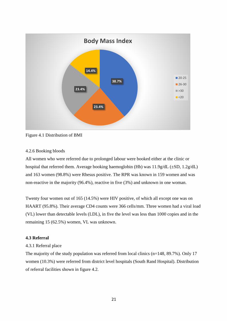
six women (23.4%) had a BMI of more than 30kg/m2.
Table 4.1 Patient demographics and other characteristics
Characteristics Number (%)
Mean (Standard deviation)
Median (Range)
N=165
p-value
Age (years)
< 20
20-35
> 35
25.2 (±5.1)
35 (21.3)
127 (76.9)
3 (1.8)
0.1
Parity
0
1
2
3
0 (0 – 3)
108 (65.5)
28 (16.9)
25 (15.2)
4 (2.4)
0.1
Gravidity 1 (1 – 4) 0.1
Gestational age (GA)
36-40 weeks
41-42 weeks
Method of determining GA
- Dates (LMP)
- Early US
- Late US
- Symphysiofundal height
38weeks 6days (±1 week 5
days)
149 (90.3)
16 (9.7)
141 (85.5)
6 (3.6)
13 (7.9)
5 (3)
0.1
BMI(kg/m2)
N=111
26.4 (±6.15)
0.1
21
Figure 4.1 Distribution of BMI
4.2.6 Booking bloods
All women who were referred due to prolonged labour were booked either at the clinic or
hospital that referred them. Average booking haemoglobin (Hb) was 11.9g/dL (±SD, 1.2g/dL)
and 163 women (98.8%) were Rhesus positive. The RPR was known in 159 women and was
non-reactive in the majority (96.4%), reactive in five (3%) and unknown in one woman.
Twenty four women out of 165 (14.5%) were HIV positive, of which all except one was on
HAART (95.8%). Their average CD4 counts were 366 cells/mm. Three women had a viral load
(VL) lower than detectable levels (LDL), in five the level was less than 1000 copies and in the
remaining 15 (62.5%) women, VL was unknown.
4.3 Referral
4.3.1 Referral place
The majority of the study population was referred from local clinics (n=148, 89.7%). Only 17
women (10.3%) were referred from district level hospitals (South Rand Hospital). Distribution
of referral facilities shown in figure 4.2.
38.7%
23.4%
23.4%
14.4%
Body Mass Index
20-25
26-30
>30
<20

22
Figure 4.2 Distribution of referral facilities
4.3.2 Reason for referral
Most of the women were referred due to prolonged active phase of labour, 77 (46.6%) and
prolonged latent phase of labour 75 (45.5%). Only 13 (7.9%) women were referred due to
prolonged second stage of labour. Some patients who were referred for prolonged active phase
also had a prolonged latent phase when analyzing their clinic records. Seven women were
referred for fetal distress and two for hypertension in pregnancy. On arrival at CMJAH they
were also found to have prolonged labour. Additional indications for referral included CPD,
MSL, low haemoglobin and maternal tachycardia.
10.3%
81.2%
3.3%
1.3%
1.3%0.6%
0.6%
0.6%
0.6%
Referral facility
South Rand Hospital
Hilbrow Community Health Center
Yeoville Clinic
Bez Valley Clinic
Jeppe Clinic
Lenasia South Clinic
OR Tambo Clinic
Witkoppen Clinic
Albert Clinic

23
Table 4.2 Indications for referral to CMJAH
Reason for referral Number (%)
N=165
Prolonged active phase
Prolonged latent phase
Prolonged second stage
77 (46.6)
75 (45.5)
13 (7.9)
4.3.3 Time taken to transfer
Average time taken to decision at the referral facility was three hours with a median of 10 hours
and a range of 0.25 to 37 hours. The average actual time taken to transfer the patient to CMJAH
was 2.5 hours with a median of 2 hours and range of 0.25 to 10.5 hours.
4.4 Progress of labour
4.4.1 Assessment on arrival at CMJAH
From 165 patients, 113 (68.5%) were in active phase of labour (≥4cm) on arrival to CMJAH, 33
(20%) were in latent phase of labour (≤3cm) and only 19 (11.5%) women arrived in second
stage of labour. The mean cervical dilatation was 5.7 (±2.4) cm.
Forty of the 75 women (53.3%) referred as prolonged latent phase, arrived in active phase of
labour, and nine of the 77 women (11.6%) referred as prolonged active phase of labour, arrived
fully dilated.
4.4.2 Duration of labour
One hundred and eleven women had prolonged latent phase. The mean duration was 16.2 (±5.9)
hours. There were no details available on the latent phase for 16 women because they arrived in
active phase of labour. A total of 92 women had prolonged active phase with a mean duration of
11.1 (±4.3) hours. The maximum duration was 25 hours. For the total sample (n=165), the
average duration of second stage of labour was 46 minutes. However, for the 31 women with
prolonged second stage the average duration was 115.9 (±78.7) minutes with the range of 45 –
360 minutes.
When comparing the duration of labour between nulliparous and multiparous women, the
difference between mean duration of latent and active phase was not statistically significant. For

24
the second stage of labour, mean duration for nulliparous and multiparous women was again not
statistically significant (p=0.73) (table 4.3).
Table 4.3 Duration of stages of labour according to parity
Stage of Labour (hours)
Mean (SD)
Latent Active 2nd stage (minutes)
Nulliparous
(n=108)
15.9 (6.24) 10.4 (4.2) 147.6 (84.9)
Multiparous (n=57) 14.8 (5.9) 11.1 (4.9) 94.5 (55.9)
p value 0.37 0.52 0.73
4.4.3 Mode of delivery
Ninety one (55.2%) women with prolonged labour had normal vaginal delivery and eight
(4.8%) had instrumental delivery. The remainder of the women (n=66, 40%) delivered by CS.
The most common indication for CS was fetal distress (n=44, 66.6%) followed by poor progress
(n=21, 31.8%) CPD (n=11; 16.6%), failed augmentation of labour (n=6; 9.1%) and failed
vacuum delivery (n=2; 3.0%). Details of patient deliveries are indicated in table 4.4.
Table 4.4 Mode of delivery
Mode of Delivery
Number (%)
N=165
Phase of prolonged labour
indicating referral
Vaginal
Delivery
Instrumental Caesarean
section Forceps
Vacuum
Latent phase 40 (43.9) 2 1 31 (47)
Active phase 45 (49.5) 0 3 30 (45.5)
Second stage 6 (6.6) 1 1 5 (7.5)
Total 91 (55) 3 5 66 (40)
25
4.5 Maternal and neonatal outcomes
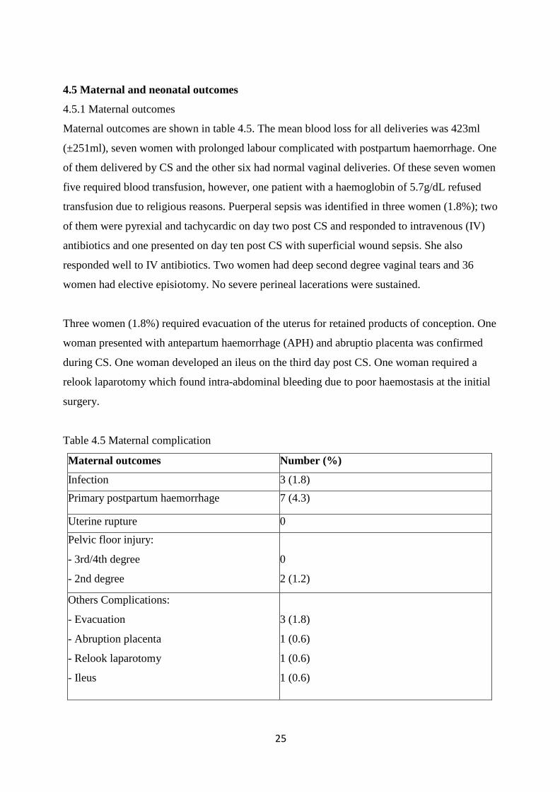
4.5.1 Maternal outcomes
Maternal outcomes are shown in table 4.5. The mean blood loss for all deliveries was 423ml
(±251ml), seven women with prolonged labour complicated with postpartum haemorrhage. One
of them delivered by CS and the other six had normal vaginal deliveries. Of these seven women
five required blood transfusion, however, one patient with a haemoglobin of 5.7g/dL refused
transfusion due to religious reasons. Puerperal sepsis was identified in three women (1.8%); two
of them were pyrexial and tachycardic on day two post CS and responded to intravenous (IV)
antibiotics and one presented on day ten post CS with superficial wound sepsis. She also
responded well to IV antibiotics. Two women had deep second degree vaginal tears and 36
women had elective episiotomy. No severe perineal lacerations were sustained.
Three women (1.8%) required evacuation of the uterus for retained products of conception. One
woman presented with antepartum haemorrhage (APH) and abruptio placenta was confirmed
during CS. One woman developed an ileus on the third day post CS. One woman required a
relook laparotomy which found intra-abdominal bleeding due to poor haemostasis at the initial
surgery.
Table 4.5 Maternal complication
Maternal outcomes Number (%)
Infection 3 (1.8)
Primary postpartum haemorrhage
- Blood transfusion required
7 (4.3)
5 (71.4)
Uterine rupture 0
Pelvic floor injury:
- 3rd/4th degree
- 2nd degree
0
2 (1.2)
Others Complications:
- Evacuation
- Abruption placenta
- Relook laparotomy
- Ileus
3 (1.8)
1 (0.6)
1 (0.6)
1 (0.6)

26
4.5.2 Neonatal outcomes
The mean weight was 3227g (±437g). Five babies were less than or equal to 2500g; 154 babies
between 2500 and 4000 g; and five (3%) were more than 4000g. The woman with the heaviest
baby (5080g) delivered vaginally after a prolonged second stage.
Out of 165, one baby was fresh still born and the mother had been referred for prolonged latent
phase of labour and prolonged ruptured of membranes. She was booked and delivered by CS,
the cord was found wrapped twice around the baby’s neck.
A total of 47 babies (28.6%) required observation in TICU, mainly due to respiratory distress.
Nine babies (5.5%) out of 164, required admission to neonatal ICU. Apgar scores of less than 7
at five minutes were found in eight babies (5.5%). No birth trauma was recorded.
Eleven out of 164 babies (6.7%) had meconium stained liqour and were admitted to TICU for
observation due to respiratory distress and possible meconium aspiration pneumonia. This
comprised 23.4% of the admissions to TICU.
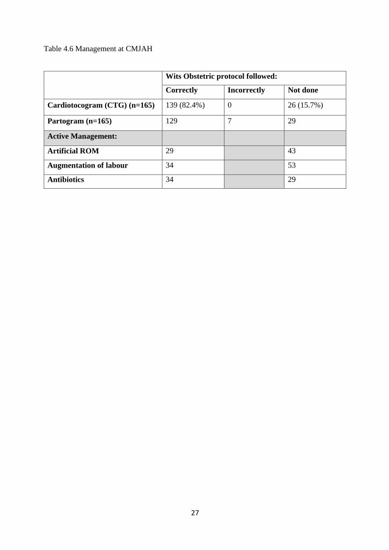
4.6 Management at CMJAH
Of the 165 women, cardiotocography (CTG) was performed in 139 women (84.2%) on
admission. Twenty seven (16.4%) women had a pathological trace requiring urgent delivery.
The partogram was plotted correctly in 129 (78.2%) women, incorrectly in seven (4.2%)
women. It was plotted at all in 29 women. Active intervention (ROM, AOL, antibiotic,
episiotomy), where the patient crossed the action line of the partogram, was instituted only in 59
(35.8%) women. In the rest of the women there was either a delay in active intervention or no
intervention at all. All steps of the protocol were correctly followed, as appropriate for the
individual woman, in 55 (33.3%) of women. In 110 (66.7%) women at least one element of the
protocol was performed incorrectly or not at all.
27
Table 4.6 Management at CMJAH
Wits Obstetric protocol followed:
Correctly Incorrectly Not done
Cardiotocogram (CTG) (n=165) 139 (82.4%) 0 26 (15.7%)
Partogram (n=165) 129 7 29
Active Management:
Artificial ROM 29 43
Augmentation of labour 34 53
Antibiotics 34 29
28
5. DISCUSSION
5.1 Characteristics of women with prolonged labour
Compared with previous studies, age was not shown to be a significant risk factor for labour
dystocia in this study (p=0.1). The majority of the population (81.2%) were between 20 and
34 years old and only nine out of 165 participants (5.4%) were of advanced maternal age. In
the study by Treacy A et al. approximately 10% of women were 35 years or older.12 This
would indicate that either, the index study population has fewer women with pregnancies at
an advanced maternal age or that there may be a lower prevalence of older women with
labour dystocia in the index study population. According to Statistics South Africa, only
3.6% of women giving birth in 2016 were between the ages of 40 and 54 years.81 Considering
the study population comprised low risk women referred from the clinic, the low number of
older women is not surprising. Older pregnant women are more likely to have medical or
obstetric conditions which necessitate management at a higher level of care both antenatally
and intrapartum.
Mothers less than 20 years have been shown to have an increased risk of chorioamnionitis
and endometritis following prolonged labour, however, in this study the three women who
developed infection were all between 20 and 30 years old.19
All the women in the study had received antenatal care. There were five women (3%) with a
diagnosis of syphilis during the pregnancy. This figure is similar to the estimated national
syphilis prevalence of 2% in antenatal clinics in South Africa (2015).74 All of the women
diagnosed with syphilis had been treated appropriately during the pregnancy at their local
clinic. Over 14% of women in this study were known to be HIV infected. This is less than
half the South African national prevalence of 30.8% HIV infected pregnant women.74
The percentage of women with obesity in this study (23.4%) is noticeably higher than the
national average according to the SADHS where roughly 15% of women between 15 and 45
years were found to be obese.14 Previous studies have showed that a BMI of greater than 30
kg/m2 increases the risk of prolonged labour, however, in this study there was no association
between BMI and labour dystocia (p=0.1).19

29
Further to this, obese women are more likely to present with prolonged labour in the first
stage rather than the second stage of labour as described by Fyfe et al.17 In the index study
there was no association found between stage of prolonged labour (latent phase, active phase
or second stage) and overweight or obesity. However, the numbers in this study are too small
to adequately explore this.
There were two women referred to CMJAH with hypertension that had developed or around
the time of labour. None of these women required magnesium sulphate infusion. The effect
of hypertension or magnesium cannot be commented upon.
A study by Okewole et al. on maternal height and likelihood of vaginal delivery, showed that
maternal height greater than 165 cm was positively associated with vaginal delivery.21 In the
index study, the average maternal height was only 160 cm which may have had an impact on
the progress of labour. Dujardin et al. have previously shown an association between short
maternal height and increased risk of CS22, in our study there were only ten women with a
height less than 150 cm, five of whom were delivered via CS. It has been shown previously
the increased prevalence of anthropoid pelvices in the African population contributes to an
increase in emergency CS for CPD.21 The study population was comprised of only African
women, however, this is merely a representation of the population served by CMJAH and the
community health care centers in central Johannesburg.
There was no significant association found between gestational age and prolonged labour
(p=0.1) as majority of the study population were near or at term. Only 16 women (9.7%)
were between 41 and 42 weeks. The average birth weight was 3227g which is less than the
expected average birth weight of 3500g. Fetal macrosomia is a recognized risk factor for
labour dystocia, however, in our study only three percent of babies in our study were
macrosomic.28
5.2 Referral factors
The majority of women (89.7%) were referred from local clinics with 17 (10.3%) women
coming from a level 1 district hospital. This indicates a problem in the referral system as
women with labour dystocia should be managed at a district level. However, there have been
several staffing and resource issues at South Rand Hospital resulting in a lack of theatre

30
facilities at this hospital. Of the 17 women referred from SRH, eight delivered vaginal, eight
needed CS and one was delivered via forceps.
A total of 659 women were referred to CMJAH during the study period, with 25% of the
referrals due to prolonged labour. This percentage is similar to a study by Perdok et al. in The
Netherlands looking at referrals from primary to secondary level facilities where 23.5% of
women referred were due to prolonged labour.82 Transport between facilities can often result
in delays in appropriate management. In this study, the average duration for transfer of a
patient was 2.5 hours. This is in comparison to the study from The Netherlands, a developed
country, where the median referral to delivery time was four hours and 40 minutes.82
5.3 Mode of delivery
Of the women referred due to prolonged first stage of labour, 55.9% delivered vaginally,
40.1% had a CS and only 3.9% had an assisted delivery. Similarly, the women with
prolonged second stage, 46.1% delivered vaginally, 38.5% via CS and 15.4% following
assisted delivery. This percentage of women delivering vaginally is similar to the finding of
Perdok et al where 58.3% delivered vaginally after prolonged first stage and 42.1% following
prolonged second stage. The biggest difference is the rate of CS section and assisted delivery.
In the Netherlands, only 14.3% of women required CS for prolonged first stage compared
with the 40.1% in this study. In women with prolonged second stage, 56.1% delivered via
assisted delivery compared to the 15.4% in this study.82 These differences may highlight the
effect of active management, correct implementation of hospital protocols or experience of
health care personnel in assisted delivery. It may also result from a difference in the antenatal
populations with regards to ethnicity and pelvic shape.23
Similar to previous studies, there was a higher proportion (65.5%) of nulliparous women in
our study.24 There were 53 out of 108 (49%) primigravid women who required CS. This is a
much higher percentage than the 15% found by Hashim et al in India.24 The duration of
active and latent phase of labour in this study was not significantly different between
primigravid (p=0.37) and multigravida women (p=0.52) as found in previous studies.24 There
was also no significant difference between the length of the second stage between primi and
multiparous women (p=0.73) which is a finding common to several previous studies.47

31
There were a number of women who arrived at CMJAH having progressed from the time at
which the decision was made to refer. Many of these women went on to deliver vaginally
with no adverse outcomes for mother or baby. This may indicate that the women were
prematurely and inappropriately referred to CMJAH. This may be as a result of being
incorrectly diagnosed in labour or from clinic staff not adhering to referral guidelines.
However, these findings lend support to the newer guidelines advocated by the ACOG that
the rate of cervical dilation is much slower than previously thought. Women may progress
more slowly but still deliver vaginally if given enough time. The data to support this was
collected in the USA by Zhang et al. and the Consortium on Safe Labour where the majority
(78.4%) of women were given epidural analgesia.83 In comparison, at CMJAH, there is no
epidural service and most women are given intramuscular Pethidine for pain control.
Allowing more time to achieve vaginal delivery decreases the number of repeat CS and the
associated complications, however, it is not justifiable to continue labouring a patient for
prolonged periods of time without adequate analgesia.79
5.4 Maternal outcomes of prolonged labour
Puerperal sepsis has previously been found to be more common in women who delivered by
CS.5 In our study, puerperal sepsis developed in only three women (1.8%), all of them
underwent CS. Two out of the three women underwent CS due to prolonged second stage of
labour of over two hours. This is in keeping with the findings of Stephansson et al. who
found increased risk of infection with increasing duration of the second stage of labour.37 The
overall rate of maternal infection was surprisingly low at 1.8% with no cases of
chorioamnionitis. This is lower than the four percent puerperal sepsis found by Maghoma and
Buchmann at Chris Hani Baragwanath Hospital.5
The most common cause of PPH in women is uterine atony with labour dystocia being a
significant risk factor.36 In this study, the majority of cases (85.7%) of PPH occurred in
women following a vaginal birth with the most noticeable cause being uterine atony. Only
one patient with primary PPH delivered by CS.
It is estimated that between one and four percent of vaginal deliveries are complicated by
obstetric anal sphincter injuries internationally.7 In this study, there were no third or fourth
degree perineal tears, only two women out of 99 (2%) who delivered vaginally, had deep

32
second degree vaginal tears. This may be due to the fact that less than five percent of women
had an assisted delivery. Only 13% of women in this study were referred due to prolonged
second stage of labour, this may also account for the low number of perineal injuries as risk
of severe perineal laceration has been found to be related to prolonged length of second stage
of labour rather than prolonged first stage of labour.55 Findings in this study were more in
keeping with those of the work done at Chris Hani Baragwanath Hospital by SM Tshabalala
which found an incidence of third or fourth degrees tears of 0.5%.56
None of the women in this study had a rupture of the uterus. Uterine rupture is a very
uncommon complication of prolonged labour in a woman with an unscarred uterus.45
Augmentation of labour with high-dose oxytocin has an increased risk of uterine rupture46, in
this study, all of the women who were augmented received low-dose oxytocin according to
the Wits protocol (Appendix D).
5.5 Fetal outcomes of prolonged labour
Fetal trauma is more common during prolonged second stage of labour due to an increase in
instrumental delivery or shoulder dystocia.5,49 However, if the 13 women referred due to
prolonged second stage, no fetal injuries were recorded.
The primary indicator of birth asphyxia was an Apgar score less than 7 at five minutes. There
were similar numbers of women with prolonged latent (2 women) and active phase (3
women) and second stage (3 women) of labour and the incidence of Apgar score less than 7
at five minutes. Five of these women underwent CS delivery and one assisted delivery due to
fetal distress diagnosed on CTG. A prospective study by Sykes et al. to assess the
relationship between the Apgar scores and the acid-base status of the babies at birth found
that only 19% of the babies with a five minute Apgar score less than 7 had severe acidosis on
cord blood gas.60 Cord blood is not routinely taken at birth for deliveries at CMJAH.
Therefore, it is not possible to determine whether the neonates with low 5 minute Apgar
score truly had birth asphyxia.
In this study, a total of 47 babies required admission to TICU mainly due to respiratory
distress. The majority (85.1%) of them were delivered following prolonged first stage of
labour, latent phase 19/47 and active phase 21/47 respectively, and a minority were delivered
33
following prolonged second stage of labour. Considering that only 13 women were referred
due to prolonged second stage of labour, the percentage of babies requiring admission is
almost double that of the babies delivered following prolonged first stage (53.8% vs 25.3%
and 27.3% respectively). This is in keeping with studies showing that prolonged second stage
of labour is associated with an increased risk of admission.62
Of the nine babies admitted to NICU, six of them were delivered following prolonged latent
phase. This accounts for the majority of NICU admissions and is contrary to the findings by
Bailit et al. who reported no differences in terms of NICU admission between women having
prolonged latent and active phases of labour.6 The poor reserve of the babies in this study
may indicate the presence of underlying issues or intrauterine growth restriction not pick up
during antenatal care. Alternatively, it may indicate the poor condition of women during
prolonged labour, for example dehydration.
5.6 Management at CMJAH
The correct diagnosis of labour is a very important part in preventing prolonged labour and
avoiding inappropriate interventions. Diagnosis of prolonged labour (latent, active or second
stage) from the referral center was confirmed in all participants on arrival at CMJAH. There
were 89 cases where there was a delay in diagnosis of prolonged labour at the clinic which
caused a further delay in referral and management. Almost a quarter of these women had
neonatal complications.
Of the 139 women who had a CTG done on arrival at CMJAH, 27 (16.4%) of the women had
a pathological trace according to the Wits protocol (Appendix G). Emergency delivered was
arranged for these women. Fourteen (51.8%) of the babies delivered following pathological
CTG intrapartum were admitted to neonatal care units (TICU or NICU) post delivery for
observation. Staff shortage, patient overload and equipment shortages may account for the
15.7% of women who did not have CTG on arrival at CMJAH. Some patients in second stage
labour may also been transferred to labour ward directly for delivery and therefore did not
have CTG done in maternity admissions.
In this study, in a tertiary referral center, the partogram was plotted correctly in 129 (78.2%)
women, incorrectly in seven (4.2%) women and not used at all in 29 women. This is

34
concerning considering the level of training of the health workers working in a tertiary level
center. A study amongst healthcare providers in Nigeria, showed that knowledge on the use
of the partograph for labour management is very low amongst nurses, midwives, and doctors
working in the primary and secondary health care levels when compared to tertiary level
care.84
The partogram was acted on appropriately (ROM, AOL, antibiotic, episiotomy) in only 59
women (35.7%). In the remainder of women there was either a delay in appropriate
management or no active intervention. Active management has been shown in several centers
to improve progress in labour and reduce the CS rate, however, the routine use of amniotomy
in all women is not recommended. Due to the high rate of HIV in the South African
population and the increased risk of mother to child transmission with increasing duration of
ROM, amniotomy is not advisable. In this study, almost 15% of women were HIV infected
and more than 60% of these women had an unknown VL. For this reason, routine amniotomy
was not used in 80% of HIV positive women with intact membranes. This lack of
intervention may have impacted on progress of labour. Amniotomy in these patients may also
have alerted health care workers to the presence of MSL. There was often in delay in
initiation of augmentation of labour. This may have been due to the shortages of nursing
staff working in labour ward resulting in a higher ratio of women to nursing staff than is
recommended.
5.7 Limitations
This study was a retrospective study over only four months. This short period of time limited
the study sample to 165 women. Maternity records were available for all women but poor
documentation limited the amount of information obtained. Data collection also relied on
record keeping from clinics. Paediatric records were not available for all babies and
information such as cord blood gas was not available which limited our interpretation of the
fetal outcomes.
5.8 Recommendation
With the controversy in the definition of labour and the small population of this study, more
research is needed to determine the definitions of prolonged labour in the South African
context. Comparative studies to evaluate the maternal and fetal outcomes in different groups

35
of women with different definitions of labour (the old and new definitions) would help to
establish a clear protocol for diagnosis and management of prolonged labour in this
population. Improving the correct usage of institutional guidelines and the partogram in
management of women referred for prolonged labour may decrease the number of resultant
adverse fetal outcomes.

36
6. CONCLUSION
There were minimal maternal adverse outcomes in women referred to CMJAH for prolonged
labour. Fetal adverse outcomes were more apparent with over a quarter of babies requiring
admission for respiratory distress and one intrauterine fetal demise. The institution protocol
for management of labour dystocia was correctly followed only in 33.3% of the study
population. This is a figure that should be address in future to help decrease the ever
increasing rate of litigation in Obstetrics in the public sector.

37
REFERENCES
1. Nystedt IH. diverse definitions of prolonged labour and its consequences with sometimes
subsequent inappropriate treatment. Pregnancy Childbirth 2014;14:233-243.
2. Friedman EA. The graphic analysis of labor. Am J Obstet Gynecol 1954;68(6):1568-75.
3. Kjærgaard H, Olsen J, Ottesen B, Dykes AK. Incidence and outcomes of dystocia in the
active phase of labor in term nulliparous women with spontaneous labor onset. Acta Obstet
Gynecol Scand 2009;88(4):402-407.
4. Zhu BP, Grigorescu V, Le T, Lin M, Copeland G, Barone M, et al. Labor dystocia and its
associations with interpregnancy interval. Am J Obstet Gynaecol 2006;195(1):121-6.
5. Maghoma J, Buchmann EJ. Maternal and fetal risks associated with prolonged latent phase
of labour. J Obstet Gynaecol 2002;22(1):16–19. doi: 10.1080/01443610120101637.
6. Bailit JL, Dierker L, Blanchard MH, Mercer BM. Outcomes of women presenting in active
versus latent phase of spontaneous labor. Obstet Gynecol 2005;105(1):77-79.
7. Allen VM, Baskett TF, O’Connell CM, McKeen D, Allen AC. Maternal and perinatal
outcomes with increasing duration of the second stage of labor. Obstet Gynecol
2009;113(6):1248-58.
8. Cavazos-Rehg PA, Krauss MJ, Spitznagel EL, Bommarito K, Madden T, Olsen MA, et al.
Maternal age and risk of labor and delivery complications. Matern Child Health J
2015;19(6):1202-1211. doi:10.1007/s10995-014-1624-7.
9. Waldenström U, Ekéus C. Risk of labor dystocia increases with maternal age irrespective
of parity: a population‐based register study. Acta Obstet Gynecol Scand 2017;96(9):1063-
1069.
10. Islam MM, Bakheit CS. Advanced maternal age and risks for adverse pregnancy
outcomes: a population-based study in Oman. Health Care Women Int 2015;36(10):1081-
1103. doi: 10.1080/07399332.2014.990560
11. Greenberg MB, Cheng YW, Sullivan M, Norton ME, Hopkins LM, Caughey AB. Does
length of labor vary by maternal age? Am J Obstet Gynecol 2007;197(4):428. e1-428.e7. doi:
12.1016/j.ajog.2007.06.058.
12. Treacy A, Robson M, O'Herlihy C. Dystocia increases with advancing maternal age. Am
J Obstet Gynecol 2006;195:760-763.
13. Bogaerts A, Witters I, Van den Bergh BR, Jans G, Devlieger R. Obesity in pregnancy:
altered onset and progression of labour. Midwifery 2013;29(12):1303-1313.

38
14. Department of Health, Medical Research Council, OrcMacro. 2007. South Africa
Demographic and Health Survey 2003. Pretoria: Department of Health.
15. Kerrigan A, Kingdon C, Cheyne H. Obesity and normal birth: A qualitative study of
clinician’s management of obese pregnant women during labour. BMC Pregnancy Childbirth.
2015;15(1):256. doi: 10.1186/s12884-015-0673-2
16. Vinayagam D, Chandraharan E. The adverse impact of maternal obesity on intrapartum
and perinatal outcomes. ISRN Obstet Gynecol 2012; Article ID 939762.
doi:10.5402/2012/939762.
17. Fyfe EM, Anderson NH, North RA, Chan EH, Taylor RS, Dekker GA, et al. Risk of first-
stage and second-stage cesarean delivery by maternal body mass index among nulliparous
women in labor at term. Obstet Gynecol 2011;117(6):1315-1322. doi:
10.1097/AOG.0b013e318217922a
18. Robinson BK, Mapp DC, Bloom SL, Rouse DJ, Spong CY, Varner MW, et al. Increasing
maternal body mass index and characteristics of the second stage of labor. Obstet Gynecol
2011;118(6):1309-1313.
19. Chin JR, Henry E, Holmgren CM, Varner MW, Branch DW. Maternal obesity and
contraction strength in the first stage of labor. Am J Obstet Gynecol 2012;207(2):129.e1-e6.
20. Norman SM, Tuuli MG, Odibo AO, Caughey AB, Roehl KA, Cahill AG. The effects of
obesity on the first stage of labor. Obstet Gynecol 2012;120(1):130-135.
doi:10.1097/AOG.0b013e318259589c
21. Okewole I, Faiola S, Fakounde A, Yoong W, Phillip H, Amer S. The relationship of
ethnicity, maternal height and shoe size, and method of delivery. J Obstet Gynaecol
2011;31(7):608-611. doi: 10.3109/01443615.2011.590907.
22. Dujardin B, Cutsem RV, Lambrechts T. The value of maternal height as a risk factor of
dystocia: a meta‐analysis. Trop Med Int Health 1996;1(4):510-521.
23. Ibison JM. Ethnicity and mode of delivery in ‘low-risk’first-time mothers, East London,
1988–1997. Eur J Obstet Gynecol Reprod Biol 2005;118(2):199-205.
24. Hashim N, Naqvi S, Khanam M, Jafry HF. Primiparity as an intrapartum obstetric risk
factor. J Pak Med Assoc 2012;62(7):694-698.
25. Bregand-White J, Kominiarek M. Hypertension and labor duration: Does it take longer?
Pregnancy Hypertension 2012;2(3):192. doi:10.1016/j.preghy.2012.04.031
26. Onwochei DN, Carvalho JCA, Luca A, Kingdom J, Balki M. Effect of magnesium sulfate
on oxytocin-induced contractility in human myometrium: an in vitro study. Can J Anesth

39
2017;64:744–775.
27. Witlin AG, Friedman SA, Sibai, BH. The effect of magnesium sulfate therapy on the
duration labor in women with mild preeclampsia at term: A randomized, double-blind,
placebo-controlled trial. Am J Obstet Gynecol 1997;176:623-627.
28. Selin L, Wallin G, Berg M. Dystocia in labour–risk factors, management and outcome: a
retrospective observational study in a Swedish setting. Acta Obstet Gynecol Scand
2008;87(2):216-21.
29. Turner MJ, Rasmussen MJ, Turner JE, Boylan PC, MacDONALD D, Stronge JM. The
influence of birth weight on labor in nulliparas. Obstet Gynecol 1990;76(2):159-63.
30. Kjærgaard H, Olsen J, Ottesen B, Nyberg P, Dykes A-K. Obstetric risk indicators for
labour dystocia in nulliparous women: a multi-centre cohort study. BMC pregnancy
childbirth 2008;8(1):45.
31. Hawkins JL. Epidural analgesia for labor and delivery. N Engl J Med 2010;362(16):1503-
1510.
32. Sng BL, Leong WL, Zeng Y, Siddiqui FJ, Assam PN, Lim Y, et al. Early versus late
initiation of epidural analgesia for labour. Cochrane Database Syst Rev [Internet]. 2014 [cited
2017 Dec 20];10:CD007238. Available from: doi.10.1002/14651858.CD007238.
33. Agrawal D, Makhija B, Arora M, Haritwal A, Gurha P. The effect of epidural analgesia
on labour, mode of delivery and neonatal outcome in nullipara of India, 2011-2014. J Clin
Diag Res 2014;8(10):OC03- OC06. doi: 10.7860/JCDR/2014/9974.4930
34. Anim‐Somuah M, Smyth R, Jones L. Epidural versus non‐epidural or no analgesia in
labour. Cochrane Database Syst Rev [Internet]. 2011 [cited 2017 Dec
20];12:CD000331. Available from: doi.10.1002/14651858.CD000331.pub3
35. Shen X, Li Y, Xu S, Wang N, Fan S, Qin X, et al. Epidural Analgesia During the Second
Stage of Labor: A Randomized Controlled Trial. Obstet Gynecol 2017;130(5):1097-1103.
36. Cunningham F, Leveno K, Bloom S, Hauth J, Rouse D, Spong C. Williams Obstetrics
23rd Edition. USA: McGraw Hill; New York; 2010.p872.
37. Stephansson O, Sandström A, Petersson G, Wikström AK, Cnattingius S. Prolonged
second stage of labour, maternal infectious disease, urinary retention and other complications
in the early postpartum period. Br J Obstet Gynaecol 2016;123(4):608-616.
38. Laughon SK, Berghella V, Reddy UM, Sundaram R, Lu Z, Hoffman MK. Neonatal and
maternal outcomes with prolonged second stage of labor. Obstet Gynecol 2014;124(1):57.

40
39. Satin AJ, Maberry MC, Leveno KJ, Sherman ML, Kline D. Chorioamnionitis: a harbinger
of dystocia. Obstet Gynecol 1992;79(6):913-5.
40. Coviello EM, Grantz KL, Huang C-C, Kelly TE, Landy HJ. Risk factors for retained
placenta. Am J Obstet Gynecol [Internet]. 2015 [cited 2017 Dec 20];213(6):864.e1-e11.
Available from: http://dx.doi.org/10.1016/j.ajog.2015.07.039
41. Nyfløt LT, Stray-Pedersen B, Forsén L, Vangen S. Duration of labor and the risk of
severe postpartum hemorrhage: A case-control study. PloS One [Internet]. 2017[cited 2017
Dec 20];12(4):e0175306. Available from: https://doi.org/ 10.1371/journal.pone.0175306.
42. National Committee on Confidential Enquiries into Maternal Deaths. Saving Mothers
Report 2011-2013: The Sixth Report of the National Committee for Confidential Enquiries
into Maternal Deaths in South Africa. Pretoria: Government Printer. 2014.
43. Vachon-Marceau C, Demers S, Goyet M, Gauthier R, Roberge S, Chaillet N, et al. Labor
dystocia and the risk of uterine rupture in women with prior cesarean. Am J Perinatol
2016;33(06):577-83.
44. Walsh CA, O’Sullivan RJ, Foley ME. Unexplained prelabor uterine rupture in a term
primigravida. Obstet Gynecol 2006;108(3, Part 2):725-7.
45. Dow M, Wax JR, Pinette MG, Blackstone J, Cartin A. Third-trimester uterine rupture
without previous cesarean: a case series and review of the literature. Am J Perinatol
2009;26(10):739-744.
46. Budden A, Chen LJ, Henry A. High‐dose versus low‐dose oxytocin infusion regimens for
induction of labour at term. Cochrane Database Syst Rev [Internet]. 2014 [cited
2017 Dec 20];10:CD009701. Available from: doi.10.1002/14651858.CD009701.pub2.
47. Caughey AB, Cahill AG, Guise J-M, Rouse DJ. Safe prevention of the primary cesarean
delivery. Am J Obstet Gynecol 2014;210(3):179-93.
48. Harrison MS, Pasha O, Saleem S, Ali S, Chomba E, Carlo WA, et al. A prospective study
of maternal, fetal and neonatal outcomes in the setting of cesarean section in low‐and middle‐
income countries. Acta Obstet Gynecol Scand 2017;96(4):410-20.
49. Radha P, Shephali T, Muhammad Fairuz A, Tee C. Maternal and perinatal morbidity after
caesarean delivery at full cervical dilatation. Singapore Med J 2012;53(10):655.
50. Govender V, Panday M, Moodley J. Second stage caesarean section at a tertiary hospital
in South Africa. J Mat Fetal Neonatal Med. 2010;23(10):1151-5.
51. Sucak A, Çelen Ş, Akbaba E, Soysal S, Moraloglu O, Danışman N. Comparison of
41
nulliparas undergoing cesarean section in first and second stages of labour: a prospective
study in a tertiary teaching hospital. Obstet Gynecol Int [Internet]. 2011 [cited 2017 Dec
20];2011:986506. Available from: doi.10.1155/2011/986506
52. Wall LL. Obstetric vesicovaginal fistula as an international public-health problem.
Lancet. 2006;368(9542):1201-1209.
53. Vangeenderhuysen C, Prual A, Ould el Joud D. Obstetric fistulae: incidence estimates for
sub‐Saharan Africa. Int J Gynecol Obstet 2001;73(1):65-6.
54. Tebeu PM, Fomulu JN, Khaddaj S, de Bernis L, Delvaux T, Rochat CH. Risk factors for
obstetric fistula: a clinical review. Int Urogynecol J 2012;23(4):387-394. doi:
10.1007/s00192-011-1622-x
55. Simic M, Cnattingius S, Petersson G, Sandström A, Stephansson O. Duration of second
stage of labor and instrumental delivery as risk factors for severe perineal lacerations:
population-based study. BMC Pregnancy Childbirth 2017;17(1):72. doi: 10.1186/s12884-
017-1251-6
56. Tshabalala SM. Risk factors, repair techniques and short term subjective outcome of
obstetrics anal sphincter injuries at Chris Hani Baragwanath Academic Hospital.
[Unpublished research report]. Johannesburg: University of the Witwatersrand; 2016.
Accessed 2017 Dec 20; available at: http://hdl.handle.net/10539/22457.
57. McClure EM, Bose CL, Garces A, Esamai F, Goudar SS, Patel A, et al. Global network
for women’s and children’s health research: a system for low-resource areas to determine
probable causes of stillbirth, neonatal, and maternal death. Mat Health Neonatol Perinatol
2015;1(1):11.
58. Pattison R, Rhoda N. Saving Babies 2012-2013: Ninth Report on perinatal care in South
Africa. Tshepesa Press, Pretoria, 2014.
59. Velaphi S, Rhoda N. Reducing neonatal deaths in South Africa-are we there yet, and what
can be done? S Afr J Child Health 2012;6(3):67-71.
60. Marrin M, Paes BA. Birth asphyxia: does the Apgar score have diagnostic value? Obstet
Gynecol 1988;72(1):120-123.
61. Saunders N, Paterson C, Wadsworth J. Neonatal and maternal morbidity in relation to the
length of the second stage of labor. Int J Gynecol Obstet 1993;40(2):189.
62. Sandström A, Altman M, Cnattingius S, Johansson S, Ahlberg M, Stephansson O.
Durations of second stage of labor and pushing, and adverse neonatal outcomes: a
population-based cohort study. J Perinatol 2017;37(3):236-242.

42
63. Aslam HA, Saleem S, Afzal R, Iqbal U, Saleem SM, Shaikh MWA, et al. Risk factors of
birth asphyxia. Italian J Pediatr 2014;40:1-9. doi: 10.1186/s13052-014-0094-2
64. Chelmow D, Kilpatrick SJ, Laros JR. Maternal and neonatal outcomes after prolonged
latent phase. Obstet Gynecol 1993;81(4):486-491
65. Lavender T, Hart A, Smyth R. Effect of partogram use on outcomes for women in
spontaneous labour at term. Cochrane Database Syst Rev [Internet]. 2014 [cited 2017 Dec
20];8:CD005461. Available from: doi:10.1002/14651858.CD005461.pub3.
66. Fatusi A, Makinde O, Adeyemi A, Orji E, Onwudiegwu U. Evaluation of health workers'
training in use of the partogram. Int J Gynecol Obstet 2008;100(1):41-44.
67. Lavender T, Alfirevic Z, Walkinshaw S. Effect of different partogram action lines on
birth outcomes: A randomized controlled trial. Obstet Gynecol 2006;108(2):295-302.
68. Wei S, Wo BL, Qi HP, Xu H, Luo ZC, Roy C, et al. Early amniotomy and early oxytocin
for prevention of, or therapy for, delay in first stage spontaneous labour compared with
routine care. Cochrane Database Syst Rev 2013;8:CD006794.
69. Hani A, Akoury GB, Caddick R, McLaughin VD. Active management of labor and
operative delivery in nulliparous women. Am J Obstet Gynecol 1988;158(2):255-258.
70. Smyth R, Markham C, Dowswell T. Amniotomy for shortening spontaneous labour.
Cochrane Database Syst Rev [Internet]. 2013 [cited 2017 Dec 20];6:CD006167. Available
from: doi.10.1002/14651858.
71. Wariki WMV, Ota E, Mori R, Wiysonge CS, Horvath H, Read JS. Interventions for
preventing mother-to-child HIV transmission: protocol of an overview of systematic reviews.
BMJ Open [Internet]. 2017 [cited 2017 Dec 20];7:e014332. Available from: doi.10.1136/
bmjopen-2016-014332.
72. Peters H, Byrne L, De Ruiter A, Francis K, Harding K, Taylor GP, et al. Duration of
ruptured membranes and mother‐to‐child HIV transmission: a prospective population‐ based
surveillance study. Br J Obstet Gynaecol 2016;123(6):975-81.
73. World Health Organisation. Consolidated guidelines on the use of antiretroviral drugs for
treating and preventing HIV infection: recommendations for a public health approach, 2nd
edition. WHO Press, Switzerland, 2016. Available from:http://www.who.int/hiv/pub/arv/arv-
2016/en/ [accessed Jan 2018.]
74. National Department of Health. The 2015 National Antenatal Sentinel HIV & Syphilis
Survey, Pretoria, 2017. Available from: www.health.gov.za/index.php/.../2015-04-30-08-21-
56?...hiv-prevalence [accessed Jan 2018].
43
75. National Department of Health. National consolidated guidelines for the prevention of
mother-to-child transmission of HIV (PMTCT) and the management of HIV in children,
adolescents and adults. Department of Health, Pretoria, 2014. Available from:
www.sahivsoc.org/Files/ART%20Guidelines%2015052015.pdf [accessed Jan 2018].
76. Basson E, Odendaal H, Grové D. Oxytocin use in South Africa-a review. S Afr Med J
2004;94(10):839-45.
77. Zhang J, Branch DW, Ramirez MM, Laughon SK, Reddy U, Hoffman M, et al. Oxytocin
regimen for labor augmentation, labor progression, perinatal outcomes. Obstet Gynecol
2011;118(2 0 1):249-256. doi:10.1097/AOG.0b013e3182220192
78. Zhang J, Duan T. The physiologic pattern of normal labour progression. Br J Obstet
Gynaecol [Internet]. 2017 [cited 2017 Dec 20]. Available from: https://doi.org/10.1111/1471-
0528.14930
79. Oladapo OT, Diaz V, Bonet M, Abalos E, Thwin SS, Souza H, et al. Cervical dilatation
patterns of ‘low‐risk’women with spontaneous labour and normal perinatal outcomes: a
systematic review. Br J Obstet Gynaecol [Internet]. 2017 [cited 2017 Dec 20]. Available
from: https://doi.org/10.1111/1471-0528.14930.
80. Cohen WR, Friedman EA. Perils of the new labor management guidelines. Am J Obstet
Gynecol 2015;212(4):420-427.
81. Statistics South Africa. Statistical release P0305, Recorded live births 2013-2015.
Statistics South Africa, Pretoria, 2016. Available from:
www.statssa.gov.za/publications/P0305/P03052015.pdf [accessed Feb 2018].
82. Perdok H, Jans S, Verhoeven C, van Dillen J, Mol BW, de Jonge A. Intrapartum referral
from primary to secondary care in the Netherlands: A retrospective cohort study on the
management of labour and outcomes. Birth 2015;42(2):156-164.
83. Zhang J, Landy HJ, Ware Branch D, Burkman R, Haberman S, Gregory K, et al.
Contemporary patterns of spontaneous labour with normal neonatal outcomes. Obstet
Gynecol 2010;116(6):1281-1287. doi: 10.1097/AOG.0b013e3181fdef6e
84. Asibong U, Okokon IB, Agan TU, Oku A, Opiah M, Essien EJ, et al. The use of the
partograph in labor monitoring: a cross-sectional study among obstetric caregivers in General
Hospital, Calabar, Cross River State, Nigeria. Int J Women Health 2014;6:873.

44
APPENDIX A: DATA SHEET
Data Collection Sheet
Research No.
Demographics and pregnancy data:
Age ___________ height ________
parity /gravidity ____________ weight at booking
gestational age ___________ By EUS/LUS/DATES/SFH bmi _________
Any maternal medical conditions/ previous CS Yes/No Specify______________________
Referral:
referral place ___________ Clinic/Hospital(specify)
Reason for referral _________________________
Stage of labour Latent/active/second stage
Times pt arrived to clinic time pt arrived to CMJAH/time of delverydecision of transfer pt arrived to CMJAHtime of delivery
Date:____________
Management at CMJAH: __________
CTG Yes/No Reactive/suspecious/pathological
Partogram used Yes/No cervical dilatation on admission _________
Partogram acted Yes/No HAB on admission ________
AROM/SROM When Yes/No
Augmentation with oxytocin Yes/No
Antibiotics given Yes/No
PROTOCOL followed YES/NO
Mode of delivery:
NVD Yes/No Episiotomy cut Yes/No
Assisted delivery Vacum/Forceps
ceasarean section Yes/No
Adverse outcomes:
Maternal: ______________________/ Unknown
infection chorioaminionitis/Peurperal sepsis/other (Within current admission)
Primary PPH Yes/No
uterine rupture ________________________
pelvic floor injuries _____________________
complications at CS
Others _____________________
Fetal :
Trauma ______________________________
Asphyxia Apgar scores ____________ Cord pH________ ABG __________
Meconium aspiration Yes/No
Admission to TU/NICU Yes/No
END Yes/No
MSB Yes/No
FSB Yes/No
Birth Weight __________ grams
HIV Status 1.Non Reactive/ 2.Reactive/ 3.Unknown
If Reactive, On HAART 1.Yes/ 2.No/ 3.Unknown
When was HAART started ___________/ Unknown
CD4 count/Viral load ____________/ Unknown
Booking Blood:
HB
RH
RPR _________
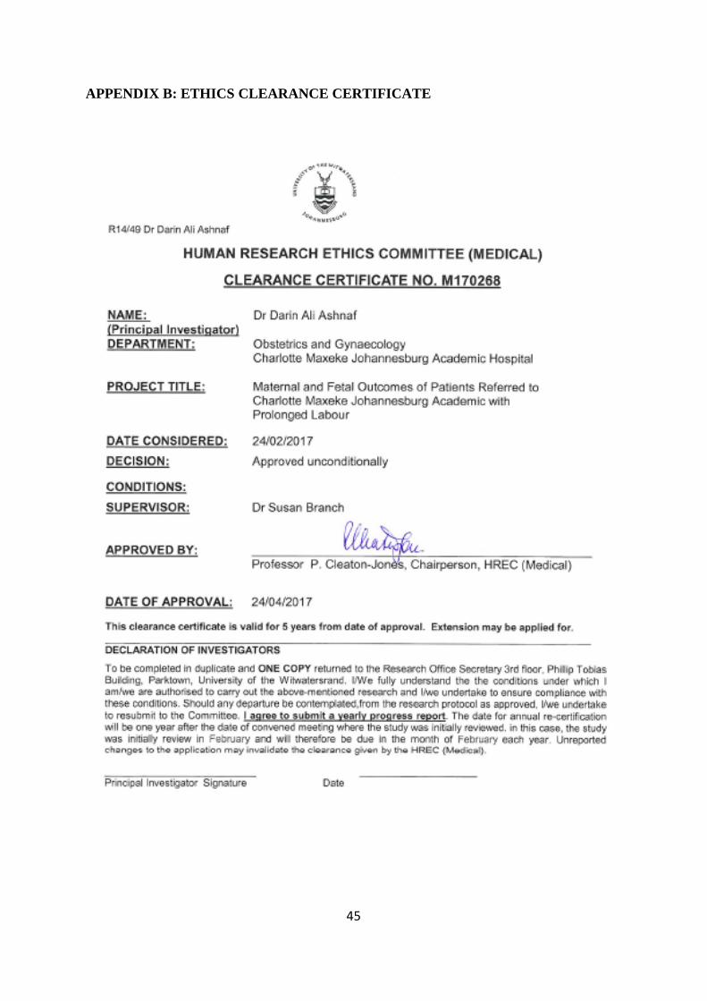
45
APPENDIX B: ETHICS CLEARANCE CERTIFICATE

46
APPENDIX C: INSTITUTIONAL PROTOCOL

47

48

49
APPENDIX D: AUGMENTATION OF LABOUR PROTOCOL

50
APPENDIX E: ANTIBIOTIC USE WITH PROLONGED RUPTURE OF
MEMBRANES

51
APPENDIX F: MANAGEMENT OF PROLONGED SECOND STAGE

52
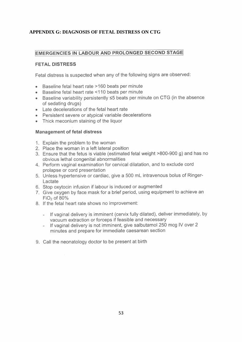
53
APPENDIX G: DIAGNOSIS OF FETAL DISTRESS ON CTG


