

MATRIX METALLOPROTEINASE 2, MATRIX METALLOPROTEINASE 9, AND
CONNECTIVE TISSUE GROWTH FACTOR IN THE EQUINE TEAR FLUID: POSSIBLE IMPLICATIONS IN CORNEAL WOUND HEALING
By
FRANCK J. OLLIVIER
A DISSERTATION PRESENTED TO THE GRADUATE SCHOOL OF THE UNIVERSITY OF FLORIDA IN PARTIAL FULFILLMENT
OF THE REQUIREMENTS FOR THE DEGREE OF DOCTOR OF PHILOSOPHY
UNIVERSITY OF FLORIDA
2004

Copyright 2004
by
Franck J. Ollivier

This work presented in this dissertation shall be a contribution to preserve sight in horses
and it is dedicated to horse owners and veterinary ophthalmologists. This dissertation is also dedicated to my family and my friends wherever they are, for
their constant support.

ACKNOWLEDGMENTS
My time as a graduate student ends with this dissertation. The years dedicated to
this work represent not only an educational but also a social experience. Many
individuals were involved in this endeavor and have been important for my success,
especially those listed below.
I would like to express my deep gratitude to my mentor and friend, Dr. Dennis
Brooks, for his support and understanding throughout these years. I thank him for having
given me the great and unique opportunity to come to Florida and pursue my education in
veterinary ophthalmology.
I would like to sincerely thank Dr. Gregory Schultz who meant a great deal for me,
my graduate education, and my project. He opened his laboratory for me and patiently
taught and helped me though many steps of my project. His positive and supportive
attitude was extraordinary.
I am also very grateful to Dr. Kirk Gelatt for sharing, with no limits, his fascinating
knowledge and interest in research and clinical veterinary ophthalmology. I am proud to
be the last ophthalmology resident he will train before a well-deserved retirement.
I am very thankful to Dr. Gysbert van Setten who guided through my project from
the other side of the Atlantic Ocean! He not only shared his tremendous knowledge on
the tear film and the cornea but also the way of questioning axioms and looking for new
ones. His suggestions and our discussions were very valuable.
iv

I owe many thanks to Dr. Stacy Andrew for sharing her indefatigable interest in
corneal research and for her precious help in my project at various stages.
I would like to thank Dr. James Farese and Dr. Sonal Tuli for being member of my
doctoral thesis committee; their advice was important in my success in this dissertation.
I owe my dear friend Dr Maria Källberg many thanks for her scientific but also
social support! I will never forget the hilarious moments as well as the tough times we go
through together.
I would like to sincerely thank Dr. Tim Blalock for his friendship, his patience and
his tremendous help in various aspects of my work.
I am very thankful to Dr Gary Stevens who helped with statistics. He friendly
taught me a lot in this field during our nice and very interesting discussions.
I am also grateful to Dr Don Samuelson and his laboratory technicians, Ms. Patricia
Lewis, and Ms. Mae Chisholm for their friendly help in the adventure of histology that I
went through during my time as a graduate student.
I would like to thank Drs. Mary Lassaline, András Komáromy, Tim Cutler and
Heidi Denis for their contributions in my project.
I owe many thanks to Ms. Suzanne Sharra-Maxwell, owner of “Pyrite farm” in
Ocala, Florida, for allowing me to perform parts of my project at her farm and for the
laughs shared together.
I also owe many thanks to the Office of Research and Graduate Studies at the
College of Veterinary Medicine, University of Florida. Associate Dean Charles Courtney
III and Mrs. Sally O’Connell helped me through the administrative procedures to
successfully finish my dissertation.
v
I would like to extend my thanks to Drs. William Dawson, Michael Goldstein,
David Moraga, Colin Serada, Cornelia Gunkel, Hendrik Nollens, as well as Mr. Harold
Sapp, Ms. Delena McTeer and Ms. Dottie Holland for all the advice, support and
friendship they have shown me throughout my time as a graduate student at the
University of Florida.
I am grateful to my friends outside of the College of Veterinary Medicine, in
Florida, Australia and in France for their understanding, and social support throughout
these years.
Last but certainly not least, I would like to thank Dr. Alain Regnier for having
given me the taste for veterinary ophthalmology and for his friendship, his endless
support and his wise advice through these years.
Finally, I would like to express my deepest gratitude to my parents Marie-Claire
and Gerard Ollivier, my brothers Philippe and Pierre, my sister Claire Ollivier for their
love and tremendous support they have offered to me over the years.
Merci beaucoup pour votre amour, votre confiance en moi et votre soutien sans
mesure et malgré la distance qui nous sépare.
vi
TABLE OF CONTENTS page ACKNOWLEDGMENTS ................................................................................................. iv
LIST OF TABLES............................................................................................................. xi
LIST OF FIGURES ......................................................................................................... xiii
KEY TO ABBREVIATIONS.......................................................................................... xvi
ABSTRACT..................................................................................................................... xix
CHAPTER
1 INTRODUCTION ........................................................................................................1
The Cornea and the Precorneal Tear Film (PTF) .........................................................1 Structure and Function of the Precorneal Tear Film .............................................1 Structure and Function of the Cornea....................................................................5
Corneal Healing and Scarring.......................................................................................9 Overview on Wound Healing and Corneal Healing..............................................9 Histological Events of Corneal Healing ..............................................................11
Epithelial wound healing..............................................................................11 Stromal and endothelial wound healing .......................................................13
Biochemical Mechanisms of Corneal Healing ....................................................15 Molecular biology of epithelial wound healing ...........................................15 Molecular biology of stromal wound healing ..............................................17
Corneal Scarring..................................................................................................19 Proteinases, Proteinase Inhibitors...............................................................................20
Proteinases in The PTF: Types and Origins ........................................................20 Serine proteinases.........................................................................................20 Matrix metalloproteinases (MMPs) .............................................................21 Origins of the proteinases.............................................................................29
Proteinases, PTF and Corneal Physiopathology..................................................30 Proteinases, Proteinase Inhibitors and the Cornea ..............................................33
Growth Factors ...........................................................................................................34 Growth Factors, PTF and Corneal Physiopathology...........................................34 Connective Tissue Growth Factor .......................................................................38
Significance to The Horse Racing Industry................................................................42 Purpose of The Study..................................................................................................43
vii
Hypotheses ..........................................................................................................43 Objectives ............................................................................................................44
2 DETECTION OF MMP-2 AND MMP-9 IN THE EQUINE TEAR FLUID,
CORNEA AND LACRIMAL GLANDS ...................................................................45
Introduction.................................................................................................................45 Materials and Methods ...............................................................................................47
Materials ..............................................................................................................47 MMP-2 and MMP-9 Western Blot......................................................................49 MMP-2 and MMP-9 Immunohistochemistry in Sections of Equine Cornea,
Equine Main Lacrimal Gland and Equine Nictitating Membrane Gland ........50 Results.........................................................................................................................51
MMP-2 and MMP-9 Western Blot: Detection of MMP-2 and MMP-9 in the Equine Tear Fluid. ...........................................................................................51
Immunohistochemistry: Immunohistochemical Localization of MMP-2 and MMP-9 in the Healthy Cornea and Lacrimal Glands of Horses......................52
Immunohistochemistry: Immunohistochemical Localization of MMP-2 and MMP-9 in Ulcerated Equine Cornea ...............................................................55
Discussion...................................................................................................................57 3 EVALUATION OF MMP-2 AND MMP-9 ACTIVITY IN THE EQUINE TEAR
FLUID.........................................................................................................................61
Introduction.................................................................................................................61 Materials and Methods ...............................................................................................63
Animals................................................................................................................63 Collection of Tear Fluid Samples........................................................................66 Determination of the Tear Fluid Flow (TFF) and Determination of the
Release of Proteolytic Activity ........................................................................67 MMP Activity Determination by Gelatin Zymography ......................................68 Image Analysis ....................................................................................................69 Statistical Analysis ..............................................................................................71
Results.........................................................................................................................71 Determination of MMP-2 and MMP-9 Activity in the Equine Tear Fluid of
Horses with Healthy Eyes................................................................................71 Determination of MMP-2 and MMP-9 Activity in the Equine Tear Fluid of
Horses Ulcerative Keratitis ..............................................................................74 Determination of TFF in Horses with Healthy Eyes and Horses with
Ulcerative Keratitis ..........................................................................................76 Determination of The “Release of Proteolytic Activity” in the Equine Tear
Fluid of Horses with Healthy Eyes and Horses with Ulcerative Keratitis.......77 Discussion...................................................................................................................78
viii

4 MATRIX METALLOPROTEINASE ACTIVITY PROFILES IN THE EQUINE TEAR FILM DURING CORNEAL HEALING IN 10 HORSES WITH ULCERATIVE KERATITIS......................................................................................85
Introduction.................................................................................................................85 Materials and Methods ...............................................................................................87
Selection of the Ten Cases ..................................................................................87 Collection of Tear Fluid Samples........................................................................88 MMP Activity Determination by Gelatin Zymography ......................................89 Image Analysis ....................................................................................................90 Statistical Analysis ..............................................................................................90
Results.........................................................................................................................92 Discussion...................................................................................................................99
5 IN VITRO INHIBITION OF MATRIX METALLOPROTEINASE ACTIVITY IN
THE TEAR FLUID OF HORSES WITH ULCERATIVE KERATITIS.................103
Introduction...............................................................................................................103 Materials and Methods .............................................................................................106
Animals..............................................................................................................106 Collection of Tear Fluid Samples......................................................................106 Determination of MMP Activity and Inhibition Tests by Gelatin
Zymography...................................................................................................107 Image Analysis ..................................................................................................110 Statistical Analysis ............................................................................................111
Results.......................................................................................................................112 Detection and Identification of Proteinases in Pooled Tears by Gelatin
Zymography...................................................................................................112 In vitro Inhibition of the Proteinases Present in the Pooled Tears ....................113
Inhibition of global proteolytic activity .....................................................113 Inhibition of proteolytic activity for each proteinase .................................115
In vitro Inhibitory Activity and the Duration of Action of Equine Serum Against the Proteinases Present in the Tear Fluid of Horses with Ulcerative Keratitis..........................................................................................................115
Discussion.................................................................................................................116 6 DETECTION OF CTGF IN THE EQUINE TEAR FLUID, CORNEA AND
LACRIMAL GLANDS ............................................................................................123
Introduction...............................................................................................................123 Materials and Methods .............................................................................................124
Materials ............................................................................................................124 CTGF Elisa Assay .............................................................................................125 Dilution Curves .................................................................................................126 CTGF Western Blot...........................................................................................127 CTGF Immunohistochemistry...........................................................................127
Results.......................................................................................................................128
ix
Detection and Quantification of CTGF in the Horse Tears...............................128 Dilution Curves – Bioequivalence.....................................................................130 CTGF Western Blot...........................................................................................131 CTGF Immunohistochemistry...........................................................................131
Discussion.................................................................................................................134 7 CONCLUSIONS ......................................................................................................137
LIST OF REFERENCES.................................................................................................143
BIOGRAPHICAL SKETCH ...........................................................................................158
x
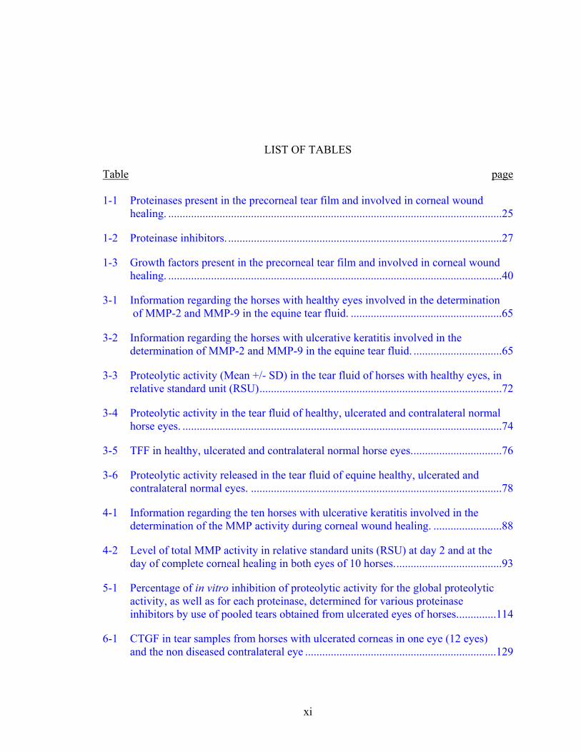
LIST OF TABLES
Table page 1-1 Proteinases present in the precorneal tear film and involved in corneal wound
healing. .....................................................................................................................25
1-2 Proteinase inhibitors. ................................................................................................27
1-3 Growth factors present in the precorneal tear film and involved in corneal wound healing. .....................................................................................................................40
3-1 Information regarding the horses with healthy eyes involved in the determination of MMP-2 and MMP-9 in the equine tear fluid. .....................................................65
3-2 Information regarding the horses with ulcerative keratitis involved in the determination of MMP-2 and MMP-9 in the equine tear fluid. ...............................65
3-3 Proteolytic activity (Mean +/- SD) in the tear fluid of horses with healthy eyes, in relative standard unit (RSU).....................................................................................72
3-4 Proteolytic activity in the tear fluid of healthy, ulcerated and contralateral normal horse eyes. ................................................................................................................74
3-5 TFF in healthy, ulcerated and contralateral normal horse eyes................................76
3-6 Proteolytic activity released in the tear fluid of equine healthy, ulcerated and contralateral normal eyes. ........................................................................................78
4-1 Information regarding the ten horses with ulcerative keratitis involved in the determination of the MMP activity during corneal wound healing. ........................88
4-2 Level of total MMP activity in relative standard units (RSU) at day 2 and at the day of complete corneal healing in both eyes of 10 horses......................................93
5-1 Percentage of in vitro inhibition of proteolytic activity for the global proteolytic activity, as well as for each proteinase, determined for various proteinase inhibitors by use of pooled tears obtained from ulcerated eyes of horses..............114
6-1 CTGF in tear samples from horses with ulcerated corneas in one eye (12 eyes) and the non diseased contralateral eye ...................................................................129
xi

6-2 CTGF from 9 tear samples from horses with ulcerated corneas in one eye (only tears from this eye were analyzed). ........................................................................130
xii

LIST OF FIGURES
Figure page 1-1: Proteinases and corneal wound healing. .................................................................18
1-2: Growth factors and corneal wound healing.............................................................18
1-3: Domain Structure of the Matrix Metalloproteinase family. .....................................29
2-1: Melting ulcer in the right eye of a 12 year old American Quarter horse. ................48
2-2: Western Blot of tear fluids from horses with ulcerated eyes (lane 3) and healthy eyes (lane 4).. ...........................................................................................................51
2-3: Western Blot of tear fluids from horses with ulcerated eyes (lane 3) and healthy eyes (lane 4). ............................................................................................................52
2-4: Immunolocalization of MMP-2 and MMP-9 in healthy equine cornea. ..................53
2-5: Immunolocalization of MMP-2 and MMP-9 in equine lacrimal gland. ..................54
2-6: Immunolocalization of MMP-2 and MMP-9 in equine nictitating membrane gland. 55
2-7: Immunolocalization of MMP-2 and MMP-9 in equine ulcerated cornea. . .............56
3-1: Fungal ulcer in the left eye of a 13 year old American Quarter horse. ....................66
3-2: Tear fluid collection in horses by glass capillary tube. ............................................67
3-3: Gelatin zymography of equine tear fluid samples....................................................70
3-4: Global MMP activity in the tear fluid of horses with healthy eyes by breed...........72
3-5: Global MMP activity in the tear fluid of horses with healthy eyes by sex. .............73
3-6: Global MMP activity in the tear fluid of horses with healthy eyes by age category. ...................................................................................................................73
3-7: Global MMP activity in the tear fluid of horses with healthy eyes and ulcerative keratitis.. ...................................................................................................................75
xiii

3-8: Proteolytic activity in the tear fluid of horses with healthy eyes and ulcerative keratitis by MMP type..............................................................................................75
3-9: TFF and global MMP activity in the tear fluid of horses with healthy eyes and ulcerative keratitis. ...................................................................................................77
4-1: Gelatin zymogram of tear fluids from case 10.. .......................................................91
4-2: Case 1 - A 6 month Thoroughbred colt presented with a melting corneal ulcer in the left eye (OS). ......................................................................................................94
4-3: Case 2 - A 17yo Arabian mare presented with a superficial corneal ulcer in the left eye (OS) and a half thickness stromal corneal ulcer in the right eye (OD). ......94
4-4: Case 3 - A 5yo Thoroughbred mare presented with a fungal keratomalacia in the left eye (OS). ............................................................................................................95
4-5: Case 4 - A 2yo Thoroughbred filly presented a deep corneal ulcer in the left eye (OS).. ........................................................................................................................95
4-6: Case 5 - A 2yo Thoroughbred filly presented a deep corneal ulcer in the right eye (OD). ........................................................................................................................96
4-7: Case 6 - A 7yo Paint gelding presented a corneal ulcer in the right eye (OD). This animal received only medical treatment ..........................................................96
4-8: Case 7 – A 2 month Thoroughbred colt presented a melting corneal ulcer in the right eye (OD). .........................................................................................................97
4-9: Case 8 – A 2 month Thoroughbred colt presented a melting corneal ulcer in the right eye (OD). .........................................................................................................97
4-10: Case 9 – A 14yo American Quarter Horse gelding presented a fungal corneal ulcer in the left eye (OS). .........................................................................................98
4-11: Case 10 – A 12yo Hanovarian stallion presented a melting corneal ulcer in the left eye (OS).. ...........................................................................................................98
5-1: Image analysis of representative zymogram gels to determine proteinase activity in pooled tears obtained from horses with an active corneal ulcer.. .........111
5-2: Gelatin zymogram of untreated pooled tears obtained from ulcerated eyes of horses......................................................................................................................112
5-3: In vitro inhibition of global proteolytic activity for various proteinase inhibitors by gelatin zymography.. .........................................................................................113
xiv
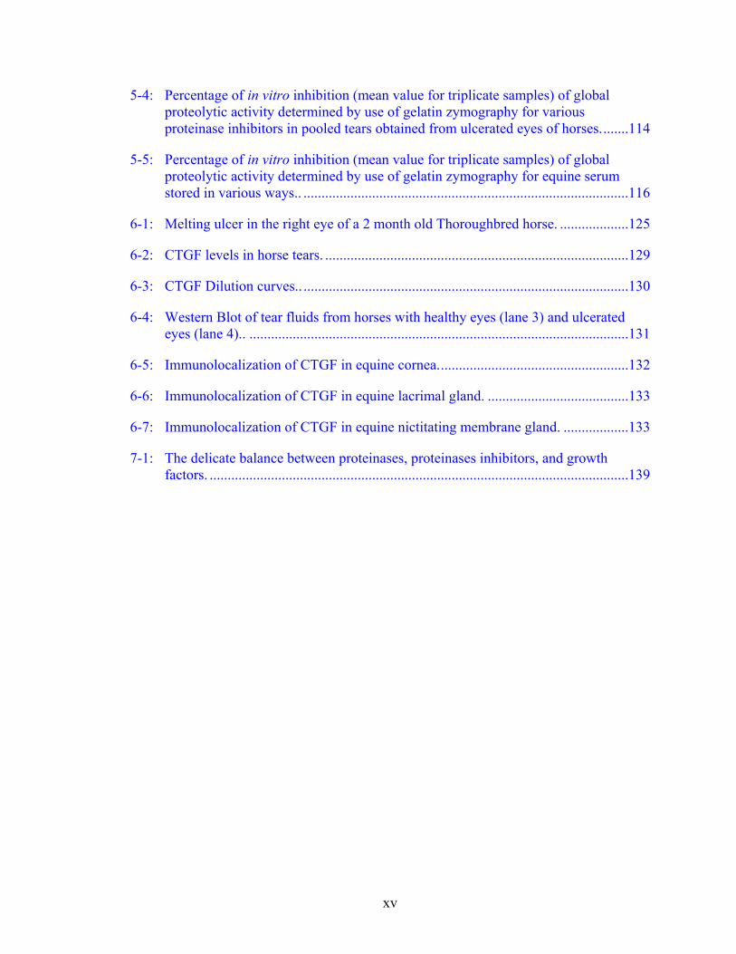
5-4: Percentage of in vitro inhibition (mean value for triplicate samples) of global proteolytic activity determined by use of gelatin zymography for various proteinase inhibitors in pooled tears obtained from ulcerated eyes of horses........114
5-5: Percentage of in vitro inhibition (mean value for triplicate samples) of global proteolytic activity determined by use of gelatin zymography for equine serum stored in various ways.. ..........................................................................................116
6-1: Melting ulcer in the right eye of a 2 month old Thoroughbred horse. ...................125
6-2: CTGF levels in horse tears. ....................................................................................129
6-3: CTGF Dilution curves.. ..........................................................................................130
6-4: Western Blot of tear fluids from horses with healthy eyes (lane 3) and ulcerated eyes (lane 4).. .........................................................................................................131
6-5: Immunolocalization of CTGF in equine cornea.....................................................132
6-6: Immunolocalization of CTGF in equine lacrimal gland. .......................................133
6-7: Immunolocalization of CTGF in equine nictitating membrane gland. ..................133
7-1: The delicate balance between proteinases, proteinases inhibitors, and growth factors. ....................................................................................................................139
xv

KEY TO ABBREVIATIONS
ARVO association for research in vision and ophthalmology
AUC area under curve
CN contralateral normal
CTGF connective tissue growth factor
DIS diseased
ECM extracellular matrix
EDTA ethylenediaminetetraacetic acid
EGF epithelial growth factor
FGF fibroblast growth factor
GAG glycosaminoglycan
GF growth factor
GFR growth factor receptor
H&E hematoxilin and eosin
HGF hepatocyte growth factor
IFN-γ Interferon gamma
IL interleukin
kDa kilodalton
KGF keratocyte growth factor
MMP matrix metalloproteinase
MT-MMP membrane-type matrix metalloproteinase
xvi

N normal
NAC N-acetylcysteine
NE neutrophil elastase
OD right eye
OS left eye
PA plasmin
PBS phosphate buffer solution
PDGF platelet-derived growth factor
PMN polymorphonuclear
PTF precorneal tear film
QH American quarter horse
RSU relative standard unit
u-PA urokinase-type plasminogen activator
t-PA tissue-type plasminogen activator
α1-PI α1-proteinase inhibitor
TB thoroughbred
TBS tris-buffered solution
TFF tear fluid flow
TGF-α transforming growth factor alpha
TGF-β transforming growth factor beta
TIMP tissue inhibitor of matrix metalloproteinase
TNF-α Tumor necrosis factor alpha
TWH Tennessee walking horse
xvii

VEGF vascular endothelial growth factor
xviii
Abstract of Dissertation Presented to the Graduate School of the University of Florida in Partial Fulfillment of the Requirements for the Degree of Doctor of Philosophy
MATRIX METALLOPROTEINASE 2, MATRIX METALLOPROTEINASE 9,AND CONNECTIVE TISSUE GROWTH FACTOR IN THE EQUINE TEAR FLUID:
POSSIBLE IMPLICATIONS IN CORNEAL WOUND HEALING By
Franck J. Ollivier
May 2004
Chair: Dennis E. Brooks Major Department: Veterinary Medicine
The goals of this study were to investigate the presence of matrix metalloproteinase
2 (MMP-2), matrix metalloproteinase 9 (MMP-9), and connective tissue growth factor
(CTGF) in the equine tear film, cornea, and lacrimal glands; to compare the levels of
MMP-2, MMP-9 , and CTGF in the tear fluid of horses with healthy eyes and horses with
ulcerative keratitis; to document the changes in MMP-2 and MMP-9 levels in horse tear
film during corneal healing; and to investigate the in vitro effects of various proteinase
inhibitors on the activity of MMP-2 and MMP-9 isolated from the equine tear film.
This study demonstrates by immunohistochemistry the expression of MMP-2
protein in the equine lacrimal gland and the gland of the nictitating membrane. The
expression of MMP-2 and MMP-9 proteins was also found in the healthy cornea of the
horse and was increased in the ulcerated equine cornea.
In this study, the level of MMP-2 and MMP-9 activity was determined in a total
330 tear fluid samples by the use of gelatin zymography. MMP proteolytic activity was
xix

detected in all tear samples and was significantly increased in the tear fluid of horses with
ulcerative keratitis (1.92 ± 1.06 RSU) compared to normal (0.62± 0.47 RSU). Based on
the collection and analysis of a total of 124 serial tear fluid samples in 10 horses with
ulcerative keratitis, we documented for the first time that total MMP activity decreases in
equine tears as corneal epithelial and stromal healing occur.
We documented, by gelatin zymography, a high amount of inhibition of equine
MMP activity in vitro by the use of potassium edatate diaminetetrataacetatic acid,
doxycycline, N-acetylcysteine, equine serum, ilomostat, and α-1-proteinase inhibitor.
The level of CTGF was determined by enzyme immunoassay in a total 64 tear fluid
samples in this study and the molecule was detected in 39 of the samples. These data
demonstrate that CTGF was present in the equine tear film. This study also indicates by
immunohistochemistry the expression of CTGF protein in the healthy cornea, the
lacrimal gland and the gland of the nictitating membrane of the horse (n=10).
xx

CHAPTER 1 INTRODUCTION
The Cornea and the Precorneal Tear Film (PTF)
The cornea is the gateway of the images into the eye. It is the most powerful
refractive ocular structure and must remain transparent. The optical properties of the
cornea include clarity, surface smoothness, and refractive index (Nishida, 1997;
Samuelson, 1999). The health of the cornea is influenced by the aqueous humor, the
intraocular pressure, the eyelids and the precorneal tear film. There are three
classifications of tear production: basic, reflex and psychic. Continuous or “basal” tears
are produced at a constant level and permit normal functioning of the precorneal tear
film. Additional tear production is stimulated by reflex response to any irritation of the
cornea, conjunctiva or nasal mucosa. Human beings are the only species for which
psychic tear stimulation has been proven.
Structure and Function of the Precorneal Tear Film
The precorneal tear film (PTF) is not truly part of the cornea but is anatomically
and functionally intimately associated with the cornea. The PTF is usually described as a
superimposition of three structurally and functionally unique layers: the outer lipid layer,
the intermediate aqueous layer, and the inner mucin layer. However, current belief is that
the layers are not so distinct and the PTF is a mucin-dominated gel (Sack et al., 2000).
The thickness of the PTF is not known in horses but has been estimated at 3 µm in human
beings (King-smith et al., 2000).
1

2
The superficial outer lipid layer of the PTF is relatively thin and composed of oily
materials (waxy and cholesterol esters) and phospholipids. This lipid layer retards the
evaporation of the underlying aqueous layer between eyelid blinks, and reduces loss of
PTF over the eyelids through increases in surface tension between the PTF and the cornea
(Chow and Gilbard, 1997; Gum et al., 1999). The lipid phase also promotes a stable and
even distribution of tear film over the cornea.
The middle layer is the aqueous tear fluid layer. It is the thickest of the three layers
and accounts for the majority of the volume. The aqueous layer is a complex mixture of
ions, small molecules, glycoproteins, and proteins including enzymes (proteinases),
immunoglobulins, cytokines, and growth factors. There is a paucity of information
regarding the composition of equine tears. The pH was recently measured in conscious
horses with indicator paper and reported to be 8.33 +/- 0.15 in the range of 8.0- 8.6
(Lowe and Crispin, 2003). In horses, the main source of tears is the orbital lacrimal gland
with a relatively minimal contribution from the nictitating membrane gland (Moore,
1990; Williams et al., 1979). Both of these glands are tubuloacinar and histologically
similar. The lacrimal gland is located dorsolaterally, in the orbit, against the supraorbital
part of the frontal bone, and the nictitating membrane gland is at the base of the T-shaped
cartilage (Samuelson, 1999). The aqueous layer flushes foreign material away from the
cornea and the conjunctiva, contributing to the mechanical barrier function. Seventy-five
percent of the PTF is evacuated through the nasolacrimal drainage system. The PTF
enters the puncta by capillary attraction and by eyelid motion which results in lower
pressure within the lacrimal sac that acts subsequently as a “lacrimal pump” (Chow and
Gilbard, 1997; Gum et al. 1999; Lemp and Wolfley, 1992). This layer also lubricates the

3
passage of the eyelids and the nictitating membrane over the cornea. Tear drainage into
the nasolacrimal system serves also to remove waste products (dissolved carbon dioxide
and lactic acid). The aqueous layer also contains antimicrobial compounds (lysozymes,
lactoferrin) and antibodies (IgA, IgG, IgM, IgGT) which chemically protect the cornea
(Chow and Gilbard, 1997; Gum et al., 1999; Lemp and Wolfley, 1992; Martin et al.,
1997; Marts et al., 1977; Nishida, 1997), and permit transfer of inflammatory cells such
as polymorphonuclear cells (PMNs) (Lemp and Wolfley, 1992). The PTF contains
soluble proteins that contribute to corneal health, and enhance defense mechanisms
(proteinases, immunoglobulins, cytokines, and growth factors). The aqueous layer also
delivers nutrients (water, glucose, electrolytes) to the avascular cornea and facilitates
transfer of atmospheric oxygen (Gum et al., 1999; Lemp and Wolfley, 1992), Finally, the
aqueous layer enhances the optical properties of the cornea by providing an optically
smooth surface and by aiding in regulating corneal hydration (Chow and Gilbard, 1997;
Gum et al., 1999; Nishida, 1997). The state of relative dehydration, or deturgescence, is
dependent upon osmotic forces between the PTF, the aqueous humor, and probably the
corneal stroma (Gum et al., 1999). The corneal surface is responsible for most of the light
refraction that occurs in the eye, because of the large difference in the indices of
refraction between air and cornea, and the angle of incidence of incoming light (Ofri,
1999). The image quality is based upon the regularity of the corneal epithelial surface and
quality of the PTF (Nishida, 1997). Furthermore, control of corneal hydration is crucial to
maintain the homogenous organization of stromal collagen lamellae and thereby corneal
transparency (Gum et al., 1999; Nishida, 1997).
4
The inner mucin layer consists of mucoproteins derived from goblet cells located in
the conjunctiva, predominantly in the fornices (Chow and Gilbard, 1997; Samuelson,
1999). The mucoproteins are bipolar molecules that resemble a gel and bind the PTF
aqueous layer (hydrophilic and lipophobic) to the epithelial surface glycocalyx (lipophilic
and hydrophobic). This ensures the stability of the PTF and permits redistribution of the
PTF after blinking, thus maintaining an optically smooth surface (Chow and Gilbard,
1997; Moore, 1990). The mucin layer also participates in the corneal defense because
bacteria and foreign bodies may be entrapped within mucoproteins and the mucin harbors
immunoglobulins (IgA) and lyzozyme.
The PTF should be perceived as a three-phase system whose components are in
dynamic equilibrium (Sack et al., 2000). The PTF serves to lubricate the ocular surface
and prevent desiccation. Tear flow, coupled with the cleansing action of the blink, serves
as a critical element in an essentially passive barrier defense, and is designed to protect
the cornea from effects of trauma, pathogens and noxious agents and to remove waste
products. Besides its nutritional value, the PTF also plays an important role in the anti-
microbial, anti-inflammatory and proteolytic activities present at the corneal surface. It is
important to note that in addition to the production of normal secretory components, the
formation of the composite, tristratified tear film depends on eyelid integrity, normal
ocular motility, and an intact blink mechanism (Sack et al., 2000). Since the cornea is in
close contact with the preocular tear film and the aqueous fluid, the various kinds of
proteinases, proteinase inhibitors, growth factors and cytokines in the tear film and
aqueous might play an important role in the turnover of the corneal cells and wound
healing of the cornea.

5
Structure and Function of the Cornea
The unique organization of the cornea permits clarity, a smooth transparent
refractive surface, tectonic strength, differential permeability, and protection (Nishida,
1997; Pepose and Ubels, 1992).
The cornea represents the most powerful refractive surface of the eye (Gum et al.,
1999; Nishida, 1997). It contributes to 48D of the approximately 60D the refractive
power of the human eye (Ofri, 1999; Pepose and Ubels, 1992). This important
contribution of the cornea is due to the large difference in refractive indices as the light
passes from air into the cornea. The cornea acts like a convex lens as it converges light
and its refractive power depends mainly on its curvature (Nishida, 1997; Ofri, 1999).
Similarly, in large eyes which are characterized by flat corneas the refractive power of
the lens is reduced (48D in humans, 43D in cats, 38-43D in dogs and only 16-20D in
horses) (Ofri, 1999).
The transmission of the light through the cornea depends on the wavelength of the
light and the angle of incidence. Obviously transmission of the light would be reduced by
corneal opacities and therefore the cornea should remain transparent (Gum et al., 1999;
Nishida, 1997; Ofri, 1999). The transparency is a crucial factor to be maintained in order
for the cornea to fulfill its required optical properties, and it is possible because of the
following structural features of the cornea: the lack of vessel and blood cells (the normal
cornea is avascular (Gum et al.,1999; Nishida, 1997; Pepose and Ubels, 1992), the lack of
pigment, the absence of keratinized cells (the corneal epithelium is simple, stratified,
squamous and non keratinized), the presence of exquisitely sensitive nerves with free
endings. Although the cornea is heavily innervated (Nishida, 1997; Pepose and Ubels,
1992), the specific arrangement of the collagen fibrils in the stroma (Nishida, 1997), the

6
presence of mechanisms that regulate the hydration of corneal stroma (Gum et al., 1999;
Nishida, 1997; Pepose and Ubels, 1992), the anatomic integrity of the epithelium and
endothelium that represent physical barriers against the influx of tears and aqueous
humor (Gum et al., 1992; Nishida, 1997; Samuelson, 1999), and the role as “pumps” of
the epithelium and endothelium for the maintenance of deturgescence in the cornea (Gum
et al., 1999; Pepose and Ubels, 1992; Samuelson, 1999). Finally, the cornea is part of the
fibrous coat of the eye and therefore it participates in the maintenance of the eyeball
shape and organization (Samuelson, 1999). The mechanical strength of the cornea is
provided by its stromal collagen matrix.
The equine cornea is 793 to 893 microns thick at the center (Andrew et al., 2001;
van der Woerdt et al., 1995) and composed of three distinct layers: the outermost
multilayered epithelium and its basement membrane, the middle stroma, the Descemet’s
membrane and the innermost endothelium.
The corneal epithelium is arranged in ten to fifteen cell layers: a single layer of
mitotically active columnar basal cells, three to six layers of wing cells, and five to ten
outer flattened layers of squamous superficial cells (the most differentiated epithelial
cells) (Samuelson, 1999; Schultz, 1997). The basal cells of the epithelium are firmly
attached to its basal lamina (i.e., its basement membrane) by hemidesmosomes,
anchoring collagen fibrils and the glycoprotein laminin. Type IV, VI and VII collagen
contribute to the basement membrane (Nishida, 1997) as well as laminin, hyaluronans,
fibrin and fibronectin (Friend et al., 1994; Klyce et al., 1998). The hemidesmosomes
attach the basal cells to the basement membrane which in turns serve to anchor the
epithelium to the stroma. The arrangement of the hemidesmosomes varies among

7
different species. The epithelial cells have good regenerative powers (basal turnover is
approximately 7 days), but after removal of the basal lamina, weeks to months may be
necessary for it to completely reestablish and, until the basement membrane is completely
reformed, the epithelium can be easily removed from the stroma (Samuelson, 1999). The
corneal epithelium is maintained by a constant cycle of shedding of superficial cells,
basal cells division, and renewal of basal cells by centripetal migration of new basal cells
originating from the limbal stem cells (Neaderland et al., 1987; Nishida, 1997; Pepose
and Ubels, 1992)
The thickest layer (90% of the corneal thickness), the corneal stroma, is composed
of a few keratocytes and fibrocytes, nerve fibers and a large amount of transparent,
almost structureless lamellae of fibrous tissue. These lamellae lie in parallel sheets and
split easily into surgical planes. Between the lamella are fixed and infrequent wandering
cells. The fixed cells are fibrocytes which are called keratocytes and the extension of
these cells contributes to formation and maintenance of the stromal lamellae. The
keratocytes synthesize collagen molecules (pro-collagen), glycosaminoglycans as well as
collagen degradative enzymes such as matrix metalloproteinases (MMPs) (Nishida,
1997). Keratocytes in the normal cornea are quiescent and serve primarily to maintain the
slow turnover of extracellular components but they may be activated easily by various
types of insults to the corneal stroma. Wandering cells are usually leukocytes that have
migrated from the limbus. The lamellae are parallel bundles of collagen fibrils, with each
lamella running the entire diameter of the cornea. All the collagen fibrils within a lamella
are parallel, but between lamellae, they vary greatly in direction. The precise overall
organization of the corneal stroma is the most important factor in maintaining corneal
8
clarity (Friend et al., 1994; Klyce et al., 1998). The mean diameter of each collagen fiber
and the mean distance between these fibers are homogenous and measure less than half of
the wavelength of visible light. This anatomic relationship is thought to be responsible for
the fact that incident ray scattered by each collagen fiber is cancelled by interference of
the other scattered ray, allowing light to pass through the cornea (Gum et al., 1999;
Nishida, 1997). If the diameter or the distance between collagen fibers varies (as in
fibrosis or edema), the cornea loses its transparency and there is a random scattering of
incident rays (Nishida, 1997). The bulk of the corneal stroma is composed of thin
uniformly positioned collagen fibrils embedded in glycosaminoglycans that form an
extracellular matrix (ECM). The stromal ECM consists of collagen fibrils (native type I,
III, V, VI and XII collagens), stromal glycosaminoglycans or GAGs (keratan sulfates,
dermatan sulfates, chondroitin sulfates), and glycoproteins. Collagen type I is the most
common. Type VI appears to play a role in cell-matrix interactions, which would be
especially important during repair. Type III and XII are both believed to be
developmental forms.
The Descemet’s membrane is a homogenous, acellular membrane forming an inner
protective boundary within the cornea. It is actually an exaggerated basement membrane
of the posterior endothelium (20 µm in thickness). To some degree, its composition is
similar to that of the trabeculum of the iridocorneal angle. This ever-thickening basement
membrane contains a number of collagen types I, III, IV, V, and VI, and also type VIII
collagen (which is not found elsewhere in the cornea), laminin, fibronectin and heparan
sulfates (Friend et al., 1994; Samuelson, 1999).The corneal endothelium rests on the
Descemet’s membrane.

9
The corneal endothelium produces Descemet’s membrane and contains an energy
dependent pump to maintain corneal deturgescence. The corneal endothelium is only one
layer of cells that does not proliferate in humans, monkeys, and cats but does proliferate
in rabbits. Endothelial cell count decreases with age and with any trauma, and a critical
loss of endothelium may lead to loss of corneal clarity.
Corneal Healing and Scarring
Horses have large, prominent eyes that are often subject to traumatic injury and
resultant corneal infection. Ulcerative keratitis is a common and often vision-threatening
condition in horses. Superficial, non-infected ulcers in horses generally heal quickly and
without complication, whereas stromal degradation in deep or infected ulcers can rapidly
and dramatically progress to corneal perforation in horses in less than 24 hours.
When the cornea is injured, multiple systems are activated, which produce a series
of complex and coordinated cellular processes that ultimately result in a healed corneal
wound. Healing of corneal wounds is an exceptionally complex process involving the
integrated actions of multiple proteinases (Table 1-1), growth factors (Table 1-3), and
cytokines produced by epithelial cells, stromal keratocytes, inflammatory cells, and
lacrimal glands. Multiple autocrine and paracrine interactions occur between epithelial
cells and activated stromal fibroblast, and the exocrine actions of factors secreted by
lacrimal gland cell into the PTF (Figures 1-1 and 1-2)
Overview on Wound Healing and Corneal Healing
Wound healing begins at the moment an injury occurs. The sequence of events in
the progress of wound healing is as follows: release of soluble chemotactic factors which
attract inflammatory cells to the injury site, influx of neutrophils and monocytes to
neutralize bacteria and/or fungi in the wound site, the debridement of connective tissue
10
matrix damage by macrophages, the initiation of neovascularisation, and the stimulation
of cell proliferation and connective tissue matrix remodeling. These events occur
sequentially until normal tissue architecture is restored. Many times this series of events
leads to restoration of normal tissue structure and functions, but sometimes fibrotic
disorders occur and scarring results in a loss of function of the particular tissue or organ.
When a corneal wound occurs, keratocytes around the wound edge die creating a
hypocellular zone (Wachtlin et al., 1999). Chemotactic factors such as PDGF, TGF-α, and
TGF-β are released and attract inflammatory cells (Table 1-3), resulting in initiation of
reepithelialisation, contraction of connective tissue and stimulation of angiogenesis
(Schultz, 1997). The first leukocytes recruited to the site of injury are neutrophils which
attack bacteria and /or fungus that may have been introduced into the tissue at the time of
injury. Levels of neutrophils begin to decline and macrophages begin to take over as the
dominant cell type in the wound. Macrophages function in the degradation and the
removal of tissue debris in preparation for reparative phases of wound healing. Activated
platelets release several growth factors which result in the recruitment of neutrophils and
monocytes. Beyond the site of injury, quiescent keratocytes become activated into
fibroblasts and migrate to the site of injury. This migration is followed by cell
proliferation, and finally deposition of ECM components. Many growth factors and
cytokines have been implicated in stimulating synthesis of ECM components, cell
proliferation and migration and angiogenesis (TGF-α, TGF-β, PDGF, FGF) (Table 1-3).
These growth factors have control over the complex processes in wound healing
involving migration, mitosis and differentiation of epithelial and stromal cells (Schultz et
al., 1992). The fibroblasts deposit additional reparative collagen and eventually

11
synthesize enough ECM to form a scar, replacing the damage tissue. All these events in
the process of inflammation must be reversed for the tissue architecture to return to
normal. For this to happen, the removal of the inflammatory mediators which were
generated must occur. Infiltration of monocytes and leukocytes must decrease in order for
this to occur. Furthermore, removal of extravasated fluid, protein, cellular debris,
granulocytes and macrophages occurs as the wound regeneration process continues
(Daoud et al., 1985).
Histological Events of Corneal Healing
Traditionally, corneal wound healing is divided into the healing of those injuries
that affect only the epithelium and those resulting in substantial loss of stroma up to the
Descemet’s membrane.
Epithelial wound healing
Three components are involved in healing of the epithelial surface of the cornea:
cell migration to cover the injured area (sliding), mitosis to reconstitute the normal
number of epithelial cells and normalization (differentiation) of the corneal epithelial
cells (Neaderland et al., 1987; Peiffer et al., 1999; Samuelson, 1999; Schultz, 1997).
In the epithelial wound healing, epithelial migration is the initial step for the
successful and complete resurfacing of defects (Neaderland et al., 1987; Peiffer et al.,
1999; Pepose and Ubels, 1992). After an epithelial injury, signals from disrupted cells or
signals generated by exposure of the basement membrane are sent to the surrounding
intact epithelial cells. Mitosis ceases (for 96 to 120 hours) and the basal cells at the
wound edge retract. Then, there is a phase during which the cell cytoskeleton and
intercellular junction of these cells are modified (Cameron, 1997). These events allow the
basal cells to begin to migrate by ameboid movement to cover the defect within an hour
12
after the injury. The edges of the cell membrane ruffle and extend pseudopodia onto the
denuded extracellular matrix (ECM), toward the center of the wound (Nishida, 1997;
Pepose and Ubels, 1992). Two types of epithelial movements for covering the denuded
area in the cornea have been observed: advance groups of sliding monolayered epithelial
basal cells and a subsequent landslide-like mass movement of the epithelium (basal and
wing cells). The sliding movement precedes the landslide-like mass movement of the
epithelium which finally covers the denuded area. During these movements it is
important that the cells remain adherent to the neighboring cells or to the ECM. The
advance group of the individual cells simply spread over a provisional ECM.
Polymorphonuclear neutrophils arrive from the tear film and begin removing remnants of
destroyed cells (Schultz, 1997). As mentioned previously, cell migration requires
coordinated changes of both the cell cytoskeleton and the cell surface attachments to
adjacent cells and substratum. Cell migration is accomplished by rapidly changing the
cytoskeleton in order to build the scaffold towards the leading edge of the cell, which
forms pseudopods, while disassembling the scaffold at the trailing edge of the cell. At the
interface between the dead and dying cells, the intercellular junctions between epithelial
cells loosen but do not completely disengage. The direction of the migration are
determined by chemical signals in both the fluid environment of the cell (chemotaxis)
and in the surface (ECM) to which the cells is attached (haptotaxis).The stimulation
factor should exist in a gradient in order to induce a cell movement and then the cell will
migrate toward the area of highest concentration (Cameron, 1997). The basal cells
migrate until contact inhibition of migration is established by physical contact with
adjacent cells (Cameron, 1997).

13
Once the injured surface has again been covered, mitosis occurs to restore the
epithelium to its normal configuration (thickness) (Pepose and Ubels, 1992; Samuelson,
1999). Mitotic replication of the basal cells begins about 24 after epithelial injury: new
basal cells are formed to replace those that are migrating forward, and the mitosis occurs
in a zone 3 to 5 mm behind the leading edge of the migrating cells (Schultz, 1997). The
mitosis continues and may result in a transient corneal epithelial hyperplasia before
normalization occurs (Peiffer et al., 1999). Repeated erosions or large defects might
overwhelm the replicative capabilities of the transient amplifying population of basal
cells adjacent to the site of injury, thus requiring participation of the permanent
replicative cells at the limbus (stem cells) (Samuelson, 1999; Peiffer et al., 1999).
When normal thickness is reestablished, the highly differentiated cellular
characteristics are re-formed (Cameron, 1997). The superficial cells terminally
differentiate by synthesizing keratin protein which helps reestablish the barrier properties
of the epithelium (Schultz, 1997). Intercellular attachments are also reestablished as well
as the basal surface contacts (formation of new hemidesmosomes in the basal cells)
(Schultz, 1997). When a corneal abrasion is limited to the epithelium and the basement
membrane is not damaged, a normal epithelium with adhesion complexes is formed soon
after healing. If the basement membrane is removed or altered, the epithelium must lay
down a new basement membrane following healing, and development of normal adhesion
complexes is delayed for several months (Pepose and Ubels, 1992).
Stromal and endothelial wound healing
Deep corneal ulcers heal with a combination of epithelial sliding and replication
(previously described), as well as stromal wound healing. Stromal healing involves the
re-synthesis and crosslinking of collagen, alterations in proteoglycans synthesis, and

14
gradual wound remodeling leading to the restoration of tensile strength (Pepose and
Ubels, 1992). The earliest event in stromal corneal wound healing appears to be the
deposition of fibrin, fibronectin and other elements of the clotting cascade into the
wound. Some keratocytes immediately adjacent to the wound margin undergo apoptosis
and others begin releasing enzymes involved in the degradation of damaged
proteoglycans and collagen lamellae at the wound edge. There is also an influx of PMNs
and monocytes in the wound within a few hours that release proteinases to cause the
proteolytic debridement of necrotic cellular and extracellular debris. Within a few hours
the adjacent keratocytes become activated and begin protein synthesis, and within 3 days
the keratocytes are able to secrete collagens and GAGs. There are also proliferation and
activation of keratocytes beyond the injury site that migrate then to the site of injury. The
proliferation, mitosis, migration and activation of the keratocytes are under influence of
many factors such as growth factors and cytokines. Once at the site on injury that has
been cleared of debris, keratocytes produce collagens and GAGs. A denervated cornea is
at risk for developing epithelial defects. Corneal nerves regenerate after corneal injury,
although the process requires months to years. The nerves regenerate from unwounded
peripheral nerve trunks. The orientation of the nerve fibers is generally random, and
corneal sensitivity is seldom returned to normal (Cameron, 1997). The healing of the
corneal stromal wound is slower than in other connective tissues presumably because of
the lack of blood vessels (Fagerholm, 2000; Schultz, 1997).
Unfortunately, the endothelial cells do not respond to cell loss as quickly as
epithelial cells, and endothelial healing varies with both age and species. For this reason
when endothelial cells are lost, the defect must be covered by the spreading of cells from

15
areas adjacent to the wound to cover the wounded area under influence of growth factors
such as TGF-β, b-FGF, and EGF (Table 1-3) (Friend et al., 1994; Samuelson, 1999).
Biochemical Mechanisms of Corneal Healing
Molecular biology of epithelial wound healing
In the epithelial wound healing, epithelial migration is the initial step for the
successful and complete resurfacing of defects. Several biochemical and synthetic events
are involved in cell migration. Migration requires energy and one of the earliest changes
that occurs during healing is the depletion of glycogen from the cells at the leading edge
of migration. Enlargement of the cells is apparently the result of an increase in cell water
content, and the actual movement of cells is calcium dependent, as calmodulin inhibition
stop migration by preventing microfilament assembly. The processes that initiate
migration are mediated by cyclic AMP. Migration of corneal epithelium during wound
repair is also accompanied by an increase in protein synthesis. A specific increase in
synthesis has been demonstrated for the cytoplasmic protein vinculin (Pepose and Ubels,
1992). The actin component is prominent within the pseudopodia of the basal cells. The
actin-containing microfilament system is attached by vinculin to the transmembrane
integrin receptors, which selectively bind to ECM proteins such as fibronectin, laminin,
and collagens (Schultz, 1997). During the first phase of epithelial wound healing,
fibronectin plays an essential role as a provisional, temporal ECM. In the normal
unwounded cornea, type IV collagen, laminin, and heparan sulfate proteoglycan are the
major components of the basement membrane but not fibronectin (Cameron, 1997;
Nishida, 1997; Schultz, 1997). Fibronectin appears shortly after the epithelial injury
(produced by adjacent cells or delivered through tears, aqueous humor). Basal cells attach
and spread over the fibronectin matrix and fibronectin disappears once the epithelial

16
wound is healed. Fibronectin stimulates epithelial migration and has chemotactic and
haptotactic activities for the corneal epithelial cells. The integrins are the receptors for
fibronectin at the surface of corneal cells. Fibronectin provides a suitable ECM for cell
attachment and migration of the epithelial cells that become more sensitive to fibronectin
through increased expression of integrin. Epithelial growth factor (EGF) and IL-6
regulate integrin expression in corneal epithelial cells (up-regulation), and by this up
regulation, they stimulate corneal cell migration. Other components of the ECM (of the
basement membrane) such as heparan sulfate and laminin play a role in wound healing by
influencing cell adhesion and migration.
The motility of the basal cells must involve the simultaneous formation and
destruction of attachments between proteins of the plasma membrane of epithelial cells
(integrin receptors) and components of the ECM such as fibronectin, and laminin.
Therefore proteinases also play a key role in epithelial cell migration by breaking down
the attachments: matrix metalloproteinases such as MMP-1, MMP-2 and MMP-9,
fibroblast and neutrophil collagenases, stromelysins, as well as serine proteases such as
plasminogen activators, plasmin, and neutrophil elastase have been shown to be involved
in wound healing (Table 1-1) (Cameron, 1997; Schultz, 1997).
Corneal epithelial cells mitosis and sliding are both strongly stimulated by EGF and
Keratocyte growth factor (KGF) that are normally present in tears, and also produced by
the corneal epithelial cells themselves following injury (Nishida, 1997; Peiffer et al.,
1999). On the other end, some member of the TGF-ß family inhibits cell proliferation and
therefore, counteracts the stimulatory effect of EGF, but most of the TGF-ß family does
not affect the migration of the epithelial cells (Table 1-3) (Nishida, 1997).
17
Molecular biology of stromal wound healing
The first step in stromal healing is similar to the epithelial healing with the
deposition of fibrin, fibronectin and other elements of the clotting cascade into the
wound. The debridement of the wound occurs very quickly after the injury. The
degradation of the necrotic cellular and extracellular debris (damaged proteoglycans and
collagen lamellae) involves many proteinases such as matrix metalloproteinases,
collagenases and urokinase-type plasminogen activator (that converts plasminogen to
plasmin that causes destruction of fibronectin). These various proteinases are produced
and released by keratocytes as well as inflammatory cells (Table 1-1).
Once the injury site has been cleared of debris, activated keratocytes produce
collagens (type I, type III), and GAGs (keratan sulfate) under the influence of growth
factors such as TGF-ß (Table 1-3). To achieve a successful wound healing of the stroma,
the fibrous components must be reestablished in a way that follows, at least to some
degree, normal development. However, the diameter and other characteristics of the
reparative collagen are quite different: the collagen diameter is larger and the individual
collagen fibers are more variable in caliber. Similarly the new proteoglycans differ in
character and proportion, and the populations of proteoglycans (i.e., GAGs) are selective
to keep the collagen fibrils organized and properly sized. Therefore, if the relative
concentrations of GAGs are substantially altered during wounding, the possibility of
reforming the fibrous architecture for needed transparency is reduced and will remain so
until the normal proteoglycan environment is rebuilt. The initial extracellular matrix of
the scar produced is the same as the final or resting scar (Schultz, 1997).

18
Figure 1-1: Proteinases and corneal wound healing.
Figure 1-2: Growth factors and corneal wound healing.

19
Corneal Scarring
As we mentioned earlier, corneal transparency is determined in large part by the
structure of the stromal ECM. When the precise structure of the stroma is disrupted, it
can never be properly restored. Corneal stroma is not regenerated but repaired: it is
replaced with unspecialized tissue that creates the scar. The strength of corneal scars and
the surrounding tissue never reaches that of the uninjured cornea. It is estimated that the
tensile strength returns to 70% of normal native (Cameron, 1997). The repair tissue
matrix contains component molecules not usually present in stroma, particularly
fibronectin, and the newly synthesized collagen fibrils are thicker than those of the
normal stroma and variable in size. Formation of the corneal scar is a dynamic process
and clinical changes can be noted up to five years after corneal wound or incision
(Eiferman, 1992). Dermatan sulfates and keratin sulfate proteoglycans from the adjacent
matrix as well as newly synthesized proteoglycans accumulate with the scar. Collagen
type I, III, V, and VI are also quite predominant within the scar (Ljubimov et al., 1998).
Some improvement in the transparency of repair tissue can occur over the long term
through the progressive remodeling of the repair tissue matrix and the MMPs appear to
be involved in this process. Remodeling results in the loss of fibrin and fibronectin and
the position of fibrils with a more uniform diameter and a more orderly arrangement.
Some of the reparative collagen and proteoglycans are selectively catabolized by specific
proteinases (Table 1-1) and new collagen and proteoglycans are then selectively
synthesized in a more advantageous orientation, quantity or proportion (Cameron, 1997).
In summary, corneal scarring is the result of abundant cell synthesis (collagen,
fibronectin, laminin) by fibroblasts which proliferate and differentiate within the

20
provisional matrix. Stromal scarring can have negative impacts on the refraction or even
the light transmission at the level of the cornea.
Proteinases, Proteinase Inhibitors
Proteinases in The PTF: Types and Origins
There are four major classes of proteinases based on the mechanism of catalysis,
including the cysteine proteinases (thiols), the aspartate proteinases (acidic), the serine
proteinases and the matrix metalloproteinases (MMPs) (Table 1-1) (Birkedal-Hansen et
al., 1993). Among these families, two play a major role in corneal physiopathology: the
serine proteinases and the MMPs (Fini et al., 1990; Fini, 1998; Matsubara et al., 1991b;
Woessner, 1999) (Figure 1-1).
Serine proteinases
The serine proteinase family includes neutrophil elastase, and the plasmin and the
plasminogen activators (Table 1-1).
Plasminogen, the inactive form of plasmin, is present in the blood plasma and in
interstitial fluids such as tears and the aqueous humor. It accumulates in inflamed tissues.
Plasmin and plasminogen are present in the healthy cornea. Conversion to the active form
occurs by cleavage at the N terminus mediated by one of the two plasminogen activators
(PA): the urokinase type (u-PA) or the tissue type (t-PA) (Stevens et al., 1992). PA can
be found at the surface of resident tissue cells and have been reported to be produced by
the cornea. However, the tear plasminogen activator seems to be predominantly of the u-
PA (Tozser and Berta, 1990). Plasmin has a wide substrate specificity and cleaves a range
of extracellular matrix substrates such as fibrin, fibronectin, laminin and it also activates
latent collagenases (MMP-1, 3, 9, 14) (Table 1-1) (Berta et al., 1990; Cejkova et al.,
1993; Salonen et al., 1987; Stevens et al., 1992). The activity of plasmin is regulated in
21
the extracellular space by plasma and cellular proteinase inhibitors (α1-proteinase
inhibitor, α2-anti plasmin, α2-macroglobulin) that are present in the interstitial fluids but
also synthesized by the resident tissue cells (Table 1-2). Elevated plasmin activity is
considered to be harmful from the point of view of the initiation (Wang et al., 1985) as
well as the development of corneal destructive processes. However, it is also suggested
that a certain level of serine proteases (plasmin and plasminogen activators) is necessary
for the repair process (Hayashi et al., 1991). The levels of plasmin activity depend on the
severity of the corneal injury (Cejkova et al., 1993).
Neutrophil elastase (NE) is the most abundant serine proteinase in human tears, is
found in dog and horse tears (Sathe et al., 1998; Strubbe et al., 2000; Watanabe et al.,
1990), and is synthesized by polymorphonuclear leukocytes and macrophages (Sakata et
al., 1997). It degrades native III and IV collagen as well as corneal ECM compounds such
as laminin, fibronectin (Barletta et al., 1996; Cejkova, 1998; Paterson et al., 1994;
Watanabe et al., 1990) (Table 1-1).
Matrix metalloproteinases (MMPs)
Enzymes of the matrix metalloproteinases (MMP) family are thought to play the
major role in ECM remodeling (Birkedal-Hansen et al., 1993; Woessner, 1999). They are
now 20 characterized members of this family of zinc containing proteinases (Table 1-1).
Substrates include essentially all known matrix molecules, including interstitial collagens
as well as the proteinases themselves or their inhibitors. MMPs are synthesized and
secreted as inactive pro-enzymes that are activated in the extracellular space by cleavage
of a portion at the N terminus (Birkedal-Hansen et al., 1993). The synthesis of the pro-
enzymes (i.e. latent forms) has been shown to be under influence of many factors
including cytokines and growth factors: IL-1, TNF-α, EGF have been shown to induce

22
the synthesis of MMP-1,-3,-9 whereas IL-4, Il-6, IFN-γ, TGF-β inhibit the synthesis of
MMP-1, -3 (Twining, 1994) (Tables 1-1 and 1-3). The pro-enzyme activation occurs by
means of proteolytic cascades, which probably differs for each enzyme and has been
partially characterized (plasmin, membrane -type MMPs) (Table 1-1). Once activated,
MMPs require Ca2+ for their stability and Zn2+ as a cofactor (Fini and Girard, 1990;
Twining, 1994). MMPs are regulated by non specific inhibitors such as α2-
macroglobulin (Table 1-2). They can also be regulated by a specific class of inhibitors,
the tissue inhibitors of MMPs (TIMPs), of which there are now 3 types (Table 1-2).
Regulatory mechanisms in the extracellular space keep MMP activity under control.
MMPs have been identified in the blood plasma and interstitial fluids. They may arrive in
tissues this route via blood or tears but most are produced by cells at the remodeling site,
either resident tissue cells or infiltrating inflammatory cells. It can be considered a
general rule that resident tissue cells will not synthesize MMPs unless there is a demand
for tissue remodeling. The process is fine tuned through reciprocal communication
between the cells and their extracellular matrix. Synthesis of MMPs has been shown to be
stimulated by a number of agents, many of which are products of inflammatory cells or
resident tissue cells. Inflammatory cells (PMNs, macrophages) have their own arsenal of
matrix degrading proteinases including serine proteinases and MMPs, and they also could
be the stimulation of the endogenous proteolytic activity. The latent forms of the MMPs
are not biologically active but it is important to consider them as they can be activated by
various factors and they can be involved in physiopathological processes (Birkedal-
Hansen et al., 1993; Woessner, 1999).
23
Five sub-families of MMPs exist according to substrate specificity: the
stromelysins, the metalloelastase, the membrane-type MMPs (MT-MMPs), the
collagenases, and the gelatinases (MMP-2, -9) (Table 1-1). The last two have been shown
to play an important role at the level of the cornea.
The collagenases (MMP-1, MMP-8, MMP-13, and MMP-18), degrade native type
I, II or III collagens. They cleave the collagen molecule into ¼ and ¾ length fragments
and stop, which is a characteristic of mammalian collagenases (Berman et al., 1971;
Berman et al., 1973; Kenney et al., 1994). Interstitial collagenase cleaves collagen type I
found in the stroma but it can not catalyze degradation of basement membrane collagens
which is the controlling step leading to stromal ulceration. For this reason, the
relationship between expression of interstitial collagenase and gelatinases is important as
they undoubtedly work together in the remodeling process (Fini et al., 1998; Matsubara et
al, 1991a).
The gelatinases A and B (MMP-2 and MMP-9) (Fini and Girard, 1990; Fini et al.,
1992) are of major importance in terms of remodeling and degradation of the corneal
stromal collagen. The origin and purpose of MMP-2 and –9 appear to differ at the corneal
level. Matrix metalloproteinase-2 is synthesized by corneal keratocytes and performs a
surveillance function in the normal cornea, becoming locally activated to degrade
collagen molecules that occasionally become damaged as a result of normal wear and
tear. (Azar et al., 1998; Matsubara et al., 1991b; Twinning, 1994) Alternatively, MMP-9
is produced by epithelial cells and polymorphonuclear neutrophils (PMNs) following
corneal wounding (Fini and Girard, 1990; Matsubara et al., 1991b). It is actually
recognized that stromal ulceration does not occur until after the epithelial basement

24
membrane disappears. It is the controlling step leading to stromal ulceration (Fini and
Girard, 1990; Fini et al., 1992; Fini et al., 1996; Mastubara et al., 1991b). Gelatinase B
(or MMP-9) plays an important role as it is able to destroys the adhesive structure of the
epithelial basement membrane (type VII and IV collagens, laminin, proteoglycans)
leading to stromal ulceration, and which delays the re-epithelialization of the injured
cornea (Fini et al., 1992; Fini et al., 1996; Kenney et al., 1994). MMP-2 and MMP-9
equally degrade gelatin (denatured collagens), native type IV, V and VII collagens,
fibronectin, elastin, and laminin (Table 1-1). This work focused on these last two MMPs.

Table 1-1: Proteinases present in the precorneal tear film and involved in corneal wound healing.
25
Type Sub-types Proteinase Origin –MW (latent/active)
Substrate StimulationActivation
MMP-1 (collagenase 1, fibroblast collagenase, interstitial collagenase)
Keratocytes Corneal fibroblasts, Macrophages 52/42 kDa
Collagen type I,II,III,VII Proteoglycans Pro-MMP 2, 9 Gelatin
IL-1 beta Plasmin MMP-3, MMP-10,MMP-7
MMP-8 (collagenase 2, neutrophil collagenase)
PMNs Macrophages 85/64 kDa
Collagen type I,II,III,VII Proteoglycans Gelatins
Neutrophil elastase
MMP-13 (collagenase 3)
52/42 kDa
Collagen type I,II,III,VII Proteoglycans Gelatins Fibronectin,tenascin
TGF alpha TGF beta IL-1 beta bFGF VEGF MMP-3
Interstitial collagenases
MMP-18 (collagenase 4)
53/42 kDa
Collagen type I
MMP-2 (gelatinase A 72-kDa gelatinase)
Keratocytes 72/66 kDa
Collagen type I,IV,V,VII Proteoglycans Gelatins Elastin, laminin, fibronectin,tenascin Pro-MMP- 9, 13
TGF alpha TGF beta bFGF Pseudomonas elastase MMP-1,MMP-7,MMP-14, MMP-15, MMP-16, MMP-24
Gelatinases
MMP-9 (gelatinase B 92-kDa gelatinase)
Corneal epithelial cells, PMNs, 92/84 kDa
Collagen type IV,V Proteoglycans Gelatins Elastin, fibronectin
TGF alpha TGF beta IL-1 beta, IL-2 bFGF, EGF MMP-1,MMP-2,MMP-3, MMP-7, plasmin
MMP-3 (stromelysin 1)
Present in the corneal stroma Macrophages 57/45 kDa
Collagen type I, II, III, IV,V, IX Proteoglycans Gelatins Fibronectin,laminin,tenascin,elastin Pro-MMP 1, 9
TGF beta, EGF, IL-1, bFGF, PDGF MMP-2 Neutrophil elastase Plasmin
MMP-10 (stromelysin 2)
54/44 kDa
Collagen typeIII, IV,V Proteoglycans Fibronectin Pro-MMP 1
TGF beta IL-1 beta VEGF, EGF Plasmin
Matrix Metallo Proteinases (MMPs)
Stromelysins
MMP-11 (stromelysin 3) 51/46 kDa
Collagen type IV Fibronectin,laminin Serine proteinase inhibitor
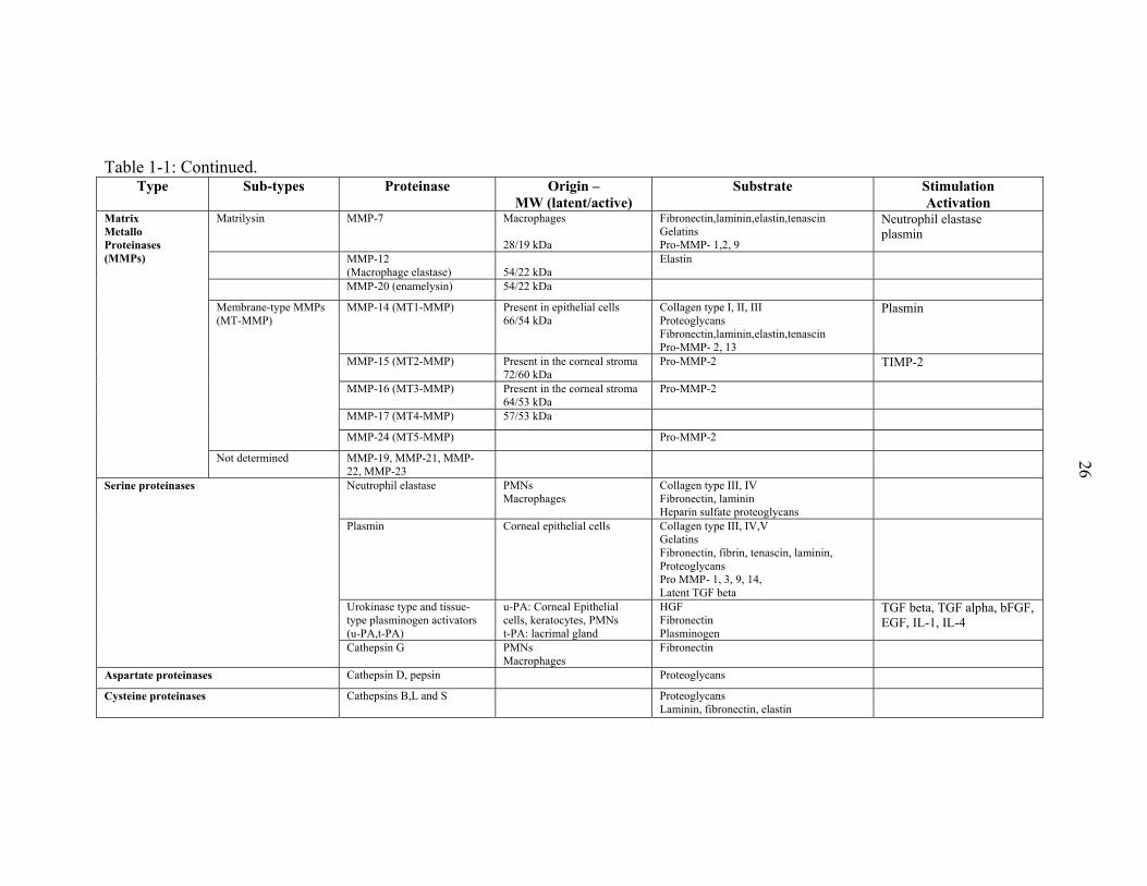
Table 1-1: Continued.
26
Type Sub-types Proteinase Origin –MW (latent/active)
Substrate StimulationActivation
Matrilysin MMP-7 Macrophages 28/19 kDa
Fibronectin,laminin,elastin,tenascin Gelatins Pro-MMP- 1,2, 9
Neutrophil elastase plasmin
MMP-12(Macrophage elastase)
54/22 kDa
Elastin
MMP-20 (enamelysin) 54/22 kDa MMP-14 (MT1-MMP) Present in epithelial cells
66/54 kDa Collagen type I, II, III Proteoglycans Fibronectin,laminin,elastin,tenascin Pro-MMP- 2, 13
Plasmin
MMP-15 (MT2-MMP) Present in the corneal stroma 72/60 kDa
Pro-MMP-2 TIMP-2
MMP-16 (MT3-MMP) Present in the corneal stroma 64/53 kDa
Pro-MMP-2
MMP-17 (MT4-MMP) 57/53 kDa
Membrane-type MMPs (MT-MMP)
MMP-24 (MT5-MMP) Pro-MMP-2
Matrix Metallo Proteinases (MMPs)
Not determined MMP-19, MMP-21, MMP-22, MMP-23
Neutrophil elastase PMNs Macrophages
Collagen type III, IV Fibronectin, laminin Heparin sulfate proteoglycans
Plasmin Corneal epithelial cells Collagen type III, IV,V Gelatins Fibronectin, fibrin, tenascin, laminin, Proteoglycans Pro MMP- 1, 3, 9, 14, Latent TGF beta
Urokinase type and tissue-type plasminogen activators (u-PA,t-PA)
u-PA: Corneal Epithelial cells, keratocytes, PMNs t-PA: lacrimal gland
HGF Fibronectin Plasminogen
TGF beta, TGF alpha, bFGF, EGF, IL-1, IL-4
Serine proteinases
Cathepsin G PMNs Macrophages
Fibronectin
Aspartate proteinases Cathepsin D, pepsin Proteoglycans Cysteine proteinases Cathepsins B,L and S Proteoglycans
Laminin, fibronectin, elastin

27
Table 1-2:- Proteinase inhibitors.
Types Proteinases Natural inhibitors Artificial inhibitorsMatrix metalloproteinases (MMPs) α2-macroglobulin (Entrapment of
the molecule) TIMP-1 (20.6 kDa) TIMP-2 (21.5 kDa, in the cornea, secreted by keratocytes) TIMP-3 (21.6 kDa) TIMP-4
Chelation of the cofactor (Ca2+ and Zn2+):
Ascorbic acid, citric acid
Disodium ethylene-diaminetetra-acetic acid (EDTA),
N-acetylcysteine (NAC)
Hydroxamic acid -dipeptides : Ilomostast
Thiol peptides
Tetracyclines (Doxycycline, oxytetracycline)
Urokinase type and tissue-type plasminogen activators (u-PA,t-PA)
PA inhibitors Phenylmethylsulfonyl fluoride (PMSF) Aminoethyl-benzne-sulfonyl fluoride (AEBSF) Corticosteroids
Neutrophil elastase α1-poteinase inhibitor α2-macroglobulin (Entrapment of the molecule)
Phenylmethylsulfonyl fluoride (PMSF) Aminoethyl-benzne-sulfonyl fluoride (AEBSF)
Serine proteases
Plasmin α1-antitrypsin α2-macroglobulin α2-antiplasmin
Aprotinin Phenylmethylsulfonyl fluoride (PMSF) Aminoethyl-benzne-sulfonyl fluoride (AEBSF)
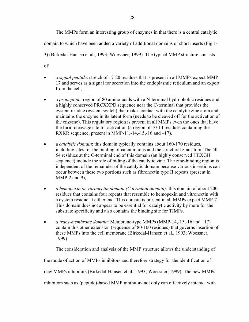
28
The MMPs form an interesting group of enzymes in that there is a central catalytic
domain to which have been added a variety of additional domains or short inserts (Fig 1-
3) (Birkedal-Hansen et al., 1993; Woessner, 1999). The typical MMP structure consists
of:
• a signal peptide: stretch of 17-20 residues that is present in all MMPs expect MMP-17 and serves as a signal for secretion into the endoplasmic reticulum and an export from the cell,
• a propeptide: region of 80 amino-acids with a N-terminal hydrophobic residues and a highly conserved PRCXXPD sequence near the C-terminal that provides the cystein residue (cystein switch) that makes contact with the catalytic zinc atom and maintains the enzyme in its latent form (needs to be cleaved off for the activation of the enzyme). This regulatory region is present in all MMPs even the ones that have the furin-cleavage site for activation (a region of 10-14 residues containing the RXKR sequence, present in MMP-11,-14,-15,-16 and –17).
• a catalytic domain: this domain typically contains about 160-170 residues, including sites for the binding of calcium ions and the structural zinc atom. The 50-54 residues at the C-terminal end of this domain (an highly conserved HEXGH sequence) include the site of biding of the catalytic zinc. The zinc-binding region is independent of the remainder of the catalytic domain because various insertions can occur between these two portions such as fibronectin type II repeats (present in MMP-2 and 9),
• a hemopexin or vitronectin domain (C terminal domain): this domain of about 200 residues that contains four repeats that resemble to hemopexin and vitronectin with a cystein residue at either end. This domain is present in all MMPs expect MMP-7. This domain does not appear to be essential for catalytic activity by more for the substrate specificity and also contains the binding site for TIMPs.
• a trans-membrane domain: Membrane-type MMPs (MMP-14,-15,-16 and –17) contain this other extension (sequence of 80-100 residues) that governs insertion of these MMPs into the cell membrane (Birkedal-Hansen et al., 1993; Woessner, 1999).
The consideration and analysis of the MMP structure allows the understanding of
the mode of action of MMPs inhibitors and therefore strategy for the identification of
new MMPs inhibitors (Birkedal-Hansen et al., 1993; Woessner, 1999). The new MMPs
inhibitors such as (peptide)-based MMP inhibitors not only can effectively interact with

29
the MMP at subsites proximal to the scissile bond but also functionally bind this critical
zinc. The effective zinc chelation in conjunction with the high affinity for the enzymes
subsites by functionality provides to the inhibition of the MMPs. The peptide-based
MMP inhibitors have been reported to have higher potency and specificity than the
previous generation of inhibitors (such as tetracyclines, N-acetyl-cysteine, EDTA).
Figure 1-3: Domain Structure of the Matrix Metalloproteinase family.
Origins of the proteinases
As already mentioned, proteinases can be produced by the corneal cells (MMP-1,
MMP-3, MMP-2, MMP-9, as well as t-PA), and the inflammatory cells such as PMNs
and macrophages (MMP-7, MMP-8, MMP-9 as well as NE and u-PA). They are
therefore called endogenous proteinases (Table 1-1). Infectious organisms also produce

30
proteinases and these constitute the exogenous proteinases (Hibbetts et al., 1999). For
example Pseudomonas aeruginosa has been shown to produces two MMPs (alkaline
protease, elastase), and Aspergillus spp and Fusarium spp have been shown to produce
serine proteinases (Gopinathan et al., 2001; Hanzi et al., 1993; Kernacki et al., 1996;
Kessler et al., 1977; Matsumoto, 2000; Zhu et al., 1990). Extra-cellular enzymes of
bacterial or fungal origin contribute to the corneal condition, either directly or indirectly,
through the activation of endogenous proteinases (Gopinathan et al., 2000; Twining et al.,
1993).
Proteinases, PTF and Corneal Physiopathology
The maintenance and the repair of the corneal matrix require a tightly coordinated
balance of ECM synthesis, degradation and remodeling where proteinases are involved as
mentioned earlier (Figure 1-1). Activities of proteolytic enzymes (endogenous
proteinases) are normally balanced by natural proteinase inhibitors (including tissue
inhibitors of MMPs or TIMPs, ∝1-proteinase inhibitor, ∝2-macroglobulin), thus
preventing excessive degradation of normal healthy tissue. Proteinases and especially
MMPs produce beneficial outcomes for corneal wounds, provided they are produced at
the right level, in the right place and at the right time but excessive levels of these
proteinases can cause severe complications. Excessive amounts of proteinases can create
an imbalance between proteinases and proteinase inhibitors, and increased amounts of
proteases can cause pathologic degradation of collagen and proteoglycans in the cornea
(Slansky et al., 1969; Twinning, 1994).
The tear film proteinases have been previously evaluated in both normal and
diseased eyes of animals and human beings (Berman et al., 1971; Berman et al., 1973;
Berman et al., 1977; Brooks 1999; Fini and Girard, 1990; Matsubara et al., 1991a;
31
Matsumoto et al., 1993; Prause, 1983a; Prause, 1983b; Strubbe et al, 2000; Tervo et al.,
1988; Tervo et al., 1991). In severely damaged corneas, proteinase activities in the tear
film were significantly increased (Berman et al., 1971; Kernacki et al., 1995; Matsubara
et al., 1991a; Prause, 1983a; Strubbe et al., 2000; Tervo et al., 1988). If infection is
present, the proteinases secreted by infectious organisms are also responsible for the
severe corneal damage that is associated with the disease (Kernacki et al., 1995; Twining
et al., 1993).
In ulcerated horse corneas, tear film levels of MMP-2, MMP-9 and neutrophil
elastase are significantly elevated when compared to age-matched normal controls
(Strubbe et al., 2000) and are hypothesized to contribute to the breakdown of stromal
collagen. Ulcerative keratitis with extensive stromal involvement displays rapid
progressive that can lead to corneal perforation in many horse eyes probably due to this
upregulated proteolytic activity (Brooks, 1999).
In the initial work done by Strubbe et al. (2000), tears were collected using glass
capillary tubes. This tear collection method allows the recording of the time needed for
collection and therefore the measurement of the tear fluid flow (TFF). The TFF has been
measured in various studies performed in human beings (Tervo et al., 1994; van Setten et
al., 1989; van Setten et al, 1990; Vesaluoma et al., 1996; Vesaluoma et al., 1997;
Vesaluoma and Tervo, 1998) but it has not been measured yet in animals and especially
in horses. By taking the TFF changes (the dilution factor) into consideration, it is possible
to give a better reflection of the changes in synthesis and release of the proteinases
between the different groups of animal studied.

32
Many studies have been performed on the changes in MMP expression in the skin
as well as MMP proteolytic activity in the wound fluids during skin wound healing in
animals and human beings (Agren, 1994; Blalock et al., 2001; Ladwig et al., 2002;
Lobmann et al., 2002; Herouy, 2001; Parks, 1999; Paul et al., 1997; Wall, 2002). Few
studies have reported the proteolytic changes in the tear film during corneal wound
healing. Changes in the tear film level of plasmin, a serine protease have been described
in animals and human beings (Barlati et al., 1990; Cejkova, 1998; Cejkova et al., 1993;
Salonen et al., 1987; Tervo et al. 1998; Tervo et al., 1989a; Tervo et al, 1989b; Tervo et
al. 1991; Vesaluoma et al., 1998; van Setten et al., 1989). The changes in level of various
MMPs in the tear film (Barro et al., 1998) as well as their expression of MMP in the
cornea during wound healing have also been described in rat and rabbit (Fini and Girard,
1992; Fini et al., 1998; Lu et al., 1999; Matsubara et al., 1991a; Ye et al., 1998). To the
author’s knowledge, there is only one study on the precise profile of the MMP activity in
tear film during corneal healing based on the collection and analysis of serial tear fluid
samples in human beings (Barro et al., 1998).
Few studies have reported the expression of MMPs in healthy and diseased
corneas. MMP-7 has been detected in healthy and wounded rat cornea (Lu et al., 1999),
MMP-3 in human corneas (Gabison et al., 2003), and MT-MMP-4, and MT-MMP-5 in
mouse corneas (Dong et al., 2000). MMP-2 and MMP-9 have been identified by
immunohistochemistry in the healthy and ulcerated corneas in humans (Gabison et al.,
2003; Kenney et al., 1998), dogs (Chandler et al., 2003), mice (Wall et al., 2002; Yang et
al., 2003), and rats (Reviglio et al., 2003; Ye and Azar, 1998) but no similar study has
been performed in horses.
33
An understanding of the physiopathologic processes as well as early diagnosis and
aggressive treatment are important to speed healing, reduce scarring, and prevent corneal
rupture in case of ulcerative corneal diseases in horses. Despite the abundant literature on
ocular proteinases, and despite the prevalence of equine ulcerative keratitis, there is very
limited information on tear film proteinases in the horse. MMPs are certainly involved in
corneal ulceration and corneal wound healing in horses as Strubbe et al. reported with
their preliminary report (Strubbe et al., 2000) and their importance needs to be further
assessed. Identification of the proteinases mainly responsible for corneal stromal
degradation would be the first logical step to allow specific inhibition
Proteinases, Proteinase Inhibitors and the Cornea
There are natural proteinase inhibitors that are present in the PTF and in the cornea.
α1-proteinase inhibitor, α2-macroglobulin and some TIMPs are found in the blood
plasma and interstitial fluids and can also be synthesized by resident tissue cells
simultaneously with the MMPs (Table 1-2, Figure 1-1). Disorders occur when there is an
imbalance between proteinases and proteinase inhibitors in favor of the proteinases.
Proteinase activity in the tear film is believed to speed up degradation of stromal
collagen, leading to rapid progression of ulcers. Normalizing proteolytic activity in the
tear film is an objective of the treatment of corneal ulcers in horses. Thus, proteinase
inhibitors have been recommended for treatment of ulcerative keratitis to reduce the
progression of stromal ulcers, speed epithelial healing, and minimize corneal scarring
(Berman, 1975; Berman, 1978; Berman et al., 1973; Berman et al., 1975; Brooks, 1999;
Tervo et al., 1992; Ward, 1999).
However, studies on activity of specific enzymes are limited. Accordingly,
recommendations for use of topically administered N-acetylcysteine (NAC), potassium
34
EDTA, and serum in horses with corneal ulcers are mostly based on anecdotal clinical
reports or extrapolations from data obtained from use of these compounds in other
species (Brooks, 1999; McLaughlin et al., 1992; Severin, 1976; Strubbe et al., 2000;
Tervo et al., 1992; Ward, 1999; Whitley and Gilger, 1999). The availability of newer
compounds such as doxycycline, a modified dipeptide that contains hydroxamic acid
(i.e., ilomostat), and α 1-proteinase inhibitor (α 1-PI), which initially have shown
considerable promise as MMP or serine-proteinase inhibitors in other species (Barletta et
al., 1996; Dursun et al., 2001; Golub et al., 1984; Perry et al., 1993; Rawal and Rawal,
1984; Schultz et al., 1992) outlines the need for specific studies on possible regulation of
enzyme activity in tears in horses with corneal disease (Table 1-2). These new
compounds have potential as potent proteinase inhibitors in horses with corneal ulcers.
Therefore, there was a need to evaluate the effectiveness of various proteinase
inhibitors for reducing the activity of equine tear film MMP-2 and -9 by in vitro testing
on samples of tear film obtained from horses with ulcerative keratitis.
Growth Factors
Growth Factors, PTF and Corneal Physiopathology
In the past 15 years, substantial progress has been made in understanding the
process of corneal maintenance and corneal wound healing at the molecular level, in
order to achieve better outcomes for clinical injuries by rationally design therapeutic
strategies that use agents to modulate corneal wound healing.
A molecular theory emerged based upon the synthesis and release of several
specific growth factors (GFs), at the site of injury which then act through autocrine and
paracrine pathways to regulate healing (Pancholi et al., 1998; Schultz, 1994; van Setten et
al., 1994). GFs are low molecular weigh polypeptides, are synthesized by a variety of cell
35
types and act locally on the cell that synthesized the GF or adjacent cells by binding to
specific, high-affinity receptors proteins located on the plasma membrane of the target
cells and they regulate many physiologic functions of these target cells (Schultz, 1997).
They are assumed to control many crucial processes of corneal wound healing
including cell migration (chemotactic effects for inflammatory cells including
macrophages, neutrophils and also for corneal cells including epithelial and endothelial
cells, keratocytes), cell proliferation (mitosis), angiogenesis (neovascularisation includes
the proliferation, migration, proteolytic activity, and capillary tube formation of the
vascular endothelial cells), and synthesis of ECM components (Casey and Li, 1997;
Imanishi et al., 2000; Schultz et al. , 1992; Vesaluaoma et al., 1996) (Table 1-3). Cell
migration, mitosis, and differentiation as well as reconstruction of adhesion complexes
are important features of corneal epithelial wound healing. In addition to regulation of the
wound healing process and fibrosis, growth factors are likely to be involved in the
maintenance of corneal cellular integrity (Imanishi et al., 2000; Schultz et al., 1992; van
Setten et al., 1998; Vesaluoma et al., 1996; Wilson et al., 1990; Wilson, 1998) ( Figure 1-
2).
The cornea is avascular and the corneal wound healing is controlled in a different
manner. Since the cornea is in close contact with the tear film and the aqueous fluid, the
various kinds of growth factors and cytokines in the tear film and aqueous play an
important role in the turnover of the corneal cells and wound healing of the cornea
(Imanishi et al., 2000; Vesaluoma et al., 1996). The tear fluid in particular has certain
functions exerted by blood components in other organs. Growth factors, mRNA or
protein expression has been shown in the lacrimal gland as well as in all corneal cell
36
layers. (Table 1-3) About 15 years ago, the first GF was identified in the PTF (van Setten
et al., 1989) and the number of GFs discovered in the PTF keeps increasing since then.
To date, the following GF have been reported to be present in the PTF of a normal or
ulcerated eye of various species: transforming growth factor-alpha (TGF-α) (van Setten et
al., 1994), fibroblast growth factor (FGF) (van Setten, 1996), epidermal growth factor
(EGF), transforming growth factor-beta 1 & 2 (TGF-β 1&2) (Gupta et al., 1996), platelet-
derived growth factor (PDGF) (Vesaluoma et al., 1997), vascular endothelial growth
factor (VEGF) (van Setten, 1997), hepatocyte growth factor (HGF) (Li et al., 1996),
keratocyte growth factor (KGF) (Wilson, 1999), and connective tissue growth factor
(CTGF) (van Setten et al., 2003). (Table 1-3).
Levels of GFs and their receptors (GFR) in a wound are crucial for its normal
healing (Gum et al., 1999; Schultz, 1997). The role of the GF is based on the balance
between the presence of the GF in the PTF and the number of GFRs at the surface of the
ocular surface. This model has been thoroughly described for EGF (van Setten et al.,
1992), and can be extended to other GFs (HGH, PDGF, TGF- β). A brief description of
this model follows hereafter. In case of a wounded cornea, the total amount of GF
released in the tear fluid is increased and the increase of the TFF is even greater.
Therefore, the concentration of GF is often decreased (Tuominen et al., 2001; van Setten
et al., 1992; Vesaluoma et al., 1996; Vesaluoma et al., 1998; Wilson, 1998; Wilson,
1999). The decrease of GF concentration induces an “up regulation” of the GFR density
on the target cells (Steinemann et al., 1990). The increased number of molecules of GF
and of GFR leads to an increase number of GF/GFR complexes that exert effect on a

37
specific aspect of cell metabolism (mitosis, synthesis, migration). The overall balance
GF/GFR system is reestablished once complete corneal wound healing is achieved.
GFs, Cytokines, proteinases, proteinases inhibitors and endocrine hormones, and
the balance between these molecules are crucial for the maintenance of the tissue
integrity and its wound healing (Schultz, 1998). The interactions between GFs and
proteinases are particularly important in the corneal wound healing process as illustrated
by the two examples hereafter:
• EGF and Plasmin, u-PA, t-PA (van Setten et al., 1992): EGF has been shown to increase the production of u-PA, and t-PA resulting in the increase of plasmin synthesis. On the other end, EGF stimulates the fibronectin synthesis which is a substrate for plasmin.
• TGF- β & u-PA, t-PA, MMP-1, MMP-3 (Twining, 1994; Vesaluoma et al., 1998): TGF- β has been shown to down-regulate the synthesis of MMP-1 and MMP-3. It has also been reported that TGF- β inhibits the production of u-PA, and t-PA. Therefore, TGF- β seems to counteract the stimulatory action of EGF on u-PA, t-PA, and plasmin synthesis. (on another note, TGF- β also counteracts the stimulatory action of EGF on corneal epithelial cells proliferation)
There are also interactions between the different GFs: EGF and TGF- β (Nishida,
1997; van Setten et al., 1992), PDGF-BB and HGF (Fagerholm, 2000; Li et al., 1996),
TGF- β and CTGF (Chen et al., 2000; Frazier et al., 1997).
Experimental studies show that growth factors influence growth of cultured corneal
cells and modulate corneal wound healing in vitro (Blalock et al., 2003; Pancholi et al.,
1998; Watanabe et al., 1993) including one using equine corneal cells (Haber et al.,
2003). The ability of the GFs to directly regulate key cellular processes such as mitosis,
migration, ECM components synthesis suggests that treatment with exogenous GF might
enhance healing of corneal wound (Schultz, 1997). Few clinical trials have been done
using growth factors. The effects on corneal healing of EGF have been clinically
38
evaluated in various species including human, primates, dogs, rabbits, and in horses
(Burling et al., 2000).
There are few reports on growth factors in veterinary ophthalmology as mentioned
earlier (Burling et al., 2000; Haber et al., 2003) and there is no information about the
presence and the role in equine PTF and equine cornea of a recently discovered growth
factor, connective tissue growth factor (CTGF).
Connective Tissue Growth Factor
Connective tissue growth factor is a secreted, cysteine-rich peptide of about 38
kilodaltons (kDa) that was originally identified as a mitogen for fibroblasts in condition
media cultures for human umbilical vein endothelial cells in 1991. This peptide is named
CTGF because it has been identified as a major chemotactic and mitogenic factor for the
cells of the connective tissue (Table 1-3), and has PDGF related biological and
immunological properties.
Cuurrently, expression of CTGF is limited to cells derived from the mesenchyme
including fibroblasts, vascular endothelial cells, smooth muscle cells, chondrocytes and
renal tubules cells (Table 1-3). Leukocytes, lymphocytes and cells derived from the
epithelium of ambryos are not known to express CTGF. Transforming growth factor β
(TGF-β) activates fibrocytes to produce CTGF (Igarashi et al., 1996; Wang et al., 2001).
CTGF can induce connective tissue cell proliferation and ECM synthesis; it has similar
but not identical properties to TGF-β, suggesting that it may function as a downstream
mediator of some action of TGF-β.
The overexpression of CTGF has been proposed to play an important role in
pathways that lead to fibrosis in various tissues: scleroderma, systemic sclerosis,
atherosclerosis, idiopathic pulmonary fibrosis, liver fibrosis, diabetic glomerulosclerosis,
39
and renal fibrosis (Chen et al., 2000; Gupta et al., 2000; Igarashi et al., 1996; Shi-wen et
al., 2000; Takehara, 2001; Wunderlich, 2000). To date, the presence of CTGF has been
reported in the human serum (Sato et al., 2000), the anterior chamber fluid (van Setten et
al., 2002; van Setten et al., 2003), and recently human tear fluid (van Setten et al., 2003)
and rat cornea (Blalock et al., 2003). Limited information is available on the expression
of CTGF in ocular tissues but these studies suggest a possible involvement of CTGF in
ocular pathology. CTGF is a fibrogenic cytokine that might be involved in corneal wound
healing and corneal fibrotic disorders as CTGF enhances fibroblasts to proliferate and
produce more collagen (Frazier et al., 1996).
Ocular surface diseases such as corneal ulceration and corneal stromal abscessation
are common devastating ocular diseases in horses. Healing of corneal ulcers in horses is
often associated with profound fibrosis and corneal scar formation that can result in
varying degrees of visual impairment (Brooks, 1999). Since no information is available
regarding CTGF in horses and since the role of CTGF appears to be so crucial in the
regulation of fibrosis in other tissues in other species, its expression and role in the equine
PTF and in the normal and fibrotic equine cornea needs to be assessed as CTGF may play
an important role in corneal scar formation in horses and might be a target for reducing
fibrosis in the future.
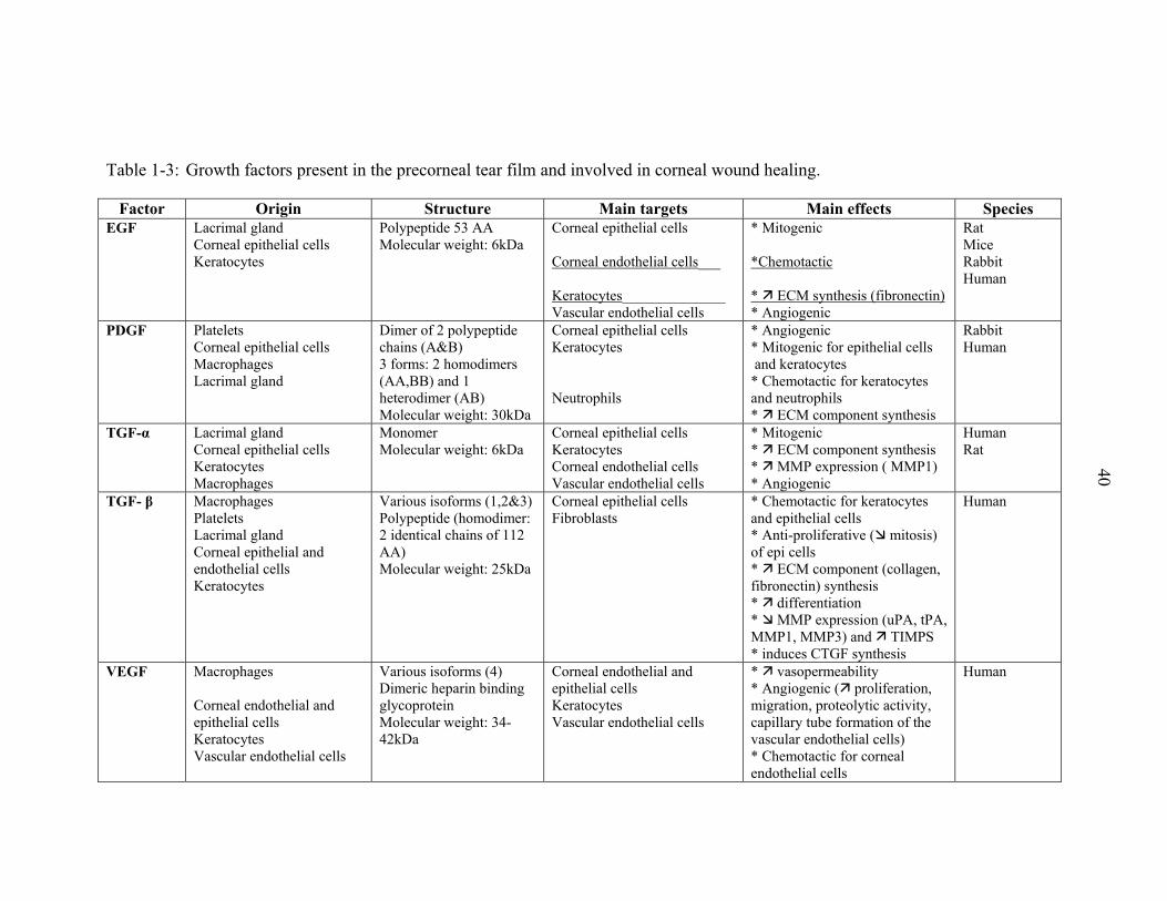
Table 1-3: Growth factors present in the precorneal tear film and involved in corneal wound healing.
40
Factor Origin Structure Main targets Main effects Species EGF
Lacrimal gland Corneal epithelial cells Keratocytes
Polypeptide 53 AA Molecular weight: 6kDa
Corneal epithelial cells Corneal endothelial cells___ Keratocytes______________ Vascular endothelial cells
* Mitogenic *Chemotactic * ECM synthesis (fibronectin) * Angiogenic
Rat Mice Rabbit Human
PDGF Platelets Corneal epithelial cells Macrophages Lacrimal gland
Dimer of 2 polypeptide chains (A&B) 3 forms: 2 homodimers (AA,BB) and 1 heterodimer (AB) Molecular weight: 30kDa
Corneal epithelial cells Keratocytes Neutrophils
* Angiogenic * Mitogenic for epithelial cells and keratocytes * Chemotactic for keratocytes and neutrophils * ECM component synthesis
Rabbit Human
TGF-α Lacrimal gland Corneal epithelial cells Keratocytes Macrophages
Monomer Molecular weight: 6kDa
Corneal epithelial cells Keratocytes Corneal endothelial cells Vascular endothelial cells
* Mitogenic * ECM component synthesis * MMP expression ( MMP1) * Angiogenic
Human Rat
TGF- β Macrophages Platelets Lacrimal gland Corneal epithelial and endothelial cells Keratocytes
Various isoforms (1,2&3) Polypeptide (homodimer: 2 identical chains of 112 AA) Molecular weight: 25kDa
Corneal epithelial cells Fibroblasts
* Chemotactic for keratocytes and epithelial cells * Anti-proliferative ( mitosis) of epi cells * ECM component (collagen, fibronectin) synthesis * differentiation * MMP expression (uPA, tPA, MMP1, MMP3) and TIMPS * induces CTGF synthesis
Human
VEGF Macrophages Corneal endothelial and epithelial cells Keratocytes Vascular endothelial cells
Various isoforms (4) Dimeric heparin binding glycoprotein Molecular weight: 34-42kDa
Corneal endothelial and epithelial cells Keratocytes Vascular endothelial cells
* vasopermeability * Angiogenic ( proliferation, migration, proteolytic activity, capillary tube formation of the vascular endothelial cells) * Chemotactic for corneal endothelial cells
Human
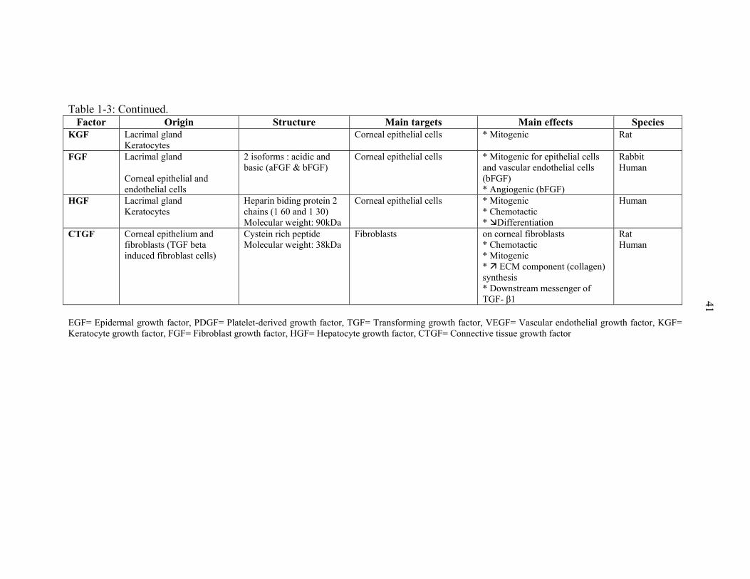
Table 1-3: Continued.
41
Factor Origin Structure Main targets Main effects Species KGF Lacrimal gland
Keratocytes Corneal epithelial cells
* Mitogenic
Rat
FGF Lacrimal gland Corneal epithelial and endothelial cells
2 isoforms : acidic and basic (aFGF & bFGF)
Corneal epithelial cells
* Mitogenic for epithelial cells and vascular endothelial cells (bFGF) * Angiogenic (bFGF)
Rabbit Human
HGF Lacrimal gland Keratocytes
Heparin biding protein 2 chains (1 60 and 1 30) Molecular weight: 90kDa
Corneal epithelial cells
* Mitogenic * Chemotactic * Differentiation
Human
CTGF Corneal epithelium and fibroblasts (TGF beta induced fibroblast cells)
Cystein rich peptide Molecular weight: 38kDa
Fibroblasts on corneal fibroblasts * Chemotactic * Mitogenic * ECM component (collagen) synthesis * Downstream messenger of TGF- β1
Rat Human
EGF= Epidermal growth factor, PDGF= Platelet-derived growth factor, TGF= Transforming growth factor, VEGF= Vascular endothelial growth factor, KGF= Keratocyte growth factor, FGF= Fibroblast growth factor, HGF= Hepatocyte growth factor, CTGF= Connective tissue growth factor

42
Significance to The Horse Racing Industry
Horses have large, prominent eyes that are often subject to traumatic injury and
resultant corneal infection. Ulcerative keratitis is a common and often vision-threatening
condition in horses. A report from the United States Department of Agriculture
mentioned that ocular problems represent the fourth most common cause of operations
(7.9%) in horses in the southern part of the United States. This followed trauma (16.2%),
leg/hoof problems (15.3%), and colic (13.1%) (NAHMS Equine study, 1998). The
Ophthalmology Service of the University of Florida Veterinary Medical Teaching
Hospital diagnosed ulcerative keratitis in 527 horses between January 1987 and October
2002. This constituted 35% of all horses evaluated for ophthalmic problems during this
period.
A simple corneal abrasion can rapidly become vision threatening if it becomes
infected with a pathogenic organism, especially in our subtropical environment.
Determination of the proteolytic activity in the normal and diseased horse eye as well as
during corneal healing and the efficacy of antiproteolytic compounds will allow us to
institute appropriate therapy earlier in the course of the disease, decrease the time
required for recovery and rehabilitation, and perhaps alleviate the need for surgical
therapies. Horses are often laid up in excess of 8 weeks during treatment of corneal
diseases. Shortening this time through prompt and adequate treatment would be of a great
economic value to the horse racing industry. A pronounced fibrovascular response is also
prominent during corneal healing in horses and the growth factors may have an important
role in this response.
An understanding of the pathophysiologic processes involving the proteinases and
the growth factors as well as early diagnosis and aggressive treatment are important for

43
quick resolution of ulcerative corneal diseases in horses and to speed healing, reduce
scarring, and prevent corneal rupture.
Purpose of The Study
The general goal of this dissertation was to understand the role that MMP-2 and
MMP-9 play in corneal ulceration and healing in horses and to assess the efficacy of
various agents that inhibit these proteases.
An additional goal of this work was to understand the role that CTGF plays in
regulating corneal scarring in horses and a long term-goal of this research is to develop
agents that reduce corneal scarring, targeting CTGF.
Hypotheses
The hypotheses were the following:
Protease levels in the tear film of horses with normal eyes and horses with corneal ulcers
• The levels of proteases in tears are increased in horses with an ulcerative keratitis (infectious or sterile) as compared to tears in a normal horse eye.
• The increased proteolytic activity present in tears from horses with ulcerative keratitis decreases as the corneal ulcer heals.
In Vitro effects of various protease inhibitors
Protease inhibitors e.g., serum, α2-macroglobulin, ethylene diamine tetraacetate,
N-acetyl-L-cysteine, tetracyclines, thiols and hydroxamic acids, α1 proteinase inhibitor
are used or represent an interesting alternative in the clinical treatment of corneal ulcers
in horses. These protease inhibitors produce a measurable reduction in MMP and NE
activity in the tear fluid of horses with ulcerative keratitis that can be measured in vitro.
CTFG levels in tear fluid from horses
• CTGF is present and detectable in the equine PTF.
44
• The level of CTGF in tear fluid is different in an ulcerated eye (infectious or sterile) as compared to a normal eye.
Localization of MMP-9, MMP-2, CTGF in the equine cornea and lacrimal glands
• MMP-9, MMP-2, CTGF are present in the corneal tissue.
• MMP-9, MMP-2, CTGF are present in the main lacrimal gland as well as then nictitating gland.
Objectives
The present work had the following goals:
• To identify the presence of MMP-2 and MMP-9 in the tear fluid, cornea and lacrimal glands of horses.
• To investigate if ulcerative keratitis has any influence on the activity of MMP-2 and MMP-9 in the equine tear fluid.
• To follow the changes in the amount of proteolytic activity in horse tear film during corneal healing and stromal remodeling.
• To investigate what in vitro effects various proteases inhibitors have on the proteolytic activity of MMP-2 and MMP-9 isolated from the equine tear fluid.
• To investigate the presence and origin of CTGF in the tear fluid, cornea and lacrimal glands of healthy horse eyes.
• To elucidate what influence ulcerative keratitis has on the presence of CTGF in the equine tear fluid.
CHAPTER 2 DETECTION OF MMP-2 AND MMP-9 IN THE EQUINE TEAR FLUID, CORNEA
AND LACRIMAL GLANDS
Introduction
The maintenance and repair of the corneal stromal extracellular matrix (ECM)
require a tightly coordinated balance of ECM synthesis, degradation and remodeling.
Proteolytic enzymes (proteinases) perform important physiological functions in the slow
turnover and remodeling of the corneal stroma. The activities of proteolytic enzymes are
normally balanced by natural proteinase inhibitors and thus prevent excessive
degradation of normal healthy tissue (Hibbetts et al., 1999; Twining et al., 1994).
Excessive levels of proteinases can create an imbalance between proteinases and
proteinase inhibitor levels, therefore causing pathological degradation of stromal collagen
and proteoglycans in the cornea ECM (Geerling et al., 1999; Matsubara et al., 1991a;
Slansky et al., 1969; Strubbe et al., 2000; Twining, 1994).
Endogenous proteinases are produced by corneal cells and inflammatory cells
(Berman et al., 1971; Kernacki et al., 1995; Matsubara et al., 1991a; Prause, 1983a;
Strubbe et al., 2000; Tervo et al., 1988). If infection is present, the proteinases secreted
by infectious organisms, called exogenous proteinases, are also responsible for corneal
damage (Gopinathan et al., 2001; Hanzi et al., 1993; Hibbetts et al., 1999; Kernacki et al.,
1995; Matsumoto, 2000; Twining et al., 1993; Zhu et al., 1990). Extra-cellular enzymes
of bacterial or fungal origin contribute to the corneal condition, either directly or
45

46
indirectly, through the activation of endogenous proteinases (Gopinathan et al., 2001;
Twining, 1993).
Among the various classes of proteinases found in the tear film and cornea of
humans and animals, two are thought to play a major role in the corneal metabolism: the
matrix metalloproteinases (MMPs) and the serine proteinases (including neutrophil
elastase) (Fini et al., 1992; Fini et al., 1998; Matsubara et al., 1991). Two of the 13
known MMPs are of major importance in terms of corneal stromal collagen remodeling
and degradation, and are the focus of this study. They include MMP-2 (72-kDa gelatinase
A) and MMP-9 (92-kDa gelatinase B) (Fini and Girard, 1990; Fini et al., 1998). Their
origin and purpose at the corneal level appear to differ.
In ulcerated horse corneas, tear film levels of MMP-2, MMP-9 and neutrophil
elastase are significantly elevated when compared to age-matched normal controls
(Strubbe et al., 2000), and are hypothesized to contribute to the breakdown of stromal
collagen. Ulcerative keratitis with extensive stromal involvement displays rapid
progression that can lead to corneal perforation in many horse eyes probably due to this
upregulated proteolytic activity (Brooks, 1999).
Few studies have reported the expression of MMPs in healthy and diseased
corneas. MMP-7 has been detected in healthy and wounded rat cornea (Lu et al., 1999),
MMP-3 in human corneas (Gabison et al., 2003), and MT-MMP-4, and MT-MMP-5 in
mousecorneas (Dong et al., 2000). MMP-2 and MMP-9 have been identified by
immunohistochemistry in the healthy and ulcerated corneas in humans (Kenney et al.,
1998; Gabison et al., 2003), dogs (Chandler et al., 2003), mice (Wall et al., 2002; Yang et

47
al., 2003), and rats (Reviglio et al., 2003; Ye and Azar, 1998) but no similar study has
been performed in horses.
MMPs are undoubtfully involved in corneal ulceration and corneal wound healing
in horses but their importance needs to be assessed. With only one preliminary study
published in horses (Strubbe et al., 2000) there is a considerable need for a thorough
investigation of the presence of MMP-2 and MMP-9 in tears and their pathophysiological
importance. The purpose of this study was to determine the presence of MMP-2 and
MMP-9 in the tear fluid of horses by Western Blot analysis, to investigate the localization
of MMP-2 and MMP-9 in healthy and pathological equine corneas, and to clarify the
possible origins of MMP-2 and MMP-9 by immunohistochemistry.
Materials and Methods
Materials
All procedures were carried out according to the ARVO statement for the Use of
Animals in Ophthalmic and Vision research and were approved by the University of
Florida Animal Care and Use Committee as well as the UF College of Veterinary
Medicine Clinical Research Review Committee.
For the detection of MMP-2 and MMP-9 in the equine tear fluid, samples were
collected from horses that were either clinical cases (Figure 2-1) from the Large Animal
Hospital at the University of Florida College of Veterinary Medicine Veterinary
Teaching Hospital or, in the cases of animals without any ocular disease, from a
commercial horse farm in Florida. The tear collections were performed after sedation of
the animal with an intravenous injection of xylazine hydrochloride (0.5 mg/kg) and
before any diagnostic procedure or any treatment. All tear fluid specimens were collected
by capillary force from the lower fornix using capillary tubes with an atraumatic tip as

48
previously described (van Setten et al., 1989). The time of collection of tear fluid samples
was not taken in this study. All samples were immediately centrifuged, transferred into
Eppendorf tubes (10µL aliquots), and stored at -80ºC until analysis. Two pools of tear
fluid (100µL) from horses with healthy eyes and horses with ulcerated were then
constituted for the identification of MMP-2 and MMP-9 on Western Blot by comparison
to human MMP-2 and MMP-9 standards.
For immunohistochemistry, five healthy corneas, five main lacrimal glands and
five nictitating membrane glands were collected from horses with normal eye following
euthanasia due to conditions not related to this project. A 2 year old Thoroughbred mare
was presented to the VMTH with corneal perforation and iris prolapse associated with a
fungal infection in her right eye. The eye was enucleated, the globe was processed. All
samples were fixated in 4% formaldehyde buffer and stored in paraffin. Hematoxylin and
Eosin (H&E) staining, and MMP-2 and MMP-9 immunostaining were done on the
healthy cornea and lacrimal glands, as well as on the diseased cornea and iris prolapse of
this clinical case.
Figure 2-1: Melting ulcer in the right eye of a 12 year old American Quarter horse. This case was involved in this study. Note the presence of hyperemic
49
conjunctiva, and a 8-9 mm melting corneal ulcer surrounded with a corneal edema.
MMP-2 and MMP-9 Western Blot
Two pools of tear fluid were formed by collection of tears from horses with healthy
eyes and horses with ulcerated eyes. The tears were centrifuged at 14,000g for 10 minutes
and the supernatant stored at – 80º C until their analysis. The samples (pools of tear fluid
from healthy and ulcerated horse eyes) as well as a molecular weight markers
(MultiMark pre-stained multicolored standards, Invitrogen, Carlsbad, CA) and 0.1 ng
of the active and latent forms of the human recombinant MMP-2 and MMP-9 were
loaded on a 5% stacking, 12% SDS-PAGE gel. The electrophoresis was run at 125 V for
1 hour in a running buffer. The proteins were electro-transferred onto a PVDF membrane
(Immobilon™-P, Millipore Corporation, Bedford, MA) in a transfer buffer at 25V at 4º C
overnight. The membrane was blocked in Tris-buffered saline (TBS) containing 10%
non-fat milk (Blotting grade blocker non-fat dry milk, BioRad Laboratories, Richmond,
CA) for one hour and then incubated in the same solution containing a dilution of 1:7000
of the mouse anti-human MMP-2 (1:20 dilution) and MMP-9 (1:25 dilution) (R&D
Systems, Mineapolis, MN) for 30 minutes at room temperature. The blot was washed
three times with 0.05% Tween-20 TBS and then incubated with rabbit anti-mouse IgG
alkaline phosphatase conjugate (Sigma, St Louis, MO) at the dilution of 1:1500 in the
blocking solution for 30 minutes at room temperature. The membrane was washed again
three times and the BCIP/NBT alkaline phosphatase substrate (Sigma, St Louis, MO) was
added and color generated. The reaction was stopped by water washing.
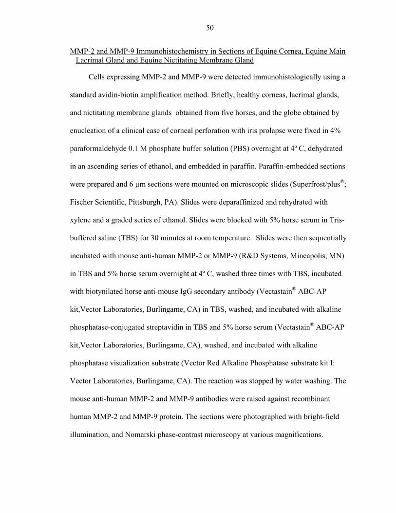
50
MMP-2 and MMP-9 Immunohistochemistry in Sections of Equine Cornea, Equine Main Lacrimal Gland and Equine Nictitating Membrane Gland
Cells expressing MMP-2 and MMP-9 were detected immunohistologically using a
standard avidin-biotin amplification method. Briefly, healthy corneas, lacrimal glands,
and nictitating membrane glands obtained from five horses, and the globe obtained by
enucleation of a clinical case of corneal perforation with iris prolapse were fixed in 4%
paraformaldehyde 0.1 M phosphate buffer solution (PBS) overnight at 4º C, dehydrated
in an ascending series of ethanol, and embedded in paraffin. Paraffin-embedded sections
were prepared and 6 µm sections were mounted on microscopic slides (Superfrost/plus®;
Fischer Scientific, Pittsburgh, PA). Slides were deparaffinized and rehydrated with
xylene and a graded series of ethanol. Slides were blocked with 5% horse serum in Tris-
buffered saline (TBS) for 30 minutes at room temperature. Slides were then sequentially
incubated with mouse anti-human MMP-2 or MMP-9 (R&D Systems, Mineapolis, MN)
in TBS and 5% horse serum overnight at 4º C, washed three times with TBS, incubated
with biotynilated horse anti-mouse IgG secondary antibody (Vectastain® ABC-AP
kit,Vector Laboratories, Burlingame, CA) in TBS, washed, and incubated with alkaline
phosphatase-conjugated streptavidin in TBS and 5% horse serum (Vectastain® ABC-AP
kit,Vector Laboratories, Burlingame, CA), washed, and incubated with alkaline
phosphatase visualization substrate (Vector Red Alkaline Phosphatase substrate kit I:
Vector Laboratories, Burlingame, CA). The reaction was stopped by water washing. The
mouse anti-human MMP-2 and MMP-9 antibodies were raised against recombinant
human MMP-2 and MMP-9 protein. The sections were photographed with bright-field
illumination, and Nomarski phase-contrast microscopy at various magnifications.
51
Results
MMP-2 and MMP-9 Western Blot: Detection of MMP-2 and MMP-9 in the Equine Tear Fluid.
Western Blot was used to compare the products detected by the antibody against
human MMP-2 and MMP-9 in the two pools of equine tear samples (normal and
diseased) to the active and latent forms of the human MMP-2 and MMP-9 standards. A
band, similar to those observed with the human latent MMP-2 standard and the human
active MMP-2 standard, 66 kDa, was noted in all the equine samples (Figure 2-2). A
band, similar to those observed with the human latent MMP-9 standard and the human
active MMP-9 standard, 83 kDa, was noted in all the equine samples (Figure 2-3).
These results indicate that there is a detectable amount of MMP-2 and MMP-9 in
the equine tear fluid.
1 2 3 4
98 kDa
52 kDa
31 kDa
Latent Active 17 kDa
Figure 2-2: Western Blot of tear fluids from horses with ulcerated eyes (lane 3) and healthy eyes (lane 4). A band similar to those observed with the human latent MMP-2 standard (lane 1) and the human active MMP-2 standard, 66 kDa (lane 2), was noted on all the equine samples (lanes 3 and 4).
Molecular Human MMP-2 Equine diseased Equine normal Weight Markers Standard tear fluid tear fluid

52
1 2 3 4
98 kDa
52 kDa
31 kDa
17 kDa Molecular Human MMP-9 Equine diseased Equine normal Weight Markers Standard tear fluid tear fluid
Figure 2-3: Western Blot of tear fluids from horses with ulcerated eyes (lane 3) and healthy eyes (lane 4). A band similar to those observed with the human latent MMP-9 standard (lane 1) and the human active MMP-9 standard, 83 kDa (lane 2), was noted on all the equine samples (lanes 3 and 4).
Immunohistochemistry: Immunohistochemical Localization of MMP-2 and MMP-9 in the Healthy Cornea and Lacrimal Glands of Horses
Immunoreactivity to MMP-2 specific antibodies was noted in the corneal
epithelium, the corneal stroma and in the corneal endothelium (Figure 2-4). In the corneal
epithelium, MMP-2 staining was concentrated in the squamous superficial epithelial cells
(Figure 2-4). The immunohistochemical staining for MMP-2 in the equine lacrimal
glands revealed specific staining for MMP-2 predominantly in the epithelial cells of the
intralobular ducts, and the intralobular connective tissue (Figure 2-5). The
immunohistochemical staining for MMP-2 in the nictitating membrane glands revealed
specific staining for MMP-2 predominantly in the acinar cells and the intralobular
connective tissue (Figure 2-6). No immunoreactivity to MMP-9 was found at the dilution
used (1:20) in the healthy cornea (Figure 2-4), the equine lacrimal gland (Figure 2-5), or
the nictitating membrane glands (Figure 2-6). No immunoreactivity was found in the

53
tissues on the sections used as controls where the primary antibody was omitted (Figures
2-4, 2-5 and 2-6).
2
111
22 2
3B CA 3 3
1
E2
D F3
111
222
33 13G H I
Figure 2-4: Immunolocalization of MMP-2 and MMP-9 in healthy equine cornea.
H&E staining of the equine cornea (A) where corneal epithelium (1), stroma (2) and Descemet’s membrane – corneal endothelium (3) can be distinguished. MMP-2: Nomarski phase-contrast 1 observation (B) and bright-field observation (C) of the negative control sections showed no staining at all. Immunohistochemical staining for MMP2 in the equine cornea (D, E, and F) showed MMP-2 specific staining in the corneal epithelium, especially in the squamous superficial cell layer (white arrow), a small amount in the stroma underneath the epithelium (black arrow) and the corneal endothelium. MMP-9: Nomarski phase-contrast 1 observation (G) and bright-field observation (H) of the negative control sections showed no staining at all. Immunohistochemical staining for MMP-9 showed no MMP-9 specific staining in the healthy equine cornea (I). Original magnifications were X100 (A, B, C, D, G, H and I) and X200 (E and F)

54
A B C
D E F
G I H
Figure 2-5: Immunolocalization of MMP-2 and MMP-9 in equine lacrimal gland.
H&E staining of the equine lacrimal gland (A) where acini (inset), intralobular ducts, and lymphocytes in the intralobular connective tissue (inset) can be distinguished. MMP-2: Nomarski phase-contrast 1 observation (B) and bright-field observation (C) of the negative control sections showed no staining at all. Immunohistochemical staining for MMP-2 in the equine lacrimal gland (D, E, and F) showed MMP-2 specific staining in the epithelial cells of the intralobular ducts (white arrow), and the intralobular connective tissue (striped arrow). MMP-9: Nomarski phase-contrast 1 observation (G) and bright-field observation (H) of the negative control sections showed no staining at all. Immunohistochemical staining for MMP-9 showed no MMP-9 specific staining in the equine lacrimal gland (I). Original magnifications were X100 (A, B, C, D, G, H and I) and X400 (E), and X1000 (inset in A and F).
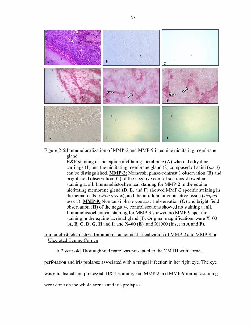
55
2 2 2 1 1 1B A C
2 1 E F
222
1 11 G H I
Figure 2-6: Immunolocalization of MMP-2 and MMP-9 in equine nictitating membrane
gland. H&E staining of the equine nictitating membrane (A) where the hyaline cartilage (1) and the nictitating membrane gland (2) composed of acini (inset) can be distinguished. MMP-2: Nomarski phase-contrast 1 observation (B) and bright-field observation (C) of the negative control sections showed no staining at all. Immunohistochemical staining for MMP-2 in the equine nictitating membrane gland (D, E, and F) showed MMP-2 specific staining in the acinar cells (white arrow), and the intralobular connective tissue (striped arrow). MMP-9: Nomarski phase-contrast 1 observation (G) and bright-field observation (H) of the negative control sections showed no staining at all. Immunohistochemical staining for MMP-9 showed no MMP-9 specific staining in the equine lacrimal gland (I). Original magnifications were X100 (A, B, C, D, G, H and I) and X400 (E), and X1000 (inset in A and F).
Immunohistochemistry: Immunohistochemical Localization of MMP-2 and MMP-9 in Ulcerated Equine Cornea
A 2 year old Thoroughbred mare was presented to the VMTH with corneal
perforation and iris prolapse associated with a fungal infection in her right eye. The eye
was enucleated and processed. H&E staining, and MMP-2 and MMP-9 immunostaining
were done on the whole cornea and iris prolapse.

56
Immunoreactivity to MMP-2 specific antibodies was noted in the corneal
epithelium, the corneal stroma and in the corneal endothelium. In the corneal epithelium,
MMP-2 staining was concentrated in the squamous superficial epithelial cells. The
immunohistochemical staining for MMP-2 was markedly increased in the epithelium and
the stroma of this cornea in comparison to the staining of a healthy cornea (Figure 2-7).
Immunoreactivity to MMP-9 specific antibodies was noted in the different layers of the
diseased cornea. The MMP-9 staining was predominant in the squamous superficial
epithelial cells (Figure 2-7) of this diseased cornea whereas the immunohistochemical
staining for MMP-9 was absent in a healthy cornea (Figures 2-4 and 2-7). No
immunoreactivity was found in the tissues on the sections used as controls where the
primary antibody was omitted (Figure 2-7).
1 1
2 2
1
2 4 4 4 4
A B C D
1 1 1 2
2 2 2 4
4 3 H
F 4 E G
1 1 2
1 2 2
2 3 L I J K3 3
2 1
2 1 1 3 4
2 2
4 4 O QM
Figure 2-7: Immunolocalization of MMP-2 and MMP-9 in equine ulcerated cornea. H&E staining of the ulcerated equine cornea (A) where corneal epithelium (1), stroma (2) and the iris prolapse (4) can be distinguished. Nomarski phase-contrast 1 observation (B) and bright-field observation of the negative control
57
sections for MMP-2 (C) and MMP-9 (D) showed no staining at all. Immunohistochemical staining of this ulcerated cornea for MMP-2 (and H) and MMP-9 (M, N, O and Q) showed an markedly increased MMP-2and MMP-9 specific staining in the corneal epithelium, especially in the squamous superficial cell layer (white arrow), and the stroma in its wholethickness (black arrow) and the corneal endothelium (striped arrow), in comparison to the immunohistochemical staining of an healthy cornea foMMP-2 (J, K, and L) and for MMP-9 (I). Original magnifications were X5(A, E, M), X100 ( B, C, D, F, I, J, and N) and X200 (G, H, K, L, O and Q) .
E, F, G
r 0
Discussion
This study documents the presence of MMP-2 and MMP-9 in the horse tear film by
comparison to human MMP Western Blot standards. These results are in accordance with
those from Strubbe et al. who first reported the presence of MMP-2 and MMP-9 in
equine tears by gelatin zymography (Strubbe et al., 2000). The present study also shows
the antigenic similarity between human and equine MMP-2 and MMP-9, and therefore
demonstrates the feasibility of localizing MMP-2 and MMP-9 proteins in other horse
ocular tissues using human MMP-2 and MMP-9 antibodies.
Proteinases can be produced by the corneal cells and the inflammatory cells such as
PMNs, macrophages (endogenous proteinases), and infectious organisms (exogenous
proteinases) (Hibbetts et al., 1999). Extra-cellular enzymes of bacterial or fungal origin
contribute to the corneal condition, either directly or indirectly, through the activation of
endogenous proteinases (Gopinathan et al., 2000; Twining et al., 1993). It seems
therefore primordial to investigate the possible origins of the proteinases in order to first
understand their physiopathological importance in corneal wound and corneal wound
healing, and then to intent to regulate the proteolytic activity.
In the present study we identified MMP-2 in the lacrimal gland and the glands of
the nictitating membrane. Both are probably a source of MMP-2 in equine tear film. The
lacrimal acini and cells of the inter-acinar ducts appear involved in the process of MMP-2
58
release into horse tear fluid. The exact mechanism of this MMP-2 release is yet to be
clarified. In this study we were not able to detect the presence of MMP-9 in the lacrimal
gland and the glands of the nictitating membrane, suggesting that these glands do not
seem to be a source of MMP-9 in horses with healthy eyes (Figures 2-5, 2-6).
MMP-2 was identified in the healthy equine cornea at various locations: mainly in
the co
ea
reliminary study, only one clinical case of a 2 year old Thoroughbred with
iris pr
ervations made in this study on the localization of MMP-2 and MMP-9 in
health
nd at
r et
rneal epithelium, but especially in the squamous superficial cell layer; a small
amount in the stroma underneath the epithelium (anterior stroma); and some in the
corneal endothelium (Figure 2-4). MMP-9 was not found in the healthy equine corn
(Figure 2-4).
In this p
olapse and associated with fungal infection was examined. Immunohistochemical
staining of this diseased cornea for MMP-2 and MMP-9 showed an increased MMP-2
and MMP-9 specific staining in the corneal epithelium, especially in the squamous
superficial cell layer, the stroma in its whole thickness, and the corneal endothelium
(Figure 2-7).
The obs
y corneas and in one diseased equine cornea correlate with those obtained in other
species. In a study on rats with healthy and wounded corneas, MMP-2 was constitutively
present in the unwounded corneal epithelium and stroma, and was upregulated after
wounding while MMP-9 was found only in the wounded rat cornea. MMP-9 was fou
the level of the basal epithelial cells as well as in the anterior stroma of the wounded
corneas (Reviglio et al., 2003; Ye and Azar, 1998). In a study of non-healing corneal
ulcer in dogs, MMP-2 and MMP-9 were found at the level of the epithelium (Chandle

59
al., 2003). In one case report of a man with corneal perforation, MMP-9 was found at the
level of the basal epithelial cells as well as in the anterior stroma (Gabison et al., 2003).
In another study performed on human corneas, MMP-2 was found in the epithelium of
the normal cornea only, upregulated in pathological corneas, but MMP-9 was not found
in normal corneas (Kenney et al., 1997). Yang et al. performed immunohistochemistry in
mice corneas and observed the presence of MMP-2 in the corneal epithelium (squamous
and basal cells), and the presence of MMP-9 in the corneal epithelium and stroma in the
normal corneas (Yang et al., 2003). They also observed that MMP-2 was upregulated and
present in the corneal epithelium, stroma and endothelium in the pathological corneas.
MMP-9 was also upregulated, mainly present in the corneal stroma and epithelium (Yan
et al., 2003).
MMP-2 a
g
nd MMP-9 (gelatinases A and B) are of major importance in terms of
remod
act
1998;
utrophils
eling and degradation of the corneal stromal collagen in horses as described in
other species (Fini and Girard, 1990; Fini and Girard, 1992). This study supports the f
that the origin and purpose of MMP-2 and –9 appear to differ at the corneal level. MMP-
2 is synthesized by corneal keratocytes and performs a surveillance function in the
normal cornea, becoming locally activated to degrade collagen molecules that
occasionally become damaged as a result of normal wear and tear. (Azar et al.,
Mastubara et al., 1991a; Twining, 1994; Wall et al., 2002; Ye and Azar, 1998).
Alternatively, MMP-9 is produced by epithelial cells and polymorphonuclear ne
(PMNs) following corneal wounding. (Fini and Girard, 1990; Mastubara et al., 1991a; Ye
and Azar, 1998)
60
It is actually recognized that stromal ulceration does not occur until after the
epithelial basement membrane disappears. It is the controlling step leading to stromal
ulceration (Fini and Girard, 1990; Fini and Girard, 1992; Mastubara et al., 1991a).
Gelatinase B (or MMP-9) plays an important role as it is able to destroys the adhesive
structure of the epithelial basement membrane (type VII and IV collagens, laminin,
proteoglycans) leading to stromal ulceration, and which delays the re-epithelialization of
the injured cornea (Fini and Girard, 1992; Fini, 1998; Kenney et al., 1994; Ye and Azar,
1998). This preliminary study reveals the importance of proteinases in corneal wound and
corneal wound healing in horses, and stresses the important need of further investigations
in order to understand the unique pathophysiology of the horse cornea.
CHAPTER 3 EVALUATION OF MMP-2 AND MMP-9 ACTIVITY IN THE EQUINE TEAR FLUID
Introduction
The Ophthalmology Service of the University of Florida Veterinary Medical
Teaching Hospital diagnosed ulcerative keratitis in 527 horses between January 1987 and
October 2002. This represented 35% of all horses evaluated for ophthalmic problems
during this period. Ulcerative keratitis is a common and often vision-threatening
condition in horses. Most superficial, non-infected ulcers in horses heal quickly and
without complication. However, stromal degradation in deep or infected ulcers can
rapidly and dramatically progress to corneal perforation in horses within 24 hours
(Brooks, 1999).
The cornea of horses manifests the most severe degree of ulcer-associated stromal
collagenolysis seen in animals (Brooks, 1999). This rapid degradation of the corneal
stroma in horses with corneal ulcers appears to be caused by various proteolytic enzymes
acting on the collagen, proteoglycans, and other components of the stromal ECM.
Microorganisms, inflammatory cells, corneal epithelial cells, and fibroblasts all produce
and release proteolytic enzymes (Hibbetts et al., 1999; Matsubara et al., 1991a;
Matsubara et al., 1991b; Twining et al., 1993). Excessive levels of proteinases can create
an imbalance between proteinases and proteinase inhibitor levels, therefore causing
pathological degradation of corneal stromal collagen and ECM proteoglycans (Geerling
et al., 1999; Matsubara et al., 1991b; Slansky et al., 1969; Strubbe et al., 2000; Twining,
1994).
61

62
The tear film proteinases have been previously evaluated in both the normal and
diseased eyes of animals and humans (Berman et al., 1971; Berman et al., 1973; Berman
et al., 1977; Brooks 1999; Fini and Girard, 1990; Matsubara et al., 1991a; Matsumoto et
al., 1993; Prause, 1983a; Prause, 1983b; Strubbe et al, 2000; Tervo et al., 1988; Tervo et
al., 1991). Proteinase activities in the tear film were significantly increased in severely
damaged corneas (Berman et al., 1971; Kernacki et al., 1995; Matsubara et al., 1991a;
Prause, 1983a; Strubbe et al., 2000; Tervo et al., 1988).
Among the various classes of proteases found in the tear film and cornea of humans
and animals, two are thought to play a major role in the corneal metabolism: the matrix
metalloproteinases (MMPs) and the serine proteinases (including neutrophil elastase)
(Fini and Girard, 1992; Fini et al., 1998; Matsubara et al., 1991a). Two of the 13 known
MMPs are of major importance in terms of corneal stromal collagen remodeling and
degradation, and are the focus of this study. They include MMP-2 (72-kDa gelatinase A)
and MMP-9 (92-kDa gelatinase B) (Fini and Girard, 1990; Fini and Girard, 1992).
Tear film levels of MMP-2, MMP-9 and neutrophil elastase are significantly
elevated in ulcerated horse corneas, when compared to age-matched normal controls
(Strubbe et al., 2000), and are hypothesized to contribute to the breakdown of stromal
collagen. In this preliminary work the tears were collected by using glass capillary tubes.
This tear collection method allows the recording of the time needed for collection and
therefore the measurement of the tear fluid flow (TFF). The TFF has been measured in
various studies performed in man (Tervo et al., 1994; van Setten et al., 1989; van Setten
et al, 1990; Vesaluoma et al., 1996; Vesaluoma et al., 1997;Vesaluoma and Tervo, 1998)
but it has not been measured yet in animals and especially in horses.
63
An understanding of the pathophysiologic processes as well as early diagnosis and
aggressive treatment are important to speed healing, reduce scarring, and prevent corneal
rupture in case of ulcerative corneal diseases in horses. Despite the abundant literature on
ocular proteinases, and despite the prevalence of equine ulcerative keratitis, there is very
limited information on tear film proteinases in the horse. MMPs are certainly involved in
corneal ulceration and corneal wound healing in horses as Strubbe et al. reported with
their preliminary report (Strubbe et al., 2000) and their importance needs to be further
assessed.
Identification of the proteinases mainly responsible for corneal stromal degradation
would be the first logical step to allow specific inhibition. Hence, with this study, we
attempted to more precisely assess the proteolytic activity level due to MMP-2 and
MMP-9 in the tear fluid of horses with healthy eyes and in horses with ulcerative keratitis
by two means. We first collected and analyzed a large number of equine tear fluid
specimens for the measurement of the MMP-2 and MMP-9 levels. We then determined
for the first time the tear fluid flow (TFF) in horses by using the same microcapillary tear
collection method. We compared the TFF rates between the healthy eyes and the
ulcerated eyes in horses, and quantified the excessive tearing that was associated with
corneal ulceration in horses. This calculation allowed us to take the tear dilution factor
into consideration while measuring the proteolytic activity in the tear fluid of horses with
healthy eyes and in horses with ulcerative keratitis.
Materials and Methods
Animals
All procedures were carried out according to the ARVO statement for the Use of
Animals in Ophthalmic and Vision research and were approved by the University of
64
Florida Animal Care and Use Committee as well as the UF College of Veterinary
Medicine Clinical Research Review Committee.
Tear film MMP activity (for MMP-2 and MMP-9) was measured by quantitative
gelatin zymography in horses that were either clinical cases presented to the
Ophthalmology Service of the University of Florida Veterinary Medicine Teaching
Hospital during 2002 or animals without any ocular disease, from a farm in Florida. A
total of 575 samples of tear fluids from about 200 horses were collected between July
2000 and March 2003. Samples were used to develop and standardize the tear fluid
collection and tear fluid analysis procedures.
A total of 103 horses were specifically involved in this part of the study. Tear fluid
was collected from both eyes of these horses (Tables 3-1 and 3-2); 65 horses had two
healthy eyes (130 tear samples), and 38 horses presented with unilateral ulcerative
keratitis (38 tear samples from ulcerated eyes and 38 tear samples from the contralateral
normal eyes). The horses presented with ulcerative keratitis had various diagnoses
including bacterial or fungal keratitis, sterile ulcerative keratitis and ulcerative keratitis of
unknown etiologic origin, all identified by a fluorescein stain uptake (Figure 3-1).

65
Table 3-1: Information regarding the horses with healthy eyes involved in the determination of MMP-2 and MMP-9 in the equine tear fluid.
Horses with healthy eyes – 65 animals
Tear fluid samples Timed samples 82 (41 OD 41 OS) Samples not timed 48 (24 OD 24 OS) Total number 130 (65 OD 65 OS) Sex Mare 57 Stallion 3 Gelding 5 Age Mean +/- SD 11.6 +/- 5.2 years 5 yo and younger 8 6 – 9 yo 19 10 – 13 yo 16 14 – 17 yo 12 18 yo and older 10 Breed TB 59 QH 3 Pony 2 TWH 1
OD= right eye, OS= left eye; TB= Thoroughbred, QH= American Quarter Horse, TWH=Tennessee Walking Horse.
Table 3-2: Information regarding the horses with ulcerative keratitis involved in the
determination of MMP-2 and MMP-9 in the equine tear fluid.
Horses with ulcerative keratitis – 38 animals Tear fluid samples Timed samples 50 (26 DIS 24 CN) Samples not timed 26 (13 DIS 13 CN) Total 76 (39 DIS 37 CN) Sex Mare 20 Stallion 11 Gelding 9 Age Mean +/- SD 8.42 +/- 8 years Breed TB 24 QH 16 Arab 1 Pinto 1 Microbiology results (bacterial or fungal growth) Negative 24 Positive 14 including 4 bacterial and 10 fungal
DIS = ulcerated, diseased eye, CN= contralateral normal eye; TB= Thoroughbred, QH= American Quarter Horse

66
A B
Figure 3-1: Fungal ulcer in the left eye of a 13 year old American Quarter horse. This case was involved in this study. Note the presence of epiphora, hyperemic conjunctiva, and a 6-7 mm fungal corneal ulcer surrounded with a corneal edema and corneal neovascularisation (A). The corneal ulcer is confirmed by a fluorescein stain uptake (B).
Collection of Tear Fluid Samples
The tear collections were performed after sedation of the animal (xylazine, 0.5
mg/kg IV), akinesia of the upper lid (lidocaine, 2ml SC), and prior to any diagnostic
procedure or treatment. Thorough ophthalmic examinations with slit-lamp
biomicroscopy, ophthalmoloscopy and photography were performed immediately after
the tear collection in order to classify the tear fluid samples in 3 categories: samples from
healthy eyes (Normal), samples from ulcerated eyes (Diseased), and samples from
contralateral healthy eyes (Contralateral Normal). Eyes with ulcerative keratitis were
identified by positive results for retention of fluorescein dye. All tear fluid specimens
were collected from the lower fornix by capillary force using 20 µL glass capillary tubes
with an atraumatic tip as previously described (Figure 3- 2) (van Setten et al., 1989).
Briefly, to avoid trauma to the conjunctiva the capillary tip was blunted with a flame and
carefully checked. The capillary was held gently in place, touching the tear fluid
meniscus, avoiding any contact with the eyelid skin (van Setten et al., 1989). We
attempted to collect tears with a minimal component due to the stimulation of the tear

67
collection. Sample collection is almost not possible without any stimulation but we aimed
to reduce the stimulating factors (light, air movement, mechanical irritation, volume
collected or simply stress) as much as possible. All samples were immediately
centrifuged, transferred into Eppendorf polypropylene microcentrifuge tubes (Brinkmann
Instruments Inc., Westbury, NY), and stored at -80ºC until analysis.
Figure 3-2: Tear fluid collection in horses by glass capillary tube.
The tear collections were performed after sedation of the animal, akinesia of the upper lid, and prior to any diagnostic procedure or treatment. Tear fluid specimens were collected from the lower fornix by capillary force using capillary tubes with an atraumatic tip as the collection should be rapid and not irritating for the ocular tissues.
Determination of the Tear Fluid Flow (TFF) and Determination of the Release of Proteolytic Activity
For 41 horses with healthy eyes and for 25 horses with ulcerative keratitis (Tables
3-1 and 3-2), the tear volume and the tear collection time were measured. The volume of
the tear fluid sample was divided by the tear collection time to yield the TFF (µl/s) in the
collection glass capillary tube as described earlier (Tervo et al., 1994; van Setten et al.,
1989; van Setten et al, 1990; Vesaluoma et al., 1996; Vesaluoma et al., 1997;Vesaluoma
and Tervo, 1998). This parameter was used to compare the tear fluid secretion rates
between healthy, diseased and contralateral normal equine eyes.

68
The release of proteolytic activity was calculated by multiplying the global MMP
activity in the tear fluid sample by the TFF in the collection capillary. As corneal
ulceration often induced a remarkable hypersecretion of tears, the use of the parameter
“release” (flow - corrected proteolytic activity or activity/time) enabled the comparison
between healthy, diseased and contralateral normal equine eyes.
Even though the microcapillary tear collection method was not quite accurate, it
offered a practical means to avoid misinterpretations due to the variable TFF rates, which
have caused significant dilution effect on the proteolytic activity in the tear fluid. The
superiority of this collection method to others techniques such as Schirmer tests strips
and the possible sources of errors of this method have been addressed by other research
teams (Berta, 1983; Tervo et al., 1991; Tervo et al., 1994; van Setten et al., 1989; van
Setten et al, 1990; Vesaluoma et al., 1996; Vesaluoma et al., 1997;Vesaluoma and Tervo,
1998) and will be discussed later.
MMP Activity Determination by Gelatin Zymography
Gelatin zymography and measurement of optical density (see Image analysis) were
used to evaluate the MMP activity in the tear fluids. Ten µL of the tear fluid samples were
mixed with an equal volume of Novex Tris-glycine SDS native sample buffer
(Invitrogen, Carlsbad, CA). Fifteen µL of the mixture were loaded into wells of pre-cast
10% Novex zymogram gelatin gels ( Invitrogen, Carlsbad, CA). Pre-stained molecular
weight standards (See Blue pre-stained standards, Invitrogen, Carlsbad, CA) and
gelatinases zymography standards for human active and latent forms of MMP-9 and
MMP-2 (Oncogen, Boston, MA) were also run on each gel. Gels were electrophoresed
at a constant voltage of 125 volts for approximately 2 hours.

69
Following electrophoresis, the gels were rinsed in distilled water and then gently
shaken in a renaturing solution of 2.7% Triton X-100 (Novex zymogram renaturing
buffer, Invitrogen, Carlsbad, CA) for 1 hour at 37ºC to reactivate MMP activities. The
gels were then incubated on a rotary shaker in a developing buffer (Novex zymogram
developing buffer, Invitrogen, Carlsbad, CA) for 24 hours at 37ºC to allow the
renatured MMPs to digest the gelatin substrate. After the digestion phase, the gels were
rinsed and stained by incubation with Coomassie blue Rapid stain (Diversified Biotech,
Boston, MA) for 1 hour. Gels were then destained with a 5% acetic acid - 7.5 % methanol
solution to maximize contrast between lytic areas and non-digested areas.
Bands of proteolytic activity appeared uncolored against a dark blue background.
The identity of the putative proteases was determined by analysis of the distance that the
bands migrated on the gels, compared with the distance for migration of molecular
weight and protease standards (Figure 3-3).
Image Analysis
The levels of pro- and active-MMP-2, and MMP-9 were measured by optical
density scanning of gelatin zymograms of the tears samples. Digital photographs of
stained gelatin zymograms were created with a GS-710 imaging densitometer (Bio-Rad,
Hercules, CA) and analyzed with the Quantity One quantification software, 4.2.1 beta
version (Bio-Rad, Hercules, CA). The four bands present in each lane of the gels were
automatically detected by the analyzer after adjusting its sensitivity for each gel. The
background was then subtracted for each lane. The image analysis produced an intensity
profile curve of each lane with the optical density (OD) function of the distance of
migration from the top of the gel (relative front). The higher the optical density, the lower
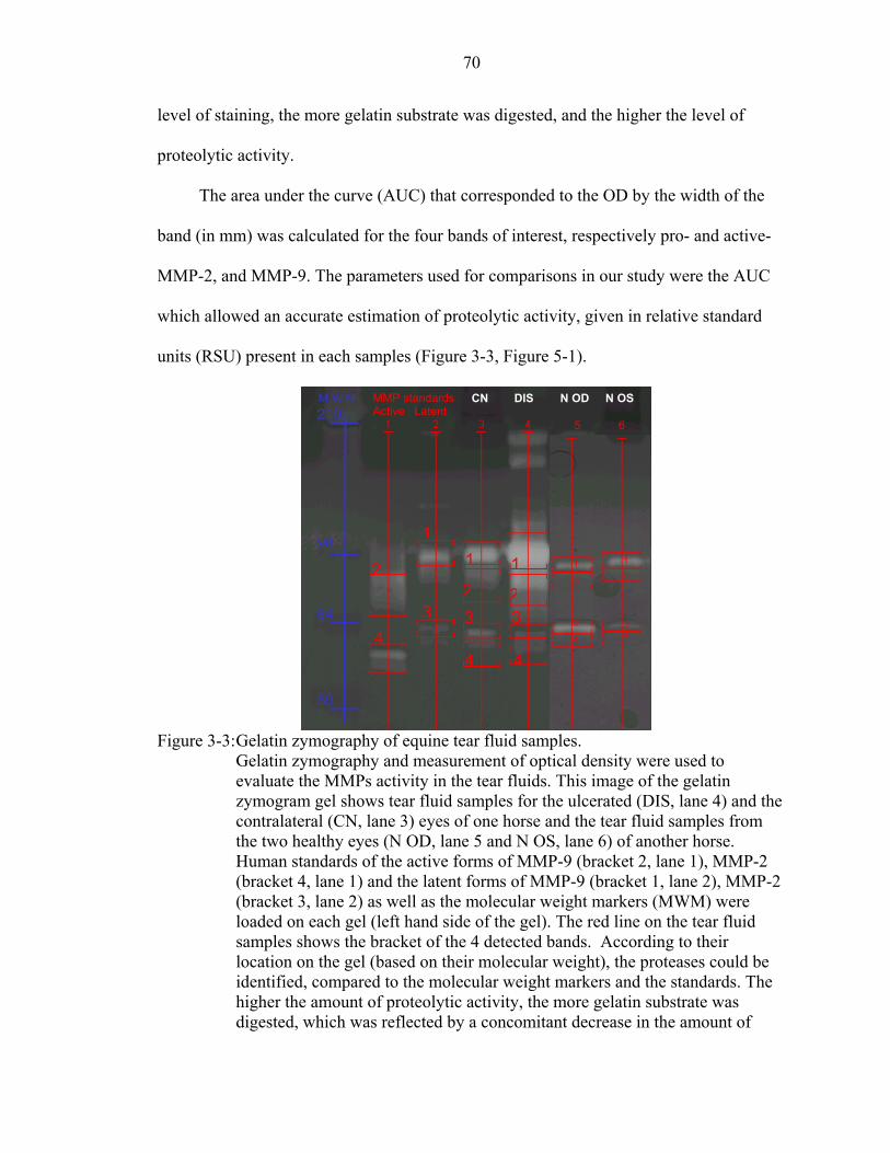
70
level of staining, the more gelatin substrate was digested, and the higher the level of
proteolytic activity.
The area under the curve (AUC) that corresponded to the OD by the width of the
band (in mm) was calculated for the four bands of interest, respectively pro- and active-
MMP-2, and MMP-9. The parameters used for comparisons in our study were the AUC
which allowed an accurate estimation of proteolytic activity, given in relative standard
units (RSU) present in each samples (Figure 3-3, Figure 5-1).
Figure 3-3: Gelatin zymography of equine tear fluid samples.
Gelatin zymography and measurement of optical density were used to evaluate the MMPs activity in the tear fluids. This image of the gelatin zymogram gel shows tear fluid samples for the ulcerated (DIS, lane 4) and the contralateral (CN, lane 3) eyes of one horse and the tear fluid samples from the two healthy eyes (N OD, lane 5 and N OS, lane 6) of another horse. Human standards of the active forms of MMP-9 (bracket 2, lane 1), MMP-2 (bracket 4, lane 1) and the latent forms of MMP-9 (bracket 1, lane 2), MMP-2 (bracket 3, lane 2) as well as the molecular weight markers (MWM) were loaded on each gel (left hand side of the gel). The red line on the tear fluid samples shows the bracket of the 4 detected bands. According to their location on the gel (based on their molecular weight), the proteases could be identified, compared to the molecular weight markers and the standards. The higher the amount of proteolytic activity, the more gelatin substrate was digested, which was reflected by a concomitant decrease in the amount of
2
4
1
3
1
3
4
21
23
4
210 M W M
98
64
50
5 6 N OD N OS MMP standard s
Active Latent CN DIS

71
stain. The zymogram gels were then scanned and the levels of each protease (or band) detected on the gel determined by measuring the optical density, transformed to a relative standard unit (RSU).The level of proteolytic activity for each of the protease detected is increased in the tear fluid samples from the diseased and the contralateral eyes in compared to the level in the healthy eyes.
Statistical Analysis
The ratios of the AUC of the band detected in the sample divided by the AUC of
the respective standard loaded on the same gel were calculated for the activity of each
individual protease (or band) detected as well as for the global proteolytic activity (sum
of all the 4 bands detected in a lane).
All analyses were run using the SAS software (SAS Institute Inc., Cary, NC) and
the tests were considered statistically significant if the p-value was less than 0.05.
The TFF as well as the ratios obtained for healthy right eyes (Normal OD) and
healthy left eyes (Normal OS) were compared by T-test analysis (SAS Proc TTEST).
Multiple linear regression analysis (SAS Proc GLM) was used to compare ratios
obtained for healthy eyes of various breeds, sex categories and age classes.
Ratios obtained for healthy eyes (N), healthy contralateral eyes (CN), and ulcerated
eyes (DIS) were compared by multiple linear regression analysis. The TFF as well as the
“release of proteolytic activity” were also compared by multiple linear regression
analysis (SAS Proc GLM).
Results
Determination of MMP-2 and MMP-9 Activity in the Equine Tear Fluid of Horses with Healthy Eyes
There was no significant difference in the global proteolytic activity (sum of the
activity for the active and latent forms of MMP-2 and MMP-9) between the right eye (N

72
OD) and the left eye (N OS) (n=65, p=0.0706) as well as for each individual protease as
illustrated in Table 3-3. Therefore, the values for the right and the left healthy eyes were
added to form one group of healthy eyes (Normal, n=130), and that was used in the
further analyses.
Table 3-3: Proteolytic activity (Mean +/- SD) in the tear fluid of horses with healthy eyes, in relative standard unit (RSU)
Eye
Global MMP
activity Latent MMP-9
Active MMP-9
Latent MMP-2
Active MMP-2
Normal OD (n=65) 0.69 ±0.51 2.22 ±1.89 0.47 ±0.53 1.16 ±2.08 0.07 ±0.12 Normal OS (n=65) 0.56 ±0.42 1.83 ±1.50 0.39 ±0.51 1.01 ±1.57 0.05 ±0.07 Normal (n=130) 0.62 ±0.47 2.02 ±1.71 0.43 ±0.52 1.09 ±1.84 0.06 ±0.10
There was no significant difference in the global proteolytic activity between the
breeds (p=0.4465) and the sex categories (p=0.1543) of the horses with healthy eyes
included in this study as illustrated in Figures 3-4 and 3-5, respectively. It is important to
note that the power to detect a difference was limited by the types of breeds and ages of
the horses available (Table 3-1, Tennessee Walking Horse n=1; stallion n=3).
0
0.2
0.4
0.6
0.8
1
1.2
1.4
Pony (n = 2) QH (n = 3) TB (n = 59) TW (n = 1)
Glo
bal M
MP
activ
ity (R
SU)
Figure 3-4: Global MMP activity in the tear fluid of horses with healthy eyes by breed.
TB= Thoroughbred, QH= American Quarter Horse, TW=Tennessee Walking Horse
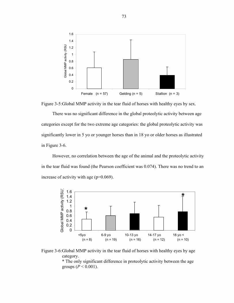
73
0
0.2
0.4
0.6
0.8
1
1.2
1.4
1.6
Female (n = 57) Gelding (n = 5) Stallion (n = 3)
Glo
bal M
MP
activ
ity (R
SU
Figure 3-5: Global MMP activity in the tear fluid of horses with healthy eyes by sex.
There was no significant difference in the global proteolytic activity between age
categories except for the two extreme age categories: the global proteolytic activity was
significantly lower in 5 yo or younger horses than in 18 yo or older horses as illustrated
in Figure 3-6.
However, no correlation between the age of the animal and the proteolytic activity
in the tear fluid was found (the Pearson coefficient was 0.074). There was no trend to an
increase of activity with age (p=0.069).
00.20.40.60.8
11.21.41.6
<5yo (n = 8)
6-9 yo (n = 19)
10-13 yo (n = 16)
14-17 yo (n = 12)
18 yo < (n = 10)
Glo
bal M
MP
act
ivity
(RS
U)
**
Figure 3-6: Global MMP activity in the tear fluid of horses with healthy eyes by age
category. * The only significant difference in proteolytic activity between the age groups (P < 0.001).

74
Determination of MMP-2 and MMP-9 Activity in the Equine Tear Fluid of Horses Ulcerative Keratitis
The global proteolytic activity in the tear fluid from diseased eyes was significantly
higher than from normal eyes (p<0.0001) and contralateral eyes (p<0.0001). The global
proteolytic activity in the tear fluid from the contralateral eyes was higher than from
normal eyes but the difference was not significant (p=0.1908) as illustrated in Table 3-4
and Figure 3-7. Similar results were obtained for each individual protease (p<0.0001)
(Table 3-4 and Figure 3-8).The proteolytic activity in the equine tear film is mainly due
to the latent forms of MMP-9 and MMP-2 (Table 3-4 and Figure 3-8).
Table 3-4: Proteolytic activity in the tear fluid of healthy, ulcerated and
contralateral normal horse eyes.
Eye status
Global MMP activity
Latent MMP-9
Active MMP-9
Latent MMP-2
Active MMP-2
Normal (n=130) 0.62 ±0.47 2.02 ±1.71 0.43 ±0.52 1.09 ±1.84 0.06 ±0.10
CN (n=37) 0.79 ±0.67 2.29 ±2.19 0.51 ±0.91 1.89 ±2.48 0.17 ±0.52 Diseased (n=39) 1.92 ±1.06* 6.30 ±4.49* 1.05 ±0.86* 9.30 ±27.57* 0.71 ±2.97*
Proteolytic activity (Mean +/- SD) in the tear fluid of healthy (Normal), ulcerated and contralateral normal (CN) eyes, in relative standard unit (RSU). The proteolytic activity in the equine tear film is mainly due to the latent forms of MMP-2 and MMP-9. * Proteolytic activity in the tear fluid from diseased eyes differed significantly (P < 0.001) from the proteolytic activity in the tear fluid from normal eyes.
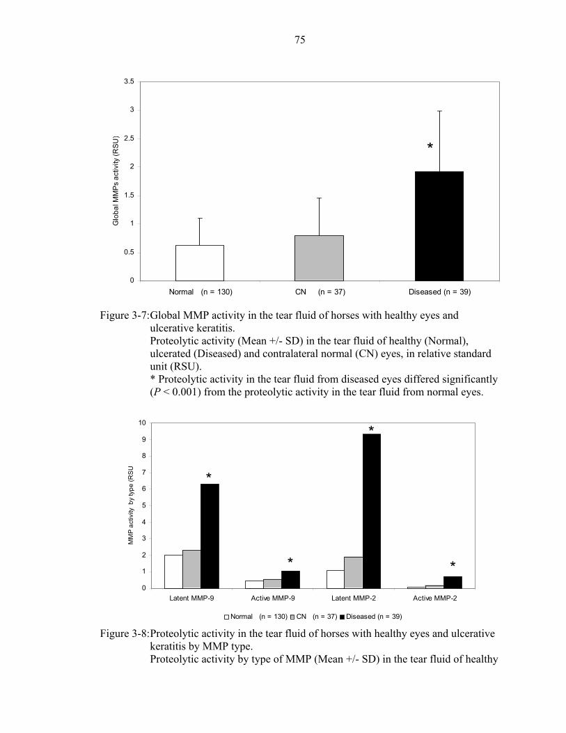
75
0
0.5
1
1.5
2
2.5
3
3.5
Normal (n = 130) CN (n = 37) Diseased (n = 39)
Glo
bal M
MPs
act
ivity
(RSU
)
*
Figure 3-7: Global MMP activity in the tear fluid of horses with healthy eyes and
ulcerative keratitis. Proteolytic activity (Mean +/- SD) in the tear fluid of healthy (Normal), ulcerated (Diseased) and contralateral normal (CN) eyes, in relative standard unit (RSU). * Proteolytic activity in the tear fluid from diseased eyes differed significantly (P < 0.001) from the proteolytic activity in the tear fluid from normal eyes.
0
1
2
3
4
5
6
7
8
9
10
Latent MMP-9 Active MMP-9 Latent MMP-2 Active MMP-2
MM
P a
ctiv
ity b
y ty
pe (R
SU
Normal (n = 130) CN (n = 37) Diseased (n = 39)
*
*
* *
Figure 3-8: Proteolytic activity in the tear fluid of horses with healthy eyes and ulcerative
keratitis by MMP type. Proteolytic activity by type of MMP (Mean +/- SD) in the tear fluid of healthy

76
(Normal), ulcerated and contralateral normal (CN) eyes, in relative standard unit (RSU). The proteolytic activity in the equine tear film is mainly due to the latent forms of MMP-2 and MMP-9. *Proteolytic activity for each protease in the tear fluid from diseased eyes differed significantly (P < 0.001) from the proteolytic activity for each protease in the tear fluid from normal eyes.
Determination of TFF in Horses with Healthy Eyes and Horses with Ulcerative Keratitis
There was no significant difference in the tear fluid flow (TFF) between the right
eye (N OD) and the left eye (N OS) (p=0.627) in the 41 healthy horses where the time
needed for the tear collection was recorded, and the TFF calculated as illustrated in Table
3-5. Therefore, the TFF values for the right and the left healthy eyes were added to form
one group of healthy eyes (Normal, n=82) and that was used in the further analyses.
The TFF in diseased eyes was significantly higher than in normal eyes (p<0.0001)
and contralateral eyes (p<0.0001) in the 25 horses with ulcerative keratitis where the time
needed for the tear collection was recorded, and the TFF calculated as illustrated in Table
3-5. The TFF in the contralateral eyes was higher in normal eyes, but the difference was
not significant (p=0.4562) as illustrated in Table 3-5.
Table 3-5: TFF in healthy, ulcerated and contralateral normal horse eyes.
Eye status
TFF (µL/s) (Mean +/- SD)
Normal OD (n = 41) 1.79 +/- 1.81 Normal OS (n = 41) 1.40 +/- 0.99
Normal (n = 82) 1.59 +/- 1.46
CN (n = 24) 2.25 +/- 2.37 Diseased (n = 26) 5.17 +/- 4.09 *
TFF (Mean +/- SD) in healthy (Normal), ulcerated (Diseased), and contralateral normal (CN) eyes, in µL per second. * TFF in diseased eyes differed significantly (P < 0.001) from TFF in normal eyes.

77
Determination of The “Release of Proteolytic Activity” in the Equine Tear Fluid of Horses with Healthy Eyes and Horses with Ulcerative Keratitis
The figure 3-9 reports the global MMP activity in the tear fluid in parallel to the
TFF and this illustrates that the proteolytic activity is maintained high in the tear fluid of
the diseased eyes in spite of a high dilution factor due to enhanced TFF.
0
1
2
3
4
5
6
7
8
9
10
Normal (n = 82) CN (n = 24) Diseased (n = 26)
TFF
(uL/
s) o
r Glo
bal p
rote
olyt
ic a
ctiv
ity (R
SU)
Total MMPs activity TFF
*
**
Figure 3-9: TFF and global MMP activity in the tear fluid of horses with healthy eyes and
ulcerative keratitis. Global proteolytic activity (Mean +/- SD) in the tear fluid of healthy (Normal), ulcerated (Diseased) and contralateral normal (CN) eyes, in relative standard unit (RSU) as well as the TFF in µL/s. The proteolytic activity is maintained high in the tear fluid of the diseased eyes in spite of a high dilution factor due to enhanced TFF. * Proteolytic activity in the tear fluid from diseased eyes differed significantly (P < 0.001) from the proteolytic activity in the tear fluid from normal eyes. ** TFF in diseased eyes differed significantly (P < 0.001) from TFF in normal eyes.
The global proteolytic activity released (global MMP activity multiplied by the
TFF) in the tear fluid from diseased eyes was significantly higher than from normal eyes
(p<0.0001) and contralateral eyes (p<0.0001). The total proteolytic activity released in
the tear fluid from the contralateral eyes was higher than from normal eyes, but the
difference was not significant (p=0.6056) as illustrated in Table 3-6.

78
Table 3-6: Proteolytic activity released in the tear fluid of equine healthy, ulcerated and contralateral normal eyes.
Eye status Mean +/- SD Normal (n = 82) 0.77 +/- 0.89 CN (n = 24) 1.39 +/- 1.73 Diseased (n = 26) 10.52 +/- 11.85*
Global MMP activity released per second (Mean +/- SD) in the tear fluid healthy (Normal), ulcerated (Diseased), and contralateral normal (CN) horse eyes, in RSU per second. * The activity released in diseased eyes differed significantly (P < 0.001) from the release in normal eyes.
Discussion
A total of 103 horses were involved in this study: 65 horses had healthy eyes and
38 horses had unilateral ulcerative keratitis.
The horses presented with ulcerative keratitis had various diagnoses including
bacterial or fungal keratitis, sterile ulcerative keratitis and ulcerative keratitis of unknown
etiologic origin, all identified by a fluorescein stain uptake. In this study, we did not take
into consideration the etiology in the measurement of the proteolytic activity in the tears;
the tear fluid samples were only split into 3 categories: tears from healthy eyes, tears
from ulcerated eyes and those from the contralateral healthy eyes. The proteinases
present in the tear film can be host-derived and microbial. The importance of the etiology
of the ulcerative keratitis on the level of proteolytic activity in the tear film remains to be
investigated.
Several of these animals had not been treated prior to their first visit at the
University of Florida Veterinary Medical Teaching Hospital, but most of them had
received ineffective therapy prior to referral to our hospital. Therefore, they did not
respond to the treatment and the ulcerative keratitis was considered still active. For this

79
reason, we did not differentiate those horses that had received medical therapy and those
that did not prior to the tear collection.
We would like to stress the importance of a proper collection method for
investigation of the tear film proteolytic activity in horses. The method of tear collection
has been reported to affect the composition of tear fluid obtained (Berta, 1983; Jones et
al., 1997; Stuchell et al., 1984; van Haeringen and Glasius, 1976; van Setten et al., 1990).
An ideal method of tear collection would permit rapid collection with no ocular irritation,
thus permitting differentiation between basal and stimulated reflex tear proteinases level;
allow efficient recovery of the tear proteins (and proteinases); and be readily applied in a
clinical setting. Conventional methods for tear collection include filter paper or Schirmer
test strips, cellulose sponges, porous polyester rods and glass capillary micropipettes
(Jones et al., 1997).
Tear collection with filter paper or Schirmer test strips is far from ideal as it has
been shown that their use leads to activation of proteolytic systems in the tear film (van
Haeringen and Glasius, 1976; van Setten et al., 1990). It seems most likely that some
micro trauma to the conjunctival and corneal epithelia is caused by the mechanical
irritation of the strips that is sufficient to activate or liberate proteinases (Stuchell et al.,
1984; van Haeringen and Glasius, 1976). Furthermore, the TFF can be measured by filter
paper or Schirmer test strips but this method does not provide an estimation of the actual
TFF, since the insertion of the filter paper into the cul-de-sac stimulates reflex
lacrimation (Mishima et al., 1966; Stuchell et al., 1984). Similar observations have been
reported with the use of cellulose sponges to collect tears (van Agtmaal et al., 1987).
80
Compared to other methods, the use of blunted capillary tubes to collect tear fluids
has been shown to cause less irritation and cellular disruption. Similarly, the use of
porous polyester rods has been shown to collect tears atraumatically with minimal ocular
irritation (Jones et al., 1997). Even if the use of capillary tubes to collect tears seems a bit
awkward in a clinical setting, it ensures a high protein recovery (Jones et al., 1997). The
microcapillary tear collection method has been used in various clinical and experimental
studies performed in man and animals (Tervo et al., 1991; Tervo et al., 1994; van Setten
et al., 1989; van Setten et al., 1990; Vesaluoma et al., 1996; Vesaluoma et al., 1997;
Vesaluoma and Tervo, 1998). It seems to be the method of choice to collect tear fluid
sample and investigate the tear film proteolytic activity (Tervo et al., 1991; van
Haeringen and Glasius, 1976; van Setten et al., 1990).
After collecting 575 tear fluid specimens over a 3 year period, we consider that we
possess a valid and standardized method to collect tears with 20µL blunted glass capillary
tubes in horses for the measurement of the equine tear film proteolytic activity.
Moreover, this microcapillary tear collection method allows the recording of the
time needed for collection and therefore the measurement of the tear fluid flow (TFF) in
the capillary. The TFF in the capillary tube has been measured in various studies
performed in man and animals (Tervo et al., 1994; van Setten et al., 1990; Vesaluoma et
al., 1996; Vesaluoma et al., 1997; Vesaluoma and Tervo, 1998) and served as a relative
value of the TFF during the sample collection. For the first time in this study, we reported
TFF values for the horse. This study documents that the TFF is increased in the eye of
horses with ulcerative keratitis as compared to healthy and contralateral normal eyes.
Similar observations have been reported in other species (Tervo et al., 1994; van Setten et

81
al. 1990; van Setten et al. 1989; Vesaluoma et al., 1996; Vesaluoma et al., 1997;
Vesaluoma and Tervo, 1998).
Although a rather gross estimate, the capillary tube method seems to be the
simplest way to have a reasonable volume correction in determination of the tear fluid
proteinases (Tervo et al., 1994; Vesaluoma et al., 1997). This is, to our understanding,
necessary in our study because of the clinically evident wide variation in the tear fluid
production between healthy and ulcerated eyes. Neglecting the tremendous variations in
the TFF rates and giving the proteolytic activity only might be more inaccurate. Since the
high TFF caused by reflex tearing in the diseased eye caused a dilution effect, we
calculated the “release of proteolytic activity” by multiplying the global MMP activity by
the TFF in the capillary (Tervo et al., 1991; Tervo et al., 1994). The parameter “release”
takes the TFF changes (the dilution factor) into consideration and probably gives a better
reflection of the changes in synthesis and release of the proteinases between the different
groups studied (healthy, diseased and contralateral normal equine eyes).
Despite the inaccuracy of the capillary tube method, we consider it a suitable
technique to collect tears for studying the proteolytic activity and the release of
proteolytic activity (i.e. activity/time) in the horse tear fluid, once the limitations of the
technique are recognized.
This study documents the proteolytic activity due to MMP-2 and MMP-9 in the tear
film of horses with healthy and ulcerated eyes.
We detected some proteolytic activity in the tear fluid of healthy horses and no
significant difference in proteolytic activity between right and left was found. These
results are in accordance with those from Strubbe et al. who first reported the presence of
82
MMP-2 and MMP-9 in equine tears by gelatin zymography (Strubbe et al., 2000) by
analyzing the tear fluids of 17 healthy horses and 23 horses with unilateral keratitis. It is
interesting to note that proteolytic activity was present even in the healthy eyes and
similar results have been reported for in humans where some proteases always appear to
be present in the tear film (Tervo et al., 1988; van Setten et al., 1990). This support the
concept that proteases and in this particular case MMP-2 and MMP-9 are involved in the
corneal physiology such as the normal maintenance of the corneal ECM (Fini and Girard,
1992; Twining, 1994). We did not observed any significant difference in the proteolytic
activity level of healthy horses between the breeds as well as between the sex categories.
The power of the statistical analysis was certainly low as some categories had very low
numbers (Table 3-1) but this study was mainly designed to look at the difference between
healthy and ulcerated eyes and we were also limited by the distribution of the horse
breeds present in Florida and the ones presented at the VMTH. Even if there was a
significant difference that we were not able to detect with the statistical analysis because
of its low power, the maximal difference in MMP activity in healthy horses observed
between breeds and between sex was 0.45 RSU (Figure 3-4) and this difference was
much smaller (approximately 3 folds) in comparison with the difference observed (Table
3-4) between healthy and ulcerated eyes (1.30 RSU). For this particular reason, we
considered that it was still valid to group all the healthy animals in one category with out
taking into account eye side, breed and sex for the comparison horse with ulcerative
keratitis. We also noticed a significant difference of activity between the two extreme age
classes: the level of proteolytic activity was significantly higher in the healthy old horses
(more than 18 years old) than in the healthy young ones (less than 5 years old). However,

83
we did not find any correlation between age and tear film proteolytic activity. To our
knowledge, age-related changes in tear film proteinases have not been evaluated in any
species except in horses by Strubbe et al. Strubbe only reported a significantly higher
level of MMP-9 in the tear fluid of young horses when compared to horses over 10 years
of age (Strubbe et al., 2000). We did not obtain similar results in our study and we
unfortunately did not find any studies to explain this phenomenon.
In our study, MMP-9 and MMP-2 were present at a significantly higher level in the
tear film of horses with ulcerative keratitis versus horses with healthy eyes. These
results are similar to findings in other species (Fini and Girard, 1990; Fini et al., 1992;
Fini et al., et 1998; Matsubara et al., 1991a), and especially those reported in horses by
Strubbe et al. (Strubbe et al., 2000) suggesting that elevation of tear film proteinases is
part of a fundamental response of the mammalian eye to corneal injury. We also observed
an increased level of proteolytic activity in the contralateral normal eye of affected horses
compared to healthy eyes but the difference was not significant in contrast with Strubbe’s
findings. The higher number of animals involved in our study might explain the different
results.
When the TFF and therefore, the dilution factor, is taken into consideration during
the proteolytic activity analysis, we observed the same trends and the differences between
the healthy, ulcerated and contralateral normal were even more pronounced. The release
of proteolytic activity was significantly higher in the ulcerated eyes compared to healthy,
and contralateral normal eyes. The release of proteolytic activity in contralateral normal
eyes was also higher than that in the in healthy eyes but not significantly.

84
This study supports the fact that MMP-2 and MMP-9 (gelatinases A and B) are of
major importance in terms of remodeling and degradation of the corneal stromal collagen
in horses as described in other species (Fini and Girard, 1990; Fini and Girard, 1992). A
particular advantage of the gelatin zymography compared to immunohistochemistry is
that both the latent (or proenzyme) and active form of MMPs, which can be distinguished
on the basis of the molecular weight, can be detected. This is possible because the
proenzymes are activated in situ presumably by the denaturation/renaturation process
during the zymography. The latent forms of the MMPs do not have any bioactivity but it
is important to quantify them as they can potentially be activated by various factors.
Corneal ulcers in horses are associated with initially high levels of tear film proteolytic
activity due to MMP-2 and MMP-9 and this study supports the use of anti-proteolytic
agents for progressive equine corneal ulcers. Treatment strategies for healing corneal
ulcers in horses should be directed towards and reducing tear film concentrations of
MMPs. Several anti-proteolytic agents have been proposed as treatments to reduce the
activity of tear proteases. Because these compounds use different mechanisms to inhibit
various families of proteases in equine tears, a combination of these inhibitors may be
indicated for the treatment of severe corneal ulcers in horses (Brooks, 1999; Hibbetts et
al., 1999). Objectives for the appropriate use of these compounds early in the course of
corneal disease in horses would be to decrease the amount of time required for recovery
and rehabilitation, reduce scarring, and potentially alleviate the need for corneal surgical
treatment.

CHAPTER 4 MATRIX METALLOPROTEINASE ACTIVITY PROFILES IN THE EQUINE TEAR
FILM DURING CORNEAL HEALING IN 10 HORSES WITH ULCERATIVE KERATITIS
Introduction
Tissue breakdown occurs with the normal metabolic activity of the cornea.
Proteolytic enzymes are important in the slow turnover and remodeling of the normal
healthy corneal stroma. The activities of these proteolytic enzymes are normally balanced
by inherent proteinase inhibitors in order to prevent excessive degradation of the normal
healthy tissue. An imbalance between proteinases and proteinase inhibitor levels due to
excessive levels of proteinases can cause pathological degradation of corneal stromal
collagen and proteoglycans (Hibbetts et al., 1999; Twining, 1994; Slansky et al., 1969).
The tear film proteinases have been previously evaluated in both normal and
diseased eyes of animals and man (Berman et al., 1971; Berman et al., 1973; Berman et
al., 1977; Brooks 1999; Fini and Girard, 1990; Matsubara et al., 1991a; Matsumoto et al.,
1993; Prause, 1983a; Prause, 1983b; Strubbe et al, 2000; Tervo et al., 1988; Tervo et al.,
1991). In ulcerated horse corneas, tear film levels of MMP-2, MMP-9 and neutrophil
elastase are significantly elevated when compared to age-matched normal controls and
are hypothesized to contribute to the breakdown of stromal collagen (Strubbe et al, 2000).
Ulcerative keratitis with extensive stromal involvement displays rapid progressive that
can lead to corneal perforation in many horse eyes probably due to this upregulated
proteolytic activity (Brooks, 1999).
85

86
Many studies have been performed on the changes in MMP expression in the skin
as well as MMP proteolytic activity in the wound fluids during skin wound healing in
animals and man (Agren, 1994; Blalock et al., 2001; Ladwig et al., 2002; Lobmann et al.,
2002; Herouy, 2001; Parks, 1999; Paul et al., 1997; Wall, 2002). Few studies have
reported the proteolytic changes in the tear film during corneal wound healing. Changes
in the tear film level of plasmin, a serine proteinase, have been described in animal and
man (Barlati et al., 1990; Cejkova, 1998; Cejkova et al., 1993; Salonen et al., 1987; Tervo
et al. 1998; Tervo et al., 1989a; Tervo et al, 1989b; Tervo et al. 1991; Vesaluoma et al.,
1998; van Setten et al., 1989). The changes in level of various MMPs in the tear film
(Barro et al., 1998) as well as their expression of MMP in the cornea during wound
healing have also been described in rat and rabbit (Fini and Girard, 1992; Fini et al.,
1998; Lu et al., 1999; Matsubara et al., 1991a; Ye et al., 1998). To the author’s
knowledge, there is only one study on the precise profile of the MMP activity in tear film
during corneal healing based on the collection and analysis of serial tear fluid samples in
man (Barro et al., 1998).
Furthermore, changes in the amount of proteolytic activity in the horse tear films
during corneal healing and stromal remodeling have never been reported. Based on the
literature and the current knowledge, we hypothesize the tear film MMP activity levels
should decrease as the corneal ulcer epithelializes and remodels. In this study, we
analyzed tear protease activity during healing of corneal ulcers in horses to test this
hypothesis.
87
Materials and Methods
Selection of the Ten Cases
All procedures were carried out according to the ARVO statement for the Use of
Animals in Ophthalmic and Vision research and were approved by the University of
Florida Animal Care and Use Committee as well as the UF College of Veterinary
Medicine Clinical Research Review Committee.
Tear film MMP activity (for MMP-2 and MMP-9) was followed serially by
quantitative gelatin zymography until complete recovery of the ulcerative keratitis in ten
horses presented to the Ophthalmology Service of the University of Florida Veterinary
Medicine Teaching Hospital during 2002. The horses had various diagnoses including
bacterial or fungal keratitis, sterile ulcerative keratitis and ulcerative keratitis of unknown
etiologic origin, all identified by a fluorescein stain uptake. Table 4-1 reports the details
of the 10 cases involved in this study (Table 4-1). The evolution of the corneal disease in
these 10 horses was monitored by ophthalmic examination including slit-lamp
biomicroscopy, fluorescein staining and photography. The complete recovery of the
ulcerative keratitis was confirmed by negative results for retention of fluorescein dye.
Treatment of the ulcerative keratitis in these 10 cases included topical
administrations of equine serum, antibiotics, antifungals, atropine, and systemic
administration of non-steroidal anti-inflammatory drugs. Surgical treatment was
performed on day 2 in addition to medical treatment in 5 cases. The surgical treatments
included various techniques such as conjunctival pedicle graft, amniotic membrane
transplantation, keratectomy, and penetrating keratoplasty (Table 4-1).
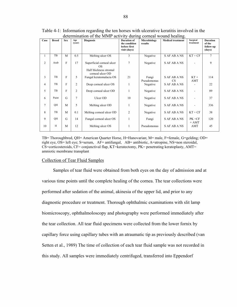
88
Table 4-1: Information regarding the ten horses with ulcerative keratitis involved in the determination of the MMP activity during corneal wound healing.
Case Breed Sex Age (year)
Diagnosis Duration of the condition before first visit (days)
Microbiology results
Medical treatment Surgical treatment
Duration of the follow-up (days)
1 TB M 0.5 Melting ulcer OS 3 Negative S AF AB A NS KT + CF 7
2 Arab F 17 Superficial corneal ulcer OS
Half thickness stromal corneal ulcer OD
7 Negative S AF AB A NS - 9
3 TB F 5 Fungal keratomalacia OS 21 Fungi Pseudomonas
S AF AB A NS CS
KT + AMT
114
4 TB F 2 Deep corneal ulcer OS 1 Negative S AF AB A NS - 22
5 TB F 2 Deep corneal ulcer OD 1 Negative S AF AB A NS - 89
6 Paint G 7 Ulcer OD 10 Negative S AF AB A NS - 37
7 QH M 5 Melting ulcer OD 1 Negative S AF AB A NS - 336
8 TB M 0.1 Melting corneal ulcer OD 2 Negative S AF AB A NS KT + CF 38
9 QH G 14 Fungal corneal ulcer OS 1 Fungi S AF AB A NS PK +CF + AMT
120
10 H M 12 Melting ulcer OS 1 Pseudomonas S AF AB A NS AMT 45
TB= Thoroughbred, QH= American Quarter Horse, H=Hanovarian; M= male, F=female, G=gelding; OD= right eye, OS= left eye; S=serum, AF= antifungal, AB= antibiotic, A=atropine, NS=non steroidal, CS=corticosteroids, CF= conjunctival flap, KT=keratectomy, PK= penetrating keratoplasty, AMT= amniotic membrane transplant Collection of Tear Fluid Samples
Samples of tear fluid were obtained from both eyes on the day of admission and at
various time points until the complete healing of the cornea. The tear collections were
performed after sedation of the animal, akinesia of the upper lid, and prior to any
diagnostic procedure or treatment. Thorough ophthalmic examinations with slit lamp
biomicroscopy, ophthalmoloscopy and photography were performed immediately after
the tear collection. All tear fluid specimens were collected from the lower fornix by
capillary force using capillary tubes with an atraumatic tip as previously described (van
Setten et al., 1989) The time of collection of each tear fluid sample was not recorded in
this study. All samples were immediately centrifuged, transferred into Eppendorf
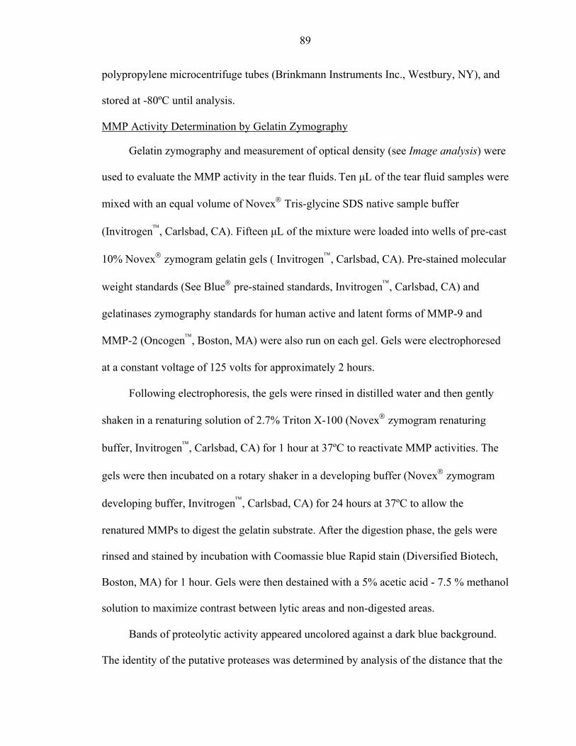
89
polypropylene microcentrifuge tubes (Brinkmann Instruments Inc., Westbury, NY), and
stored at -80ºC until analysis.
MMP Activity Determination by Gelatin Zymography
Gelatin zymography and measurement of optical density (see Image analysis) were
used to evaluate the MMP activity in the tear fluids. Ten µL of the tear fluid samples were
mixed with an equal volume of Novex Tris-glycine SDS native sample buffer
(Invitrogen, Carlsbad, CA). Fifteen µL of the mixture were loaded into wells of pre-cast
10% Novex zymogram gelatin gels ( Invitrogen, Carlsbad, CA). Pre-stained molecular
weight standards (See Blue pre-stained standards, Invitrogen, Carlsbad, CA) and
gelatinases zymography standards for human active and latent forms of MMP-9 and
MMP-2 (Oncogen, Boston, MA) were also run on each gel. Gels were electrophoresed
at a constant voltage of 125 volts for approximately 2 hours.
Following electrophoresis, the gels were rinsed in distilled water and then gently
shaken in a renaturing solution of 2.7% Triton X-100 (Novex zymogram renaturing
buffer, Invitrogen, Carlsbad, CA) for 1 hour at 37ºC to reactivate MMP activities. The
gels were then incubated on a rotary shaker in a developing buffer (Novex zymogram
developing buffer, Invitrogen, Carlsbad, CA) for 24 hours at 37ºC to allow the
renatured MMPs to digest the gelatin substrate. After the digestion phase, the gels were
rinsed and stained by incubation with Coomassie blue Rapid stain (Diversified Biotech,
Boston, MA) for 1 hour. Gels were then destained with a 5% acetic acid - 7.5 % methanol
solution to maximize contrast between lytic areas and non-digested areas.
Bands of proteolytic activity appeared uncolored against a dark blue background.
The identity of the putative proteases was determined by analysis of the distance that the
90
bands migrated on the gels, compared with the distance for migration of molecular
weight and protease standards (Figure 4-1).
Image Analysis
The levels of pro- and active- MMP-2, and MMP-9 were measured by optical
density scanning of gelatin zymograms of the tears samples. Digital photographs of
stained gelatin zymograms were created with a GS-710 imaging densitometer (Bio-Rad,
Hercules, CA) and analyzed with the Quantity One quantification software, 4.2.1 beta
version (Bio-Rad, Hercules, CA). The four bands present in each lane of the gels were
automatically detected by the analyzer after adjusting its sensitivity for each gel. The
background was then subtracted for each lane triplicate. The image analysis produced an
intensity profile curve of each lane with the optical density (OD) function of the distance
of migration from the top of the gel (relative front). The higher the optical density, the
lower level of staining, the more gelatin substrate was digested, and the higher the level
of proteolytic activity.
The area under the curve (AUC) that corresponded to the OD by the width of the
band (in mm) was calculated for the four bands of interest, respectively pro- and active-
MMP-2, and MMP-9. The parameters used for comparisons in our study were the AUC
which allowed an accurate estimation of proteolytic activity, given in relative standard
units (RSU) present in each samples (Figure 4-1). The graphs represent the tear film
MMP activity profile in RSU in both eyes for each horse function of the time (Figures 4-
2 to 4-11).
Statistical Analysis
The ratios of the AUC of the band detected in the sample divided by the AUC of
the respective standard loaded on the same gel were calculated for the activity of each
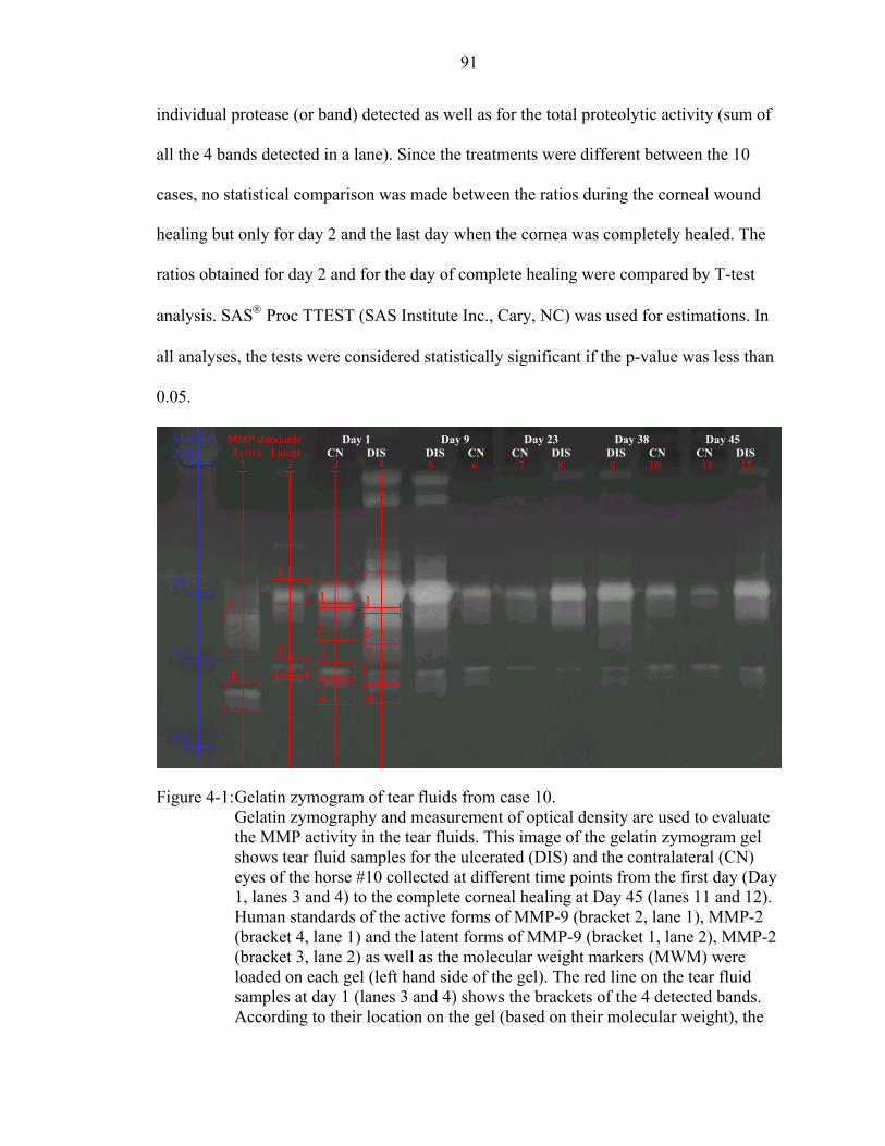
91
individual protease (or band) detected as well as for the total proteolytic activity (sum of
all the 4 bands detected in a lane). Since the treatments were different between the 10
cases, no statistical comparison was made between the ratios during the corneal wound
healing but only for day 2 and the last day when the cornea was completely healed. The
ratios obtained for day 2 and for the day of complete healing were compared by T-test
analysis. SAS Proc TTEST (SAS Institute Inc., Cary, NC) was used for estimations. In
all analyses, the tests were considered statistically significant if the p-value was less than
0.05.
Figure 4-1: Gelatin zymogram of tear fluids from case 10.
Gelatin zymography and measurement of optical density are used to evaluate the MMP activity in the tear fluids. This image of the gelatin zymogram gel shows tear fluid samples for the ulcerated (DIS) and the contralateral (CN) eyes of the horse #10 collected at different time points from the first day (Day 1, lanes 3 and 4) to the complete corneal healing at Day 45 (lanes 11 and 12). Human standards of the active forms of MMP-9 (bracket 2, lane 1), MMP-2 (bracket 4, lane 1) and the latent forms of MMP-9 (bracket 1, lane 2), MMP-2 (bracket 3, lane 2) as well as the molecular weight markers (MWM) were loaded on each gel (left hand side of the gel). The red line on the tear fluid samples at day 1 (lanes 3 and 4) shows the brackets of the 4 detected bands. According to their location on the gel (based on their molecular weight), the
210 M W M MMP standards Day 38 Day 45 Day 1 Day 9 Day 23
Active Latent
98
64
50
2
4
1
3
1
3
4
2
1
23
4
CN DIS DIS CN CN DIS 5 6 7 8 9 10
DIS CN 11 12
CN DIS

92
proteases could be identified. The zymogram gels are then scanned and the levels of each protease (or band) detected on the gel determined by measuring the optical density, transformed to a relative standard unit (RSU), and the values for each eye and time point reported in figures 2 to 11.
Results
Total tear film MMP activity decreased as corneal healing progressed compared to
levels measured on Day 1 (Figures 4-2 to 4-11). The general trend was a decrease.
However, different patterns of tear film MMP activity were observed. There was an
increase in proteolytic activity after keratectomy (case 3, Figure 4-4) and penetrating
keratoplasty (case 9, Figure 4-10) but not after amniotic membrane transplant or
conjunctival graft surgery (cases 1, 8 and 10, Figure 4-2, 4-9 and 4-11 respectively).
Table 4-2 reports the level of MMP activity in RSU for both eyes of each animal as
well as the mean and standard deviation (SD) for the group of 10 horses at Day 1 and at
day of complete corneal healing. The mean of the total MMP activity (±SD) measured in
the tear fluid of the ulcerated eye (2.44 ±1.44) of the 10 horses was significantly higher
than the one in the contralateral eye (0.81 ± 0.68) (p=0.006) on the first day of admission
to the VMTH. The mean MMP activity in these ulcerated eyes significantly decreased (-
82.4%) between the first of admission and the day when the ulcer was completely healed
(p=0.0002). The level activity in the healed eye (0.43 ± 0.17) was not significantly
different to the one in the contralateral eye (0.36 ± 0.18) at the day of complete corneal
healing (p=0.374). The level of MMP activity in the contralateral eye decreased from
0.81 ± 0.68 to 0.36 ± 0.18, although this 56.0% decrease was not significant (p=0.069)
(Table 4-2).

93
Table 4-2: Level of total MMP activity in relative standard units (RSU) at day 2 and at the day of complete corneal healing in both eyes of 10 horses.
Case
Eye
Eye status
Total MMP activity (RSU) at
Day 2
Total MMP activity (RSU) at
Day of complete healing
% change in Total MMP activity
1 OS D 0.83 0.52 - 37.7 OD CN 0.7 0.43 - 38.0
2 OS D 0.59 0.12 - 80.0 OD D 0.47 0.23 - 51.6
3 OD CN 0.21 0.34 + 63.3 OS D 4.26 0.41 - 90.3
4 OD CN 0.44 0.68 + 55.2 OS D 2.53 0.33 - 86.7
5 OS CN 0.49 0.54 + 10.8 OD D 2.75 0.51 - 81.3
6 OD D 3.27 0.37 - 88.8 OS CN 0.89 0.13 - 85.4
7 OD D 2.53 0.67 -73.4 OS CN 1.63 0.36 - 77.7
8 OD D 1.53 0.66 - 56.9 OS CN 0.27 0.34 + 27.8
9 OD CN 2.22 0.27 - 87.6 OS D 3.51 0.54 - 84.7
10 OD CN 0.47 0.10 -77.6 OS D 4.61 0.37 - 91.9
Mean ± SD (D) CI
2.44 ±1.44 1.48 – 3.41
0.43 ± 0.17 0.32 – 0.54
- 82.4
Mean ± SD(CN) CI
0.81 ± 0.68 0.29 – 1.33
0.36 ± 0.18 0.22 – 0.50
- 56.0
OS= left eye, OD= right eye; D= diseased eye, CN=contralateral normal. CI = 95% confidence interval

94
Total MMPs activity - Case 1
0
0.1
0.2
0.3
0.4
0.5
0.6
0.7
0.8
0.9
1 2 3 7
Days
MM
Ps a
ctiv
ity (R
SU)
OD OS*
CF
*= ulcerated eye= Fluorescein staining negative
a b
c dFigure 4-2: Case 1 - A 6 month Thoroughbred colt presented with a melting corneal ulcer
in the left eye (OS). This animal received a medical treatment, and then a conjunctival graft (CF) on the day after admission. The pictures on the right illustrate the clinical evolution of the ulceration: day 1 (a), day 1 with fluorescein (b), day 2 (c), and day 45 (d).
Total MMPs activity - Case 2
0
0.1
0.2
0.3
0.4
0.5
0.6
0.7
1 5 9
Days
MM
Ps a
ctiv
ity (R
SU)
OD* OS*
a b
*= ulcerated eye= Fluorescein staining negative
Figure 4-3: Case 2 - A 17yo Arabian mare presented with a superficial corneal ulcer in the left eye (OS) and a half thickness stromal corneal ulcer in the right eye (OD). This animal received only a medical treatment. The pictures on the right illustrate the clinical evolution of the ulceration: day 1 OD (a), day 1 OS (b), day 5 OD (c), and day 5 OS (d).
c d

95
Total MMPs activity - Case 3
00.5
11.5
22.5
33.5
44.5
1 2 4 7 11 12 114
Days
MM
Ps a
ctiv
ity (R
SU)
OD OS*
KT - AMT
*= ulcerated eye= Fluorescein staining negative
a b
Figure 4-4: Case 3 - A 5yo Thoroughbred mare presented with a fungal keratomalacia in the left eye (OS). This animal received medical treatment followed by a keratectomy and amniotic membrane transplantation (K+AMT) on the second day. The pictures on the right illustrate the clinical evolution of the ulceration: day 1 (a), day 2 (b), day 7 (c), and day 114 (d).
c d
Total MMPs activity - Case 4
00.5
11.5
22.5
33.5
4
1 2 3 4 6 7 22
Days
MM
Ps a
ctiv
ity (R
SU)
OD OS*
a b
*= ulcerated eye= Fluorescein staining negative
Figure 4-5: Case 4 - A 2yo Thoroughbred filly presented a deep corneal ulcer in the left eye (OS). This animal received only medical treatment. The pictures on the right illustrate the clinical evolution of the ulceration: day 1 (a), day 2 (b), day 7 (c), and day 22 (d).
c d
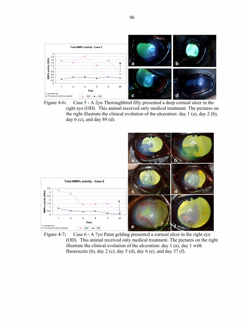
96
Total MMPs activity - Case 5
00.20.40.60.8
11.21.41.61.8
2
1 2 4 5 6 89
Days
MM
Ps a
ctiv
ity (R
SU)
OD* OS*= ulcerated eye
= Fluorescein staining negative Figure 4-6: Case 5 - A 2yo Thoroughbred filly presented a deep corneal ulcer in the
right eye (OD). This animal received only medical treatment. The pictures on the right illustrate the clinical evolution of the ulceration: day 1 (a), day 2 (b), day 6 (c), and day 89 (d).
a b
c d
a b
Total MMPs activity - Case 6
00.5
11.5
22.5
33.5
1 2 5 6 7 37
Days
MM
Ps a
ctiv
ity (R
SU)
OD* OS
c d
*= ulcerated eye= Fluorescein staining negative e f Figure 4-7: Case 6 - A 7yo Paint gelding presented a corneal ulcer in the right eye
(OD). This animal received only medical treatment. The pictures on the right illustrate the clinical evolution of the ulceration: day 1 (a), day 1 with fluorescein (b), day 2 (c), day 5 (d), day 6 (e), and day 37 (f).

97
Tobal MMPs activity - Case 7
0
0.5
1
1.5
2
2.5
3
1 2 3 336
Days
MM
Ps a
ctiv
ity (R
SU)
OD* OS*= ulcerated eye
= Fluorescein staining negative
a b
Figure 4-8: Case 7 – A 2 month Thoroughbred colt presented a melting corneal ulcer in the right eye (OD). This animal received medical treatment, and a conjunctival graft (CF) on the second day after admission. The pictures on the right illustrate the clinical evolution of the ulceration: day 1 (a), day 1 with fluorescein (b), day 3 (c), and day 38 (d).
Tobal MMPs activity - Case 8
0
0.5
1
1.5
2
2.5
3
1 2 3 4 38
Days
MM
Ps a
ctiv
ity (R
SU)
OD* OS
CF
*= ulcerated eye= Fluorescein staining negative
a b
c d
Figure 4-9: Case 8 – A 2 month Thoroughbred colt presented a melting corneal ulcer in the right eye (OD). This animal received medical treatment, and a conjunctival graft (CF) on the second day after admission. The pictures on the right illustrate the clinical evolution of the ulceration: day 1 (a), day 1 with fluorescein (b), day 3 (c), and day 38 (d).
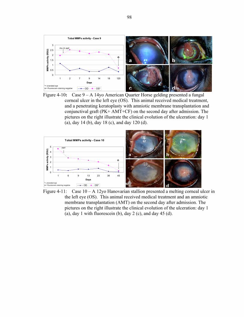
98
Tobal MMPs activity - Case 9
0
0.5
1
1.5
2
2.5
3
1 2 7 9 14 18 120
Days
MM
Ps a
ctiv
ity (R
SU)
OD OS*
PK-CF-AMT
*= ulcerated eye= Fluorescein staining negative
Figure 4-10: Case 9 – A 14yo American Quarter Horse gelding presented a fungal corneal ulcer in the left eye (OS). This animal received medical treatment, and a penetrating keratoplasty with amniotic membrane transplantation and conjunctival graft (PK+ AMT+CF) on the second day after admission. The pictures on the right illustrate the clinical evolution of the ulceration: day 1 (a), day 14 (b), day 18 (c), and day 120 (d).
a b
c d
Tobal MMPs activity - Case 10
0
1
2
3
4
5
1 6 9 13 23 38 45
Days
MM
Ps
activ
ity (R
SU)
OD OS*
AMT
*= ulcerated eye= Fluorescein staining negative
a b
Figure 4-11: Case 10 – A 12yo Hanovarian stallion presented a melting corneal ulcer in
the left eye (OS). This animal received medical treatment and an amniotic membrane transplantation (AMT) on the second day after admission. The pictures on the right illustrate the clinical evolution of the ulceration: day 1 (a), day 1 with fluorescein (b), day 2 (c), and day 45 (d).
c d

99
Discussion
Our results of MMP activity in the tear film of horses with ulcerative keratitis prior
to initiation of therapy are similar to results reported by Strubbe et al. that MMP-9 and
MMP-2 are present at a significantly higher level in the tear film of horses with ulcerative
keratitis (Strubbe et al., 2000). The majority of host-derived and microbial tear film
enzymes are MMPs. Enzymes of bacterial or fungal origin (i.e. exogenous proteinases)
can contribute to ulcerative keratitis, either directly or indirectly, through the activation of
corneal proteinases (i.e. endogenous proteinases) (Berman et al., 1971; Berman et al.,
1973; Berman et al., 1977; Brooks, 1999; Fini and Girard, 1990; Hibbetts et al., 1999;
Matsubara et al., 1991a; Matsumoto et al., 1993; Prause, 1983a; Prause, 1983b; Slansky
et al., 1969; Strubbe et al, 2000; Tervo et al., 1988; Tervo et al., 1991). Bacteria were
isolated from two cases (cases 3 and 10) and fungi isolated from two cases (cases 3 and
9).
Multiple studies have reported elevated levels of proteinases in tears from animals
and humans with ulcerative keratitis (Berman et al., 1971; Berman et al., 1973; Berman et
al., 1977; Brooks, 1999; Fini and Girard, 1990; Hibbetts et al., 1999; Matsubara et al.,
1991a; Matsumoto et al., 1993; Prause, 1983a; Prause, 1983b; Slansky et al., 1969;
Strubbe et al, 2000; Tervo et al., 1988) and changes in proteinase activity during the
healing process of the ulcerated cornea but precise MMP activity profiles based on the
collection and the analysis of many serial tear fluid samples have not been reported in
either animals or humans. We document for the first time that total MMP activity
decreases in equine tears as the corneal epithelial and stromal healing occur.
These results of corneal wound healing are in accordance with those previously
reported of skin wound healing (Agren, 1994; Blalock et al., 2001; Herouy, 2001;
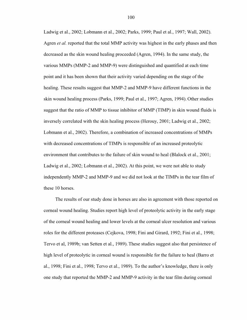
100
Ladwig et al., 2002; Lobmann et al., 2002; Parks, 1999; Paul et al., 1997; Wall, 2002).
Agren et al. reported that the total MMP activity was highest in the early phases and then
decreased as the skin wound healing proceeded (Agren, 1994). In the same study, the
various MMPs (MMP-2 and MMP-9) were distinguished and quantified at each time
point and it has been shown that their activity varied depending on the stage of the
healing. These results suggest that MMP-2 and MMP-9 have different functions in the
skin wound healing process (Parks, 1999; Paul et al., 1997; Agren, 1994). Other studies
suggest that the ratio of MMP to tissue inhibitor of MMP (TIMP) in skin wound fluids is
inversely correlated with the skin healing process (Herouy, 2001; Ladwig et al., 2002;
Lobmann et al., 2002). Therefore, a combination of increased concentrations of MMPs
with decreased concentrations of TIMPs is responsible of an increased proteolytic
environment that contributes to the failure of skin wound to heal (Blalock et al., 2001;
Ladwig et al., 2002; Lobmann et al., 2002). At this point, we were not able to study
independently MMP-2 and MMP-9 and we did not look at the TIMPs in the tear film of
these 10 horses.
The results of our study done in horses are also in agreement with those reported on
corneal wound healing. Studies report high level of proteolytic activity in the early stage
of the corneal wound healing and lower levels at the corneal ulcer resolution and various
roles for the different proteases (Cejkova, 1998; Fini and Girard, 1992; Fini et al., 1998;
Tervo et al, 1989b; van Setten et al., 1989). These studies suggest also that persistence of
high level of proteolytic in corneal wound is responsible for the failure to heal (Barro et
al., 1998; Fini et al., 1998; Tervo et al., 1989). To the author’s knowledge, there is only
one study that reported the MMP-2 and MMP-9 activity in the tear film during corneal
101
wound healing based on analysis of serial tear fluid samples (Barro et al., 1998). In this
study, Barro et al. showed that enhanced and persistent MMP-2 and MMP-9 activity in
tears correlated with corneal damage and graft failure (Barro et al., 1998).
It is also interesting to note that the MMP activity decrease during the corneal
wound healing is the most pronounced for the three cases with positive cultures (cases 3,
9 and 10). This might underline the participation of the microorganisms in the proteolytic
activity detected in the tear film as mentioned earlier.
Our data support the use of aggressive therapy to rapidly reduce the activity of tear
proteases in case of ulcerative keratitis. Equine serum, which contains strong and broad
protease inhibitors such as α2-macroglobulin and α1-proteinase inhibitor, was used
topically in all the cases presented here. Various other anti-protease compounds are also
available (N-acetylcysteine or NAC, potassium edatate diaminetetrataacetatic acid or
EDTA) that utilize different mechanisms to inhibit different families of proteases present
in equine tears. A combination of inhibitors may be indicated for severe corneal
ulceration in horses (Brooks, 1999; Hibbetts et al., 1999).
Medical and surgical treatments of the corneal ulcers in these horses lead to a
reduction in tear film proteolytic activity that corresponded with the improvement in the
clinical signs of corneal ulceration. Corneal ulcers in horses are associated with initially
high levels of tear film proteolytic activity which decrease as the ulcers heal.
Measurement of MMP activity in the equine tear film might represent a way to monitor
the progression of corneal healing in horses with ulcerative keratitis. Treatment strategies
for healing corneal ulcers in horses should be directed towards reducing microbial
activity, decreasing uveitis, and reducing tear film concentrations of MMPs as reduced

102
MMP activity is associated with improvement of the clinical signs of ulcerative keratitis
in horses.

CHAPTER 5 IN VITRO INHIBITION OF MATRIX METALLOPROTEINASE ACTIVITY IN THE
TEAR FLUID OF HORSES WITH ULCERATIVE KERATITIS
Introduction
Ulcerative keratitis is a common and often vision-threatening condition in horses.
The Ophthalmology Service of the University of Florida Veterinary Medical Teaching
Hospital diagnosed ulcerative keratitis in 527 horses between January 1987 and October
2002. This constituted 35% of all horses evaluated for ophthalmic problems during this
period. Superficial, non-infected ulcers in horses generally heal quickly and without
complication, whereas stromal degradation in deep or infected ulcers can rapidly and
dramatically progress to corneal perforation in horses within 24 hours (Brooks, 1999). A
pronounced fibrovascular response is also prominent during corneal healing in horses. An
understanding of the pathophysiologic processes as well as early diagnosis and
aggressive treatment are important for quick resolution of ulcerative corneal diseases in
horses and to speed healing, reduce scarring, and prevent corneal rupture.
Proteolytic enzymes perform important physiologic functions in normal tissues,
such as turnover and remodeling of the corneal stroma. Activities of proteolytic enzymes
are normally balanced by natural proteinase inhibitors, thus preventing excessive
degradation of normal healthy tissue. Excessive amounts of proteinases can create an
imbalance between proteinases and proteinase inhibitors, and increased amounts of
proteases are believed to cause pathologic degradation of collagen and proteoglycans in
the cornea (Slansky et al., 1969; Twining, 1994).
103
104
The cornea of horses manifests the most severe degree of ulcer-associated stromal
collagenolysis seen in animals (Brooks, 1999). This rapid degradation of the corneal
stroma in horses with corneal ulcers appears to be caused by various proteolytic enzymes
acting on the collagen, proteoglycans, and other components of the stromal extracellular
matrix. Microorganisms, inflammatory cells, corneal epithelial cells, and fibroblasts all
produce and release proteolytic enzymes (Hibbets et al., 1999; Matsubara et al., 1991a;
Matsubara et al., 1991b; Twining et al., 1993).
Two important families of enzymes that affect the cornea are the matrix
metalloproteinases (MMPs) and serine proteinases (including neutrophil elastase) (Fini
and Girard, 1990; Fini et al., 1998; Matsubara et al., 1991a). Two MMPs (MMP-2 [72-kd
gelatinase A] and MMP-9 [92-kd gelatinase B]) (Fini and Girard, 1990; Fini and Girard,
1992) are of major importance in terms of remodeling and degradation of the corneal
stromal collagen. The origin and purpose of MMP-2 and –9 appear to differ at the corneal
level. Matrix metalloproteinase-2 is synthesized by corneal keratocytes and performs a
surveillance function in the normal cornea, becoming locally activated to degrade
collagen molecules that occasionally become damaged as a result of normal wear and tear
(Azar et al., 1998; Matsubara et al., 1991b; Twining, 1994). Alternatively, MMP-9 is
produced by epithelial cells and polymorphonuclear neutrophils (PMNs) following
corneal wounding (Fini and Girard, 1990; Matsubara et al., 1991b).
Proteinases have been evaluated in the tear film of normal and diseased eyes of
humans and other animals (Berman et al., 1971; Berman et al., 1977; Berman et al., 1973;
Fini and Girard, 1990; Matsubara et al., 1991a; Prause, 1983; Strubbe et al, 2000; Tervo
et al., 1998). In severely damaged corneas, proteinase activities in the tear film were

105
significantly increased (Berman et al., 1971; Kernacki et al., 1995; Matsubara et al.,
1991a; Prause, 1983a; Tervo et al., 1998). In another study (Strubbe et al., 2000) in
horses, higher amounts of MMP-2, MMP-9, and neutrophil elastase were found in the
tear film of ulcerated eyes, compared with values for the tear film of age-matched normal
control horses.
Proteinase activity in the tear film is believed to speed up degradation of stromal
collagen, leading to rapid progression of ulcers. Normalizing proteolytic activity in the
tear film is an objective of the treatment of corneal ulcers in horses. Thus, proteinase
inhibitors have been recommended for treatment of ulcerative keratitis to reduce the
progression of stromal ulcers, speed epithelial healing, and minimize corneal scarring
(Berman, 1975; Berman, 1978; Berman et al., 1973; Berman et al., 1975; Brooks, 1999;
Tervo et al., 1992; Ward, 1999).
However, studies on activity of specific enzymes are limited. Accordingly,
recommendations for use of topically administered N-acetylcysteine (NAC), potassium
EDTA, and serum in horses with corneal ulcers are mostly based on anecdotal clinical
reports or extrapolations from data obtained from use of these compounds in other
species (Brooks, 1999; McLaughlin et al., 1992; Severin, 1976; Strubbe et al., 2000;
Tervo et al., 1992; Ward, 1999; Whitley and Gilger, 1999). The availability of newer
compounds such as doxycycline, a modified dipeptide that contains hydroxamic acid
(i.e., ilomostat), and α1-proteinase inhibitor (α1-PI), which initially have shown
considerable promise as MMP or serine-proteinase inhibitors in other species (Barletta et
al., 1996; Dursun et al., 2001; Golub et al., 1984; Perry et al., 1993; Rawal and Rawal,
1984; Schultz et al., 1992), outlines the need for specific studies on possible regulation of

106
enzyme activity in tears in horses with corneal disease. These new compounds have
potential as potent protease inhibitors in horses with corneal ulcers.
Therefore, in the study reported here, we evaluated the effectiveness of various
protease inhibitors for reducing the activity of MMP-2 and -9 in vitro in samples of tear
film obtained from horses with ulcerative keratitis.
Materials and Methods
Animals
All procedures were carried out according to the ARVO statement for the Use of
Animals in Ophthalmic and Vision research and were approved by the University of
Florida Animal Care and Use Committee as well as the UF College of Veterinary
Medicine Clinical Research Review Committee.
Tear samples were collected from the eyes of 34 horses with ulcerative keratitis.
Each horse was evaluated by members of the Ophthalmology Service of the University of
Florida Veterinary Medical Teaching Hospital during 2002. The horses had been
diagnosed with various conditions, including bacterial or fungal keratitis, sterile
ulcerative keratitis, and ulcerative keratitis of unknown etiologic origin.
Collection of Tear Fluid Samples
Horses were sedated, and akinesia of the upper eyelid was achieved prior to
collection of tear samples. Samples were obtained before any diagnostic procedure or
application of treatment; however, immediately after collection of tear samples, thorough
ophthalmic examinations that included slit-lamp biomicroscopy, tonometry, and
ophthalmoscopy were performed. Samples were collected only from eyes with ulcerative
keratitis, as identified by positive results for retention of fluorescein dye. All tear samples
were collected from the lower fornix by capillary force, using capillary tubes with an

107
atraumatic tip as described elsewhere (van Setten et al., 1989). Time of collection of each
sample was not recorded. All samples were immediately centrifuged, transferred into
polypropylene microcentrifuge tubes (Eppendorf tubes, Brinkmann Instruments Inc.,
Westbury, NY), and stored at –80oC until analysis. Tear samples collected from the eyes
of the 34 horses were pooled to yield a volume of 1,500µL.
Determination of MMP Activity and Inhibition Tests by Gelatin Zymography
Gelatin zymography and measurement of optical density (OD) were used to
evaluate MMP activity in untreated pooled tear samples and to conduct inhibition tests of
MMP activity in treated tear samples. Aliquots (10 µL) of the pooled tears were mixed
with an equal volume of SDS-sample buffer (Novex tris-glycine SDS native sample
buffer (2X), Invitrogen, Carlsbad, CA). Then, fifteen µL of the mixture was loaded into
wells of precast 10% zymogram gelatin gels (10% Novex zymogram gelatin gel,
Invitrogen, Carlsbad, CA). Untreated samples were constituted on each gel in 3 lanes that
remained untreated. Treated samples were constituted in 3 lanes that were treated with a
protease-inhibitor compound (triplicate pattern). Prestained molecular-weight standards
(See Blue prestained standards, Invitrogen, Carlsbad, CA) and gelatinase-zymography
standards for active (Active MMP-2 and active MMP-9 enzymes, Oncogen, Boston, MA)
and latent (Proenzyme MMP-2 and MMP-9, Oncogen, Boston, MA) forms of human
MMP-2 and -9 were also assayed on each gel.
Inhibitory compounds tested in the study included EDTA, doxycycline, NAC,
ilomostat, α1-PI, and fresh equine serum. A solution containing 0.2% EDTA was
obtained by filling an evacuated, EDTA-containing, 10-mL blood-collection tube
(Vacutainer EDTA, Becton-Dickinson, Franklin Lakes, NJ) with 10 mL of developing
buffer. Doxycycline 1% (Doxy 100, American Pharmaceutical Partners Inc, Los Angeles,

108
CA), NAC 20% (Acetylcysteine 20%, Abbott Laboratories, North Chicago, IL), and 1%
α1-PI were diluted with developing buffer to obtain concentrations of 0.1% doxycycline,
10% NAC, and 0.1% α1-PI, and 0.5% α1-PI, respectively. Ilomostat (2 mg) was
dissolved in 50 µL of dimethyl sulfoxide, and then 20 mL of developing buffer was
added to achieve a final concentration of 0.1% ilomostat. Ilomostat and 1% α1-PI were
provided by 1 of the investigators (GSS).
Blood samples were collected from the jugular vein of a horse into dry, sterile, 7-
mL blood-collection tubes that did not contain an anticoagulant but did contain gel and
clot activator (Vacutainer SST Plus, Becton-Dickinson, Franklin Lakes, NJ). Serum was
separated by centrifugation of the tubes at 1000 g for 8 minutes. Serum was harvested
and maintained at room temperature (19ºC); it was used undiluted for in vitro inhibition
testing on the day on which it was obtained. Inhibition tests were also conducted with
undiluted fresh serum that was stored at cool temperature (4ºC) during 1 to 8 days, and
frozen serum (-20ºC).
Gels were electrophoresed at a constant voltage of 125 volts for approximately 2
hours. After electrophoresis, gels were rinsed in distilled water and then gently shaken in
a renaturing solution of 2.7% Triton X-100 (Novex zymogram renaturing buffer (10X),
Invitrogen, Carlsbad, CA) for 1 hour at 37oC to reactivate MMP activities. Following the
renaturation phase, gels were cut to form 2 sets of triplicate lanes. One set of triplicate
lanes was not treated with an inhibitor (untreated samples), whereas the set was treated
with one of the inhibitors (treated samples). Gels were then incubated on a rotary shaker
in developing buffer (Novex zymogram developing buffer (10X), Invitrogen, Carlsbad,
CA) for 24 hours at 37oC to allow the MMPs to digest the gelatin substrate. Inhibitors,

109
except for the equine serum, were added to the developing buffer during this 24-hour
digestion phase to provide inhibitory effects. To test for inhibitory effects of equine
serum, gels were incubated in undiluted pure equine serum for 2 hours at 37oC prior to
being incubated in the developing buffer for 24 hours at 37oC.
After the digestion phase, gels were rinsed and stained by incubation with
Coomassie blue (Diversified Biotech, Boston, MA) for 1 hour. Gels were then destained
with a solution of 5% acetic acid-7.5 % methanol to maximize contrast between lytic
areas and non-digested areas.
Bands of proteolytic activity appeared uncolored against a dark-blue background.
Inhibition of protease activity was manifested as a lack of band formation. Identity of
putative proteases was determined by analysis of the distance that bands migrated on the
gels, compared with the distance for migration of molecular weight and protease
standards.
Investigators were careful to obtain a homogenous pool of tears for use in
evaluating the untreated samples and the samples treated with the various inhibitors. An
equal volume of the pooled tear sample was loaded into each well; therefore, the same
amount of protein and MMPs were contained in the untreated and treated samples. Thus,
a difference in MMP activity observed in a gel between an untreated and a treated sample
did not result from a difference in the concentration of loaded MMPs; instead it was
considered to be the difference in proteolytic activity attributable to the inhibitor. We
considered the comparison between the untreated and treated samples to be valid, and we
believed that the gels represented the actual in vitro inhibitory effects of the tested
compounds.

110
Image Analysis
Digital photographs of stained gelatin zymograms were created by use of an
imaging densitometer (GS-710 Calibrated imaging densitometer, Bio-Rad Laboratories,
Hercules, CA) and analyzed by use of quantification software (Quantity One
quantification software, 4.2.1 beta version, Bio-Rad Laboratories, Hercules, CA).
Sensitivity of the analyzer was adjusted for each gel, and bands in each lane of the gels
were then automatically detected by the analyzer. Background data was then subtracted
for each triplicate set of lanes. Image analysis produced an intensity pattern of each lane
by use of the optical density (OD) function for the distance of migration from the top of
the gel (relative front). The higher the amount of proteolytic activity, the more gelatin
substrate was digested, which was reflected by a concomitant decrease in the amount of
stain. Area under the curve (AUC) that corresponded to the OD multiplied by the width
of the band (in mm) was calculated for each band. Variables used for comparisons in the
study were the AUC, which allowed an accurate estimation of proteolytic activity in each
sample (untreated and treated), as well as the inhibition obtained with the various
inhibitors. Image analysis provided a report listing all the bands detected on a gel and the
AUC for each band (Figure 5-1).

111
Figure 5-1: Image analysis of representative zymogram gels to determine proteinase
activity in pooled tears obtained from horses with an active corneal ulcer. Each gel on the right side of the figure (lane 1, untreated control sample; lane 2, treated with 0.1% α1-proteinase inhibitor [PI]) was scanned by use of an imaging densitometer. Notice that 7 bands were detected on the gels. Intensity patterns generated by the image analysis software for the 2 lanes (lane 1, red tracing; lane 2, green tracing) are indicated on the left side of the figure. The higher the intensity of the band, the higher the optical density (OD), and, therefore, the higher the proteolytic activity. Area under the curve (AUC) that corresponds to the OD multiplied by the width of the band was calculated for each band. Use of the AUC allowed an accurate estimation of proteolytic activity in each pair of samples (untreated and treated), and comparison of the AUCs allowed estimation of the inhibition rate for 0.1% α1-PI in relation to the untreated sample. Relative front = Proportional distance of migration from the top of the gel.
Statistical Analysis
Ratios of AUC with inhibitor (i.e., treated sample) to AUC without inhibitor (i.e.,
untreated sample) for global proteolytic activity (i.e., sum of all bands detected in a lane),
as well as for activity of each protease (or band) detected, were calculated and compared
by use of multiple-regression analysis. A statistical program (Proc GLM, version 8.01,
SAS Institute Inc, Cary, NC) was used for estimations. For all analyses, results were
considered significant at values of P < 0.05.

112
Results
Detection and Identification of Proteinases in Pooled Tears by Gelatin Zymography
All triplicate lanes of untreated pooled tears had 7 bands (Figures 5-1 and 5-2). On
the basis of the migration of the bands and standards on the gels, we determined that
bands 4, 5, 6, and 7 corresponded to activity of the latent form of MMP-9, active form of
MMP-9, latent form of MMP-2, and active form of MMP-2, respectively (Figure 5-2).
Thus, pooled tears obtained from ulcerated eyes of horses contained the latent and active
forms of MMP-2 and -9.
Aggregates of MMP-2
Pro MMP-9 Act MMP-9 Pro MMP-2 Act MMP-2
Figure 5-2: Gelatin zymogram of untreated pooled tears obtained from ulcerated eyes of
horses. Pooled tears were loaded on each gel in triplicate and incubated without inhibitor (untreated samples) or with various proteinase inhibitors (treated samples) to assess effects on proteolytic activity of the proteinases in the pooled tears. Molecular weight markers are indicated on the left side of the gel. Each of the 7 bands detected are indicated (red brackets) in the untreated lane on the right side. On the basis of their location on the gel and their molecular weight, proteinases could be identified. MMP = Matrix metalloproteinase. Pro MMP-9 = Latent form of MMP-9. Act MMP-9 = Active form of MMP-9. Pro MMP-2 = Latent from of MMP-2. Act MMP-2 = Active form of MMP-2.
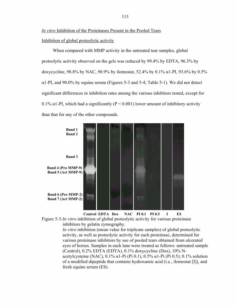
113
In vitro Inhibition of the Proteinases Present in the Pooled Tears
Inhibition of global proteolytic activity
When compared with MMP activity in the untreated tear samples, global
proteolytic activity observed on the gels was reduced by 99.4% by EDTA, 96.3% by
doxycycline, 98.8% by NAC, 98.9% by ilomostat, 52.4% by 0.1% α1-PI, 93.6% by 0.5%
α1-PI, and 90.0% by equine serum (Figures 5-3 and 5-4; Table 5-1). We did not detect
significant differences in inhibition rates among the various inhibitors tested, except for
0.1% α1-PI, which had a significantly (P < 0.001) lower amount of inhibitory activity
than that for any of the other compounds.
Band 1 Band 2
Band 3
Band 4 (Pro MMP-9) Band 5 (Act MMP-9)
Band 6 (Pro MMP-2) Band 7 (Act MMP-2) Control EDTA Dox NAC PI 0.1 PI 0.5 I ES Figure 5-3: In vitro inhibition of global proteolytic activity for various proteinase
inhibitors by gelatin zymography. In vitro inhibition (mean value for triplicate samples) of global proteolytic activity, as well as proteolytic activity for each proteinase, determined for various proteinase inhibitors by use of pooled tears obtained from ulcerated eyes of horses. Samples in each lane were treated as follows: untreated sample (Control), 0.2% EDTA (EDTA), 0.1% doxycycline (Dox), 10% N-acetylcysteine (NAC), 0.1% α1-Pi (Pi 0.1), 0.5% α1-Pi (Pi 0.5); 0.1% solution of a modified dipeptide that contains hydroxamic acid (i.e., ilomostat [I]), and fresh equine serum (ES).

114
Table 5-1: Percentage of in vitro inhibition of proteolytic activity for the global proteolytic activity, as well as for each proteinase, determined for various proteinase inhibitors by use of pooled tears obtained from ulcerated eyes of horses.
Band
EDTA 0.2%
Doxycycline 0.1%
NAC 10%
α1 PI 0.1%
α1 PI 0.5%
Ilomostat 0.1%
Fresh serum
1 96.3 86.1 94.3 46.8* 94.5 89.1 90.7 2 99.9 96.3 99.2 22.5* 87.0 95.1 87.4 3 96.6 96.5 97.8 44.6* 96.3 92.1 63.6 4 99.8 98.4 99.8 55.3* 94.9 99.7 91.4 5 98.6 95.9 98.7 46.6* 92.3 99.1 82.4 6 100 95.0 97.0 78.8* 90.5* 98.4 95.9 7 98.5 98.8 96.3 77.8* 70.7* 100 96.6
Total 99.4 96.3 98.8 52.4* 93.6 98.9 90.0
Values reported represent mean value for triplicate samples. Band 4 represented the latent form of matrix metalloproteinase MMP-9. Band 5 represented the active form of MMP-9. Band 5 represented the latent form of MMP-2. Band 7 represented the active form of MMP-2. *Inhibition rate for this compound differed significantly (P < 0.001) from the inhibition rates for the other compounds. EDTA = 0.2% EDTA. Dox = 0.1% Doxycycline. NAC = 10% N-acetylcysteine. 0.1 PI = 0.1% α1-proteinase inhibitor. 0.5 PI = 0.5% α1-proteinase inhibitor. I = 0.1% Solution of a modified dipeptide that contains hydroxamic acid (i.e., ilomostat). ES = Fresh equine serum.
0
10
20
30
40
50
60
70
80
90
100
Per
cent
age
of in
hibi
tion
EDTA Dox NAC Ilo A1Pi0.1%
Figure 5-4: Percentage of in vitro inhibition proteolytic activity determined bproteinase inhibitors in pooled teInhibitors used were as follows: (black bar), 10% N-acetylcysteinright of bar), 0.1% α1-PI (diagon
*
A1Pi 0.5% Fresh equine serum
(mean value for triplicate samples) of global y use of gelatin zymography for various ars obtained from ulcerated eyes of horses. 0.2% EDTA (white bar), 0.1% doxycycline e (diagonal lines from upper left to lower al lines from lower left to upper right of bar),
115
0.5% α 1-PI (dotted bar); 0.1% ilomostat (vertical lines), and fresh equine serum (horizontal lines). *Inhibition rate for this compound differed significantly (P < 0.001) from the inhibition rates for the other compounds.
Inhibition of proteolytic activity for each proteinase
We also assessed the inhibitory effects of these various compounds detected by
gelatin zymography on each individual proteinase present in the tear pool (Figure 5-3;
Table 5-1). It is interesting that inhibition was greatest for the latent form of MMP-9
(band 4) and least for the active form of MMP-2 (band 7) for EDTA, NAC, and 0.5% α1-
PI, whereas the inhibition was greatest for active form of MMP-2 (band 7) for
doxycycline, 0.5% α1-PI, ilomostat, and the fresh serum. There was significant difference
in the inhibition rates for each individual band between the 0.1% α1-PI and any other of
the tested compounds (P < 0.05). Furthermore, the inhibition of the latent and active
MMP2 by 0.5% α1-PI was significantly lower than those for the other tested compounds
(p < 0.05).
In vitro Inhibitory Activity and the Duration of Action of Equine Serum Against the Proteinases Present in the Tear Fluid of Horses with Ulcerative Keratitis
There was no difference in MMP inhibition between fresh serum (-90.0% activity)
and frozen serum, i.e. -20ºC (-90.9% activity). No difference in MMP inhibition between
the serum kept at room temperature, i.e. 19ºC (-92.0 ± 4.9% activity) or in the
refrigerator, i.e. 4ºC (-90.6 ± 2.8% activity) was detected during the eight day trial
(Figure 5-5).
Inhibitory activity of serum towards tear film MMPs did not significantly change
over the eight-day test period.

116
82
84
86
88
90
92
94
96
98
100
1 2 3 5 8
Time (day)
Perc
enta
ge o
f inh
ibiti
on
Serum RTSerum CoolSerum frozen
Figure 5-5: Percentage of in vitro inhibition (mean value for triplicate samples) of global
proteolytic activity determined by use of gelatin zymography for equine serum stored in various ways. Serum RT = serum stored at room temperature (i.e. 19ºC), Serum Cool = serum stored in the refrigerator (i.e. 4ºC), Serum frozen = serum stored in the freezer (i.e. -20ºC).
Discussion
Analysis of the results we obtained for the untreated (control) samples confirmed
those reported elsewhere (Strubbe et al., 2000) in which MMP-2 and -9 are found in
substantially high amounts in the tear film of horses with ulcerative keratitis. The
majority of host-derived and microbial enzymes in the tear film are MMPs (Barletta et
al., 1996; Fini et al., 1992; Fini et al., 1998; Matsumoto, 2000). Enzymes of bacterial or
fungal origin (i.e., exogenous proteases) can contribute directly or indirectly to ulcerative
keratitis through the activation of corneal proteinases (i.e., endogenous proteinases)
(Brooks, 1999; Gopinathan et al., 2001; Matsumoto, 2000; O’Brien, 1997; Slansky et al.,
1969). In the study reported here, we assessed only the inhibitory effects of the various
compounds on the global proteolytic activity that can be detected in ulcerated eyes of
horses without distinguishing between proteolytic activity attributable to endogenous
117
proteinases and activity attributable to exogenous proteinases. Thus, we did not take into
account the specific cause of the corneal ulcers.
Proteinase inhibitors have been recommended and used in human and veterinary
ophthalmology for many years. Their use remains controversial, because the efficacy and
local toxic effects of these compounds are frequently questioned. In the study reported
here, we documented in vitro a high amount of inhibition of equine MMP activity by the
use of EDTA, doxycycline, NAC, equine serum, ilomostat, and α1-PI. For each of these
proteinase inhibitors, the proposed mechanisms of inhibition, the current
recommendations for use in human and veterinary ophthalmology, and the adverse
effects that have been reported with each can be used to justify treatment of horses with
ulcerative keratitis.
We observed a large reduction in the in vitro activity of equine MMPs with 0.2%
EDTA. Doxycycline, EDTA, and NAC are metal-chelating agents, and they inhibit
MMPs by chelation of zinc or calcium that MMPs require as a cofactor and stabilizing
ion, respectively (Twining, 1994; Ward, 1999). By chelating calcium ions, EDTA
interferes with the stability of the MMPs and also decreases the stimulation for the
migration of the PMNs to the site of a corneal ulcer. The PMNs release powerful
proteinases that are also responsible for breakdown of stromal collagen. EDTA interferes
with attachment of opsonized zymosan to the cell membrane of PMNs, leaving the PMNs
in a resting, inactivated, granulated state (Pfister et al., 1984). In addition, EDTA has
been recommended for the treatment of ulcers associated with corneal collagenolysis
(Berman, 1978; Pfister and Pfister, 1997; Ward, 1999); however, corneal tolerance and
efficacy of EDTA are subjects of controversy (Corbett et al., 2001; Furrer et al., 1999). In

118
rabbits, treatment of ulcerated corneas with 0.1% EDTA after keratectomy did not
significantly affect the rate of re-epithelialization (Schultz, 1997). Furthermore, EDTA
seems to be tolerated well when used at concentrations ranging from 0.05% to 0.2% for
treatment of corneal ulcers in horses (Brooks, 1999). However, the long-term use of
EDTA may impair formation of tight junction complexes between epithelial cells as a
result of the requirement of calcium ions for stable tight junctions.
In the study reported here, we documented in vitro inhibition of equine MMPs by
0.1% doxycycline. Tetracyclines inhibit MMP activity independent of their antimicrobial
properties (Golub et al., 1984). The proposed mechanism of action of these antimicrobial
agents is that tetracyclines bind to zinc and calcium cations that are essential for the
MMPs. Tetracyclines form a binding complex with the MMPs, resulting in reduced
enzyme activity (Golub et al., 1984; Rawal and Rawal, 1984; Smith et al., 1999).
Tetracyclines, and especially doxycycline, inhibit the synthesis of MMPs in human
vascular endothelial cells (Rawal and Rawal, 1984). Tetracyclines, such as minocycline
and doxycycline, inhibit the breakdown of various connective tissues (i.e., skin, bone, and
cornea) mediated by excessive collagenolytic activity (Golub et al., 1984), and they have
been used specifically for ophthalmic treatments (Golub et al., 1984; Rawal and Rawal,
1984). Doxycycline promotes healing of persistent ulcers and epithelial defects in
humans (Dursun et al., 2001; Perry et al., 1993) and inhibits alkali-induced corneal ulcers
in rabbits (Seedor et al., 1987). Tetracyclines are recommended for use in ophthalmic
conditions in animals (Ward, 1999; Whitley and Gilger, 1999) but we are not aware of
any information on the use of doxycycline in horses.
119
In our study, 10% NAC proved to be effective for inhibiting in vitro MMP activity
in the equine tear film. This supports the fact that NAC is commonly used as an MMP
inhibitor to treat humans and other animals with corneal ulcers (Slansky et al., 1972;
Ward, 1999). Application of NAC at concentrations of 2% to 10 % every 1 to 4 hours has
been recommended for treatment of dogs (Kanao et al., 1993; Ward, 1999; Whitley and
Gilger, 1999) and horses (Andrew et al., 1998; Schmidt, 1997; Ward, 1999; Whitley and
Gilger, 1999). More specifically, equine serum and 10% NAC has been recommended for
the treatment of horses wit severe corneal ulcers (Brooks, 1999). A thiol, NAC contains a
sulfhydryl group that binds irreversibly or removes the intrinsic metal cofactor (i.e., zinc)
of the MMPs. It may also reduce one or more disulfide bonds of an enzyme (Slansky et
al., 1970; Woessner, 1991). Also, NAC inhibits MMP production at the transcriptional
level (Galis et al., 1998). In one study (Galis et al., 1998), NAC suppressed MMP-9
synthesis in macrophages. In another study (Thermes et al., 1991), effects of
acetylcysteine on rabbit conjunctival and corneal surfaces were examined. Investigators
did not detect signs of ocular toxicosis for various concentrations tested, but 20% NAC
caused some superficial necrosis and dose-related disruption in the mucus layer of the
tear film. Adverse effects were not found for the rate of re-epithelialization of the rabbit
corneas in which 10% and 20% NAC were applied as treatment for a superficial
epithelial ulcer (Petroutsos et al., 1982).
In the study reported here, we detected high amounts of in vitro inhibition of MMP
activity attributable to equine serum. The α 2-macroglobulin is a nonspecific protease
inhibitor that reduces the activity of proteinases of all 4 major proteinase classes (i.e.,
serine [including neutrophil elastase], aspartic, thiol, and metalloproteinases in human

120
and rabbit corneas) (Berman, 1975; Hibbetts, 1999; Twining, 1994). The α2-
macroglobulin is a tetrameric molecule composed of 2 pairs of identical disulfide-linked
subunits. Each subunit contains a region that binds the enzymes and allows the
proteinases to cleave peptide bonds. This cleavage leads to a change in conformation of
the α 2-macroglobulin, resulting in entrapment of the proteinase within the inhibitor
(Hibbetts, 1999; Twining, 1984; Woessner, 1991). This particular mechanism tenaciously
binds 2 protease molecules/ α 2-macroglobulin molecule; thus, α 2-macroglobulin is one
of the strongest known inhibitors of MMPs. Although α 2-macroglobulin mRNA and
protein are found in the cornea (Prause, 1983a; Twining, 1994; Twining et al., 1994a),
this multifunctional inhibitor is found at much higher quantities in the blood (Prause,
1983a). For this reason, topical application of autologous serum (Bron et al., 2002;
Stangogiannis et al., 2002) (1 or 2 drops every 1 to 2 hours) has been recommended for
the treatment of corneal ulcers in humans (La Lau, 1979) and other animals (Brooks,
1999; Ward, 1999; Whitley and Gilger, 1999). Blood collected into dry, sterile containers
that do not contain anticoagulants will clot rapidly and yield serum that can be separated
by centrifugation (Ward, 1999). The serum can be used immediately or stored in a
refrigerator or a freezer until needed, and in our experience, its inhibitory effect remains
high even after several days of storage (Figure 5-5). However, it is recommended that
serum be discarded if not used within 5 days, because it may provide an excellent
medium for bacterial growth should it become contaminated (Brooks, 1999; McLaughlin
et al., 1992; Ward, 1999).
We observed a dose-dependent decrease in proteolytic activity of the MMPs in the
pooled tears of horses for α1-PI at concentrations of 0.1 and 0.5%. It is possible that the
121
dose-related reduction of activity of the MMPs attributable to this serine proteinase
inhibitor is not a result of direct inhibition of MMPs by α1-PI; instead, it may result from
α1-PI acting as a substrate for the MMPs and competing with the gelatin from the gels.
Also called α1-antitryspin, α1-PI is also found in and synthesized by the cornea, (Berman
et al., 1973; Twining et al., 1994b) but it is found at a higher concentration in the blood
(Berman et al., 1973; Ward, 1999). Furthermore, α1-PI is part of the serpin family of
inhibitors that inhibit serine proteinases, such as neutrophil elastase (Berman et al., 1973).
The α1-PI exerts its action on proteinases through a mechanism similar to that for α 2-
macroglobulin. It has the ability to form tight complexes with proteinases and, therefore,
reduces proteinase activity (Berman, 1975). It has been documented that α1-PI is a
critical substrate for MMP-9 in vivo, and MMP-9 acts upstream to regulate neutrophil
elastase activity by inactivating α1-PI (Liu et al., 2000).
During the past decade, efforts have been made to design synthetic inhibitors of
proteinases (Barletta et al., 1996; Burns et al., 1990a; Burns et al., 1990b; Schultz et al.,
1992). Among those that have been reported, ilomostat appears to be promising for the
treatment of rapid degradation of the corneal stroma (Schultz et al., 1992). It is more
effective in vitro against MMPs in rabbits, compared with the classic chelating agents
(Hao et al., 1999a; Hao et al., 1999b). Ilomostat also decreases Pseudomonas alkaline
proteinase activity in vitro (Barletta et al., 1996) and is effective in reducing corneal
destruction following alkali burns in rabbits (Schultz et al., 1992) or intrastromal
injection of pseudomonal culture broth (Barletta et al., 1996). In the study reported here,
we reported that use of 0.1% ilomostat caused a high amount of inhibition of in vitro
activity of MMPs in the equine tear film. Because the structure of MMPs is highly

122
conserved among animal species, it is reasonable to expect that the inhibitory effects for
this synthetic MMP inhibitor in rabbits would be seen in other domestic animals.
Multiple studies have found increased amounts of proteases in tears from humans
and other animals with active corneal ulcers. Several agents have been proposed as
treatments to reduce the activity of tear proteases, but we are not aware of any reports
comparing the relative efficacy of the agents. This places practitioners in a difficult
position of not knowing which treatment may be superior. In the study reported here, the
relative efficacies of 5 protease inhibitors and equine serum were determined for the
inhibition of MMP activity in pooled tears obtained from horses with active corneal
ulcers. Analysis of our results indicates that EDTA and ilomostat, followed by NAC and
doxycycline, are the most effective inhibitors in vitro and are likely to cause the fewest
adverse effects. Because these compounds use different mechanisms to inhibit various
families of proteases in equine tears, a combination of these inhibitors may be indicated
for the treatment of severe corneal ulcers in horses. Objectives for the appropriate use of
these compounds early in the course of corneal disease in horses would be to decrease the
amount of time required for recovery and rehabilitation, reduce scarring, and potentially
alleviate the need for corneal surgical treatment. Our results that indicate some readily
available substances are effective inhibitors of proteases in tears of eyes with active
corneal ulcers provide practitioners with a possible valuable adjunctive treatment for
vision-threatening diseases of the eyes.

CHAPTER 6 DETECTION OF CTGF IN THE EQUINE TEAR FLUID, CORNEA AND LACRIMAL
GLANDS
Introduction
Transforming growth factor beta (TGF-β) activates fibrocytes to produce
connective tissue growth factor (CTGF) (Berman et al., 1973; Igarashi et al., 1996).
Overexpression of CTGF apparently plays an important role in fibrosis of various tissues
(Chen et al., 2000; Gupta et al., 2000; Shi-wen et al., 2000; Takehara, 2001; Wunderlich,
2000). CTGF may be involved in corneal wound healing via induction of fibroblast
proliferation and collagen production (Fraxier et al., 1996). After the report of CTGF
presence in human serum (Sato et al., 2000) and its detection in the aqueous humor and
tear fluid (van Setten et al., 1992; van Setten et al., 1996), CTGF has been suggested to
play a role in ocular wound healing in concert with other proteins, growth factors and
enzymes (Schultz et al., 1992; van Setten, 1998). However, one problem in previous
studies has been the varying stability of CTGF in the fluids investigated, resulting in
difficulties to estimate the correct level of protein concentration. With only one
preliminary study so far published there is a considerable need for further thorough
investigation on the presence of CTGF in tears and its pathophysiological importance. In
melting diseases of the ocular surface such as keratitis and stromal ulceration swift
remodeling and reestablishment of tectonic stability is urgently desired. Here CTGF may
play an important role. Healing of corneal ulcers in horses, on the other hand, is often
associated with profound fibrosis and corneal scar formation that can result in varying
123

124
degrees of visual impairment (Brooks, 1999). The purpose of this study was hence to
determine if CTGF was also present in the tear fluid of horses, to investigate possible
alterations of concentration during corneal pathology, and to clarify its possible origin.
Materials and Methods
Materials
All procedures were carried out according to the ARVO statement for the Use of
Animals in Ophthalmic and Vision research and were approved by the University of
Florida Animal Care and Use Committee as well as the UF College of Veterinary
Medicine Clinical Research Review Committee.
For the analysis of CTGF concentration in tear fluid samples were collected from
65 eyes of 44 horses that were either clinical cases (Figure 6-1) from the Large Animal
Hospital at the University of Florida College of Veterinary Medicine Veterinary
Teaching Hospital or, in case of animals without any ocular disease, from a farm in
Florida. In total 32 samples were collected from normal eyes; 21 samples from eyes with
corneal ulceration, and 12 samples from the unaffected contralateral eyes of horses with
ulcers.
The tear collections were performed after sedation of the animal with an
intravenous injection of xylazine hydrochloride (0.5 mg/kg) and before any diagnostic
procedure or any treatment. All tear fluid specimens were collected by capillary force
only using capillary tubes with an atraumatic tip as previously described (van Setten et
al., 1989) from the lower fornix. The time of collection of all tear fluid sample was not
taken in this study. All samples were immediately centrifuged, transferred into Eppendorf
tubes (10µL aliquots), and stored at -80ºC until analysis.

125
For the identification of CTGF in western blot two pools of tear fluid (100µL) from
horses with healthy eyes and horses with ulcerated eyes were analyzed and compared
with human CTGF.
For immunohistochemistry, five healthy corneas, five main lacrimal glands and
five nictitating membrane glands were collected from horses with normal eye following
euthanasia due to condition not related to this project. All samples were fixated in 4%
formaldehyde buffer and stored in paraffin.
Figure 6-1: Melting ulcer in the right eye of a 2 month old Thoroughbred horse.
This case was involved in this study. Note the presence of hyperemic conjunctiva, corneal neovascularization at the periphery, and a 5-6 mm melting corneal ulcer with mild corneal edema and white opacification in the deep stroma in the center of the ulcer.
CTGF Elisa Assay
CTGF levels were determined using an enzyme immunoassay kit (CTGF Elisa)
developed for human tear samples. This assay is a non-commercially available,
quantitative "sandwich" enzyme immunoassay technique (Tamatani et al., 1998). Briefly,
96-well microtiter plates were coated with an affinity-purified polyclonal antibody
specific for human CTGF at 37 C for one hour (a goat IgG against human CTGF). After
washing, standards and samples, rediluted to a volume of 50 µl, were added to the

126
microtiter plates and incubated for 1 hour at room temperature. After another series of
washes, a biotin-linked polyclonal antibody specific for human CTGF was added to the
wells to bind to the CTGF molecules bound by the first antibody. After an incubation
time of 1 hour at room temperature in the dark, alkaline-phosphate conjugated
streptavidin was then added and incubated for an additional hour. P-nitrophenyl
phosphate, the substrate for alkaline-phosphate, was added to the wells until the color
change developed (about 30-45 minutes). Optical densities were obtained by reading the
samples at 405 nm with a spectrophotometer Biotek FL340™ Microplate reader (Biotek
instruments, Winooski, VT). The detection limit of the assay was approximately 0.1
ng/ml CTGF.
Dilution Curves
A comparison of the human CTGF standard curve with an equine “standard” curve
was made in order to assess the bio-equivalence, and to be able to interpret the results
obtained for the equine samples using a human Elisa assay.
The human standard curve was obtained by analyzing various samples obtained by
serial dilutions of the human CTGF provided with the human Elisa kit. The initial human
CTGF standard was at a concentration of 100 ng/ml and corresponded to 100% of the
product in the sample tested. It was then diluted to obtain the final concentrations of 50,
10, 1.0, and 0.1 ng/ml. To obtain an equine dilution curve, tear samples from 10 horses
(20 eyes) were pooled and an identical serial dilution of the 200 µl mixture was made.
The diluted samples were analyzed with the human Elisa kit. The equine pool without
any dilution corresponds to 100% of product in the samples although its CTGF
concentration was not known.

127
CTGF Western Blot
The two pools of tear fluid were formed by collection of tears from horses with
healthy eyes and horses with ulcerated eyes. The tears were centrifuged at 14,000g for 10
minutes and the supernatant stored at – 80º C until their analysis. The samples (pools of
tear fluid from healthy and ulcerated horse eyes) as well as a molecular weight markers
(MultiMark pre-stained multicolored standards, Invitrogen, Carlsbad, CA) and 50 ng
of human recombinant CTGF were loaded on a 5% stacking, 12% SDS-PAGE gel. The
electrophoresis was run at 125 V for 1 hour in a running buffer. The proteins were
electrotransferred onto a PVDF membrane (Immobilon™-P, Millipore Coporation,
Bedford, MA) in a transfer buffer at 25V at 4º C overnight. The membrane was blocked
in Tris-buffered saline (TBS) containing 10% non-fat milk (Blotting grade blocker non-
fat dry milk, BioRad Laboratories, Richmond, CA) for one hour and then incubated in the
same solution containing a dilution of 1:7000 of the goat anti-human CTGF for 30
minutes at room temperature. The blot was washed three times with 0.05% Tween-20
TBS and then incubated with rabbit anti-goat IgG alkaline phosphatase conjugate (Sigma,
St Louis, MO) at the dilution of 1:7500 in the blocking solution for 30 minutes at room
temperature. The membrane was washed again three times and the BCIP/NBT alkaline
phosphatase substrate (Sigma, St Louis, MO) was added and color generated. The
reaction was stopped by water washing.
CTGF Immunohistochemistry
Cells expressing CTGF were detected immunohistologically using a standard
avidin-biotin amplification method (Blalock et al., 2003). Briefly, corneas, lacrimal
glands, and nictitating membrane glands were obtained from five horses, fixed in 4%
paraformaldehyde 0.1 M phosphate buffer solution (PBS) overnight at 4º C, dehydrated

128
in an ascending series of ethanol, and embedded in paraffin. Paraffin-embedded sections
were prepared and 6 µm sections were mounted on microscopic slides (Superfrost/plus®;
Fischer Scientific, Pittsburgh, PA). Slides were deparaffinized and rehydrated with
xylene and a graded series of ethanol. Slides were blocked with 10% horse serum in Tris-
buffered saline (TBS) for 30 minutes at room temperature. Slides were then sequentially
incubated with goat anti-human CTGF in TBS and 10% horse serum overnight at 4º C,
washed three times with TBS, incubated with biotinylated horse anti-goat IgG secondary
antibody (Vectastain® ABC-AP kit,Vector Laboratories, Burlingame, CA) in TBS,
washed, and incubated with alkaline phosphatase-conjugated streptavidin in TBS and
10% horse serum (Vectastain® ABC-AP kit,Vector Laboratories, Burlingame, CA),
washed, and incubated with alkaline phosphatase visualization substrate (Vector Red
Alkaline Phosphatase substrate kit I: Vector Laboratories, Burlingame, CA). The reaction
was stopped by water washing. The goat anti-human CTGF antibody was raised against
recombinant human CTGF protein and purified with a CTGF-affinity column (Frazier et
al., 1996). The antibody predominantly recognizes antigenic determinants on the N-
terminal sequence of CTGF. The sections were photographed with bright-field
illumination, and Nomarski phase-contrast microscopy at various magnifications.
Results
Detection and Quantification of CTGF in the Horse Tears
The human Elisa kit was able to detect the CTGF in the equine sample. CTGF was
detected in 23 normal unaffected eyes (72%) and 8 normal contralateral eyes (67%), with
the mean CTGF levels (± SEM) being 51.5 ± 19.2 and 13.4 ± 3.9 ng/ml respectively.
CTGF was found in 8 of the 21 eyes with corneal ulcers (38%) with the mean CTGF
level (± SEM) being 26.3 ± 14.8 ng/ml (Figure 6-2 & Tables 6-1 and 6-2). In contrast to

129
eyes with corneal ulceration, often showing CTGF levels below detection limit, eyes with
corneal laceration (n=3) did always show CTGF in their tears (mean 85.3 ng/ml ± 38.8
ng/ml).
0
20
40
60
80
100
120
140
160
180C
TGF
conc
entr
atio
n (n
g/m
l)
Contralateral eyes Ulcerated eyes Normal eyes
8/12 8/21 23/32
Figure 6-2: CTGF levels in horse tears.
Mean CTGF levels and standard deviations for 12 tear samples from the contralateral eyes of horses with corneal ulceration (CTGF was detected in 8 out of the 12 samples), 21 tear samples from the eyes of horses with corneal ulcers and lacerations (CTGF was detected in 8 out of the 21 samples), and 32 tear samples from the eyes of horses without any corneal condition in both eyes (CTGF was detected in 23 out of the 32 samples). The vertical lines indicate the standard deviation range.
Table 6-1: CTGF in tear samples from horses with ulcerated corneas in one eye (12 eyes) and the non diseased contralateral eye
Diseased eye Contralateral eye Tear
sample # Diagnosis CTGF level
(ng/ml) Tear
sample # CTGF level
(ng/ml) 1 Melting ulcer ND 1’ 30.40 2 Ulcer 1.07 2’ 19.73 3 Melting ulcer ND 3’ 15.73 4 Melting ulcer 23.97 4’ 15.39 5 Melting ulcer ND 5’ 0.79 6 Melting ulcer 6.10 6’ 30.31 7 Laceration 151.88 7’ 12.36 8 Superficial ulcer ND 8’ 36.32 9 Ulcer 9.07 9’ ND
10 Melting ulcer 283.07 10’ ND 11 Melting ulcer ND 11’ ND 12 Melting ulcer ND 12’ ND

130
Table 6-2: CTGF from 9 tear samples from horses with ulcerated corneas in one eye (only tears from this eye were analyzed).
Tear sample #
Diagnosis CTGF level (ng/ml)
1 Corneal laceration 86.95 2 Corneal laceration 17.77 3 Deep stromal ulcer ND 4 Melting ulcer ND 5 Desmetocoele ND 6 Ulcer ND 7 Ulcer ND 8 Superficial ulcer ND 9 Melting ulcer ND
Dilution Curves – Bioequivalence
In figure 6-3 a dilution curve shows the optical density (values obtained from the
microplate reader) represented as a function of the percentage of protein in the sample
tested. The human standard curve was very linear with a correlation coefficient of 0.9978
whereas the equine dilution curve was slightly less linear with a correlation coefficient of
0.865. However, the main difference between the human CTGF standard curve and the
horse pool dilution curve was regarding their slopes: the slope being 0.0076 and 0.0019
in the human and horse respectively (Figure 6-3).
y = 0.0076x + 0.042R2 = 0.9978
y = 0.0019x + 0.0758R2 = 0.865
00.10.20.30.40.50.60.70.80.9
0 20 40 60 80 100 120
Percentage of product in sample
Opt
ical
den
sity
Figure 6-3: CTGF Dilution curves.
A serial dilution of a human CTGF (Ο) was made and used as the standard for this Elisa assay (0, 0.1, 1.0, 5.0, 10, 50 & 100 ng/ml). The

131
concentration of 100 ng/mL of human CTGF corresponds to 100% of product in the samples in the graph. An identical serial dilution of a pool of horse tear samples is presented ( ).
CTGF Western Blot
Western blot showed the two characteristic CTGF bands on all the equine samples,
similar to those observed around 38 kDa with the human CTGF standard (Figure 6-4).
These results indicate that the detectable amount of CTGF in the equine tear fluid is
indeed CTGF.
Figure 6-4: Western Blot of tear fluids from horses with healthy eyes (lane 3) and
ulcerated eyes (lane 4). The two characteristic CTGF bands around 38 kDa were noted on all the equine samples (lanes 3 and 4), similar to those observed with the human CTGF standard (lane 2).
CTGF Immunohistochemistry
Immunoreactivity to CTGF specific antibodies was noted in the corneal epithelium
and in the corneal endothelium whereas staining was absent in the corneal stromal
(Figure 6-5). In the corneal epithelium, staining was concentrated in the squamous
superficial epithelial cells (Figure 6-5).
The immunohistochemical staining for CTGF in the equine lacrimal glands and the
nictitating membrane glands revealed specific staining for CTGF predominantly in the
acinar cells, the epithelial cells of the intralobular ducts, and the intralobular connective
tissue (Figures 6-6 and 6-7). In the acinar cells, the staining was concentrated close to the
1 2 3 4
52 kDa
31 kDa
Molecular Weight Human CTGF Equine normal Equine diseased Markers Standard tear fluid tear fluid
132
basally located nuclei, opposite to the lumen (Figures 6-6 and 6-7). No immunoreactivity
was detected in the control sections (Figures 6-5, 6-6 and 6-7).
Figure 6-5: Immunolocalization of CTGF in equine cornea.
H&E staining of the equine cornea (A) where corneal epithelium (1), stroma (2) and Descemet’s membrane – corneal endothelium (3) can be distinguished. Nomarski phase-contrast 1 observation (B) and bright-field observation (C) of the negative control sections showed no staining at all. Immunohistochemical staining for CTGF in the equine cornea (D, E, F) showed CTGF specific staining in the corneal epithelium, especially in the squamous superficial cell layer (white arrow) and the corneal endothelium (black arrow). Original magnifications were X100 (A, B, C, D) and X200 (E, F) .
D
A
E
CB
F
1
3
2
1
3
2
1
2
3
133
A B C
D EF
Figure 6-6: Immunolocalization of CTGF in equine lacrimal gland. H&E staining of the equine lacrimal gland (A) where acini (inset), intralobular ducts, lymphocytes in the intralobular connective tissue (inset) can be distinguished. Nomarski phase-contrast 1 observation (B) and bright-field observation (C) of the negative control sections showed no staining at all. Immunohistochemical staining for CTGF in the equine lacrimal gland (D, E, F) showed CTGF specific staining in the acinar cells (white arrow), the epithelial cells of the intralobular ducts (black arrow), and the intralobular connective tissue (striped arrow). In the acinar cells, the staining was concentrated close to the basally located nuclei (white arrow), opposite to the lumen. Original magnifications were X100 (A, B, C, D) and X400 (E), and X1000 (inset in A, F).
1 2 1 2 1 2
CA B
1 2 D E F
Figure 6-7: Immunolocalization of CTGF in equine nictitating membrane gland. H&E staining of the equine nictitating membrane (A) where the hyaline cartilage (1) and the nictitating membrane gland (2) composed of acini (inset) can be distinguished. Nomarski phase-contrast 1 observation (B) and bright-field observation (C) of the negative control sections showed no staining at all. Immunohistochemical staining for CTGF in the equine nictitating membrane gland (D, E, F) showed CTGF specific staining in the acinar cells (white arrow), the epithelial cells of the intralobular ducts

134
(black arrow), and the intralobular connective tissue (striped arrow). Original magnifications were X100 (A, B, C, D) and X400 (E), and X1000 (inset in A, F).
Discussion
This study identifies CTGF in the horse tear film. CTGF was present at detectable
levels in approximately 70% of the tears from normal horse eyes and tears from the
normal, contralateral eyes of horses with corneal ulcers. The absence of detectable CTGF
in the remaining tears fluid samples could indicate the total absence of any CTGF in tears
of some horses or a concentration below detection limit. Furthermore, similarly as
reported earlier for human tear fluid (van Setten et al., 2003), the processing of the equine
tear samples involving centrifugation and storage at -80ºC until analysis could have
affected the data. However, we do believe the comparison between our samples is valid
as all samples have been handled and stored in the same manner. The observation that
CTGF was found in about 40% of the tears from ulcerated horse eyes at a lower level
than in the normal eyes (Figure 6-2) might reflect a dilution effect resulting from
increased tear volume, such as shown for EGF (van Setten, 1990). It also could indicate a
significantly enhanced receptor binding to the surface at specific stages of the healing
period, finally it may be the result of the temporary exhaustion of CTGF reservoir in
lacrimal gland cells. The latter may also be the explanation for decreased levels of CTGF
found in non-ulcerated sister eyes of horses with corneal ulceration in one eye. The
simultaneous presence of enzymes in corneas with corneal ulceration possibly may also
contribute to reduced CTGF levels in tears due to proteolysis. The importance of
proteolytic enzymes and their regulatory inhibitors in tears has been investigated
extensively (Berman et al., 1973; Berman et al., 1988; Kueppers, 1971; Matrisian, 1990;
Matsubara et al., 1991b; Tervo et al., 1988; Thorїg et al., 1984; Thorїg et al., 1983; Tsung
135
and Holly, 1981; Twining, 1994; Twining et al., 1994a; Twining et al., 1994b; Twining et
al., 1994c; van Haeringen and Thorїg, 1986).
In the present study we identified CTGF in the lacrimal gland and the glands of the
nictitating membrane, both probably a major source of CTGF. The localization of the
specific staining is very similar to that shown for TGF-α (Schultz et al., 1992; van Setten
et al., 1996). According to our results, both the acini and cells of the inter-acinar ducts are
involved in the process of CTGF release into tear fluid. The exact mechanisms of CTGF
release has yet to be clarified. CTGF was also identified in the equine healthy cornea
which correlates with the findings in previous studies performed other species (Blalock et
al., 2003).
The present study also shows a strong antigenic similarity between human and
equine CTGF, and therefore demonstrates the feasibility of measuring CTGF protein in
horse tears using the human CTGF Elisa. There was, however, an incomplete equivalence
between the data obtained for human and horse samples as the standard curves were not
parallel (Figure 6-3). The values obtained for the horse samples using this human Elisa
kit may indicate slightly lower concentrations of CTGF found in the horse tears than a
test for horse specific CTGF might do.
The occurrence of CTGF specific mRNA in retrocorneal membranes and
subepithelial membranes has been recently reported (Wunderlich et al., 2000) and
suggests an involvement of CTGF in various wound-healing events in the eye.
Considering tear fluid composition as a rough mirror of ocular surface conditions, the
current study supports the suggested importance of CTGF in corneal pathology. Its
presence in the tears of other horses with ulcers and during healing phases of ulcers
136
deserves further investigation. The identification of other growth factors in the horse tear
fluid that can influence CTGF production or release such as TGF-β in the human tear
fluid (Gupta et al., 2000; Igarashi et al., 1996; Vesaluoma et al., 1997; Wang et al., 2001)
would be helpful to understand the mechanism of corneal wound healing and scarring. If
not generally, at least in horses with high CTGF levels this growth factor could be of
major importance for physiology or wound healing events of the equine cornea. Possibly
the CTGF like growth factors are the stromal equivalent to EGF (van Setten et al., 1992)
in corneal wound healing.
CHAPTER 7 CONCLUSIONS
When the cornea is injured, multiple systems are activated which produce a series
of complex and coordinated cellular processes that ultimately result in a healed corneal
wound. Healing of corneal wounds is an exceptionally complex process involving the
integrated actions of multiple growth factors, cytokines, and proteinases produced by
epithelial cells, stromal keratocytes, inflammatory cells, and lacrimal glands. Multiple
autocrine and paracrine interactions occur between epithelial cells and activated stromal
fibroblasts, and the exocrine actions of factors secreted by lacrimal gland cell into the
PTF. For this reason the tear film can be described as the mirror of the event occurring at
the corneal surface.
When a corneal wound occurs, keratocytes around the edge die by apoptosis and
chemotactic factors including growth factors such as PDGF, TGF-α, TGF-β are released
and attract inflammatory cells, resulting in the initiation of re-epithelialization,
contraction of connective tissue, and stimulation of angiogenesis (Schultz, 1997).
Proteinases are involved in the early phase of corneal wound healing as they play a role
in the epithelium cell migration by breaking down the cellular attachments (Cameron,
1997; Schultz et al., 1993). Beyond the site of injury, quiescent keratocytes become
activated into fibroblasts and migrate to the site of injury. This migration is followed by
cell proliferation, and finally deposition of fibronectin, laminin, heparin sulfate and other
ECM components. Many growth factors and cytokines have been implicated in
stimulating synthesis of these components, as well as cell proliferation, migration and
137

138
also differentiation of epithelial and stromal cells and angiogenesis (TGF-α, TGF-β,
PDGF, FGF) (Schultz et al. 1992).The fibroblasts deposit the reparative collagen and
eventually synthesize enough ECM to form a scar replacing the damaged tissue.
Corneal transparency is determined in large part by the structure of the stromal
ECM. In case of corneal injury, corneal stroma is not regenerated but repaired. It is
instead replaced with unspecialized tissue that creates the scar (Cameron, 1997). The
repair tissue matrix contains component molecules particularly fibronectin, not usually
present in stroma, and the newly synthesized collagen fibrils are thicker and more
variable in size than those of the normal stroma. All these events in the process of wound
healing must be reversed for the tissue architecture to return to normal. The removal of
the inflammatory mediators which were generated initially must first occur. Infiltration of
monocytes and leukocytes must decline, and then removal of extravasated fluid, protein,
cellular debris, granulocytes and macrophages occurs as the wound regeneration process
continues. Some improvement in the transparency of repair tissue can occur over the long
term through the progressive remodeling of the repair tissue matrix and the MMPs appear
to be involved in this process to remove the provisional ECM.
A delicate balance between protein synthesis and proteolysis is present in corneal
wound healing with many factors including growth factors, proteinases and proteinases
inhibitors involved (Figure 7-1). Any imbalance can lead to fibrotic processes (inhibitors
> proteinases) or to excessive tissue destruction (proteinases > inhibitors) (Figure 7-1).
The major complications of corneal trauma, infection and surgery in horses are
corneal liquefaction (i.e., corneal melting) and corneal scarring. A vast amount of
research on stromal wound healing and scar formation exists in human and other species
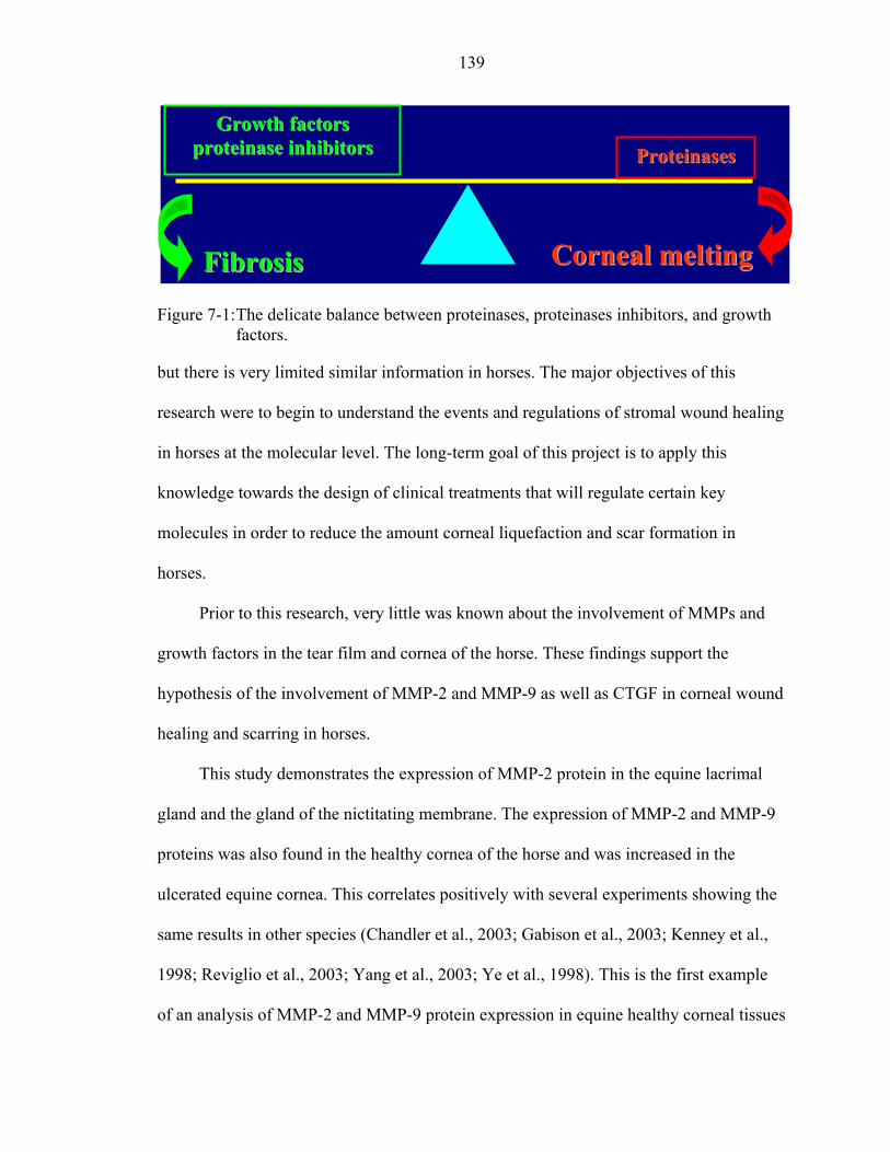
139
GGrroowwtthh ffaaccttoorrss pprrootteeiinnaassee iinnhhiibbiittoorrss PPrrootteeiinnaasseess
CCoorrnneeaall mmeellttiinnggFFiibbrroossiiss
Figure 7-1: The delicate balance between proteinases, proteinases inhibitors, and growth factors.
but there is very limited similar information in horses. The major objectives of this
research were to begin to understand the events and regulations of stromal wound healing
in horses at the molecular level. The long-term goal of this project is to apply this
knowledge towards the design of clinical treatments that will regulate certain key
molecules in order to reduce the amount corneal liquefaction and scar formation in
horses.
Prior to this research, very little was known about the involvement of MMPs and
growth factors in the tear film and cornea of the horse. These findings support the
hypothesis of the involvement of MMP-2 and MMP-9 as well as CTGF in corneal wound
healing and scarring in horses.
This study demonstrates the expression of MMP-2 protein in the equine lacrimal
gland and the gland of the nictitating membrane. The expression of MMP-2 and MMP-9
proteins was also found in the healthy cornea of the horse and was increased in the
ulcerated equine cornea. This correlates positively with several experiments showing the
same results in other species (Chandler et al., 2003; Gabison et al., 2003; Kenney et al.,
1998; Reviglio et al., 2003; Yang et al., 2003; Ye et al., 1998). This is the first example
of an analysis of MMP-2 and MMP-9 protein expression in equine healthy corneal tissues

140
as well as in an injured equine cornea. MMP-2 and MMP-9 are of major importance in
terms of remodeling and degradation of the corneal stromal collagen in horses as
described in other species (Fini and Girard, 1990; Fini et al., 1992). This study documents
the fact that the origin and purpose of MMP-2 and –9 appear to differ at the corneal level.
In this study, the level of MMP-2 and MMP-9 activity was determined in a total
330 tear fluid samples by the use of gelatin zymography. Activity was detected in all tear
fluids samples, and it was concluded that MMP-2 and MMP-9 are constant components
of the equine tear fluid. TFF rate and MMP proteolytic activity were significantly
increased in the tear fluid of horses with ulcerative keratitis. Based on the collection and
analysis of a total of 124 serial tear fluid samples in 10 horses with ulcerative keratitis,
we also documented for the first time that total MMP activity decreases in equine tears as
the corneal epithelial and stromal healing occur.
Since the long term goal of this project is to develop agents that will reduce corneal
melting with minimal side effects, initial experiments using various anti-proteolytic
agents were performed. We documented a high amount of inhibition of equine MMP
activity in vitro by the use of potassium edatate diaminetetrataacetatic acid (EDTA),
doxycycline, N-acetylcysteine (NAC), equine serum (that contains various inhibitors
including α2-macroglobulin and α1-proteinase inhibitor), ilomostat, and α1-PI. These
anti-protease compounds utilize different mechanisms to inhibit different families of
proteases present in equine tears and a combination of inhibitors may be indicated for
severe corneal ulceration in horses (Brooks, 1999; Hibbetts et al., 1999).
Further animal studies will be performed in the future to confirm that these anti-
proteolytic agents have similar effect in vivo. Other studies need to be conducted in order
141
to investigate the regulation of MMP activity directly by targeting the MMP active site
(pro-enzyme activation and active enzyme inhibition), but also indirectly by targeting the
MMP synthesis (transcriptional regulation) at the level of the equine cornea and tear film
The level of CTGF was determined by Elisa assay in a total 64 tear fluid samples in
this study. Since this molecule was detected in 39 of the samples, we concluded that
CTGF is a component of the equine tear fluid. This data demonstrates that CTGF was
present in the equine tear film. This study also indicates the expression of CTGF protein
in the healthy cornea, the lacrimal gland and the gland of the nictitating membrane of the
horse. CTGF has been shown to be synthesized and secreted into tears by lacrimal gland
cells (van Setten et al., 2002) and has been found in the cornea of other species (Blalock
et al., 2003).
These findings are important but further studies are needed. The identification of
other growth factors in the horse tear fluid that can influence CTGF production or release
such as TGF-β in the human tear fluid (Gupta et al., 2000; Igarashi et al., 1996;
Vesaluoma et al., 1997, Wang et al., 2001) would be helpful to understand the
mechanism of corneal wound healing and scarring in horses, and to develop anti-fibrotic
therapy in horses. It is currently known in other species that the transforming growth
factor beta family plays a dominant role in the regulation of stromal fibrosis (Chen et al.,
2000). It has recently been shown that CTGF is involved in the process because of its
regulation by TGF-β (Blalock et al., 2003). CTGF induces scar formation by mediating
many effects of TGF beta on ECM production and corneal scarring. Future studies should
for example investigate the role of TGF-β in equine corneal scarring and then search for
agents inhibiting these molecules in order to reduce the corneal scarring in horses with

142
minimal side effects. Steroids can reduce the corneal scarring but their use in horse is not
without severe risk. Some non specific anti-cancer drugs haven been used to treat corneal
scarring such as 5 fluorouracil and mitomycin C, but they cause serious side effect such
as epithelial defects and endothelial cell damage (Lee, 1994; Khaw et al., 1993). Recent
studies using ribosome therapy targeting CTGF have shown that the ribozyme reduced
the expression of CTGF and its biological effects, and might be an interesting alternative
to consider in horses.
In summary, these experiments have made tremendous progress in developing and
evaluating the role of MMP-2, MMP-9, and CTGF in the process of corneal wound
healing and scarring in horses. However, the lack of information on the molecular
regulation of corneal scarring in horses appear to be almost infinite in extent, promising
many exciting years in research on MMPs and growth factors and corneal wound healing
and scarring in horses.

LIST OF REFERENCES
Agren M S. Gelatinase activity during wound healing. Brit J Derm. 1994; 131: 634-640.
Andrew SE, Brooks DE, Smith PJ, Gelatt KN, Chmielewski NT, Whittaker CJ. Equine ulcerative keratomycosis: visual outcome and ocular survival in 39 cases (1987–1996). Equine Vet J. 1998; 30: 109–116.
Andrew SE, Willis AM, Anderson DE. Density of corneal endothelial cells, corneal thickness, and corneal diameters in normal eyes of llamas and alpacas. Am J Vet Res. 2002; 63: 326-9.
Azar DT, Pluznik D, Jain S, Khoury JM. Gelatinase B and A expression after laser in situ keratomileusis and photorefractive keratectomy. Arch Ophthalmol. 1998; 116: 1206–1208.
Barlati S, Marchina E, Quaranta CA, Vigasio F, Semeraro F. Analysis of fibronectin, plasminogen activators and plasminogen in tear fluid as markers of corneal damage and repair. Exp Eye Res. 1990; 51: 1-9.
Barletta JP, Angella G, Balch KC, Stern GA, van Setten GB, Schultz GS. Inhibition of pseudomonal ulceration in rabbit corneas by a synthetic matrix metalloproteinase inhibitor. Invest Ophthalmol Vis Sci. 1996; 37: 20–28.
Barro CD, Romanet JP, Fdili A, Guillto M, Morel F. Gelatinase concentration in tears of corneal-grafted patients. Curr Eye Res 1998; 17: 174-182.
Berman M. Collagenase inhibitors: rationale for their use in treating corneal ulceration. Int Ophtalmol Clin. 1975; 15: 49–66.
Berman M. Regulation of collagenase. Therapeutic considerations. Trans Ophthalmol Soc. 1978; 98: 397–405.
Berman M, Dohlman CH, Gnadinger M. Characterization of collagenolytic activity in the ulcerating cornea. Exp Eye Res. 1971; 11: 255–257.
Berman MB, Manabe R. Corneal collagenases: evidence for zinc metalloenzymes. Ann Ophthalmol. 1973; 5: 1193–1195.
Berman MB, Barber JC, Talamo RC, Langley CE. Corneal ulceration and the serum antiproteases. I. Alpha 1-antitrypsin. Invest Ophthalmol Vis Sci. 1973; 12: 759–770.
143
144
Berman M, Gordon J, Garcia LA, Gage J. Corneal ulceration and the serum antiproteases. II. Complexes of corneal collagenases and alpha-macroglobulins. Exp Eye Res. 1975; 20: 231–244.
Berman M, Leary R, Gage J. Latent collagenase in the ulcerating rabbit cornea. Exp Eye Res. 1977; 25: 435–445.
Berman MB, Kenyon K, Hayashi K, L'Hernault N. The pathogenesis of epithelial defects and stromal ulceration. In: The Cornea: Transactions of the World Congress on the Cornea III (ed. Cavanagh HD) Raven Press: New York, 1988; 35-43.
Berta A. Collection of tear samples with or without stimulation. Am J Ophthalmol. 1983; 96: 115-6.
Berta A, Tozser J, Holly F J. Determination of plasminogen activator activities in normal and pathological human tears. The significance of tear plasminogen activators in the inflammatory and traumatic lesions of the cornea and the conjunctiva. Acta Ophthalmol. 1990; 68: 508-514.
Birkedal-Hansen H, Moore WGI, Bodden MK, Windsor LJ, Birkedal-Hansen B, DeCarlo A, Engler JA. Matrix Metalloproteinases: A review. Crit Rev Oral Biol Med. 1993; 4: 197-250.
Blalock TD, Varela JC, Gowda S, Tang Y, Chen C, Mast BA, Schultz GS. Ischemic skin wound healing models in rats. Wounds. 2001; 13: 35-43.
Blalock TD, Duncan MR, Varela JC, Goldstein MH, Tuli SS, Grotendorst GR, Schultz GS. Connective tissue growth factor expression and action in human corneal fibroblast cultures and rat corneas after photorefractive keratectomy. Invest Ophthalmol Vis Sci. 2003; 44: 1879-87.
Bron AM, Lafontaine PO, Garcher C. Treatment of persistent corneal epithelial defects by autologous serum. In: Proceedings Annu Meet Assoc Res Vis Ophthalmol. IVOS: Bethesda, 2002; 1693.
Brooks DE. Equine ophthalmology. In: Veterinary Ophthalmology 3rd edn (ed. Gelatt KN) Lippincott Williams & Wilkins: Philadelphia, 1999; 1053-1116.
Burling K, Seguin MA, Marsh P, Brinkman K, Madigan J, Thurmond M, Moon-Massat P, Mannis M, Murphy CJ. Effect of topical administration of epidermal growth factor on healing of corneal epithelial defects in horses. Am J Vet Res. 2000; 61: 1150-5.
Burns FR, Gray RD, Paterson CA. Inhibition of alkali-induced corneal ulceration and perforation by a thiol peptide. Invest Ophthalmol Vis Sci. 1990a; 31: 107–114.
145
Burns FR, Paterson CA, Gray RD, Wells JT. Inhibition of Pseudomonas aeruginosa elastase and Pseudomonas keratitis using a thiol-based peptide. Antimicrob Agents Chemother. 1990b; 34: 2065–2069.
Cameron JD. Corneal response to injury. In: Cornea 1st edn (ed. Krachmer JH, Mannis MJ, Holland EJ) Mosby: St. Louis, 1997; 163-182.
Casey R, Li WW. Factors controlling ocular angiogenesis. Am J Ophthalmol. 1997; 124: 521-9.
Cejkova J. Enzyme histochemistry of corneal wound healing. Hist and Histopat. 1998; 13: 553-564.
Cejkova J, Lojda Z, Dropcova S, Kadlecova D. The histochemical pattern of mechanically or chemically injured rabbit cornea after aprotinin treatment: relationships with the plasmin concentration of the tear fluid. Histochem J. 1993; 25: 438-445.
Chandler HL, Kusewitt DF, Colitz CM. Enhanced protease production in refractory corneal ulcers In: Proceedings Annu Meet Assoc Res Vis Ophthalmol. IVOS: Bethesda, 2002; 889.
Chen MM, Lam A, Abraham JA, Schreiner GF, Joly AH. CTGF expression is induced by TGF- beta in cardiac fibroblasts and cardiac myocytes: a potential role in heart fibrosis. J Mol Cell Cardiol. 2000; 32: 1805-19.
Chow CY, Gilbard JP. Tear film. In: Cornea, 1st edn. (ed. Krachmer JH, Mannis MJ, Holland EJ) Mosby: St. Louis, 1997; 49-60.
Corbett MC, O’Bart DP, Patmore AL, Marshall J. Effect of collagenase inhibitors on corneal haze after PRK. Exp Eye Res. 2001; 72: 253–259.
Daoud AS, Fritz KE, Jarmolych J, Frank AS. Role of macrophages in regression of atherosclerosis. Ann N Y Acad Sci. 1985; 454: 101-14.
Dong Z, Ghabrial M, Katar M, Fridman R, Berk RS. Membrane-type matrix metalloproteinase in mice intracorneally infected with Pseudomonas aeruginosa. Invest Ophthalmol Vis Sci. 2000; 41: 4189-4194.
Dursun D, Kim MC, Solomon A, Pflugfelder SC. Treatment of recalcitrant recurrent corneal erosions with inhibitors of matrix metalloproteinase-9, doxycycline and corticosteroids. Am J Ophthalmol. 2001; 132: 8–13.
Eiferman RA. Corneal wound healing and its pharmacological modification after radial keratectomy. In: Refractive keratectomy for myopia and astigmatism. (ed. Waring GO) Mosby: Chicago, 1992.

146
Fagerholm P. Wound healing after photorefractive keratectomy. J Cataract Refract Surg. 2000, 26: 432-447.
Fini ME, Girard MT. Expression of collagenolytic/gelatinolytic metalloproteinases by normal cornea. Invest Ophthalmol Vis Sci. 1990; 31: 1779–1788.
Fini ME, Girard MT, Matsubara M. Collagenolytic/gelatinolytic enzymes in corneal wound healing. Acta Ophthalmol. 1992; Suppl: 26-33.
Fini ME, Parks WC, Rinehart WB, Girard MT, Matsubara M, Cook JR, West-Mays JA, Sadow PM, Burgeson RE, Jeffrey JJ, Raizman MB, Krueger RR, Zieske JD. Role of matrix metalloproteinases in failure to re-epithelialize after corneal injury. Am J Pathol. 1996; 149: 1287-1302.
Fini ME, Cook JR, Mohan R. Proteolytic mechanisms in corneal ulceration and repair. Arch Dermatol Res. 1998; 290 Suppl: S12-S23.
Frazier K, Williams S, Kothapalli D, Klapper H, Grotendorst GR. Stimulation of fibroblast cell growth, matrix production, and granulation tissue formation by connective tissue growth factor. J Invest Dermatol. 1996; 107: 404-11.
Friend J, Hassell JR. Biochemistry of the cornea. In: The cornea 3rd edn (ed. Smolin G, Thoft RA) Little, Brown and Company: Boston, 1994; 47-62.
Furrer P, Mayer JM, Plazonnet B, Gurny R. Ocular tolerance of preservatives on the murine cornea. Eur J Pharm Biopharm. 1999; 47: 105–112.
Gabison EE, Chastang P, Menashi S, Mourah S, Doan S, Oster M, Mauviel A, Hoang-Xuan T. Late corneal perforation after photorefractive keratectomy associated with topical diclofenac: involvement of matrix metalloproteinases. Ophthalmology. 2003; 110: 1626-31.
Galis ZS, Asanuma K, Godin D. N-acetyl-cysteine decreases the matrix-degrading capacity of macrophage-derived foam cells: new target for antioxidant therapy? Circulation. 1998; 97: 2445–2453.
Geerling G, Joussen AM, Daniels J T, Mulholland B, Khaw PT, Dart JK. Matrix metalloproteinases in sterile corneal melts. Ann NY Acad Sci. 1999; 878: 571-574.
Golub LM, Ramamurthy N, McNamara TF, Gomes B, Wolff M, Casino A, Kapoor A, Zambon J, Cianco S, Perry H. Tetracyclines inhibit tissue collagenase activity. J Perdiodontal Res. 1984; 19: 651–655.
Gopinathan U, Ramakrishna T, Willcox M, Rao CM, Balasubramanian D, Kulkarni A, Vemuganti GK, Rao G. Enzymatic, clinical and histologic evaluation of corneal tissues in experimental fungal keratitis in rabbits. Exp Eye Res. 2001; 72: 433–442.

147
Gum GG, Gelatt KN, Ofri R. Physiology of the eye. In: Veterinary Ophthalmology 3rd edn (ed. Gelatt KN) Lippincott Williams & Wilkins: Philadelphia, 1999; 151-181.
Gupta A, Monroy D, Ji Z, Yoshino K, Huang A, Pflugfelder SC. Transforming growth factor beta-1 and beta-2 in human tear fluid. Curr Eye Res. 1996; 15: 605-614.
Gupta S, Clarkson MR, Duggan J, Brady HR. Connective tissue growth factor: potential role in glomerulosclerosis and tubulointerstitial fibrosis. Kidney Int. 2000; 58: 1389-99.
Haber M, Cao Z, Panjwani N, Bedenice D, Li WW, Provost PJ. Effects of growth factors (EGF, PDGF-BB and TGF-beta 1) on cultured equine epithelial cells and keratocytes: implications for wound healing. Vet Ophthalmol. 2003; 6: 211-7.
Hanzi M, Shimizu M, Hearn V M, Monod M. A study of the alkaline proteases secreted by different Aspergillus species. Mycoses. 1993; 36: 351-356.
Hao JL, Nagano T, Nakamura M, Kumagai N, Nishida T. Galardin inhibits collagen degradation by rabbit keratocytes by inhibiting the activation of pro-matrix metalloproteinases. Exp Eye Res. 1999a; 68: 565–572.
Hao JL, Nagano T, Nakamura M, Kumagai N, Nishida T. Effect of Galardin on collagen degradation by Pseudomonas aeruginosa. Exp Eye Res. 1999b; 69: 595–601.
Hayashi K, Berman M, Smith D, el Ghatit A, Pease S, Kenyon K R. Pathogenesis of corneal epithelial defects: role of plasminogen activator. Curr Eye Res. 1991; 10: 381-398.
Herouy Y. Matrix metalloproteinases in skin pathology (Review). Int J Mol Med. 2001; 7: 3-12.
Hibbetts K, Hines B, Williams D. An overview of proteinases inhibitors. J Vet Intern Med. 1999; 13: 302–308.
Igarashi A, Nashiro K, Kikuchi K, Sato S, Ihn H, Fujimoto M. Connective tissue growth factor gene expression in tissue sections from localized scleroderma, keloid, and other fibrotic skin disorders. J Invest Dermatol. 1996; 106: 729-33.
Imanishi J, Kamiyama K, Iguchi I, Kita M, Sotozono C, Kinoshita S. Growth factors: importance in wound healing and maintenance of transparency of the cornea. Prog Retin Eye Res. 2000; 19: 113-29.
Jones D T, Monroy D, Pflugfelder S C. A novel method of tear collection: comparison of glass capillary micropipettes with porous polyester rods. Cornea. 1997; 16: 450-458.
Kanao S, Kouzuki S, Tsuruno M. Clinical application of 3% N-acetylcysteine eye drops in corneal diseases in dogs. J Jpn Vet Med Assoc. 1993; 46: 487–491.

148
Kenney MC, Chwa M, Opbroek AJ, Brown DJ. Increased gelatinolytic activity in keratoconus keratocyte cultures. A correlation to an altered matrix metalloproteinase-2/tissue inhibitor of metalloproteinase ratio. Cornea. 1994; 13: 114-124.
Kenney MC, Chwa M, Alba A, Saghizadeh M, Huang ZS, Brown DJ. Localization of TIMP-1, TIMP-2, TIMP-3, gelatinase A and gelatinase B in pathological human corneas. Curr Eye Res. 1998; 17: 238-246.
Kernacki KA, Hobden JA, Hazlett LD, Fridman R, Berk RS. In vivo bacterial protease production during Pseudomonas aeruginosa corneal infection. Invest Ophthalmol Vis Sci. 1995; 36: 1371–1378.
Kernacki KA, Fridman R, Hazlett LD, Lande MA, Berk RS. In vivo characterization of host and bacterial protease expression during Pseudomonas aeruginosa corneal infections in naive and immunized mice. Curr Eye Res. 1997; 16: 289-297.
Kessler E, Mondino BJ, Brown SI. The corneal response to Pseudomonas aeruginosa: histopathological and enzymatic characterization. Invest Ophthalmol Vis Sci. 1977a; 16: 116-125.
Kessler E, Kennah HE, Brown SI. Pseudomonas protease. Purification, partial characterization, and its effect on collagen, proteoglycan, and rabbit corneas. Invest Ophthalmol Vis Sci. 1977b; 16: 488-497.
Khaw PT, Doyle JW, Sherwood MB, Grierson I, Schultz G, McGorray S. Prolonged localized tissue effects from 5-minute exposures to fluorouracil and mitomycin C. Arch Ophthalmol. 1993; 111: 263-7.
Klyce SD, Buerman RW. Structure and function of the cornea. In: The cornea 2nd edn (ed. Kaufman HE, Barron BA, McDonald MB) Butterworth-Heinemann: Boston, 1998; 3-30.
King-smith PE, Fink BA, Fogt N, Nichols KK, Hill RM, Wilson GS. The thickness of the human precorneal tear film: evidence from reflection spectra. Invest Ophthalmol Vis Sci. 2000; 41: 3348-3359.
Kueppers F. Proteinase inhibitor in human tears. Biochim Biophys Acta. 1971; 229: 845-849.
La Lau C. Continuous lavage in (chemical) corneal lesions. Documenta Ophthalmol. 1979; 46: 215–218.
Ladwig GP, Robson MC, Liu R, Kuhn MA, Muir DF, Schultz GS. Ratios of activated matrix metalloproteinase-9 to tissue inhibitor of matrix metalloproteinase-1 in wound fluids are inversely correlated with healing of pressure ulcers. Wound Repair and Regeneration. 2002: 26-37.

149
Lee DA. Antifibrosis agents and glaucoma surgery. Invest Ophthalmol Vis Sci. 1994; 35: 3789-91.
Lemp MA, Wolfley DE. The lacrimal apparatus. In: Adler’s physiology of the eye 9th edn. (ed. Hart WM) Mosby: St. Louis, 1992; 18-28.
Li Q, Weng J, Mohan RR. Hepatocyte growth factor and hepatocyte growth factor receptor in the lacrimal gland, tears, and cornea. Invest Ophthalmol Vis Sci. 1996; 37: 27-739.
Liu Z, Zhou X, Shapiro SD, Shipley JM, Twining SS, Diaz LA, Senior RM, Werb Z. The serpin alpha 1-proteinase inhibitor is a critical substrate for gelatinase B/MMP9 in vivo. Cell. 2000; 02: 647–655.
Lobmann R, Ambrosch A, Schultz G, Waldmann K, Schiweck S, Lehnert H. Expression of matrix-metalloproteinases and their inhibitors in the wounds of diabetic and non-diabetic patients. Diabetologia. 2002; 45: 1011-1016.
Lowe RC, Crispin SM. Normal equine tear pH as measured with pH paper. In: Programs and abstracts of the joint meeting of BrAVO/ECVO/ESVO/ISVO. Cambridge, UK: June 2003.
Lu P C, Ye H, Maeda M, Azar DT. Immunolocalization and gene expression of matrilysin during corneal wound healing. Invest Ophthalmol Vis Sci. 1999; 40: 20-27.
Ljubimov AV, Alba SA, Burgeson RE, Ninomiya Y, Sado Y, Sun TT, Nesburn AB. Extracellular changes in human corneas after radial keratotomy. Exp Eye Res. 1998; 67: 265-272.
Martin E, Molleda J M, Ginel P J, Novales M, Lucena R, Lopez R. Total protein and immunoglobulin concentrations in equine tears. Zentralbl Veterinarmed A. 1997; 44: 461-465.
Marts BS, Bryan GM, Prieur DJ. The Schirmer tear test measurement and lysozyme concentration of equine tears. J Eq Med Surg. 1977; 1: 427-430.
Mastumoto K. Proteases in bacterial keratitis. Cornea. 2000; S19: 160–164.
Matrisian LM. Metalloproteinases and their inhibitors in matrix remodeling. Trends Genet. 1990; 6: 121-125.
Matsubara M, Girard MT, Kublin CL, Cintron C, Fini ME. Differential roles for two gelatinolytic enzymes of the matrix metalloproteinase family in the remodeling cornea. Dev Biol. 1991a; 147: 425–439.
Matsubara M, Zieske JD, Fini ME. Mechanism of basement membrane dissolution preceding corneal ulceration. Invest Ophthalmol Vis Sci. 1991b; 32: 3221–3237.
150
Matsumoto K, Shams NB, Hanninen L A, Kenyon KR. Cleavage and activation of corneal matrix metalloproteases by Pseudomonas aeruginosa proteases. Invest Ophthalmol Vis Sci. 1993; 34: 1945-1953.
McLaughlin SA, Gilger BC, Whitley RD. Infectious keratitis in horses: evaluation and management. Compend Contin Educ Pract Vet. 1992; 14: 372–379.
Mishima S, Gasset A, Klyce SD Jr, Baum JL. Determination of tear volume and tear flow. Invest Ophthalmol. 1966; 5: 264-76.
Moore CP. Qualitative tear film disease. Vet Clin North Am Small Anim Pract. 1990; 20: 565-581.
NAHMS Equine’98 Study. Part I: Baseline reference of 1998 Equine Health and Management. United States Department of Agriculture - Animal and Plant Health Inspection Service – National Animal Health Monitoring system – August 1998. Available at: http://www.aphis.usda.gov/vs/ceah/cahm/Equine/eq98des1.htm.
Neaderland MH, Riis RC, Rebhun WC, Erb HN. Healing of experimentally induced corneal ulcers in horses. Am J Vet Res. 1987; 48: 427-30.
Nishida T. Cornea. In: Cornea 1st edn (ed. Krachmer JH, Mannis MJ, Holland EJ) Mosby: St. Louis, 1997; 3-27.
O’Brien TP. Bacterial keratitis. In: Cornea 1st edn (ed. Krachmer JH, Mannis MJ, Holland EJ) Mosby: St. Louis, 1997; 1139–1190.
Ofri R. Optics and physiology of the vision. In: Veterinary Ophthalmology 3rd edn (ed. Gelatt KN) Lippincott Williams & Wilkins: Philadelphia, 1999; 182-216.
Pancholi S, Tullo A, Khaliq A, Foreman D, Boulton M. The effects of growth factors and conditioned media on the proliferation of human corneal epithelial cells and keratocytes. Graefes Arch Clin Exp Ophthalmol. 1998 ; 236: 1-8.
Parks WC. Matrix metalloproteinases in repair. Wound Repair and Regeneration. 1999; 7: 423-432.
Paterson CA, Wells JG, Koklitis PA, Higgs GA, Docherty AJ. Recombinant tissue inhibitor of metalloproteinases type 1 suppresses alkali-burn-induced corneal ulceration in rabbits. Invest Ophthalmol Vis Sci. 1994; 35: 677-684.
Paul RG, Tarlton JF, Purslow PP, Sims TJ, Watkins P, Marshall F, Ferguson MJ, Bailey AJ. Biomechanical and biochemical study of a standardized wound healing model. Int J Biochem Cell Biol. 1997; 29: 211-220.
Peiffer RL, Wilcock BP, Dubielzig RR, Render, JA, Whiteley HE. Fundamentals of veterinary ophthalmic pathology. In: Veterinary Ophthalmology 3rd edn (ed. Gelatt KN) Lippincott Williams & Wilkins: Philadelphia, 1999; 355-425.
151
Pepose JS, Ubels JL. The cornea. In: Adler’s physiology of the eye 9th edn (ed. Hart WM) Mosby: St. Louis, 1992; 29-70.
Perry HD, Hodes LW, Seedor JA, Golub LM. Effect of doxycycline hyclate on corneal epithelial wound healing in the rabbit alkali-burn model. Preliminary observations. Cornea. 1993; 12: 379–382.
Petroutsos G, Guimaraes R, Giraud JP, Pouliquen Y. Effect of acetylcysteine (Mucomyst) on epithelial wound healing. Ophthalmic Res. 1982; 14: 241–248.
Pfister RR, Haddox JL, Dodson RW, Deshazo WF. Polymorphonuclear leukocytic inhibition by citrate, other metal chelators, and trifluoperazine. Evidence to support calcium binding protein involvement. Invest Ophthalmol Vis Sci. 1984; 25: 955–970.
Pfister RR, Pfister DA. Alkali injuries of the eye. In: Cornea 1st edn (ed. Krachmer JH, Mannis MJ, Holland EJ) Mosby: St. Louis, 1997; 1443–1452.
Prause JU. Serum albumin, serum antiproteases and polymorphonuclear leucocyte neutral collagenolytic protease in the tear fluid of patients with corneal ulcers. Acta Ophthalmol. 1983a; 61: 272–282.
Prause JU. Serum antiproteases and polymorphonuclear leucocyte neutral collagenolytic protease in the tear fluid of patients with corneal ulcers treated with n-butylcyano acrylate glue. Acta Ophthalmol. 1983b; 61: 283-291.
Rawal SY, Rawal YB. Non-antimicrobial properties of tetracyclines—dental and medical implications. West Indian Med. 1984; 50: 105–108.
Reviglio VE, Rana TS, Li QJ, Ashraf MF, Daly MK, O'Brien TP. Effects of topical nonsteroidal anti-inflammatory drugs on the expression of matrix metalloproteinases in the cornea. J Cataract Refract Surg. 2003: 29: 989-997.
Sack R A, Beaton A, Sathe S, Morris C, Willcox M, Bogart B. Towards a closed eye model of the pre-ocular tear layer. Prog Retin Eye Res. 2000; 19: 649-668.
Sakata M, Sack R A, Sathe S, Holden B, Beaton A R. Polymorphonuclear leukocyte cells and elastase in tears. Curr Eye Res. 1997; 16: 810-819.
Salonen E M, Tervo T, Torma E, Tarkkanen A, Vaheri A. Plasmin in tear fluid of patients with corneal ulcers: basis for new therapy. Acta Ophthalmol. 1987; 65: 3-12.
Samuelson D. Ophthalmic anatomy. In: Veterinary Ophthalmology 3rd edn (ed. Gelatt KN) Lippincott Williams & Wilkins: Philadelphia, 1999; 31-150.

152
Sathe S, Sakata M, Beaton AR, Sack RA. Identification, origins and the diurnal role of the principal serine protease inhibitors in human tear fluid. Curr Eye Res. 1998; 17: 348-362.
Sato S, Nagaoka T, Hasegawa M, Tamatani T, Nakanishi T, Takigawa M. Serum levels of connective tissue growth factor are elevated in patients with systemic sclerosis: association with extent of skin sclerosis and severity of pulmonary fibrosis. J Rheumatol. 2000; 27: 149-54.
Schmidt GM. Problem oriented ophthalmology. Part 4. Corneal ulceration. Mod Vet Pract. 1977; 58: 25–28.
Schultz GS. Modulation of corneal wound healing. In: Cornea 1st edn (ed. Krachmer JH, Mannis MJ, Holland EJ) Mosby: St. Louis, 1997; 183–196.
Schultz G, Rotatori DS, Clark W. EGF and TGF-alpha in wound healing and repair. J Cell Biochem. 1991; 45: 346-52
Schultz G, Nasser C, Grant M, Khaw P, Mackay S. Effects of growth factors on corneal wound healing. Acta Ophthalmol. 1992a; 70: 60-66.
Schultz GS, Strelow S, Stern GA, Chegini N, Grant MB, Galardy RE, Grobelny D, Khaw PT. Treatment of alkali-injured rabbit corneas with a synthetic inhibitor of matrix metalloproteinases. Invest Ophthalmol Vis Sci. 1992b; 33: 3325–3331.
Seedor JA, Perry HD, McNamara TF, Golub LM. Systemic tetracycline treatment of alkali-induced corneal ulceration in rabbit. Arch Ophthalmol. 1987; 105: 268–271.
Severin GA. Chapter 10: Cornea. In: Veterinary ophthalmology notes 2nd ed (ed. Severin GA) Colorado State University Press: Fort Collins, 1976; 318-328.
Shi-wen X, Pennington D, Holmes A, Leask A, Bradham D, Beauchamp JR. Autocrine overexpression of CTGF maintains fibrosis: RDA analysis of fibrosis genes in systemic sclerosis. Exp Cell Res. 2000; 259: 213-24.
Sivak J M, Fini M E. MMPs in the eye: emerging roles for matrix metalloproteinases in ocular physiology. Prog Retin Eye Res. 2002; 21: 1-14.
Slansky HH, Berman MB, Dohlman CH, Rose J. Cysteine and acetylcysteine in the prevention of corneal ulcerations. Ann Ophthalmol. 1970; 2: 488–491.
Slansky HH, Gnadinger MC, Itoi M, Dohlman CH. Collagenase in corneal ulcerations. Arch Ophthalmol. 1969; 82: 108–111.
Smith GN Jr, Mickler EA, Hasty KA. Specificity of inhibition of matrix metalloproteinase activity by doxycycline: relationship to structure of the enzyme. Arthritis Rheum. 1999; 42: 1140–1146.
153
Stangogiannis C, Stangogiannis E, Orellana M, Pifano I. Epithelial promoting agents vs. autologous serum in reepithelialization after experimental mechanical debridement in vivo In: Proceedings Annu Meet Assoc Res Vis Ophthalmol. IVOS: Bethesda, 2002; 1680.
Steinemann TL, Thompson HW, Maroney KM, Palmer CH, Henderson LA,Malter JS, Clarke D, Bromberg B, Kunkle M, Beuerman RW. Changes in epithelial epidermal growth factor receptor andlacrimal gland EGF concentration after corneal wounding. Invest Ophthalmol Vis Sci. 1990; 31: 55.
Stevens JD, Marshall JM, Benjamin L, Cederholm-Williams SA, Bron AJ. Plasminogen activator in human tears. Eye. 1992; 6: 653-658.
Strubbe DT, Brooks DE, Schultz GS, Willis-Goulet H, Gelatt KN, Andrew SE, Kallberg ME, Mackay EO, Collante WR. Evaluation of tear film proteinases in horses with ulcerative keratitis. Vet Ophthalmol. 2000; 3: 111–119.
Stuchell RN, Feldman JJ, Farris RL, Mandel ID. The effect of collection technique on tear composition. Invest Ophthalmol Vis Sci. 1984 Mar; 25: 374-7.
Takehara K. Growth regulation of skin fibroblasts. J Dermatol Sci. 2001; 24: S70-S77.
Tamatani T, Kobayashi H, Tezuka K, Sakamoto S, Suzuki K, Nakanishi T. Establishment of the enzyme-linked immunosorbent assay for connective tissue growth factor (CTGF) and its detection in the sera of biliary atresia. Biochem Biophys Res Commun. 1998; 251: 748-52.
Tervo T, Salonen E-M, Vaheri A, Immonen I, van Setten G-B, Himberg JJ, Tarkkanen A. Elevation of tear fluid plasmin in corneal disease. Acta Ophthalmol. 1988; 66: 393-399.
Tervo T, Tervo K, van Setten GB. Plasminogen activator and its inhibitor in the experimental corneal wound. Exp Eye Res. 1989a; 48: 445-449.
Tervo T, van Setten GB. Aprotinin for Inhibition of plasmin on the ocular surface: principles and clinical observations. In: Healing Processes of the Cornea. (eds. Beuerman RW, Crosson CE, Kaufman HE ) Portfolio Publishing Company of Texas: The Woodlands, 1989b; 151-163.
Tervo T, van Setten G, Joutsimo L, Tarkkanen A. Recommendations on the collection of tear fluid for investigations of the fibrinolytic system. Exp Eye Res. 1991; 53: 809.
Tervo T, van Setten GB, Tervo K, Tarkkanen A. Experience with plasmin inhibitors. Acta Ophthalmol. 1992; 202: 47–53.
Tervo T, Virtanen T, Honkanen N, Harkonen M, Tarkkanen A. Tear fluid plasmin activity after excimer laser photorefractive keratectomy. Invest Ophthalmol Vis Sci. 1994; 35: 3045-50.
154
Thermes F, Molon-Noblot S, Grove J. Effects of acetylcysteine on rabbit conjunctival and corneal surfaces. Invest Ophthalmol Vis Sci. 1991; 32: 2958–2964.
Thörig L, Wijngaards G, van Haeringen NJ. Immunological characterization and possible origin of plasminogen activator in human tear fluid. Ophthalmic Res. 1983; 15: 268-276.
Thörig L, van Haeringen NJ, Wijngaards G. Comparison of enzymes of tears, lacrimal gland fluid and lacrimal gland tissue in rat. Exp Eye Res. 1984; 38: 605-609.
Tozser J, Berta A. Urokinase-type plasminogen activator in rabbit tears. Comparison with human tears. Exp.Eye Res. 1990; 51: 33-37.
Tsung PK, Holly FJ. Protease activities in human tears. Cur Eye Res. 1981; 1: 351-355
Tuominen IS, Tervo TM, Teppo AM, Valle TU, Gronhagen-Riska C, Vesaluoma MH. Human tear fluid PDGF-BB, TNF-alpha and TGF-beta1 vs corneal haze and regeneration of corneal epithelium and subbasal nerve plexus after PRK. Exp Eye Res. 2001; 72: 631-41.
Twining SS. Regulation of proteolytic activity in tissues. Crit Rev Biochem Mol Biol. 1994; 29: 315–383.
Twining SS, Kirschner SE, Mahnke LA, Frank DW. Effect of Pseudomonas aeruginosa elastase, alkaline protease, and exotoxin A on corneal proteinases and proteins. Invest Ophthalmol Vis Sci. 1993; 34: 2699–2712.
Twining SS, Fukuchi T, Yue BY, Wilson PM, Boskovic G. Corneal synthesis of alpha 1-proteinase inhibitor (alpha 1-antitrypsin). Invest Ophthalmol Vis Sci. 1994a; 35: 458–462.
Twining SS, Fukuchi T, Yue BY, Wilson PM, Loushin G. Alpha 2-macroglobulin is present in and synthesized by the cornea. Invest Ophthalmol Vis Sci. 1994b; 35: 3226–3233.
Twining SS, Fukuchi T, Yue BY, Wilson PM, Zhou X. Alpha 1-antichymotrypsin is present in and synthesized by the cornea. Cur Eye Res. 1994c; 13: 433-439.
van Agtmaal EJ, van Haeringen NJ, Bloem MW, Schreurs WH, Saowakontha S. Recovery of protein from tear fluid stored in cellulose sponges. Curr Eye Res. 1987; 6: 585-8.
van der Woerdt A, Gilger BC, Wilkie DA, Strauch SM. Effect of auriculopalpebral nerve block and intravenous administration of xylazine on intraocular pressure and corneal thickness in horses.Am J Vet Res. 1995; 56: 155-8.

155
van Haeringen NJ, Glasius E. The origin of some enzymes in tear fluid, determined by comparative investigation with two collection methods. Exp Eye Res. 1976; 22: 267-72.
van Haeringen NJ, Thörig L. Enzymatic composition of tears. In: The Preocular Tear Film. (Ed. Holly FJ) Dry Eye Institute Inc., Lubbock, 1986; 522-527.
van Setten GB. Vascular endothelial growth factor (VEGF) in normal human corneal epithelium: detection and physiological importance. Acta Ophthalmol. 1997; 75: 649-652.
van Setten GB. Tear film deficiency in dry eye disease: altered presence of growth factors to the ocular surface. In: Ojo seco - dry eye (ed. Juan Murube del Castillo) in: Mesa Redonda: 73rd Congress of the Spanish Society of Ophthalmology, Granada, 1998; 105-109.
van Setten GB, Salonen EM, Vaheri A, Beuermann RW, Hietanen J, Tarkkanen A, Tervo T. Plasmin and plasminogen activator activities in tear fluid during corneal wound healing after anterior keratectomy. Curr Eye Res. 1989; 8: 1293-1298.
van Setten G B, Stephens R, Tervo T, Salonen E M, Tarkkanen A, Vaheri A. Effects of the Schirmer test on the fibrinolytic system in the tear fluid. Exp Eye Res. 1990a; 50: 135-141.
van Setten GB. Epidermal growth factor in human tear fluid: increased release but decreased concentrations during reflex tearing. Cur Eye Res. 1990b; 9: 79-83.
van Setten GB, Tervo T, Tervo K, Tarkkanen. Epidermal growth factor (EGF) in ocular fluids: presence, origin and therapeutical considerations. Acta Ophthalmol. 1992; 202: 54-9.
van Setten GB, Macauley S, Humphreys-Beher M, Chegini N, Schultz G. Detection of transforming growth factor-alpha mRNA and protein in rat lacrimal glands and characterization of transforming growth factor-alpha in human tears. Invest Ophthalmol Vis Sci. 1996; 37: 166-73
van Setten GB, Blalock TD, Grotendorst G, Schultz GS. Detection of connective tissue growth factor (CTGF) in human aqueous humor. Ophthalmic Res. 2002; 34: 306-8
Vesaluoma M, Teppo AM, Gronhagen-Riska C, Tervo T. Platelet-derived growth factor-BB (PDGF-BB) in tear fluid: a potential modulator of corneal wound healing following photorefractive keratectomy. Curr Eye Res. 1996; 16: 825-831.
Vesaluoma M, Teppo AM, Gronhagen-Riska C, Tervo T. Increased release of tumour necrosis factor-alpha in human tear fluid after excimer laser induced corneal wound. Br J Ophthalmol. 1997; 81a: 145-9.

156
Vesaluoma M, Teppo AM, Gronhagen-Riska C, Tervo T. Release of TGF-beta 1 and VEGF in tears following photorefractive keratectomy. Curr Eye Res. 1997b; 16: 19-25.
Vesaluoma M, Tervo T. Tear fluid changes after photorefractive keratectomy. Advances in Exp Med Biol. 1998a; 438: 515-521.
Vesaluoma MH, Tervo TT. Tenascin and cytokines in tear fluid after photorefractive keratectomy. J Refract Surg. 1998b; 14: 447-54.
Wachtlin J, Langenbeck K, Schrunder S, Zhang EP, Hoffmann F. Immunohistology of corneal wound healing after photorefractive keratectomy and laser in situ keratomileusis. J Refract Surg. 1999; 15: 451-8.
Wall SJ, Bevan D, Thomas DW, Harding KG, Edwards DR, Murphy G. Differential expression of marix metalloproteinases during impaired wound healing of the Diabetes Mouse. J Invest Derm. 2002; 119: 91-98.
Wang HM, Berman M, Law M. Latent and active plasminogen activator in corneal ulceration. Invest Ophthalmol Vis Sci. 1985; 26: 511-524.
Wang S, Denichilo M, Brubaker C, Hirschberg R. Connective Tissue growth factor in tubulointerstitial injury of diabetic nephropathy. Kidney Int. 2001; 60: 96-105.
Ward D. Ocular pharmacology. In: Veterinary Ophthalmology 3rd edn (ed. Gelatt KN) Lippincott Williams & Wilkins: Philadelphia, 1999; 336–354.
Watanabe H, Hattori S, Katsuda S, Nakanishi I, Nagai Y. Human neutrophil elastase: degradation of basement membrane components and immunolocalization in the tissue. J Biochem Tokyo. 1990; 108: 753-759.
Whitley RD, Gilger BC. Diseases of the canine cornea and sclera. In: Veterinary Ophthalmology 3rd edn (ed. Gelatt KN) Lippincott Williams & Wilkins: Philadelphia, 1999; 635–671.
Williams RD, Manning JP, Peiffer RL. The Schirmer tear test in the equine: normal values and the contribution of the gland of the nictitating membrane. J Eq Med Surg. 1979; 3: 117-119.
Wilson SE, Lloyd SA, Kennedy RH. Basic fibroblast growth factor (FGFb) and epidermal growth factor (EGF) receptor messenger RNA production in human lacrimal gland. Invest Ophthalmol Vis Sci. 1991; 32: 2816-2820.
Wilson SE, Li Q, Mohan RR, Tervo T, Vesaluoma M, Bennett GL. Lacrimal gland growth factors and receptors: lacrimal fibroblastic cells are a source of tear HGF. Adv Exp Med Biol. 1998; 438: 625-628.

157
Wilson SE, Liang Q, Kim WJ. Lacrimal gland HGF, KGF, and EGF mRNA levels increase after corneal epithelial wounding. Invest Ophthalmol Vis Sci. 1999; 40: 2185-2190.
Woessner JF. Matrix metalloproteinases and their inhibitors in connective tissue remodeling. FASEB J. 1991; 5: 2145–2154.
Woessner FJ: MMP inhibition. From the Jurassic to the third millennium. Ann NY Acad Sci. 1999; 878: 388.403.
Wunderlirch K, Senn BC, Reiser P, Pech M, Flammer J, Meyer P. Connective Tissue growth factor in retrocorneal membranes and corneal scars. Ophthalmologica. 2000; 214: 341-346.
Yang YN, Bauer D, Wasmuth S, Steuhl KP, Heiligenhaus A. Matrix metalloproteinases (MMP-2 and 9) and tissue inhibitors of matrix metalloproteinases (TIMP-1 and 2) during the course of experiemental necrotizing herpetic keratitis. Exp Eye Res. 2003; 77: 227-237.
Ye HQ, Azar DT. Expression of gelatinases A and B, and TIMPs 1 and 2 during corneal wound healing. Invest Ophthalmol Vis Sci. 1998; 39: 913-921.
Zhu WS, Wojdyla K, Donlon K, Thomas PA, Eberle HI. Extracellular proteases of Aspergillus flavus. Fungal keratitis, proteases, and pathogenesis. Diagn Microbiol Infect Dis. 1990; 13: 491-497.

BIOGRAPHICAL SKETCH
Franck Jean Ollivier was born on January 6, 1972, in La Tronche, France. He
attended elementary and secondary schools in France. Franck attended the National
School of Veterinary Medicine in Lyon, France, from 1992-1996. After attending
veterinary school, Franck did an ophthalmology internship in the National School of
Toulouse under the mentorship of Dr. Alain Regnier. After passing the national exam for
veterinary ophthalmology in 1997, he worked as a research project manager in the R&D
department for a French veterinary pharmaceutical company, in Sydney, Australia, from
1997 to 1999. Franck returned to Toulouse to finish his thesis titled “Experimental Study
on the Ocular Tolerance in Rabbit of a Veterinary Cyanoacrylate Tissue Adhesive
(Vetbond®)” and he earned his Dr. Med. Vet. degree in 1999. He worked as a veterinary
ophthalmologist in a private referral practice in Grenoble, France, in 1999-2000.
In 2000, Franck became a graduate student at the College of Veterinary Medicine,
University of Florida, Gainesville, Florida, with duties as a teaching and research
assistant under the leadership of Dr. Dennis E. Brooks. In 2003, Franck started his
residency in comparative ophthalmology at the College of Veterinary Medicine,
University of Florida, Gainesville, Florida, under the mentorship of Dr. Dennis E. Brooks
and Dr. Kirk N. Gelatt, which he will finish in 2006. His special interests include the tear
film, cornea, anti-proteolytic treatment, and corneal graft.
158


