

ORIGINAL PAPER
Mechanical deconstruction of lignocellulose cell wallsand their enzymatic saccharification
Ingrid C. Hoeger • Sandeep S. Nair •
Arthur J. Ragauskas • Yulin Deng •
Orlando J. Rojas • J. Y. Zhu
Received: 3 November 2012 / Accepted: 15 January 2013
� Springer Science+Business Media Dordrecht (outside the USA) 2013
Abstract Laboratory mechanical softwood pulps
(MSP) and commercial bleached softwood kraft pulps
(BSKP) were mechanically fibrillated by stone grind-
ing with a SuperMassColloider�. The extent of fibril-
lation was evaluated by SEM imaging, water retention
value (WRV) and cellulase adsorption. Both lignin
content and mechanical treatment significantly
affected deconstruction and enzymatic saccharifica-
tion of fibrillated MSP and BSKP. Fibrillation of MSP
and BSKP cell walls occurs rapidly and then levels off;
further fibrillation has only limited effect on cell wall
breakdown as measured by water retention value and
cellulase adsorption. Complete (100 %) saccharifica-
tion can be achieved at cellulase loading of 5 FPU/g
glucan for BSKP after only 15 min fibrillation with
energy input of 0.69 MJ/kg. However, the presence of
lignin in MSP affects the extent of fibrillation produc-
ing fibrils mainly above 1 lm. Lignin binds nonpro-
ductively to cellulases and blocks cellulose thereby
reducing its accessibility. As a result, the cellulose
saccharification efficiency of MSP fibrils (6 h of
fibrillation, energy input of 13.33 MJ/kg) was only
55 % at same cellulase loading of 5 FPU/g glucan.
Keywords Cell wall deconstruction � Enzymatic
hydrolysis � Saccharification � Lignocelluloses � Size
reduction � Grinding � Nanocellulose � Nanofibers �Nanofibrils � Lignocellulose nanofibrils � Biofuel
Introduction
Lignocellulose is the most abundant renewable material
on earth (Perlack et al. 2005). Traditional utilizations of
lignocelluloses include structure materials, hog fuel, and
paper fibers. Proper deconstruction or fractionation of
cell wall components can facilitate the development of a
variety of high value materials. For example, structural
carbohydrates can be enzymatically saccharified to
monomeric sugars which can be used as building blocks
to produce biofuels and a variety of chemicals (Bozell
This work is conducted on official government time of Zhu
while Hoeger and Nair were visiting scientists at the USDA
Forest Service, Forest Products Laboratory.
I. C. Hoeger � O. J. Rojas
Department of Forest Biomaterials, North Carolina State
University, Raleigh, NC, USA
S. S. Nair � A. J. Ragauskas � Y. Deng
Institute of Paper Science and Technology,
Georgia Institute of Technology, Atlanta, GA, USA
A. J. Ragauskas
Department of Chemistry and Biochemistry,
Georgia Institute of Technology, Atlanta, GA, USA
Y. Deng
Department of Chemical and Biomolecular Engineering,
Georgia Institute of Technology, Atlanta, GA, USA
J. Y. Zhu (&)
USDA Forest Service, Forest Products Laboratory,
Madison, WI, USA
e-mail: [email protected]
123
Cellulose
DOI 10.1007/s10570-013-9867-9

and Petersen 2010). However, a pretreatment step such
as dilute acid (Sun and Cheng 2002), ammonia (Gupta
and Lee 2009), Alkaline green liquor (Koo et al. 2011),
SPORL (Zhu et al. 2009b), etc., is required to remove the
natural resistance of lignocellulose cell wall to microbial
deconstruction—recalcitrance (Himmel et al. 2007; Zhu
et al. 2011b) for efficient enzymatic saccharification.
Conventional physical pretreatment through mechanical
size reduction of lignocelluloses to the level of fiber or
fiber bundles—Class I size reduction (Leu and Zhu
2012) has always been applied in conjunction with a
thermo-chemical pretreatment but it is not capable of
achieving good saccharification when applied alone
(Zhu et al. 2009a; Zhu 2011). Mechanical size reduction
can proceed beyond Class I size reduction resulting in
complete breakup of the cell wall to micro- or nano-
fibrils—Class II size reduction (Leu and Zhu 2012)—to
achieve complete saccharification without thermo-
chemical pretreatment. Conclusions similar to the class
II size reduction concept and the efficacy of breakdown
cell wall to nanofibrils for complete saccharification of
wood cellulose were also reported by Endo and co-
workers (Endo 2010; Lee et al. 2010). Although
mechanical energy consumption is very high for Class
II size reduction, it can avoid undesirable compounds
produced in thermo-chemical pretreatment and thereby
facilitate downstream processing. It also can produce a
very pure and chemically unmodified lignin for value-
added co-product development. Therefore, evaluation of
mechanical cell wall deconstruction through Class II
size reduction has practical significance.
This study evaluates the direct enzymatic sacchar-
ification of fibrils produced by Class II size reduction
of chemical and mechanical softwood pulps using a
commercial stone disk grinder. Similar evaluation
using hardwood was conducted by Endo (2010);
however, the fiber source used in the present inves-
tigation (softwoods) is much more recalcitrant and
with higher lignin content than hardwood. Such
evaluation is different from studies of the dynamics
of enzymatic hydrolysis of nanofibrillated cellulose
films produced from bleached and unbleached kraft
birch fibers with very low lignin content (Ahola et al.
2008; Martin-Sampedro et al. 2012). Such evaluation
is also different from those studies that carried out
wood size reduction after thermochemical pretreat-
ment of wood chips, such as alkaline green liquor (Koo
et al. 2011), sodium hydroxide (Zhu et al. 2009a), hot
compressed water (Lee et al. 2010), dilute acid and
SPORL (Zhu et al. 2010), that removed a significant
amount of cell wall components to enhance enzymatic
saccharification. The objective of this study is to
uncover the relationship among the degree of mechan-
ical fibrillation, the associated energy consumption
and the enzymatic digestibility of fibrillated lignocel-
lulose fibrils. We also examined the effect of lignin
removal on enzymatic saccharification by comparing
fibrils produced from BSKP and MSP sources.
Materials and methods
A bleached softwood (loblolly pine) kraft dry lap pulp
sample (BSKP) was obtained from a commercial
source. The chemical composition of the BSKP is
listed in Table 1. A sample of lodgepole pine was used
to produce laboratory mechanical pulp. The sample
was harvested from the Sulphur Ranger District,
Arapaho–Roosevelt National Forest, Colorado, as
described previously (Luo et al. 2010; Zhu et al.
2011a). The trees were about 100 years old, with a
diameter of approximately 25 cm at breast height; the
logs were debarked at site and then wrapped in plastic
bags and shipped to USDA Forest Service, Forest
Products Laboratory, Madison, Wisconsin. The wood
logs were then chipped and the wood chips were
screened to remove all particles greater than 38 mm
and less than 6 mm in length. The accepted chips had a
thickness range from 3 to 8 mm.
Endoglucanase (Fibercare�), and a mixture of
cellulases (Cellic CTec2) with an activity of approx-
imately 150 FPU/mL were kindly provided by Novo-
zymes North America (Franklinton, NC). All other
chemicals used were ACS reagent grade from Sigma
to Aldrich (St. Louis, MO).
Table 1 Chemical compositions of the bleached softwood kraft pulp (BSKP) and mechanical softwood pulp (MSP)
Klason lignin (%) Arabinan (%) Glactan (%) Glucan (%) Xylan (%) Mannan (%)
BSKP 0.2 0.6 0.3 79.6 9.2 5.9
MSP 29.2 2.1 4.2 39.1 6.0 10.0
Cellulose
123

Mechanical pulping of lodgepole pine chips
The lodgepole pine wood chips were pre-steamed
(Andritz Sprout-Bauer Atmospheric Refiner, Spring-
field, OH) for 10 min to increase their moisture. The
chips were refined in a mechanical refiner (Sprout–
Waldron Operation, Koppers Company, Muncy, PA)
operating at atmospheric pressure. The two disk-plates in
the refiner have a D2-B505 pattern. The disk plate gap
used in the first pass was 0.51 and 0.18 mm in the second
pass. A vibratory screen (Cooper Crouse-Hinds, Hous-
ton, Texas) with mesh opening size of 0.15 mm was used
to remove fiber bundles. The obtained fiber suspension is
denoted here as Mechanical Softwood Pulp (MSP).
Mechanical nanofibrillation
The BSKP sample at 2 % solids was soaked in
deionized water for 24 h and then disintegrated using a
lab disintegrator (TMI, Ronkonkoma, NY) after
10,000 revolutions before mechanical fibrillation
using a SuperMassColloider (Model: MKZA6-2,
DISK Model: MKGA6-80#, Masuko Sangyo Co.,
Ltd, Japan). The MSP sample was used directly after
refining and screening for similar mechanical fibrilla-
tion, using the SuperMassColloider. As described
previously (Wang et al. 2012b), pulp was fed contin-
uously by gravity to the Colloider consisting of two
stone grinding disks rotating at 1,500 rpm. The gap of
the two disks was set at -100 lm. The zero gap was
determined right at the contact position before loading
pulp. The presence of pulp between the disks ensured
that there was no direct contact between the two disks
even at the negative setting. Fibrillated samples were
collected periodically and the time–dependent energy
consumption was recorded using a power meter
(Model KWH-3 Energy Meter, Load Control Inc.,
Sturbridge, MA). To avoid mold growth the samples
were treated with Kathon CP/ICP II (Rohm and Haas
company, Bellefonte, PA) at a dose of 10 lL/mL of
the fibrillated suspension.
Characterization of the nanofibrillated material
Water retention values (WRV)
Water retention value (WRV) was used to indirectly
asses the total internal pore and interfibril surface areas
and substrate accessibility to cellulase (Luo and Zhu
2011). A modification of the TAPPI standard Method
256 (TAPPI 2009) was used to determine WRV for
both of the unfibrillated pulp and the fibrillated
sample. Samples at 4 % solids, with an oven dry
(od) weight of 0.25 g, were centrifuged at 900g for
30 min (Model EXD, International equipment CO,
Boston, MA). The samples were weighed and oven
dried at 105 �C until reaching a constant weight. The
WRV was determined as the amount of water held by
the fibers/fibrils upon centrifugation relative to the od
weight of the substrate.
Characterization of cellulose nanofibrils (CNF)
from BKSP
The morphology of lignin free cellulose nanofibrils
(CNF) from BSKP was analyzed by scanning electron
microscope (LEO EVO 40 SEM, Carl Zeiss NTS,
Peabody, Massachusetts) at 15 kV. Drops of the NFC
sample at approximately 0.1 % consistency were dried
on polished aluminum mounts and were sputter-coated
with gold to provide adequate conductivity.
The CNF crystallinity was measured by a FT-
Raman spectroscopic method (Agarwal et al. 2010)
using approximately 0.15 g of air-dried sample pressed
into a pellet with a Bruker MultiRam spectrometer
(Bruker Instruments Inc., Billerica, MA). Crystallinity
index was calculated using the Raman spectral inten-
sities at two wavenumbers (cm-1) as
CrRaman ¼ I380=I1096ð Þ � 0:0286½ �=0:0065 ð1Þ
Characterization of lignocellulose nanofibrils (LCNF)
from MSP
The morphology of the lignocellulose nanofibrils
(LCNF) was analyzed by field emission scanning
electron microscope (FE-SEM, JEOL, 6400F, Pea-
body, MA, USA) operating at 10 kV. Drops of LCNF
suspension at approximately 0.1 % consistency were
air dry onto clean silicon wafers and then fixed on
carbon tape and coated with a layer of Au/Pt. The
diameter distribution was obtained from at least 300
fibers or fibrils randomly selected and the image
analyzed using a Revolution software (4pi Analysis
Inc. Durham, NC). The crystallinity of the LCNF
samples were determined by X-ray diffraction with a
diffractomerter (Rigaku SmatLab) equipped with a
monochromater using a Cu Ka radiation at 40 kV and
Cellulose
123
44 mA. The scans were performed 5–50� 2h with a
step size of 0.05� and a count time of 15 s at each step.
The crystallinity was calculated using the Segal
method (Segal et al. 1959) with the (002) crystal
plane corresponding to 22.5� (2h) and after subtracting
the amorphous contribution at 21�(2h):
Crystallinity ¼ 1� Iamorphous=I½002�� �� �
� 100 ð2Þ
Cellulose accessibility
Cellulose accessibility to cellulase for each fibrillated
sample was evaluated from the extent of endoglucanase
binding. A commercial grade endoglucanase, Fibercare�
was used and its concentration in the suspension was
determined by UV–Vis spectrometry (Liu et al. 2011;
Wang et al. 2012a). Each sample of 0.1 g was treated with
endoglucanase loading equivalent to 50 mg protein/L in
110 mL of acetate buffer (pH 4.8) at 4 �C. The sample
suspension was circulated through a quartz cuvette of
10 mm optical path length using a flow loop and a
peristaltic pump as describe previously (Liu et al. 2011).
The free enzyme concentration in the solution was
continuously monitored by a UV–Vis spectrometer
(Model 8453 Agilent, Palo Alto, CA) at wavelength
291 nm. The second derivate method was applied to
correct for spectral interferences from light scattering by
small particles and absorption by any lignin leached
during the experiments (Chai et al. 2001; Liu et al. 2011).
Enzymatic saccharification of CNF and LCNF
Enzymatic hydrolysis experiments were carried out at
a substrate solids consistency of 1 % (w/v) in sodium
acetate buffer of pH 4.8 at 50 �C in an incubator
(Excella E25, New Brunswick Scientific, Edison, NJ)
at 200 rpm. A commercial cellulase cocktail (CTec2)
at different loadings of 2, 5, 10 and 15 FPU/g glucan
was used. Hydrolysates were sampled periodically at
0.5, 1, 2, 3, 5, 7, 9, 24, and 48 h for glucose analysis.
The aliquots were centrifuged at 12,000 rpm (Sorvall
Legend Micro 17 centrifuge, ThermoFisher Scientific)
for 5 min, and the glucose in the supernatant was
measured using a biochemistry analyzer (YSI 2700,
YSI Incorporated, Yellow Springs, OH). The substrate
enzymatic digestibility (SED), defined as the percent
of glucan in the solid substrate enzymatically saccha-
rified to glucose, was used to represent cellulose
saccharification efficiency.
Results and discussion
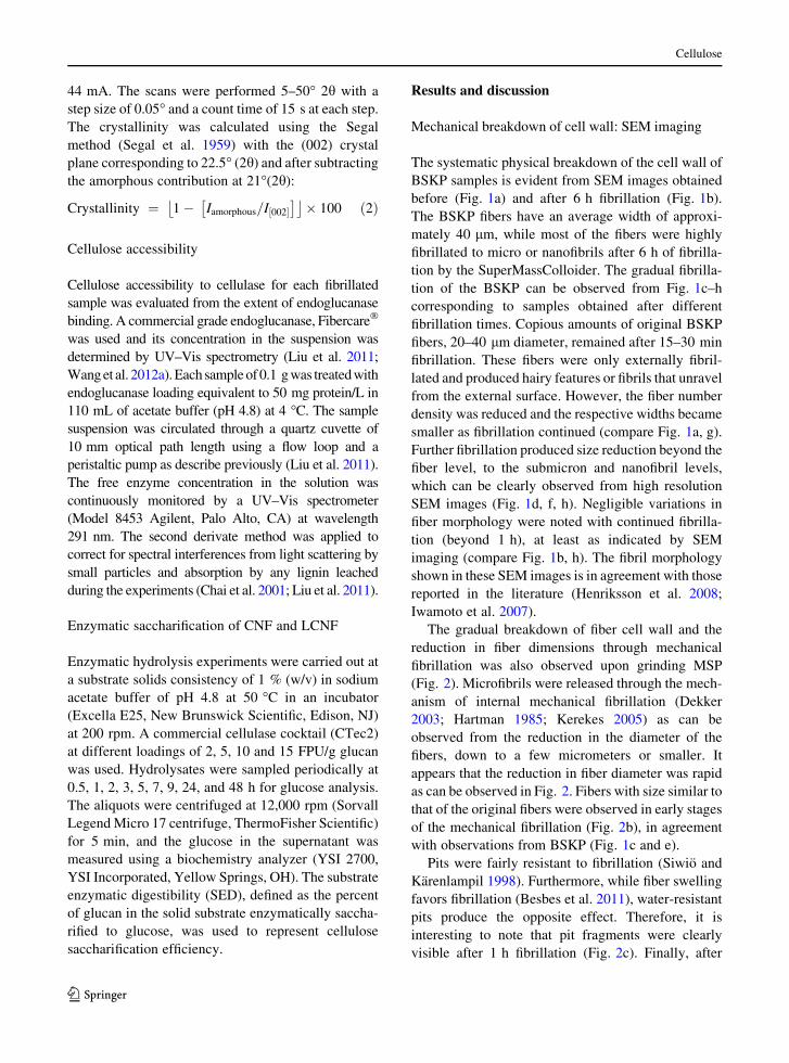
Mechanical breakdown of cell wall: SEM imaging
The systematic physical breakdown of the cell wall of
BSKP samples is evident from SEM images obtained
before (Fig. 1a) and after 6 h fibrillation (Fig. 1b).
The BSKP fibers have an average width of approxi-
mately 40 lm, while most of the fibers were highly
fibrillated to micro or nanofibrils after 6 h of fibrilla-
tion by the SuperMassColloider. The gradual fibrilla-
tion of the BSKP can be observed from Fig. 1c–h
corresponding to samples obtained after different
fibrillation times. Copious amounts of original BSKP
fibers, 20–40 lm diameter, remained after 15–30 min
fibrillation. These fibers were only externally fibril-
lated and produced hairy features or fibrils that unravel
from the external surface. However, the fiber number
density was reduced and the respective widths became
smaller as fibrillation continued (compare Fig. 1a, g).
Further fibrillation produced size reduction beyond the
fiber level, to the submicron and nanofibril levels,
which can be clearly observed from high resolution
SEM images (Fig. 1d, f, h). Negligible variations in
fiber morphology were noted with continued fibrilla-
tion (beyond 1 h), at least as indicated by SEM
imaging (compare Fig. 1b, h). The fibril morphology
shown in these SEM images is in agreement with those
reported in the literature (Henriksson et al. 2008;
Iwamoto et al. 2007).
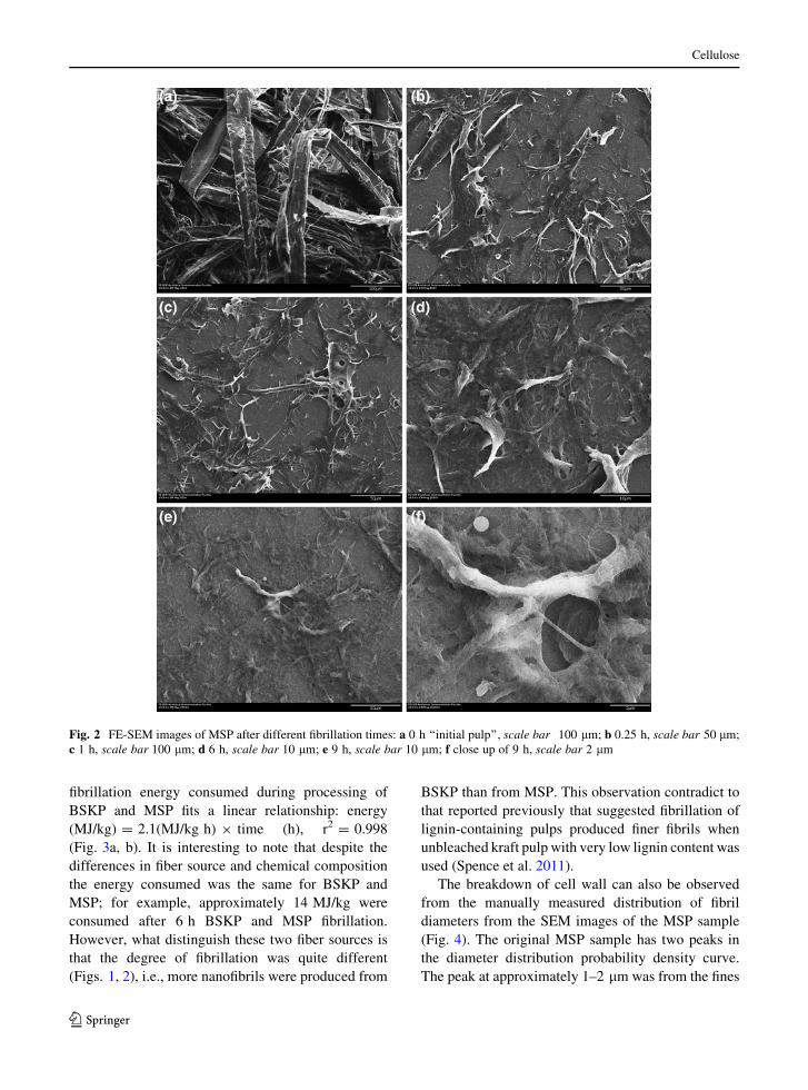
The gradual breakdown of fiber cell wall and the
reduction in fiber dimensions through mechanical
fibrillation was also observed upon grinding MSP
(Fig. 2). Microfibrils were released through the mech-
anism of internal mechanical fibrillation (Dekker
2003; Hartman 1985; Kerekes 2005) as can be
observed from the reduction in the diameter of the
fibers, down to a few micrometers or smaller. It
appears that the reduction in fiber diameter was rapid
as can be observed in Fig. 2. Fibers with size similar to
that of the original fibers were observed in early stages
of the mechanical fibrillation (Fig. 2b), in agreement
with observations from BSKP (Fig. 1c and e).
Pits were fairly resistant to fibrillation (Siwio and
Karenlampil 1998). Furthermore, while fiber swelling
favors fibrillation (Besbes et al. 2011), water-resistant
pits produce the opposite effect. Therefore, it is
interesting to note that pit fragments were clearly
visible after 1 h fibrillation (Fig. 2c). Finally, after
Cellulose
123

fibrillation for 6 h, the microfibers reached a fairly
uniform diameter, which can be confirmed by
the narrow diameter distribution around 1.75 lm
(Fig. 2d, e).
Interestingly, the energy consumption of mechan-
ical fibrillation by the SuperMassColloider was found
to depend on the fibrillation time alone and indepen-
dent of the chemical composition of the fibers. The
Fig. 1 SEM images of BSKP at different fibrillation times: a 0 h; b 6 h; c, d 0.25 h; e, f 0.5 h; g, h 1 h. Note the difference of the scalebars, 100 lm for a, c, e, and g; 1 lm for b, d, f, and h
Cellulose
123
fibrillation energy consumed during processing of
BSKP and MSP fits a linear relationship: energy
(MJ/kg) = 2.1(MJ/kg h) 9 time (h), r2 = 0.998
(Fig. 3a, b). It is interesting to note that despite the
differences in fiber source and chemical composition
the energy consumed was the same for BSKP and
MSP; for example, approximately 14 MJ/kg were
consumed after 6 h BSKP and MSP fibrillation.
However, what distinguish these two fiber sources is
that the degree of fibrillation was quite different
(Figs. 1, 2), i.e., more nanofibrils were produced from
BSKP than from MSP. This observation contradict to
that reported previously that suggested fibrillation of
lignin-containing pulps produced finer fibrils when
unbleached kraft pulp with very low lignin content was
used (Spence et al. 2011).
The breakdown of cell wall can also be observed
from the manually measured distribution of fibril
diameters from the SEM images of the MSP sample
(Fig. 4). The original MSP sample has two peaks in
the diameter distribution probability density curve.
The peak at approximately 1–2 lm was from the fines
Fig. 2 FE-SEM images of MSP after different fibrillation times: a 0 h ‘‘initial pulp’’, scale bar 100 lm; b 0.25 h, scale bar 50 lm;
c 1 h, scale bar 100 lm; d 6 h, scale bar 10 lm; e 9 h, scale bar 10 lm; f close up of 9 h, scale bar 2 lm
Cellulose
123

produced from mechanical refining during producing
MSP. The second peak at approximately 45 lm
represents fiber diameter, as shown in Fig. 2a. With
only 15 min fibrillation, the second peak disappeared
and the distribution peaked at 0.75–1.75 lm, which
suggests the MSP fibers were delaminated into
macrofibrils of approximately 1 lm. However, fibers
with diameter 5–35 lm still exist (Figs. 2b, 4).
Continued fibrillation eliminated large fibrils, above
5 lm, resulting in fibrils with diameters between 0.75
and 1.75 lm (6 h data in Fig. 4). It appears that the
selected stone disk (MKGA6-80#) in the SuperMass-
Collider is not able to further break the crosslinked
lignin barrier to produce fibrils with widths of\1 lm.
This is quite evident from the fact that the fibril
diameter distribution density below 1 lm was essen-
tially unchanged even after 6 h fibrillation. This is also
supported by SEM imaging (Fig. 2). The absence of
lignin facilitated mechanical fibrillation of BSKP to
produce fibrils with diameters in the nanoscale
(Fig. 1), much smaller than those observed in Fig. 2
of MSP fibrils under same energy consumption.
Degree of fiber fibrillation (cell wall breakdown)
Water retention value (WRV) is a measure of fiber
swelling capacity. It quantifies the amount of water in
fiber pores and between fibers after elimination (via
centrifugation) of free water inside the cell lumen (Luo
et al. 2011; Weise et al. 1996). WRV has been used to
measure the degree of micro fibrillation or homoge-
nization of cellulose (Cheng et al. 2007). WRV
increased rapidly in the first hour of fibrillation for
both the BSKP and MSP samples (Fig. 5). WVR
reached a plateau for BSKP thereafter. This suggests
that pores are almost completely accessible so that no
more water-accessible surface can be created through
continued fibrillation. This is corroborated by the
similar morphologies of the fibrillated samples
observed in SEM imaging (Fig. 1h, b). For the MSP
sample, WRV continued to increase but much slowly
after 1 h fibrillation (Fig. 5). WRV of the MSP sample
is lower than that of the BSKP under the same
fibrillation time. This indicates that the extent of
fibrillation of BSKP is much severe than that of MSP
at a given fibrillation time (see SEM images in
Figs. 1h, 2c). Furthermore the cross-linked lignin in
MSP sample is more hydrophobic and reduces fibril
flexibility to swell. While the BSKP has good swelling
and flexibility due to decreased heteropolysaccharides
Fig. 3 Effect of fibrillation on enzymatic saccharification
efficiency, represented by fibrillated substrate enzymatic
digestibility (SED), and energy consumption for fibrillation.
a MSP; b BSKP
Fig. 4 Comparisons of MSP fibril diameter distribution prob-
ability density functions at three different fibrillation times
Cellulose
123

content which reduced the charge density of the fibers
(Laine and Stenius 1997).
Overall, fibrillation reduces the crystallinity of the
fibrils by about 15–25 %. This is in agreement with
other reports (Iwamoto et al. 2007). The continuous
shearing by the grinding stones can destroy the cellulose
crystals and shorten the chain length resulting in smaller
crystals (Wang et al. 2012b). This can be seen from the
measured crystallinity index of the BSKP fibrils
(Fig. 6). The crystallinity index of the BSKP fibers
was 57 % and reduced to approximately 46 % after 6 h
fibrillation. The rate of reduction in crystallinity was
rapid in the first 2 h and then decreased, suggesting the
crystal structure of BSKP was well destroyed in early
stage of fibrillation. The measured crystallinity index of
the MSP fibers was only 47 % due to the presence of a
significant amount of lignin (29.2 %) that is amorphous.
Furthermore, the rate of reduction in crystallinity index
was slow in the early stage of fibrillation, but increased
after 4 h fibrillation. This is opposite to that observed
from MSKP and suggests that mechanical fibrillation
energy was used to breakdown the crosslinked lignin
and hemicelluloses in the early stage fibrillation. The
cellulose crystal structure can then be rapidly destroyed.
Enzymatic hydrolysis of nanofibrillated BSKP
and MSP
It is known that mechanical size reduction can enhance
enzymatic saccharification of lignocellulosic sub-
strates (Dasari and Berson 2007; Zhu et al. 2009a,
2010). However, most studies that use conventional
size reduction resulted in two main conclusions: (1)
there is a significant effect of substrate size but limited
maximal saccharification when using a lignocellulose
that is not thermo-chemically or biologically treated or
modified (Dasari and Berson 2007; Zhu et al. 2009a);
(2) there is a limited or reduced effect of size reduction
but with significant saccharification efficiency, e.g.,
50 % or high, when using fairly well pretreated
substrates, for example, by dilute acid, sulfite, or-
ganosolv (Zhu et al. 2010). In a previous study, we
proposed the concept of two classes of plant biomass
size reduction (Leu and Zhu 2012). Class I merely
increases the external surface area by producing fibers
or fiber bundles without significant breakup of fiber
cell walls. Class II completely breaks down fibers to
render cell walls fully accessible to enzymes. The
efficacy of Class II size reduction for enzymatic
saccharification of untreated wood was corroborated
by Endo using direct milling of Eucalyptus wood
powder (Endo 2010). Solid-state NMR measurements
indicated the domain (fibril) size was approximately
5 nm in the Eucalyptus milled substrate. However, the
SEM images of the milled softwood pulps shown in
Figs. 1 and 2 indicate both Class I and Class II size
reduction with fibril size bigger than 5 nm. This
discrepancy is partly due to the indirect NMR method
used by Endo (Endo 2010). The effect of the two
classes of size reduction on wood cellulose sacchar-
ification can be clearly observed from the enzymatic
hydrolysis of the MSP fibrils. The MSP was produced
Fig. 5 Effect of fibrillation time on water retention values
(WRVs) of BSKP and MSP
Fig. 6 Effect of fibrillation time on crystallinities of BSKP and
MSP
Cellulose
123
from mechanical pulping of lodgepole pine wood
chips without any thermo-chemical pretreatment, as
described in the materials and methods section. The
cellulose enzymatic saccharification efficiency of
MSP at cellulase loading of 10 FPU/g glucan, repre-
sented by SED, was only approximately 6 % without
mechanical fibrillation by the SuperMassColloider
(Fig. 3a). SED was significantly increased by fivefold,
to 30 % after just 15 min fibrillation time (Class I size
reduction). The Class I size reduction enables signif-
icant increase in hydrolysis, but only reached unsat-
isfactory SED of 30 %. SEM images indicated that the
cell wall was only partially broken and large fibers
were still visible (Figs. 2b and 4). Continued increase
in SED was observed with further fibrillation—Class
II size reduction. SED approached 60 % after fibril-
lation for 6 h. The fibers were completely fibrillated to
become macrofibrils (Fig. 2d). Even at cellulase
loading of 5 FPU/g glucan, a SED of 60 % after
48 h hydrolysis was achieved for samples fibrillated
for 6 h. A cellulase loading of 5 FPU/g glucan is low
compared to cellulase loading reported in most studies
using softwoods. This suggests that efficient sacchar-
ification of untreated softwood is possible at relatively
low cellulase loadings without any thermo-chemical
pretreatments by using mechanical fibrillation alone
when the cell wall is completely broken down to
nanofibrils, agree with those reported by Endo using a
Eucalyptus (hardwood) (Endo 2010). This verifies the
concept of Class II size reduction to reconcile
conflicting results reported in the literature on the
effect of plant biomass size reduction on enzymatic
saccharification.
BSKP was produced through chemical pulping and
bleaching. However, the effect of mechanical fibrilla-
tion on enzymatic saccharification of BSKP showed
similar results as discussed above for MSP (Fig. 3b).
At a cellulase loading of only 2 FPU/g glucan, SED
was increased from approximately 27–60 % after 1 h
fibrillation with most fibers fibrillated into nanofibrils
(Fig. 1g, h) and render fiber cell wall completely
accessible to cellulase. SED at 48 h was 94 %
with only 15 min fibrillation at cellulase loading of
5 FPU/g glucan. The difference in cellulose sacchar-
ification at the same cellulase loading of 5 FPU/g
glucan between fibrillated BSKP and fibrillated MSP
is primarily a result of nonproductive cellulase binding
to lignin (in MSP fibrils) (Lan and Zhu 2012;
Mansfield et al. 1999; Sewalt et al. 1997) and lignin
blockage or coverage effect (Mansfield et al. 1999;
Wang et al. 2012c) to reduce cellulose accessibility to
cellulase. Spherical particles were observed in the
MSP fibrils (Fig 3f). These particles most likely are
Pseudo-lignin. It is possible that the separated amor-
phous lignin by mechanical grinding in the Super-
MassCollider can be agglomerated and re-precipitated
(Hu et al. 2012; Kallavus and Gravitis 1995). The
unseparated lignin can cover the cellulose microfibrils
as hypothesized by Endo and co-workers (Lee et al.
2010). These phenomena will affect cellulose
accessibility.
The above mechanistic discussions of the effects of
two classes of size reduction on enzymatic sacchar-
ification can be supported by quantitative measure-
ments. Mechanical fibrillation breaks down cell walls
and increases cellulose accessibility as can be seen
from the increase in the amounts of cellulase bound
with fibrillation time (Fig. 7). The amount of CTec2
bound to the unfibrillated BSKP (fibrillation
time = 0) was significant because cellulose in the
bleached pulp is highly accessible to cellulase. CTec2
binding curves for both BSKP and MSP fibrils were
increased rapidly within the first hour of fibrillation
suggesting rapid cell wall breakdown and further
confirmed by SEM imaging (Figs. 1, 2). CTec2
binding to BKSP fibrils reached a plateau while
binding to MSP fibrils increased slowly with further
fibrillation. The cellulase binding curves shown in
Fig. 7 are very similar to WRV curves (Fig. 5),
confirming cellulase binding is directly related to
accessible surface area as measured by WRV, in
agreement with a recent study (Lee et al. 2010). The
fact that CTec2 binding was near zero for the MSP
sample at zero fibrillation time indicate: (1) external
surface plays a minor role in cellulose accessibility
(Luo and Zhu 2011; Sinitsyn et al. 1991; Wang et al.
2012a); (2) the MSP sample has limited pores
accessible to cellulase due to lignin coverage (Leu
and Zhu 2012; Wang et al. 2012c). The continued
increase in CTec2 binding to MSP fibrils (Fig. 7)
indicates mechanical fibrillation alone can break down
cell wall to make cellulose completely accessible. It is
simply a coincidence that the amount of CTec2
binding to the MSP fibrils is approximately the same
as that bound to KSKP fibrils after extended fibrilla-
tion, e.g. 6 h. Because the amounts of cellulase
binding shown in Fig. 6 include both the productive
(to cellulose) and nonproductive (to lignin and other
Cellulose
123
components), it is apparent that significantly more
cellulase were bound productively to BSKP with zero
lignin content and hemicellulose content of 15.1 %
than to MSP that has a lignin content of 29.2 % and
hemicellulose content of 16 % (Table 1). This
explains the difference in SED between MSP and
BSKP fibrils at the same CTec2 loading of 5FPU/g
glucan (Fig. 3a, b).
Conclusions
Mechanical size reduction of plant biomass can be
categorized into two classes: Class I reduces plant
biomass to the level of fibers or fiber bundles with
limited cell wall breakdown; Class II significantly
breaks down fiber cell walls to produce macro or
nanofibrils that renders cell wall highly or completely
accessible to cellulase. Low degree of fibrillation, i.e.,
Class I size reduction, has a significant effect on
enhancing enzymatic saccharification of a mechanical
softwood pulp but with a overall low saccharification
efficiency. This is because Class I size reduction
merely increases fiber external surface without break-
ing down the cell wall structure and therefore with
limited increase in cellulose accessibility evidenced
by measured cellulase adsorption. Further fibrillation
to the level of Class II size reduction significantly
breaks down cell walls to macro or nanofibrils that
made most fibrils accessible to cellulase resulting in
significant saccharification of cellulose. The transition
from Class I to Class II size reduction in the
SuperMassCollider was rapid when the starting mate-
rials were wood fibers. Extended fibrillation has
limited effect on cell wall breakdown as measured
by water retention value and cellulase adsorption. This
is probably due to the fact that fibrillation reached the
limit of the stone used. Lignin plays a significant role
in protecting cell wall. Under the same fibrillation
energy input, the degree of cell wall breakdown for the
mechanical softwood pulp is much less severer than
that for a bleached softwood kraft pulp. The presence
of lignin also prevented efficient cellulose saccharifi-
cation by blocking cellulose as well as nonproduc-
tively binding to cellulase.
Acknowledgments This work was sponsored by the USDA
Forest Service R&D special funding on Cellulose Nano-
Materials (2012).
References
Agarwal UP, Reiner RS, Ralph SA (2010) Celulose I crystal-
linity determination using FT-Raman spectroscopy: uni-
variate and multivariate methods. Cellulose 17(4):721–733
Ahola S, Turon X, Osterberg M, Laine J, Rojas OJ (2008)
Enzymatic hydrolysis of native cellulose nanofibrils and
other cellulose model films: effect of surface structure.
Langmuir 24(20):11592–11599
Besbes I, Alila S, Boufi S (2011) Nanofibrillated cellulose from
TEMPO-oxidized eucalyptus fibres: effect of the carboxyl
content. Carbohydr Polym 84(3):975–983
Bozell JJ, Petersen GR (2010) Technology development for the
production of biobased products from biorefinery carbo-
hydrates: the US department of energy’s ‘‘top 10’’ revis-
ited. Green Chem 12(4):539–554
Chai X-S, Zhu JY, Li J (2001) A simple and rapid method to
determine hexeneuronic acid groups in chemical pulps.
J Pulp Paper Sci 27(5):165–170
Cheng Q, Wang S, Rials TG, Lee SH (2007) Physical and
mechanical properties of polyvinyl alcohol and polypro-
pylene composite materials reinforced with fibril aggre-
gates isolated from regenerated cellulose fibers. Cellulose
14(6):593–602
Dasari RK, Berson RE (2007) The effect of particle size on
hydrolysis reaction rates and rheological properties in
cellulosic slurries. Appl Biochem Biotechnol 137:289–299
Dekker J (2003) New insights in beating leading to innovative
beating techniques. PIRA 2003 refining conference,
Stockholm, Sweden
Endo T (2010) Bioethanol production from woods with the aid
of nanotechnology. Synthesiology 4(2):270–281
Gupta R, Lee YY (2009) Pretreatment of hybrid poplar by
aqueous ammonia. Biotechnol Prog 25:357–364
Hartman RR (1985) Mechanical treatment of pulp fibers for
paper property development. In: Punton V (ed) The 8th
fundamental research symposium: paper making materials.
Fig. 7 Effect of fibrillation time on the amount of cellulase
binding to fibrillated substrates
Cellulose
123

Mechanical Engineering Publications Limited, Oxford,
pp 413–442
Henriksson M, Berglund LA, Isaksson P, Lindstrom T, Nishino TD
(2008) Cellulose nanopaper structures of high toughness.
Biomacromolecules 9:1579–1585
Himmel ME, Ding SY, Johnson DK, Adney WS, Nimlos MR,
Brady JW, Foust TD (2007) Biomass recalcitrance: engi-
neering plants and enzymes for biofuels production. Sci-
ence 315(5813):804–807
Hu F, Jung S, Ragauskas A (2012) Pseudo-lignin formation and
its impact on enzymatic hydrolysis. Bioresour Technol
117:7–12
Iwamoto S, Nakagaito AN, Yano H (2007) Nano-fibrillation of
pulp fibers for the processing of transparent nanocompos-
ites. Appl Phys Mater Sci Process 89:461–466
Kallavus U, Gravitis J (1995) A comparative investigation of the
ultrastructure of steam exploded wood with light, scanning
and transmission electron-microscopy. Holzforschung
49(2):182–188
Kerekes RJ (2005) Characterizing refining actions: linking the
process to refining results. In: PIRA 2005 refining confer-
ence, Barcelona, Spain
Koo BW, Treasure TH, Jameel H, Phillips RB, Chang HM, Park S
(2011) Reduction of enzyme dosage by oxygen delignifica-
tion and mechanical refining for enzymatic hydrolysis of
green liquor-pretreated hardwood. Appl Biochem Biotechnol
165(3–4):832–844
Laine J, Stenius P (1997) Effect of charge on the fiber and paper
properties. Pap Puu 79:257–266
Lan TQ, Lou H, Zhu JY (2012) Enzymatic saccharification of
lignocelluloses should be conducted at elevated pH
5.2–6.2. BioEner Res. doi:10.1007/s12155-012-9273-4
Lee SH, Chang F, Inoue S, Endo T (2010) Increase in enzyme
accessibility by generation of nanospace in cell wall
supramolecular structure. Bioresour Technol 101(19):
7218–7223
Leu S-Y, Zhu JY (2012) Substrate related factors affecting
enzymatic saccharification of lignocelluloses: our recent
understanding. BioEner Res. doi:10.1007/s12155-012-
9276-1
Liu H, Zhu JY, Chai XS (2011) In situ, rapid, and temporally
resolved measurements of cellulase adsorption onto lig-
nocellulosic substrates by UV-vis spectrophotometry.
Langmuir 27(1):272–278
Luo X, Zhu JY (2011) Effects of drying-induced fiber hornifi-
cation on enzymatic saccharification of lignocelluloses.
Enzym Microb Technol 48(1):92–99
Luo X, Gleisner R, Tian S, Negron J, Horn E, Pan XJ, Zhu JY
(2010) Evaluation of mountain beetle infested lodgepole
pine for cellulosic ethanol production by SPORL pre-
treatment. Ind Eng Chem Res 49(17):8258–8266
Luo X, Zhu JY, Gleisner R, Zhan HY (2011) Effect of wet
pressing-induced fiber hornification on enzymatic sac-
charification of lignocelluloses. Cellulose 18:1339–1344
Mansfield SD, Mooney C, Saddler JN (1999) Substrate and
enzyme characteristics that limit cellulose hydrolysis.
Biotechnol Prog 15:804–816
Martin-Sampedro R, Filpponen I, Hoeger IC, Zhu JY, Laine J,
Rojas OJ (2012) Rapid and complete enzyme hydrolysis
of ligocellulosic nanofibrils. ACS Macro Lett 1(1):
1321–1325
Perlack RD, Wright LL, Turhollow A, Graham RL, Stokes B,
Erbach DC (2005) Biomass as feedstock for a bioenergy
and bioproducts industry: The technical feasibility of a
billion-ton annual supply. Oak ridge national laboratory
report, ORNL/TM-2005/66, US Dept of Energy
Segal L, Creely JJ, Martin AE, Conrad CM (1959) An empirical
method for estimating the degree of crystallinity of native
cellulose using the X-ray diffractometer. Text Res J
29:786–794
Sewalt VJH, Glasser WG, Beauchemin KA (1997) Lignin
impact on fiber degradation.3. Reversal of inhibition of
enzymatic hydrolysis by chemical modification of lignin
and by additives. J Agric Food Chem 45(5):1823–1828
Sinitsyn AP, Gusakov AV, Vlasenko EY (1991) Effect of
structural and physico-chemical features of cellulosic
substrates on the efficiency of enzymatic hydrolysis. Appl
Biochem Biotechnol 30:43–59
Siwio J, Karenlampil P (1998) Pits as natural irregularities in
softwood fibers. Wood Fiber Sci 30:27–39
Spence KL, Venditti RA, Rojas OJ, Habibi Y, Pawlak JJ
(2011) A comparative study of energy consumption and
physical properties of microfibrillated cellulose produced
by different processing methods. Cellulose 18(4):1097–
1111
Sun Y, Cheng JY (2002) Hydrolysis of lignocellulosic materials
for ethanol production: a review. Bioresour Technol
83(1):1–11
TAPPI (2009) TAPPI test methods. Technical Association of
the Pulp and Paper Industry, Atlanta
Wang QQ, He Z, Zhu Z, Zhang Y-HP, Ni Y, Luo XL, Zhu JY
(2012a) Evaluations of cellulose accessibilities of ligno-
celluloses by solute exclusion and protein adsorption
techniques. Biotechnol Bioeng 109(2):381–389
Wang QQ, Zhu JY, Gleisner R, Kuster TA, Baxa U, McNeil SE
(2012b) Morphological development of cellulose fibrils of
a bleached eucalyptus pulp by mechanical fibrillation.
Cellulose 19(5):1631–1643
Wang ZJ, Zhu JY, Gleisner R, Chen KF (2012c) Ethanol pro-
duction form poplar wood the rough enzymatic sacchari-
fication and fermentation by dilute acid and SPORL
pretreatments. Fuel 95:606–614
Weise U, Maloney T, Paulapuro H (1996) Quantification of
water in difference states of interaction with wood pulp
fibres. Cellulose 3:189–202
Zhu JY (2011) Physical pretreatment—woody biomass size-
reduction—for forest biorefinery. In: Zhu JY, Zhang X,
Pan XJ (eds) Sustainable production of fuels, chemicals,
and fibers from forest biomass. American Chemical Soci-
ety, Washington, pp 89–107
Zhu J, Wang G, Pan X, Gleisner R (2009a) Specific surface to
evaluate the efficiencies of milling and pretreatment of
wood for enzymatic saccharification. Chem Eng Sci
64(3):474–485
Zhu JY, Pan XJ, Wang GS, Gleisner R (2009b) Sulfite pre-
treatment (SPORL) for robust enzymatic saccharification
of spruce and red pine. Bioresour Technol 100(8):2411–
2418
Zhu W, Zhu JY, Gleisner R, Pan XJ (2010) On energy con-
sumption for size-reduction and yield from subsequent
enzymatic sacchrification of pretreated lodgepole pine.
Bioresour Technol 101(8):2782–2792
Cellulose
123

Zhu JY, Luo X, Tian S, Gleisner R, Negrone J, Horn E (2011a)
Efficient ethanol production from beetle-killed lodgepole
pine using SPORL technology and Saccharomyces cere-
visiae without detoxification. Tappi J 10(5):9–18
Zhu JY, Verrill SP, Liu H, Herian VL, Pan XJ, Rockwood DL
(2011b) On polydispersity of plant biomass recalcitrance
and its effects on pretreatment optimization for sugar
production. BioEner Res 4(3):201–210
Cellulose
123


