
Mr
DD
a
ARAA
KLLRM
1
midcs
2
pcicw22
aoes
mno
2h
CASE REPORT – OPEN ACCESSInternational Journal of Surgery Case Reports 5 (2014) 152–154
Contents lists available at ScienceDirect
International Journal of Surgery Case Reports
j ourna l h om epage: www.caserepor ts .com
esh fistulation into the rectum after laparoscopic ventral meshectopexy�
ayo Adeyemo ∗
epartment of Gastrointestinal Surgery, Wye Valley Hospital NHS Trust, Hereford HR1 1BX, United Kingdom
r t i c l e i n f o
rticle history:eceived 7 December 2013ccepted 16 December 2013vailable online 31 December 2013
a b s t r a c t
INTRODUCTION: Laparoscopic ventral mesh rectopexy (LVMR) is an effective method of management offunctional disorders of the rectum including symptomatic rectal intussusception, and obstructed defae-cation. Despite the technical demands of the procedure and common use of foreign body (mesh), theincidence of mesh related severe complications of the rectum is very low.
eywords:aparoscopic ventral mesh rectopexyVMR complicationectal fistulaesh complication
PRESENTATION OF CASE: A 63 year old woman presented with recurrent pelvic sepsis following a mesh rec-topexy. Investigations revealed fistulation of the mesh into the rectum. She was treated with an anteriorresection.DISCUSSION: The intraoperative findings and management of the complication are described. Risk factorsfor mesh attrition and fistulation are also discussed.CONCLUSION: Chronic sepsis may lead to ‘late’ fistulation after mesh rectopexy.
blish
© 2013 The Authors. Pu. Introduction
Laparoscopic ventral mesh rectopexy (LVMR) is an effectiveethod of management of functional disorders of the rectum
ncluding symptomatic rectal intussusception, and obstructedefaecation.1,2 Despite the technical demands of the procedure andommon use of foreign body (mesh), the incidence of mesh relatedevere complications of the rectum is very low.
. Presentation of case
A 63-year old lady presented with a three month history ofrogressively worsening recurrent pelvic pain. There was no asso-iated rectal bleeding or change in bowel habits, but there had beenntermittent rectal discharge. Each episode resolved quickly afterommencement of broad spectrum antibiotics, but the episodesere becoming more frequent. Past medical history included type
diabetes mellitus and a laparoscopic mesh rectopexy performed4 months earlier.
A CT scan during a previous episode showed chronic sepsisround the sacral promontory (in the area of the anchored tail
f the radio-opaque mesh). General, abdominal and digital rectalxaminations were unremarkable. Rigid sigmoidoscopy demon-trated normal rectal mucosa, but there was a local concentration of� This is an open-access article distributed under the terms of the Creative Com-ons Attribution-NonCommercial-No Derivative Works License, which permits
on-commercial use, distribution, and reproduction in any medium, provided theriginal author and source are credited.∗ Tel.: +44 7961034412; fax: +44 01432 364102.
E-mail address: [email protected]
210-2612/$ – see front matter © 2013 The Authors. Published by Elsevier Ltd on behalf
ttp://dx.doi.org/10.1016/j.ijscr.2013.12.012
ed by Elsevier Ltd on behalf of Surgical Associates Ltd. All rights reserved.
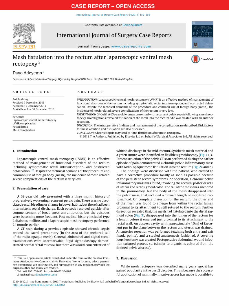
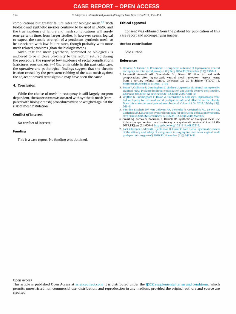
whitish discharge in the mid-rectum. Synthetic mesh material anda green suture were identified on flexible sigmoidoscopy (Fig. 1). 3-D reconstruction of the pelvic CT scan performed during the earlierepisode of pain demonstrated a chronic pelvic inflammatory masswith radio-opaque mesh fistulation into the rectum (Fig. 2a and b).
The findings were discussed with the patient, who elected tohave a corrective procedure locally as soon as possible becauseof the persistent severe symptoms. At operation, a chronic pelvicinflammatory mass was found, involving the rectum, posterior wallof uterus and rectosigmoid colon. The tail of the mesh was anchoredto the promontory, but the body of the mesh disappeared intothe pelvic mass, that included a ‘bowed’ length of mobilized rec-tosigmoid. On complete dissection of the rectum, the other endof the mesh was found to emerge from within the rectal lumenproximal to its attachment to still sutured to the rectum. Furtherdissection revealed that, the mesh had fistulated into the distal sig-moid colon (Fig. 3), disappeared into the lumen of the rectum fora length before it emerged just proximal to its attachment to therectal wall. An abscess cavity with approximately 10 ml of faecu-lent pus in the plane between the rectum and uterus was drained.An anterior resection was performed (excising both entry and exitfistula points), and a stapled anastomosis fashioned. A coveringloop ileostomy was created. Postoperative abdominal wound infec-tion cultured proteus sp. (similar to organisms cultured from thedrained pelvic abscess).
3. Discussion
While mesh rectopexy was described many years ago, it hasgained popularity in the past 2 decades. This is because the success-ful application of minimally invasive access has made it possible to
of Surgical Associates Ltd. All rights reserved.

CASE REPORT – OPEN ACCESSD. Adeyemo / International Journal of Surgery Case Reports 5 (2014) 152–154 153
Fr
owocfdro
(rbg
ig. 1. Endoscopic picture showing the fistulated mesh and adjacent suture in theectal lumen.
ffer the procedure to the very elderly and other patient groupshere the risks of a major laparotomy outweighed the benefits
f the surgery.3,4 Nevertheless, the technical demands of the pro-edure in the confined bony pelvic cavity are recognized, and riskactors for failure (non-correction of functional symptoms) are wellocumented.2,5 Significant immediate, short-term and long-termectal complications after mesh rectopexy have been reported, butn the whole are uncommon.
The most serious complications of LVMR are mesh relatedinfection, extrusion and erosion). While a systematic review
eported similar rates of mesh complications and failure foriologic and synthetic LVMR,5 a larger systematic review ofynaecological organ prolapse mesh repair reported no mesh Fig. 3. Specimen showing the adherent mesh fistula into the rectum.Fig. 2. (a) and (b) Coronal & sagittal CT scans demonstrating the mesh fistula into the rectum.
– O1 f Surg
cbtetbm
at(tft
4
dpr
C
F
1
2
3
4
5
6
OTpc
CASE REPORT54 D. Adeyemo / International Journal o
omplications but greater failure rates for biologic mesh.6,7 Bothiologic and synthetic meshes continue to be used in LVMR, andhe true incidence of failure and mesh complications will surelymerge with time, from larger studies. It however seems logicalo expect the tensile strength of a persistent synthetic mesh toe associated with low failure rates, though probably with moreesh related problems (than the biologic mesh).Given that the mesh (synthetic, combined or biological) is
nchored to or in close proximity to the rectum sutured duringhe procedure, the reported low incidence of rectal complicationsstrictures, erosions, etc.) ∼1% is remarkable. In this particular case,he operative and pathological findings suggest that the chronicriction caused by the persistent rubbing of the taut mesh againsthe adjacent bowed rectosigmoid may have been the cause.
. Conclusion
While the choice of mesh in rectopexy is still largely surgeonependent, the success rates associated with synthetic mesh (com-ared with biologic mesh) procedures must be weighed against theisk of mesh fistulation.
onflict of interest
No conflict of interest.
unding
This is a case report. No funding was obtained.
7
pen Accesshis article is published Open Access at sciencedirect.com. It is distribermits unrestricted non commercial use, distribution, and reproductredited.
PEN ACCESSery Case Reports 5 (2014) 152–154
Ethical approval
Consent was obtained from the patient for publication of thiscase report and accompanying images.
Author contribution
Sole author.
References
. D’Hoore A, Cadoni’ R, Penninckx F. Long-term outcome of laparoscopic ventralrectopexy for total rectal prolapse. Br J Surg 2004;91(November (11)):1500–5.
. Badrek-Al Amoudi AH, Greenslade GL, Dixon AR. How to deal withcomplications after laparoscopic ventral mesh rectopexy: lessons learntfrom a tertiary referral centre. Colorectal Dis 2013;15(June (6)):707–12,http://dx.doi.org/10.1111/codi.12164.
. Boons P, Collinson R, Cunningham C, Lindsey I. Laparoscopic ventral rectopexy forexternal rectal prolapse improves constipation and avoids de novo constipation.Colorectal Dis 2010;12(June (6)):526–32. Epub 2009 April 10.
. Wijffels N, Cunningham C, Dixon A, Greenslade G, Lindsey I. Laparoscopic ven-tral rectopexy for external rectal prolapse is safe and effective in the elderly.Does this make perineal procedures obsolete? Colorectal Dis 2011;13(May (5)):561–6.
. Van den Esschert JW, van Geloven AA, Vermulst N, Groenedijk AG, de Wit LT,Gerhards MF. Laparoscopic ventral rectopexy for obstructed defecation syndrome.Surg Endosc 2008;22(December (12)):2728–32. Epub 2008 March 5.
. Smart NJ, Pathak S, Boorman P, Daniels IR. Synthetic or biological mesh usein laparoscopic ventral mesh rectopexy – a systematic review. Colorectal Dis2013;15(June (6)):650–4, http://dx.doi.org/10.1111/codi.12219.
. Jia X, Glazener C, Mowatt G, Jenkinson D, Fraser C, Bain C, et al. Systematic reviewof the efficacy and safety of using mesh in surgery for uterine or vaginal vaultprolapse. Int Urogynecol J 2010;21(November (11)):1413–31.
uted under the IJSCR Supplemental terms and conditions, whichion in any medium, provided the original authors and source are


