

November 2017
Multiple infantile
hemangiomas in a preterm
infant
SWISS SOCIETY OF NEONATOLOGY
Heschl K, Romaine Arlettaz R, Department of Neonatology
(HK, AR), University Hospital Zurich, Switzerland
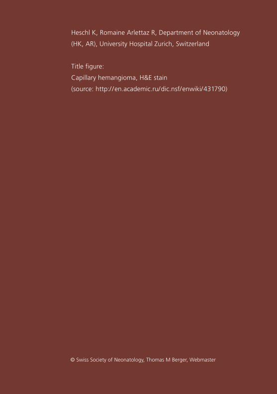
Title figure:
Capillary hemangioma, H&E stain
(source: http:/ /en.academic.ru/dic.nsf/enwiki/431790)
© Swiss Society of Neonatology, Thomas M Berger, Webmaster

Hemangiomas represent the most common tumor of
infancy with the highest incidence in preterm infants
(1). They are usually not present at birth but develop
in the first few weeks of life and proliferate in the
following weeks and months. The etiology is not yet
fully understood but vascular endothelial growth factor
(VEGF) may play an important role. Multiple heman
giomas are defined by five or more heman giomas, are
usually smaller in size and more likely to be associated
with visceral hemangiomas (2).
INTRODUCTION
3
CASE REPORT
4
We present a male preterm infant born to a healthy
31yearold G1/P1 by Cesarean section at 28 4 /7 weeks
due to preeclampsia.
The baby had a birthweight of 1100 g (P 25 – 50). Apgar
scores were 6, 8 and 8 at 1, 5 and 10 minutes, respec
tively, and the arterial umbilical cord pH was 7.27. He
was transferred to the neonatal intensive care unit with
mild respiratory distress on nasal CPAP and required a
maximal FiO2 of 0.3. His postnatal course – apart from
an episode of lateonset sepsis – was un remarkable.
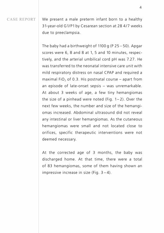
At about 3 weeks of age, a few tiny hemangiomas
the size of a pinhead were noted (Fig. 1 – 2). Over the
next few weeks, the number and size of the hemangi
omas increased. Abdominal ultrasound did not reveal
any intestinal or liver hemangiomas. As the cuta neous
hemangiomas were small and not located close to
orifices, specific therapeutic interventions were not
deemed necessary.
At the corrected age of 3 months, the baby was
discharged home. At that time, there were a total
of 83 hemangiomas, some of them having shown an
impressive increase in size (Fig. 3 – 4).
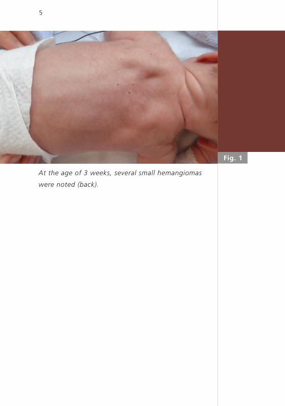
Fig. 1
5
At the age of 3 weeks, several small hemangiomas
were noted (back).
Fig. 2
6
At the age of 3 weeks, several small hemangiomas
were noted (chest).
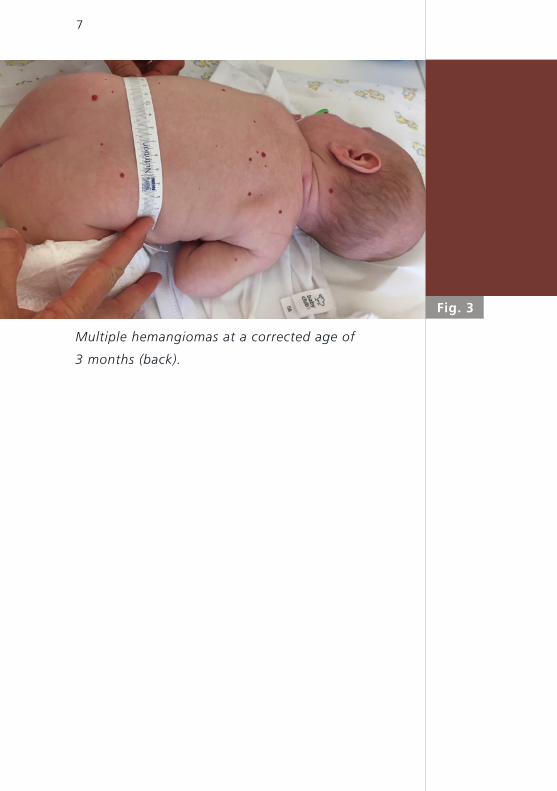
Fig. 3
7
Multiple hemangiomas at a corrected age of
3 months (back).
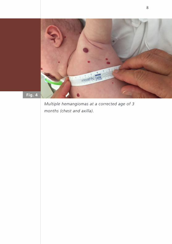
Fig. 4
8
Multiple hemangiomas at a corrected age of 3
months (chest and axilla).
9
DISCUSSIONThis case report of a premature infant with multiple
cutaneous hemangiomas without visceral involve
ment illustrates a common problem in this population.
About 25% of preterm infants develop hemangiomas
during the neonatal period compared to around 5%
of term infants (3). Infantile hemangiomas are clas
sified as focal or localized, segmental, indeterminate
and multifocal infantile hemangiomas (4). Multiple
hemangiomas are mainly located on the skin, but
these infants have a higher risk of internal heman
giomas, which are particularly located in the liver.
Ulceration and bleeding are the typical complications
of cutaneous hemangiomas. These infants require
regular clinical and sonographic followup including
precise documentation of the lesions (5).
Infantile hemangiomas are vascular tumors. Initially,
there is a proliferating phase in the first six to nine
months of age, followed by an involution phase over
years which ultimately leads to a residual, mostly fibro
tic mass. The differential diagnosis includes other vas
cular tumors, as well as other vascular malfor mations,
which in contrast to infantile hemangiomas are usually
present at birth.
The exact pathogenesis of multiple infantile hemangi
omas is not yet fully understood. There are different
hypotheses, however, vascular endothelial growth fac
tor A (VEGFA), an important molecule in angiogenesis
and vasculogenesis, likely plays a role. In addition, cell

10
embolization from the placenta, somatic mutations in
a gene mediating endothelial cell proliferation, endo
thelial progenitor cell and hypoxia have all been impli
cated (6 – 9).
According to the European Propranolol In the Treat
ment of Complicated Haemangiomas (PITCH) study,
there are three main indications for treatment:
1) periocular location with threat to vision, 2) location
on the face with risk of cosmetic disfigurement and
3) risk for ulceration and bleeding, seen for example
in perianal hemangiomas (10). Overall, treatment is
rarely required and longterm outcome is good.
Therapeutic options include systemic or local treat
ment with beta blockers, cryotherapy, or laser coa
gulation. Since beta blocker treatment has become
available, surgery is now only rarely required. Cortico
steroids are generally not used in infantile hemangio
mas. Modifying VEGFA action could be a promising
therapeutic option in the future, but, at this point,
results of randomised controlled trials are still lacking.
Propranolol therapy for infantile hemangiomas was
used in 2008 for the first time and since then has
rapidly become the first line treatment. Its effect on
infantile hemangiomas was discovered by chance:
LéautéLabrèze et al. used corticosteroids to treat a
patient with a nasal hemangioma. During this treat
ment, the hemangioma stabilized in size but the
11
patient developed hypertrophic obstructive cardiomy
opathy, which in turn was treated with propranolol.
The treatment team observed rapid involution of the
hemangioma (11).
Oral propranolol therapy should be started with a
dose of 1 mg/kg/day in the hospital setting to monitor
heart rate, blood pressure and glucose concentrations.
If well tolerated, it can be increased to maintenance
dose of 2 mg/kg/day (12). The main side effects are
mild and include sleep disturbance and cool extre
mities. At higher doses, fatigue and bradycardia can
occur. Treatment response depends on timing and
duration of treatment. There is no consensus regar
ding the optimum treatment duration, but studies
suggest that the risk for rebound growth decreases
after 12 months (12). Rebound growth has been
reported to occur in up to 20% of patients. Risk factors
for rebound growth are short duration of propranolol
therapy, female gender, deep infantile hemangiomas
and abrupt discontinuation of therapy (13).
In summary, infantile hemangiomas are very common
in premature infants. Usually, they are not present
at birth but proliferate in the first few months with
a peak around the age of 3 – 6 months, followed by
a stable phase for another 2 years and a regression
phase until the age of 4 – 5 years. The pathogenesis
of infantile hemangioma is still not fully understood.
Whether active treatment is required depends on

12
localization and size of the lesions as well as the risk
for complications. Overall, only a small proportion of
patients needs therapy. Local or systemic propranolol
is the first line treatment. It is safe and highly effec
tive, but compliance is important and parents should
be informed about the potential and usually mild side
effects.
1. Goelz R, Poets CF. Incidence and treatment of infantile
haeman gioma in preterm infants. Arch Dis Child Fetal Neonatal
Ed 2015;100:F85F91 (Abstract)
2. Horii KA, Drolet BA, Frieden IJ, et al. Prospective study
of the frequency of hepatic hemangiomas in infants with
multiple cutaneous infantile hemangiomas. Pediatr Dermatol
2011;28:245 – 253 (Abstract)
3. Jacobs AH, Walton RG. The incidence of birthmarks in the
neonate. Pediatrics 1976;58:218–222 (Abstract)
4. Dasgupta R, Fishman SJ. ISSVA classification. Semin Pediatr
Surg 2014; 23:158 – 161 (Abstract)
5. Haggstrom AN, Drolet BA, Baselga E, et al. Prospective study
of infantile hemangiomas: clinical characteristics predicting
complications and treatment. J Pediatr 2006;118:882 – 887
(Abstract)
6. Chen TS, Lawrence F, Friedlander SF. Infantile hemangio
mas: an update on pathogenesis and therapy, Pediatrics
2013;131:99 – 108 (Abstract)
7. Mihm MC Jr, Nelson JS. Hypothesis: the metastatic niche theory
can elucidate infantile hemangioma development. J Cutan
Pathol 2010;37:83 – 87 (Abstract)
8. Walter JW, North PE, Waner M, et al. Somatic mutation
of vascular endothelial growth factor receptors in juvenile
hemangioma. Genes Chromosomes Cancer 2002;33:295 – 303
(Abstract)
9. Greenberger S, Bischoff J. Infantile hemangioma –
mechanism(s) of drug action on a vascular tumor. Cold Spring
Harb Perspect Med 2011;1:a006460 (Abstract)
REFERENCES
13
10. Wedgeworth E, Glover M, Irvine AD, et al. Propranolol in the
treatment of infantile haemangiomas: lessons from the Euro
pean Propranolol In the Treatment of Complicated Haemangio
mas (PITCH) taskforce survey. Br J Dermatol 2016;174:594 – 601
(Abstract)
11. LéautéLabrèze C, Dumas de la Roque E, Hubiche T, Boralevi F,
Thambo JB, Taïeb A. Propranolol for severe hemangiomas of
infancy. N Engl J Med 2008;358:2649 – 2651 (Abstract)
12. Drolet BA, Frommelt PC, Chamlin SL, et al. Initiation and use
of propranolol for infantile hemangioma: report of a consensus
conference. Pediatrics 2013;131:128 – 140 (Abstract)
13. Shah SD, Baselga E, McCuaig, et al. Rebound growth of
infantile hemangiomas after propranolol therapy. Pediatrics
2016;137:e20151754 (Abstract)
14


SUPPORTED BY
CONTACT
Swiss Society of Neonatology
www.neonet.ch
con
cep
t &
des
ign
by
mes
ch.c
h

![InfantileHemangiomasMasqueradingas …strawberry-colorednodule,whiledeeporbitalhemangiomas typically present as a fluctuant, compressible bluish mass [2]. Infantile hemangiomas are](https://static.documents.pub/doc/80x56/6103541b09e789301341c088/infantilehemangiomasmasqueradingas-strawberry-colorednodulewhiledeeporbitalhemangiomas.jpg)


