

NF-Y Recruits Both Transcription Activator and Repressorto Modulate Tissue- and Developmental Stage-SpecificExpression of Human c-Globin GeneXingguo Zhu1, Yongchao Wang1, Wenhu Pi1, Hui Liu2, Amittha Wickrema2, Dorothy Tuan1*
1 Department of Biochemistry and Molecular Biology, Medical College of Georgia and College of Graduate Studies, Georgia Health Sciences University, Augusta, Georgia,
United States of America, 2 Department of Medicine, University of Chicago, Chicago, Illinois, United States of America
Abstract
The human embryonic, fetal and adult b-like globin genes provide a paradigm for tissue- and developmental stage-specificgene regulation. The fetal c-globin gene is expressed in fetal erythroid cells but is repressed in adult erythroid cells. Themolecular mechanism underlying this transcriptional switch during erythroid development is not completely understood.Here, we used a combination of in vitro and in vivo assays to dissect the molecular assemblies of the active and therepressed proximal c-globin promoter complexes in K562 human erythroleukemia cell line and primary human fetal andadult erythroid cells. We found that the proximal c-globin promoter complex is assembled by a developmentally regulated,general transcription activator NF-Y bound strongly at the tandem CCAAT motifs near the TATA box. NF-Y recruits toneighboring DNA motifs the developmentally regulated, erythroid transcription activator GATA-2 and general repressorBCL11A, which in turn recruit erythroid repressor GATA-1 and general repressor COUP-TFII to form respectively the NF-Y/GATA-2 transcription activator hub and the BCL11A/COUP-TFII/GATA-1 transcription repressor hub. Both the activator andthe repressor hubs are present in both the active and the repressed c-globin promoter complexes in fetal and adulterythroid cells. Through changes in their levels and respective interactions with the co-activators and co-repressors duringerythroid development, the activator and the repressor hubs modulate erythroid- and developmental stage-specifictranscription of c-globin gene.
Citation: Zhu X, Wang Y, Pi W, Liu H, Wickrema A, et al. (2012) NF-Y Recruits Both Transcription Activator and Repressor to Modulate Tissue- and DevelopmentalStage-Specific Expression of Human c-Globin Gene. PLoS ONE 7(10): e47175. doi:10.1371/journal.pone.0047175
Editor: Ramani Ramchandran, Medical College of Wisconsin, United States of America
Received July 18, 2012; Accepted September 10, 2012; Published October 10, 2012
Copyright: � 2012 Zhu et al. This is an open-access article distributed under the terms of the Creative Commons Attribution License, which permits unrestricteduse, distribution, and reproduction in any medium, provided the original author and source are credited.
Funding: This work was supported by the National Institutes of Health (HL 89519 and MD003383 to D.T., CA98550 to A.W.), and the Giving Tree Foundation toA.W. The funders had no role in study design, data collection and analysis, decision to publish, or preparation of the manuscript.
Competing Interests: The authors have declared that no competing interests exist.
* E-mail: [email protected]
Introduction
The human b-like globin genes, consisting of embryonic e-, fetal
Gc- and Ac- and adult d- and b-globin genes, are expressed in
erythroid cells and undergo an ordered developmental switching
program: e-globin gene is expressed in early embryos until ,7
weeks in gestation, when it is switched off and c-globin genes are
switched on; at the time of birth, c-globin genes are switched off
and b-globin gene is switched on. The mechanisms of transcrip-
tional activation and silencing of human c-globin genes have been
under intensive investigation because of the clinical importance of
c-globin gene re-activation in the adult erythroid cells of sickle cell
disease and b-thalassemia patients in ameliorating the symptoms
of the diseases. Many pharmacological compounds have been
found to re-activate fetal c-globin gene in adult erythroid cells [1].
However, the mechanism by which these compounds re-activate
c-globin gene is not fully understood, as the molecular mechanism
of c-globin gene activation and subsequent inactivation during
erythroid development has not been clearly established.
Large-scale sequence analysis of human promoters finds that over
60% of annotated human promoters contain the CCAAT motifs,
which are located frequently in the proximal promoter regions, 40–
120 bases upstream of the TATA box [2,3]. The CCAAT motifs are
present in the proximal promoter regions of all globin genes
(GenBank U01317, NG000006 and X14061). The CCAAT motif
binds transcription factor NF-Y and plays a critical role in
transcriptional activation and developmental switching of c-globin
genes [4–6]. NF-Y is a ubiquitously expressed protein complex
composed of NF-YA, -YB and -YC subunits; YB and YC first
associate through their histone fold domains to form a dimer, which
thenrecruitsYA–theregulatory subunit; the trimericNF-Ythrougha
DNA binding domain in YA binds to the CCAAT motif with
specificity and affinity among the highest for DNA-binding
transcription factors [3,7,8]. Among globin promoters, the proximal
c-globin promoter uniquely contains two tandem CCAAT motifs; in
addition, it contains DNA motifs that bind erythroid transcriptional
activators: GATA-2, as shown in this study, and the CP2/NF-E4
complex [9] and also general transcription repressors: BCL11A
[10,11] and COUP-TFII, an orphan nuclear receptor [12–15]
(Fig. 1A). Base mutations in the DNA motifs in the proximal c-globin
promoter cause hereditary persistence of fetal hemoglobin (HPFH)
[16–20], indicating the functional importance of these DNA motifs
and the transcription factors recruited by them in regulating c-globin
gene expression. However, how these DNA-binding transcription
factors interact to assemble the active c-globin proximal promoter
complex in fetal erythroid cells and the inactive promoter complex in
adult erythroid cells is largely unknown.
PLOS ONE | www.plosone.org 1 October 2012 | Volume 7 | Issue 10 | e47175

In this study, we show that the developmentally-regulated, general
activator NF-Y stably bound at the tandem CCAAT motifs served as
an anchor to assemble a developmentally-regulated activator hub,
NF-Y/GATA-2, which recruited co-activators CBP and MLL2 to
modify histones [21,22] and members of the Mediator complex [23]
and basal transcription machinery to transcribec-globin mRNA and
a repressor hub, BCL11A/COUP-TFII/GATA-1, which recruited
co-repressorHDAC1toantagonize theactivitiesof theactivatorhub.
Themolecularassembliesof theactiveandinactiveproximalc-globin
promoter complexes shed light on the molecular mechanism of c-
globin gene activation and repression during erythroid development
and suggest potential mechanisms of pharmacological compounds in
re-activating the repressed c-globin gene in adult erythroid cells.
Materials and Methods
Human Cells and Ethics StatementNormal human fetal livers ,100 days in age were obtained
from University of Washington Birth Defects Research Labora-
tory, which collects fetal tissues from aborted fetuses at regional
hospital and clinics with protocols approved by the Human
Subjects IRB of the University of Washington (Approval
No. 11449) and provides the fetal tissues for research conducted
by NIH-supported scientists. Human adult erythroid cells were
grown from CD34+ cells isolated from peripheral blood samples of
growth factor mobilized healthy donors as described, except
without the FACS sorting step [24]. The human cells were
obtained commercially from AllCells and processed with protocols
approved by Human Assurance Committee of Georgia Health
Sciences University (HAC file #10-09-064). K562 cells, obtained
from ATCC, were cultured as described [25].
Plasmids and Lentiviral PlasmidsFor construction of plasmids and packaging of recombinant
lentiviruses, see Methods S1.
Transfection and TransductionTransfection of GFP-reporter plasmids and transcription factor
expression plasmids into K562 cells was carried out by electro-
poration. The level of GFP expression was determined after 48
hours from FACS dot-plots with correction for transfecion
Figure 1. Expression profiles of human globin genes and transcription factors that bind to the proximal c-globin promoter inhuman fetal and adult erythroid cells. A. Sequence of the proximal c-globin promoter, which is identical in both GC- and Ac-globin promoters.Shaded and/or underlined bases: DNA motifs that bind transcription factors as marked. Numbers in parentheses: first base positions in the motifsrelative to the transcription start site. B. Transcription profiles of globin genes and transcription factors determined by quantitative real-time RT-PCRin total cellular RNAs isolated from human fetal liver, K562, and adult erythroid cells cultured from CD34+ cells for 3–14 days and non-erythroidhuman fetal brain cells, FL, K, D3–D14 and FB, respectively. In BCL11A panel, the PCR primer pair amplified all 4 different isoforms detected in Fig. 1C.The level of 18S ribosomal RNA set at 106 served as the reference for comparison. RNA levels in K562, fetal liver and D14 cells were averages of twoseparate RNA preparations; RNA levels in other cells were averages of two RT-PCR reactions. C. Protein expression profiles of transcription factorsdetermined by Western blots. Numbers in left margin: sizes of proteins in Kd.doi:10.1371/journal.pone.0047175.g001
Molecular Assembly of c-Globin Promoter Complex
PLOS ONE | www.plosone.org 2 October 2012 | Volume 7 | Issue 10 | e47175

efficiencies as described [25,26]. For transduction protocol of
GFP-lentiviruses into primary erythroid cells, FACS analysis and
sorting of the transduced GFP-fluorescent cells, see Methods S1.
RNA Isolation, RT-PCR, Western Blots, Transfection andEMSA
RNA isolation, RT-PCR, Western blots, transfection and
EMSA were carried out as described [25–27]. Human fetal brain
RNA was from Biochain (Cat. # R124414).
ChIP and Re-ChIPChIP and Re-ChIP were carried out as described [25,28]. In
ChIP, 106 cells were used for each antibody pull-down and the
pulled down chromatin was quantified by PCR.
In vivo and in vitro co-IP, EMSA Probes, PCR Primers andAntibodies
See Methods S1.
Results
Expression Profiles of Human Globin Genes andTranscription Factors that Bind to the Proximal c-globinPromoter in Primary Human Fetal and Adult ErythroidCells
To study regulation of tissue- and developmental stage
transcription of c-globin gene, we investigated the proximal c-
globin promoter complex in human fetal liver erythroid cells, in
which the c-globin promoter is active, and adult erythroid cells
cultured from CD34+ hematopoietic stem/progenitor cells for up
to 14 days, during which, the progenitor cells undergo terminal
differentiation to become mature, adult erythroid cells [24], in
which the c-globin gene is silenced. To molecularly characterize
the cell systems, we determined the expression profiles in these
primary cells of globin genes and transcription activators NF-Y,
GATA-2 and CP2/NF-E4 and repressors COUP-TFII, GATA-1
and BCL11A that bind to the proximal c-globin promoter
(Fig. 1A).
The transcription profiles of globin mRNAs in fetal liver and
adult erythroid cells demonstrated that these primary erythroid
cells recapitulated the developmental globin gene switching
program: In fetal liver erythroid cells, c-globin genes were
expressed at a high level but b-globin gene at a very low level,
at ,1% that of c-globin genes; in contrast, in adult erythroid cells
cultured for 14 days from CD34+ cells, c-globin genes were
suppressed and expressed at ,1% that of the actively expressed b-
globin gene (Fig. 1B, c- and b-globin gene panels). In parallel with
the decline of c-globin gene expression during fetal to adult
erythroid development, expression of transcription activators NF-
YA, the regulatory and limiting subunit of NF-Y [3], GATA-2 and
NF-E4 also declined (Fig. 1B and 1C). However, expression of
repressors COUP-TFII, GATA-1 and BCL11A remained rela-
tively stable or increased during fetal to adult erythroid
development (Fig. 1C). The expression profile of BCL11A protein
with differently-sized isoforms was complex: In fetal erythroid
cells, only the median-sized isoforms were expressed at low levels;
in Day 14 adult erythroid cells, the long-, median- and short-
isoforms were all more abundantly expressed (Fig. 1B).
The human erythroleukemia K562 cells expressed c- but not b-
globin gene (Fig. 1B, top panels), like fetal erythroid cells, and the
expression profiles of the transcription factors were also similar to
those of fetal erythroid cells (Fig. 1B and 1C). Therefore, K562
cells were used in place of fetal erythroid cells for subsequent
transfection experiments due to the availability of large numbers of
easily transfectable cells.
CCAAT and GATA Motifs Activate and GGCCGG MotifRepresses c-globin Promoter Activity in Erythroid CellsRegardless of their Developmental Stages
To functionally dissect the proximal c-globin promoter, we
focused on three DNA motifs near the TATA box: the tandem
CCAAT motifs with flanking bases, which bind both activator NF-
Y and repressor COUP-TFII, the 273 GATA site, whose
functional significance was unknown until this study and the
256 GGCCGG motif, which binds repressor BCL11A [10,11]
(Fig. 1A). To determine the contribution of these motifs to
promoter activity by transfection assays, we generated two sets of
GFP reporter plasmids containing either the short 0.13 kb
proximal promoter or the long 1.3 kb c-globin promoter spanning
the distal as well as the proximal promoter regions (Fig. 2A). In
these wildtype (Wt) plasmids, mutations were introduced into the
tandem CCAAT motifs to eliminate the binding site for NF-Y,
which also eliminated the binding site for COUP-TFII, as the two
binding sites overlapped at the CCAAT motif (Fig. 1A), or into the
GATA or the GGCCGG motifs to eliminate binding of GATA
factors or BCL11A (Fig. 2A). The plasmids and recombinant
lentiviruses were transfected/transduced into K562 and primary
adult erythroid cells, respectively.
The results showed that mutations of the CCAAT or the GATA
motif significantly reduced the activities of the c-globin promoter
in both K562 and D7–14 primary adult erythroid cells (Fig. 2B
and 2C). Thus, these two motifs bound transcription activators in
both K562 and adult erythroid cells. Since the CCAAT mutation
eliminated the binding of both activator NF-Y and repressor
COUP-TFII, the drastically reduced activity of the mutant
CCAAT promoter indicated that the CCAAT motif bound
predominantly activator NF-Y over repressor COUP-TFII in both
K562 and adult erythroid cells.
In contrast, base mutations in the GGCCGG motif drastically
increased c-globin promoter activity in both K562 and Day 7–14
adult erythroid cells (Fig. 2B and 2C), indicating that this motif
was a repressive site in both K562 and adult erythroid cells. Since
K562 cells, like fetal erythroid cells, expressed only BCL11A-M
isoforms, these isoforms thus also bound to the GGCCGG motif
and repressed c-globin promoter activity. The results indicated
that CCAAT and GATA motifs bound transcription activators
and GGCCGG motif bound transcription repressors in erythroid
cells regardless of their developmental stages.
NF-Y Bound at the Tandem CCAAT Motifs Recruits andStabilizes Binding of GATA-2 and BCL11A to theNeighboring GATA and GGCCGG Motifs, whichInherently cannot Bind or Bind Weakly the RespectiveTranscription Factors
As it had not been shown whether NF-Y and BCL11A indeed
bound to the CCAAT and GGCCGG motifs and which GATA
factor(s) bound to the GATA motif in primary human erythroid
cells, we carried out electrophoretic mobility shift assays (EMSAs)
with nuclear extracts from human fetal liver, K562 cells and Day
14 adult human erythroid cells, using as probe the proximal c-
globin promoter of 94 bases from the CCAAT motifs to the CP2/
NF-E4 site (Fig. 1A). With nuclear extracts from fetal liver and
K562 cells, the probe generated three bands (Fig. 3 A and 3B,
lanes 1). The top band was generated by NF-Y binding to the
CCAAT motifs: the competitor spanning the tandem CCAAT
motifs abolished most of the top band and NF-YA antibody
Molecular Assembly of c-Globin Promoter Complex
PLOS ONE | www.plosone.org 3 October 2012 | Volume 7 | Issue 10 | e47175

completely super-shifted this band (Fig. 3A and 3B, lanes 3 and 7);
however, antibodies to C/EBPc or C/EBPd, which could
potentially bind to the CAAT motif, did not supershift the band
(not shown). A small portion of this band contained BCL11A,
since BCL11A antibody supershifted ,30% of the NF-Y band
(Fig. 3A and 3B, lanes 11; Fig. S1A). The GGCCGG motif serving
as competitor, however, did not appreciably diminish the intensity
of the NF-Y/BCL11A band (Fig. 3A and 3B, lanes 6), indicating
that the GGCCGG motif by itself, even at 100x molar excess than
the probe, was unable to bind significant amount of BCL11A to
diminish the intensity of the NF-Y/BCL11A band. The
GGCCGG motif did not bind Sp1, since Sp1 antibody did not
Figure 2. Function of the tandem CCAAT, GATA and GGCCGG motifs in regulating proximal c-globin promoter activity, determinedin GFP reporter plasmids by transfection/transduction assays. A. Plasmid maps of Wt and mutant long 1.3 kb and short, proximal 0.13 kb c-globin promoter coupled to GFP gene. Mutated bases in the motifs were shown in parentheses. B. Transfection into K562 cells of GFP plasmidscontaining the long and the short promoters, left and right panels respectively. Numbers on top of the bars: fluorescence levels of GFP expressedfrom the mutant plasmids compared to those from Wt plasmids set at 100. Values were averages of two independent transcfection experiments. C.Transduction into adult erythroid cells of recombinant lentiviruses containing the Wt and mutant proximal c-globin promoters coupled to GFP gene.Cells were transduced on day 3 and GFP fluorescence was determined on day 7, 10 and 14 of culture, as shown in the 3 panels respectively. The GFPlevel in control cells transduced with the wildtype 0.13 kb c-globin promoter-GFP lentivirus was set at 100. Values were averages of two independenttransduction experiments.doi:10.1371/journal.pone.0047175.g002
Molecular Assembly of c-Globin Promoter Complex
PLOS ONE | www.plosone.org 4 October 2012 | Volume 7 | Issue 10 | e47175

supershift the NF-Y/BCL11A band (Fig. 3A&B, lane 10). Thus,
NF-Y bound at the tandem CCAAT motifs appeared to recruit
and stabilize binding of BCL11A to the GGCCGG site. On the
other hand, the proximal c-globin promoter with mutated
GGCCGG motif bound much less BCL11A than did the Wt c-
promoter probe (Fig. S1B), indicating cooperation of the
GGCCGG motif with the CCAAT motif in recruiting BCL11A
to the proximal promoter. Since the NF-YA antibody completely
super-shifted the NF-Y/BCL band (Fig. 3A and 3B, lanes 3 and 7),
NF-Y and BCL11A appeared to co-exist in the same protein
complex.
As NF-Y and COUP-TFII bound to the cognate sites that
overlap at the distal CCAAT motif (Fig. 1A), the relative strengths
of their binding were determined by competition EMSAs using as
competitors the distal CCAAT motif (dCCAAT), which contained
the COUP-TFII binding site, and the proximal CCAAT motif
(pCCAAT), which did not contain the COUP-TFII site. The
results showed that competitors dCCAAT and d+p CCAAT
spanning both the COUP-TFII and NF-Y sites were 10% more
efficient competitors than pCCAAT and E1CCAAT, spanning
only the NF-Y site (Compare the NF-Y/COUP-TFII band
intensities of lanes 3–6, Fig. 3D). Quantification of the NF-Y/
COUP-TFII band intensities showed that the distal CCAAT motif
was occupied predominantly by NF-Y, since COUP-TFII binding
constituted only ,10% of the NF-Y band (Fig. S1C).
The faster migrating band below the NF-Y band was generated
by GATA-1 and -2, since the (GATA)7 competitor that binds
GATA-1 and -2 [29] abolished this band, and both GATA-1 and -
2 antibodies supershifted this band (Fig. 3A and 3B, lanes 5, 8 and
9), whereas GATA-3 antibody did not do so (not shown). Again,
the oligonucleotide competitor spanning the 273 GATA site
failed to diminish the intensity of the GATA band (Fig. 3A and 3B,
lanes 4), and when used as a probe it did not bind the GATA
factors to generate a GATA band (Fig. S1D). Thus, the 273
GATA site in the absence of the tandem CCAAT motifs was
unable to bind the GATA factors. However, the proximal c-globin
promoter with mutated GATA motif bound much less GATA-2/
21 than did the Wt c-promoter probe (Fig. S1D), indicating
cooperation of the GATA motif with the CCAAT motif in
recruiting GATA-2/21 to the proximal promoter.
It was curious to note that although GATA-2 binding to the
273 GATA site required NF-Y bound at the neighboring
CCAAT motif, suggesting that the GATA-2 EMSA band should
also contain NF-Y bound at the neighboring CCAAT motif, yet
the GATA-2 EMSA band did not appear to contain NF-Y, since
the NF-Y antibody did not abolish or super-shift the GATA band
(see Fig. 3A and 3B, lanes 7–9). One possible explanation for this
apparent paradox was that the DNA/protein interaction detected
in EMSA was not in a static state but in a dynamic equilibrium of
protein/DNA and protein/protein interactions, as discussed
previously [25]. In this network of dynamic association and
dissociation reactions, NF-Y was first recruited to the CCAAT
motif in the c-globin promoter probe; the NF-Y protein/CCAAT
DNA complex then recruited GATA-2 to the adjacent weak 273
GATA site to form a stable GATA-2 protein/GATA DNA
complex; NF-Y was subsequently dissociated from the neighboring
CCAAT site to bind to other un-occupied CCAAT sites in the free
promoter probe, since the free probe was present in a large
molecular excess over the transcription factor molecules in the
EMSA reaction. Thus, the excess free probe was able to drive the
dynamic EMSA reactions to an equilibrium, where the occupied
probes contained predominantly only one bound protein per
probe to generate either the GATA-2/21 or the NF-Y band
observed in the EMSA gels.
In D14 adult erythroid cells, the proximal promoter probe
generated only an NF-Y band, since the tandem CCAAT motifs as
competitors abolished the band and NF-Y antibody completely
super-shifted the band (Fig. 3E, lanes 3 and 7). Approximately
40% of the NF-Y band contained BCL11A, since BCL11A
antibody supershifted ,40% of the band (Fig. 3E, lanes 7 and 11;
Fig. S1A). Even though D14 adult erythroid cells expressed
abundant GATA-1 protein (Fig. 1C), GATA-1 was not recruited
by NF-Y to the 273 GATA site to generate a GATA band
(Fig. 3E, lanes I, 5, 8 and 9), indicating that in the absence of a
detectable level of GATA-2 in D14 cells (Fig. 1C), GATA-1 was
not recruited to the 273 GATA site. Thus, GATA-1 recruitment
appeared to require GATA-2 bound at the 273 GATA site.
Indeed, addition to D14 nuclear extract of myc-tagged GATA-2,
but not myc-tagged GATA-1, generated a clear GATA band
(Fig. 3G, lanes 2 and 6). As this GATA band was completely
abolished by either GATA-2 or -1 antibody (Fig. 3G, lanes 3&4
and 7&8), GATA-2 and -1 appeared to form a hetero-dimer at the
273 GATA site.
With both K562 and D14 nuclear extracts, the mutant proximal
promoter probe containing mutated CCAAT motifs generated no
NF-Y band as anticipated, but also no GATA band and only two
faint bands that were super-shifted by BCL11A and CP2
antibodies (Fig. 3C and 3F, lanes 3, 7–11; Fig. S1A). This again
demonstrated that in the absence of NF-Y bound at the CCAAT
motifs, the mutant promoter containing normal GATA and
GGCCGG motifs was unable to efficiently bind GATA-2/21 and
BCL11A in both K562 and D14 erythorid cells.
To further investigate the role of NF-Y in recruiting GATA-2
and/or -1, we carried out ID-EMSA with K562 nuclear extract
from which NF-Y, GATA-2 or -1 was immuno-depleted (Fig. 3H,
top). In ID-EMSA with the NF-YA depleted (DNF-YA) nuclear
extract, the proximal c-globin promoter probe generated neither
the NF-Y band nor the GATA bands (Fig. 3H, lane 5), although
GATA-2 and -1 proteins were present in the DNF-YA extract
(Fig. 3H, top left panel). This result again confirmed that
recruitment of GATA-2/21 to the 273 GATA site required
NF-Y bound at the neighboring CCAAT motifs. In contrast, the
DGATA-1 and -2 nuclear extracts generated the NF-Y band with
the c-globin promoter probe (Fig. 3H, lanes 9 and 13), indicating
that NF-Y binding to the promoter probe did not require presence
of either GATA-2 or -1. Addition of myc-tagged GATA-1 or -2 to
these nuclear extracts regenerated the GATA band (Fig. 3G, lanes
12 and 16).
Together, the EMSA results indicated a hierarchical binding
order of the transcription factors to the proximal promoter: NF-Y
bound strongly at the CCAAT motifs formed a stable anchor that
subsequently recruited GATA-2 and BCL11A to their cognate
sites; the bound GATA-2 in turn recruited GATA-1. The results
also indicated that the proximal c-globin promoter bound both
transcription activator NF-Y and repressor BCL11A in both fetal
liver and adult erythroid cells, regardless of whether the c-globin
promoter was active or repressed in the respective erythroid cells.
NF-Y and GATA-2 Activate and GATA-1 and BCL11ARepress c-globin Promoter Activity
To determine the functional roles of NF-Y, GATA-2 and -1 and
BCL11A on c-globin promoter activity, we over-expressed or
knocked-down by siRNA the respective transcription factors and
measured the resultant effects on transcription of the endogenous
c-globin mRNA in K562 and in primary adult erythroid cells. The
transcription factors were either over-expressed or siRNA knocked
down with a co-expressed GFP selectable marker gene in a
lentiviral vector for NF-YA, GATA-1 and -2 or as a GFP fusion
Molecular Assembly of c-Globin Promoter Complex
PLOS ONE | www.plosone.org 5 October 2012 | Volume 7 | Issue 10 | e47175

Figure 3. Identification of transcription factors that bind to the proximal c-globin promoter as determined by EMSA. A, B & E. Wtproximal c-globin promoter probe with nuclear extracts from human fetal liver, K562 cells and D14 adult erythroid cells, respectively. Top labels:Competitors and antibodies used in EMSAs. Self, self CCAAT, self GATA, (GATA)7 and self GC were respectively 100x molar excess of unlabeled probe,distal 2115 and proximal 288 CCAAT motifs, the 273 GATA motif, seven tandem GATA motifs (29), and GC-rich bases spanning GGCCGG motif.Margin labels: bands generated by the transcription factors as marked. C & F. Mutant CCAAT proximal promoter probe with K562 and D14 nuclearextracts; all other labels were the same as in A. D. Binding affinity of COUP-TFII to its cognate site overlapping the distal CCAAT motif in the proximal
Molecular Assembly of c-Globin Promoter Complex
PLOS ONE | www.plosone.org 6 October 2012 | Volume 7 | Issue 10 | e47175

gene in a plasmid vector for BCL11A. The transfected K562 cells
and the transduced Day 14 adult erythroid cells were sorted by
FACS (Fig. 4A). The sorted fluorescent cells were then used for
Western blot to assess the degree of over-expression or knockdown
of the respective transcription factors (Fig. 4B) and qRT-PCR to
determine the subsequent effects on transcription of the endog-
enous c-globin mRNA. The protein and RNA analyses showed
that over-expression of NF-YA, either the long NF-YAL or the
short NF-YAS isoform (see Fig. 1C), and of GATA-2 activated
while over-expression of GATA-1 suppressed transcription of c-
globin mRNA in both K562 and D14 adult erythroid cells (Fig. 4C
and 4D; NF-YAS results not shown). Thus, NF-Y and GATA-2
were activators while GATA-1 was a repressor of c-globin
promoter activity in both K562 and D14 cells independent of
the developmental stages of the cells. In addition, over-expression
of each of the four BCL11A isoforms–XL, L, M and S–in K562
cells all suppressed transcription of c-globin mRNA, with the XL
isoform being the most effective repressor (Fig. 4C), in agreement
with earlier findings [10,30]. Consistent with the over-expression
results, knockdown of NF-Y and GATA-2 conversely suppressed
whereas knockdown of GATA-1 conversely activated transcription
of c-globin mRNA (Fig. 4C). Thus, NF-Y and GATA-2 were
activators and GATA-1 and BCL11A were repressors of c-globin
promoter activity.
Over-expression of Transcription Activator NF-Y or GATA-2 or Repressor BCL11A or GATA-1 Changes the in vivoAssembly of the Proximal c-globin Promoter Complex
To determine whether changes in transcription of c-globin
mRNA induced by the over-expressed transcription activators or
repressors were correlated with corresponding changes in the in
vivo assembly of the proximal c-globin promoter complex, we
carried out ChIP assays on the sorted fluorescent cells over-
expressing each of the transcription factors. ChIP results showed
that over-expression of NF-YA, the regulatory and limiting subunit
of NF-Y [3], increased occupancy of NF-Y on the proximal c-
globin promoter (Fig. 4E). As a result, GATA-2 occupancy also
increased (Fig. 4E), due likely to increased recruitment by NF-Y of
GATA-2 to the 273 GATA site, even though the level of GATA-2
was not increased by over-expression of NF-Y (Fig. 4B, first panel).
Associated with the increased occupancies of activators NF-Y and
GATA-2, occupancies increased also for the following co-
activators (Fig. 4E): CBP with histone acetyltransferase activity
[21] that can bind to GATA-2 [31], MLL2 with histone
methyltransferase activity [22], which could be recruited to the
globin gene locus by the transcription activators [32] to methylate
lysine 4 in histone 3 and generate the H3K4me3 chromatin mark
associated with actively transcribed gene locus, and Mediator 1, a
member of the Mediator complex that has been shown to interact
with GATA-2 in mouse erythroid and thyroid cells [33,34] and
may mediate signal transmission between the activators and the
basal transcription machinery. Occupancies of TBP and Pol II
that could be recruited by NF-Y, GATA-2 and Mediator 1 to the
TATA box [3,35] also increased at the proximal c-globin
promoter (Fig. 4E). Thus, over-expression of NF-Y increased
occupancy of NF-Y, which in turn increased occupancies of
GATA-2, the co-activators and TBP and Pol II, resulting in
increased transcription of c-globin mRNA (Fig. 4C).
To determine whether co-activators MLL2 and Mediator 1
indeed regulated transcription of c-globin gene, the expression of
MLL2 or Mediator 1 was knocked down by the respective siRNAs
in K562 cells. Transcription of c-globin mRNA was reduced by
60–80% as a result of the knockdown of either MLL2 or Mediator
1 (Fig. S2), indicating the critical importance of MLL2 and MED 1
in transcriptional regulation of c-globin promoter activity in
human erythroid cells.
Over-expression of NF-YA increased occupancies also of
repressor BCL11A and co-repressor HDAC1 at the proximal
promoter (Fig. 4E), due likely to recruitment of BCL11A by the
increased occupancy of NF-Y, since NF-Y bound at the tandem
CCAAT motifs was able to interact with and recruit BCL11A to
the neighboring GGCCGG motif, as shown by EMSA (Fig. 3A,
3B and 3E); increased occupancy of BCL11A in turn recruited
more HDAC1 due likely to BCL11A/HDAC1 interaction [30].
Since BCL11A-M, the major isoform detectable in K562 cells
(Fig. 1C), was a weak transcriptional repressor (Fig. 4C), presence
of the weak repressor complex BCL11A-M/HDAC1 in the
proximal promoter complex did not overcome the activity of the
induced activator complex NF-Y/GATA-2, which activated
transcription of c-globin mRNA at a higher level as a result of
NF-Y over-expression (Fig. 4C). Consistent with these findings,
knockdown of NF-YA in K562 cells decreased occupancy of not
only NF-Y but also GATA-2; however, NF-Y knockdown caused
not a decrease in occupancy of BCL11A as anticipated from NF-
Y/BCL11A interaction but a slight increase in BCL11A
occupancy (Fig. S3A). This was likely due to strong interaction
of BCL11A with COUP-TFII [11], whose occupancy at the
proximal c-globin promoter increased as a result of NF-Y
knockdown (Fig. S3A). Since COUP-TFII binding site overlapped
with the NF-Y binding site in the proximal c-globin promoter
(Fig. 1A), COUP-TFII was able to bind competitively to the
proximal promoter at a higher level as a result of the decrease in
occupancy of NF-Y. Thus, NF-Y knockdown decreased occupan-
cies of activators NF-Y and GATA-2 and co-activator MLL2 but
increased occupancies of repressors COUP-TFII and BCL11A
and co-repressor HDAC1 at the proximal promoter (Fig. S3A),
leading to transcriptional suppression of c-globin mRNA (Fig. 4C).
On the other hand, over-expression of strong repressor
BCL11A-XL in K562 cells not only increased occupancies of
repressors BCL11A and COUP-TFII and co-repressor HDAC1
but also reduced occupancies of activators NF-Y and GATA-2 and
co-activators CBP, MLL2 and MED1 and TBP and Pol II
(Fig. 4E). The combination of the strong repressor complex BCL-
XL/COUP-TFII/HDAC1 with the weakened activators/co-
activators complex thus drastically reduced transcription of c-
globin mRNA due to BCL-XL over-expression (Fig. 4C).
c-globin promoter: Competition EMSAs of proximal c-globin promoter probe spanning the tandem CCAAT motifs to the GATA site (base positions:2120 to 262 in Fig. 1A) with K562 nuclear extract. Competitors d, p and d+p CCAAT: distal and proximal CCAAT motif and distal+proximal CCAATmotifs in the c-globin promoter; E1CCAAT: CCAAT motif in the ERV-9 LTR enhancer upstream of the b-globin gene locus (25). For sequences of thecompetitors, see Methods S1. G. Binding affinities of GATA-1 and -2 to the proximal c-globin promoter: EMSA of Wt proximal promoter probe withD14 nuclear extract. Lanes 2 & 6, 3 & 7, 4 & 8: myc-tagged GATA-1, G-1(myc), and myc-tagged GATA-2, G-2(myc), added respectively to D14 nuclearextract alone, and with GATA-1 or -2 antibody. H. Binding of GATA-1 and -2 to the proximal c-globin promoter required presence of NF-Y determinedby EMSA with immuno-depleted K562 nuclear extract (ID-EMSA). Upper panel: Western blots of K562 nuclear extracts immuno-depleted withantibody to NF-YA, GATA-1 and -2, DNF-YA, D GATA-1 and DGATA-2 respectively. Lower panel: EMSA of Wt proximal promoter probe spanning thetandem CCAAT motifs to the 273 GATA site with wildtype, DNF-YA, D GATA-1 and DGATA-2 K562 nuclear extracts respectively.doi:10.1371/journal.pone.0047175.g003
Molecular Assembly of c-Globin Promoter Complex
PLOS ONE | www.plosone.org 7 October 2012 | Volume 7 | Issue 10 | e47175

Over-expression of activator GATA-2 and repressor GATA-1
induced respective changes in occupancies of the transcription
activators, repressors, co-activators and co-repressors (Fig. 4F)
similar to the respective changes induced by over-expression of
NF-Y and BCL11A (Fig. 4E). Notably, over-expression of GATA-
2 increased occupancy not only of GATA-2 but also of GATA-1
on the proximal c-globin promoter (Fig. 4F), even though GATA-
1 expression remained the same in cells over-expressing GATA-2
as in control cells (Fig. 4B, 2nd panel from left). Thus, over-
expression of GATA-2 resulted in higher occupancy of not only
Figure 4. Over-expression of NF-YA, GATA-2, -1 or BCL11A activates or represses c-globin promoter activity and inducescorresponding changes in the in vivo assembly of proximal c-globin promoter complex. A. Fluorescent K562 cells over-expressing NF-YAlong isoform (YAL) with co-expressed GFP sorted by FACS. Lower right quadrant: sorted GFP-fluorescent cells comprising 33% of total cell populationused for analyses in panels B–E. FACS sorting of K562 and D14 cells over-expressing the other three transcription factors (not shown). B. Westernblots of proteins isolated from K562 cells over-expressing NF-YAL, GATA-2, -1 or BCL-XL and K562 cells in which NF-YA or GATA-1 was knocked downby the respective siRNA, siNF-YA or siGATA-1. +Vector: control K562 cells trasduced or transfected by the empty vector. C & D. Effects of over-expression/knockdown of NF-YAL, GATA-2, -1 or various isoforms of BCL11A on the transcription level of endogenous c-globin mRNA in K562 andD14 adult erythroid cells determined by real-time RT-PCR. The level of c-globin mRNA in K562 and D14 cells transduced/transfected with the vectorwas set at 100. Levels of c-globin mRNA in test samples were averages of two independently transduced/transfected cells. Effects of over-expressingNF-YA short isoform, NF-YAS, on transcription of c-globin mRNA in K562 and D14 cells were similar to the effects of over-expressing NF-YAL (notshown). E & F. ChIP assays of the endogenous c-globin promoter in K562 cells over-expressing NF-YAL, BCL-XL, GATA-2 or -1. The NF-YA and BCL11Aantibodies used in the ChIP assays recognized all isoforms of the respective transcription factor (Fig. 1C) and should pull down chromatin associatedwith all the isoforms of NF-YA and BCL11A. ChIP values were averages of two independent pull-down assays.doi:10.1371/journal.pone.0047175.g004
Molecular Assembly of c-Globin Promoter Complex
PLOS ONE | www.plosone.org 8 October 2012 | Volume 7 | Issue 10 | e47175

GATA-2 but also GATA-1. Paradoxically, over-expression of
GATA-1 decreased occupancy of GATA-1 as well as of GATA-2
(Fig. 4F). Since GATA-2 expression was suppressed by GATA-1
over-expression (Fig. 4B, 3rd panel from left), the decreased cellular
level of GATA-2 reduced occupancy of GATA-2, which led in
turn to decreased recruitment of GATA-1, despite GATA-1 over-
expression. Thus, GATA-1 recruitment to the proximal c-globin
promoter depended on GATA-2 occupancy, in agreement with
earlier EMSA results (Fig. 3E and 3G).
It could be argued that the changes in chromatin occupancies
due to over-expression of GATA-2 and -1 might not have
occurred at the proximal c-globin promoter as was interpreted
above but occurred at the multiple GATA sites in the distal
promoter immediately upstream of the proximal promoter, since
the in vivo ChIP assays might not be able to differentiate between
the distal and proximal GATA sites separated by short distances of
,100 DNA bases. To address this question, we carried out ChIP
assays of transfected plasmid containing no distal promoter and
only the 0.13 kb proximal c-globin promoter linked to the GFP
reporter gene. The results showed that over-expression of GATA-
1 or -2 caused similar changes in the occupancies of transcription
factors and co-factors on the transfected 0.13 kb proximal
promoter as on the endogenous c-globin proximal promoter
(compare Fig. S3B and Fig. 4F). These results supported the
original interpretation that changes in the levels of transcription
factors caused changes in the assembly of the proximal c-globin
promoter complex.
In summary, the ChIP assays showed that changes in the levels
of transcription activators or repressors induced distinct changes in
the proximal c-globin promoter complex in correlation with the
induced activation or repression of c-globin promoter activity:
Over-expression of activators induced higher occupancies of the
activators and co-activators but also of the repressor and co-
repressor, which were apparently weak and did not prevent the
activators/co-activators from activating transcription of c-globin
mRNA. On the other hand, over-expression of repressors not only
increased occupancies of the repressors and co-repressor but also
reduced occupancies of activators and co-activators; thus, the
combination of a strong repressor complex with a weakened
activator complex repressed transcription of c-globin mRNA.
Both activators and repressors were present in both the activated
and the repressed c-globin promoter complex, due at least in part
to interaction between the activator and the repressor: NF-Y with
BCL11A and GATA-2 with GATA-1, as was indicated also by
EMSA results (Fig. 3A, 3B and 3E). Thus, c-globin promoter
activity appeared to be determined by the relative abundance/
strengths of the activator/co-activator complex vs. the repressor/
co-repressor complex.
Molecular Assemblies of the Active and the RepressedProximal c-globin Promoter Complexes in Human Fetaland Adult Erythroid Cells
To verify the molecular assembly of the activated and repressed
proximal c-globin promoter complexes obtained in K562 cells due
to over-expression of transcription activators NF-Y and GATA-2
or repressors BCL11A and GATA-1 (Fig. 4), we examined by
ChIP the in vivo assembly of the active and the repressed proximal
c-globin promoter complexes in primary human fetal and adult
erythroid cells, in which the transcription activators and repressors
were developmentally regulated (see Fig. 1B and 1C). In fetal liver
erythroid cells, as compared to human fetal brain cells in which
the globin gene locus is transcriptionally inactive, the active,
proximal c-promoter bound activators NF-Y, GATA-2 and NF-
E4 and also repressors BCL11A and COUP-TFII (Fig. 5A). The
activators recruited co-activators CBP and MLL2 to generate high
levels of active chromatin marks H3K4Ac and H3K4Me3, and
Mediator 1, TBP and Pol II (Fig. 5A) to actively transcribe c-
globin gene. However, the repressors BCL11A and COUP TFII
appeared unable to recruit a significant level of HDAC1 in fetal
erythroid cells (Fig. 5A).
In contrast, in D14 adult erythroid cells, which expressed lower
levels of activators NF-Y and GATA-2 but higher levels of
BCL11A and GATA-1 as compared to fetal erythroid cells (Fig. 1B
and 1C), the repressed c-globin promoter bound much lower
levels of NF-Y and GATA-2 and -1 and co-activators CBP and
MLL2 and TBP and Pol II but much higher levels of BCL11A and
HDAC1 (Fig. 5A). Even though GATA-1 was expressed at a
higher level in adult D14 erythroid cells than in fetal erythroid
cells, GATA-1 occupancy was lower in D14 cells than in fetal
erythroid cells (Fig. 5A), due again to dependence of GATA-1
recruitment on GATA-2 occupancy, which was low since GATA-
2 protein was expressed at an undetectable level in adult D14
erythroid cells (Fig. 1C). It was curious that even though GATA-2
expression was undetectable in D14 erythroid cells by Western
blot, GATA-2 was detected by in vivo ChIP assay to be present in
the repressed proximal c-globin promoter complex, in opposition
to the findings of the in vitro EMSA that the proximal c-globin
promoter probe did not bind GATA-2 or -1 in the D14 nuclear
extract (Fig. 3E). An explanation for the different in vivo and in vitro
results could be that even though GATA-2 was expressed at a very
low level in adult D14 erythroid cells, the expressed GATA-2
protein existed in vivo in the nucleus in discreet nuclear structures
[36] with relatively high local concentrations of GATA-2 that were
detectable by the ChIP assays. However, when the nuclear
structure was disrupted to make the protein/nuclear extracts for
Western blot and EMSA, the homogenized GATA-2 concentra-
tion in the bulk extracts could be too low to be detectable by either
of the two in vitro methods. In both fetal and adult erythroid cells,
ChIP assays showed that the levels of NF-E4 and SIRT1–an
HDAC that associates with BCL11A [37]–did not change
significantly in the proximal promoter complexes (Fig. 5A). These
proteins appeared not to be involved in the developmental
repression of the c-globin promoter in D14 adult erythroid cells
and were not further investigated.
In summary, ChIP assays in primary fetal and adult erythroid
cells demonstrated that the molecular assemblies of the proximal
promoter complexes, as measured by the relative occupancies,
therefore abundance/strengths, of the activators/co-activators vs.
the repressors/co-repressor, correlated with the developmental
expression profiles of the transcription factors and with c-globin
promoter activities in fetal and adult erythroid cells.
To examine the in vivo interactions of NF-YA, GATA-2, and -1
and BCL11A among one another and with other proteins in the
active and the repressed c-globin proximal promoter complexes,
we next carried out re-ChIP assays of the chromatin initially pulled
down by the antibody to each of these four factors from fetal and
adult erythroid cells in the first ChIP. In fetal erythroid cells, the
re-ChIP results showed that activators NF-YA and GATA-2
associated with each other and with the co-activators at higher
levels than with repressors BCL11A and Coup-TFII and co-
repressor HDAC1 (Fig. 5B & 5C, left panels), whereas the
repressors BCL11A and Coup-TFII associated with each other
and with co-repressor HDAC1 at higher levels than with the
activators and the co-activators (Fig. 5D & 5E, left panels), except
for BCL11A, which also associated at a high level with NF-YA
(Fig. 5E, left panel, BCL11A and NF-YA lanes), and for GATA-1,
which associated at high levels with both activators NF-YA and
Molecular Assembly of c-Globin Promoter Complex
PLOS ONE | www.plosone.org 9 October 2012 | Volume 7 | Issue 10 | e47175

Molecular Assembly of c-Globin Promoter Complex
PLOS ONE | www.plosone.org 10 October 2012 | Volume 7 | Issue 10 | e47175

GATA-2 and repressor BCL11A (Fig. 5B–E, left panels, GATA-1
lanes).
In re-ChIP of the repressed c-globin promoter complex in Day
14 adult erythroid cells, the activators appeared to associate at
lower levels with the co-activators but at higher levels with the
repressors and the co-repressor (Fig. 5B & 5C, right panels),
whereas the repressors BCL11A and CoupTFII still associated
with each other and with the co-repressor at higher levels than
with the activators and the co-activators (Fig. 5D and 5E, right
panels), again except for BCL11A, which associated at a high level
with NF-YA (Fig. 5E, right panel, BCL11A and NF-YA lanes), and
for GATA-1, which associated at high levels with both activators
NF-YA and GATA-2 and repressor BCL11A (Fig. 5B–E, right
panels, GATA-1 lanes).
In summary, ChIP assays showed that in the active c-globin
promoter complex of fetal erythroid cells, occupancies of
transcription activators NF-YA, GATA-2 and co-activators CBP,
MLL2, Med 1, TBP and pol II were higher than those of
transcription repressors BCL11A and CoupTFII and co-repressor
HDAC1; however, in the repressed c-globin promoter complex of
D14 adult erythroid cells, occupancies of transcription activators
and co-activators were lower than those of transcription repressors
and co-repressor (Fig. 5A). Re-ChIP assays confirmed that in the
active promoter complex, the activators associated at higher levels
with co-activators than with repressors and co-repressors (Fig. 5B
& 5C, left panels); in the repressed c-globin promoter complex, the
activators associated at higher levels with the repressors and co-
repressor than with the co-activators (Fig. 5B & 5C, right panels).
In contrast, the repressors and co-repressor associated preferen-
tially with each other in both the active and the repressed
promoter complexes (Fig. 5D and 5E, right and left panels).
Through cross interactions of the activators with the repressors,
NF-Y with BCL11A and GATA-2 with GATA-1, both the
activators/co-activators and the repressors/co-repressor were
present in both the active and the repressed c-globin promoter
complexes in fetal and adult erythroid cells (Fig. 5A and 5B).
Protein Interaction Network in the Assembly of theActivator and the Repressor Hubs
Since ChIP and re-ChIP assays showed association of the
transcription factors and co-factors with c-globin promoter DNA
but did not reveal whether the component proteins interacted with
one another, we next carried out in vivo and in vitro co-
immunoprecipitation (co-IP) to investigate the protein interaction
network in the proximal promoter complex. For in vivo co-IP, the
four key transcription factors, NF-YA, GATA-2 and -1 and BCL-
XL were expressed with different tags from plasmids separately
transfected into K562 cells. The proteins associated with each of
the tagged transcription factors were pulled down by the specific
antibodies for each of the tags. Western blots of pulled-down
proteins showed particular protein association patterns: Activators
NF-YA and GATA-2 associated with each other and with co-
activators CBP and MLL2 and TBP and pol II but not with co-
repressor HDAC1; in addition, activators NF-YA and GATA-2
cross-associated with repressors BCL11A and GATA-1 respec-
tively (Fig. 6A, 1st and 2nd columns from left). In contrast,
repressor BCL-XL associated only with co-repressor HDAC1 but
not with the co-activators, and repressor COUP-TFII associated
only with BCL11A (Fig. 6A, 4th column).
Whether these in vivo associations between two target proteins
were direct interactions between the two proteins or indirect
association through common third protein partners were assessed
by in vitro co-IP of individual pairs of differently tagged proteins
expressed in and isolated from K562 and/or 293 cells. The in vitro
Figure 5. Molecular assemblies of the active and repressed proximal c-globin promoter complexes in human fetal and adulterythroid cells. A. ChIP assays of the proximal c-globin promoter complexes in fetal brain, fetal liver and adult D14 erythroid cells, FB, FL and D14,respectively. Values were averages of two independent pull-down assays. B–E. Re-ChIP assays of fetal liver and D14 adult erythroid cells, left and rightpanels respectively: The antibodies to the respective transcription factors used in first ChIP is shown on the Y-axis. The amount of c-globin promoterpulled down by antibodies in the 1st ChIP was set at 100 to serve as the reference for comparing the amount of promoter pulled down in the 2ndChIP by antibodies to proteins shown on the X-axis. Values were averages of two independent re-ChIP assays.doi:10.1371/journal.pone.0047175.g005
Figure 6. Protein interaction network in the proximal c-globinpromoter complex. A. Western blots of proteins co-immunoprecip-itated with tagged NF-YA, GATA-2 or -1 or BCL11A-XL expressed fromplasmids transfected into K562 cells. B. Pair-wise in vitro interactionsbetween purified transcription factors and co-factors. Pair-wiseinteractions within the red frame: in vitro interactions betweentranscription factors. Pair-wise interactions within the blue frame: invitro interactions between transcription factors and co-factors. ++, +, 2:strong, average or no interaction between the proteins. Interactionsmarked with *: the proteins in the pair-wise interaction were isolatedfrom both K562 and HEK293 transfected with the expression plasmidsfor the target proteins. Otherwise, the proteins were isolated fromtransfected HEK293 cells.doi:10.1371/journal.pone.0047175.g006
Molecular Assembly of c-Globin Promoter Complex
PLOS ONE | www.plosone.org 11 October 2012 | Volume 7 | Issue 10 | e47175
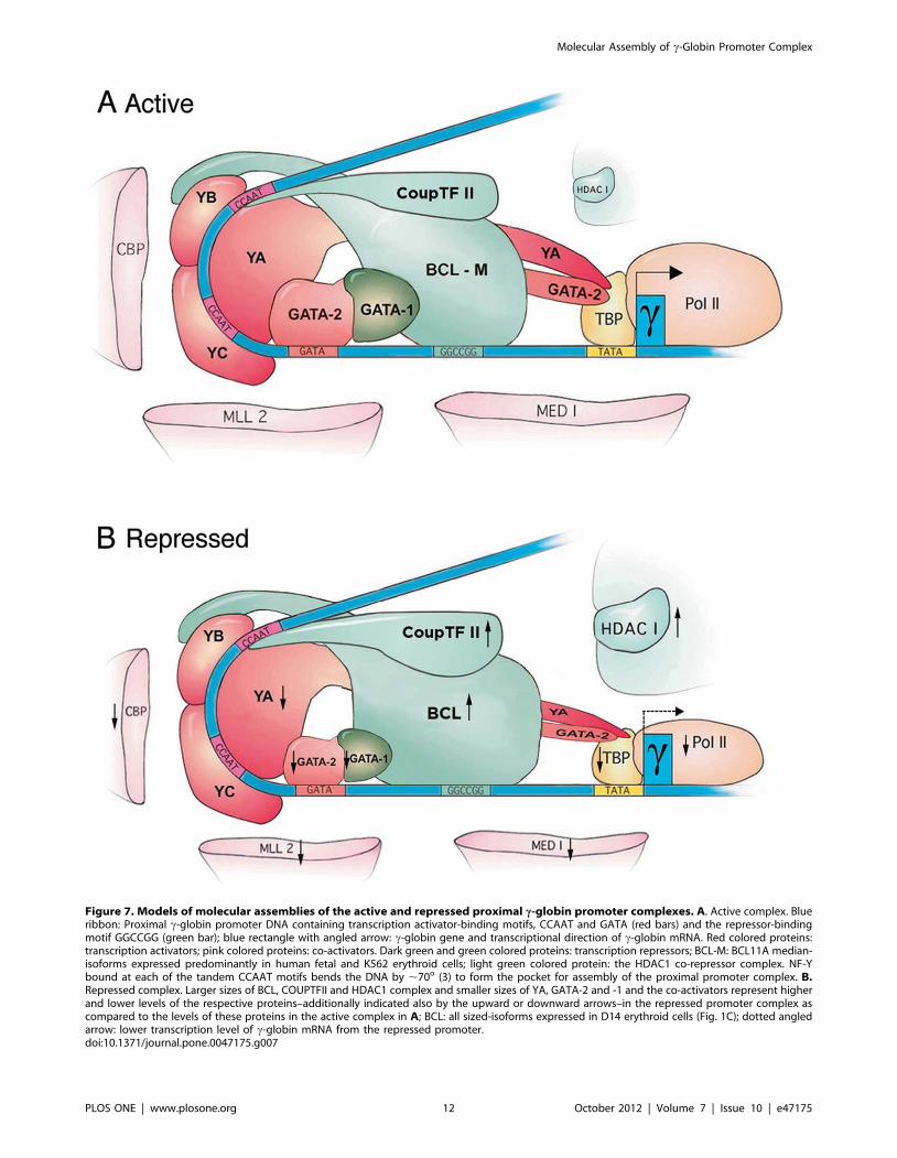
Figure 7. Models of molecular assemblies of the active and repressed proximal c-globin promoter complexes. A. Active complex. Blueribbon: Proximal c-globin promoter DNA containing transcription activator-binding motifs, CCAAT and GATA (red bars) and the repressor-bindingmotif GGCCGG (green bar); blue rectangle with angled arrow: c-globin gene and transcriptional direction of c-globin mRNA. Red colored proteins:transcription activators; pink colored proteins: co-activators. Dark green and green colored proteins: transcription repressors; BCL-M: BCL11A median-isoforms expressed predominantly in human fetal and K562 erythroid cells; light green colored protein: the HDAC1 co-repressor complex. NF-Ybound at each of the tandem CCAAT motifs bends the DNA by ,70o (3) to form the pocket for assembly of the proximal promoter complex. B.Repressed complex. Larger sizes of BCL, COUPTFII and HDAC1 complex and smaller sizes of YA, GATA-2 and -1 and the co-activators represent higherand lower levels of the respective proteins–additionally indicated also by the upward or downward arrows–in the repressed promoter complex ascompared to the levels of these proteins in the active complex in A; BCL: all sized-isoforms expressed in D14 erythroid cells (Fig. 1C); dotted angledarrow: lower transcription level of c-globin mRNA from the repressed promoter.doi:10.1371/journal.pone.0047175.g007
Molecular Assembly of c-Globin Promoter Complex
PLOS ONE | www.plosone.org 12 October 2012 | Volume 7 | Issue 10 | e47175

co-IPs confirmed that most of the protein pairs that associated in
vivo also associated in vitro (Fig. 6B), except that BCL11A, which
associated in vivo with both NF-YA and -YB (Fig. 6A), associated in
vitro only with NF–YB (Fig. 6B).
Together, the co-IPs revealed specific protein interactions: (1).
Transcription activators NF-Y and GATA-2 interacted with each
other to form an apparent activator hub. (2). Repressor BCL11A
interacted with COUP-TFII with strong affinity and with GATA-
1 (Fig. 6B) to form an apparent repressor hub. (3). The activator
and the repressor hubs cross-associated through interactions
between activator NF-Y and repressor BCL11A, and activator
GATA-2 and repressor GATA-1. (4). The activator hub interacted
with co-activators CBP, MLL2 and Mediator 1, and also TBP and
pol II in the basal transcription machinery but not with co-
repressor HDAC1, while the repressor hub interacted with co-
repressor HDAC1 but not the co-activators. Incorporating these
and earlier results (Fig. 2,3,4,5,6), the molecular assemblies of the
active and the repressed proximal c-globin promoter complexes
were depicted in Fig. 7.
Discussion
In this study, we showed the pivotal role of NF-Y in assembling
the active and the repressed proximal c-globin promoter
complexes (Fig. 7): Transcription activator NF-Y stably bound at
the CCAAT motifs with affinity reported to be among the highest
for DNA binding proteins [3,7,8] recruited and stabilized binding
of activator GATA-2 to the neighboring GATA motif to form the
activator hub, NF-Y/GATA-2; NF-Y also recruited and stabilized
binding to the neighboring GGCCGG motif of repressor
BCL11A, which in turn interacted with COUP-TFII bound at
its cognate site overlapping the distal CCAAT motif and with
GATA-1 recruited by GATA-2 to form the repressor hub,
BCL11A/COUP-TFII/GATA-1. The interaction between
BCL11A and COUP-TFII in the repressor hub was consistent
with earlier reports that BCL11A was originally identified and
cloned by its interaction with COUP-TFII and was initially named
CTIP1, COUP-TFII Interacting Protein 1 [38] and that it was a
transcription repressor with cognate DNA binding motif
GGCCGG [11]. The association between BCL11A and GATA-
1 has also been previously reported [30]. Due apparently to cross-
interaction of NF-Y with BCL11A and GATA-2 with GATA-1,
both the activator and the repressor hubs were present in both the
active and the repressed c-globin promoter complexes. However,
through their relative levels in the proximal promoter complex and
their respective interactions with the co-activators and co-
repressor, the activator and the repressor hubs together modulated
activation or repression of c-globin promoter activity during
erythroid development.
Our findings shed light on the underlying mechanisms of tissue-
and developmental stage-specific transcription of c-globin gene. In
fetal erythroid cells, which expressed at high levels the develop-
mentally regulated, ubiquitous activator NF-Y and erythroid
activator GATA-2 (Fig. 1C), NF-Y interacted with GATA-2 to
assemble a strong activator hub that recruited co-activators CBP
and MLL2 to remodel promoter chromatin, and Mediator 1, TBP
and pol II to activate transcription of c-globin mRNA from the
proximal promoter; the fetal erythroid cells also expressed at very
low levels repressor BCL11A-M isoforms (Fig. 1C), which
assembled apparently a weak repressor hub BCL-M/COUP-
TFII/GATA-1 that interacted only weakly with the HDAC
repressor complex. The strong activator hub predominated over
the weak repressor hub to confer erythroid- and fetal stage-specific
transcription of c-globin mRNA (Fig. 1B). In D14 adult erythorid
cells, which expressed lower levels of activators NF-Y and no
detectable GATA-2 but much higher levels of all repressor
BCL11A isoforms (Fig. 1C), the strong repressor hub predomi-
nated over the weakened activator hub to repress transcription of
c-globin mRNA in adult erythroid cells (Fig. 1B). In non-erythroid
cells such as brain cells, which expressed a high level of NF-Y and
very low to non-detectable levels of GATA-2 (Fig. 1B), but high
levels of BCL11A [11] and COUP-TFII [39], the very weak
activator hub combined with an apparently strong repressor hub
further repressed transcription of c-globin mRNA in non-
erythroid cells (Fig. 1B).
Our finding that BCL11A bound to the GGCCGG motif in the
proximal c-globin promoter was consistent with earlier studies
[10,11]. However, it has been reported that BCL11A does not
bind to the c-globin promoter but binds instead to the locus
control region (LCR) far upstream of c-globin genes and the
intergenic region 39 of Ac globin gene, thus repressing c-globin
gene transcription indirectly by long-range interactions [30,40].
The difference in findings may be due in part to sensitivity
differences between the techniques of ChIP-chip using micro-
arrays as the quantification tool [40,41] and the ChIP-qPCR using
real-time PCR as the quantification tool employed in this report.
In a recent study on global gene expression profiles in human
erythroid cells, a side-by side comparison between microarrays
and qPCR as quantification tools shows that qPCR is more
sensitive in detecting changes in expression levels of genes,
including b-globin gene, throughout erythroid development [42].
The human b-globin gene locus contains four GGCCGG binding
sites for BCL11A, located respectively near the HS3 site in the
LCR, in the proximal promoters of Gc- and Ac-globin genes and
39 of the Ac-globin gene (GenBank U01317). BCL11A bound to
the strong LCR HS3 site at a sufficiently high level detectable by
both ChIP-chip and ChIP-qPCR, while it bound to the relatively
weak c-globin promoter site at a lower level detectable only by
ChIP-qPCR (Fig. S4). Thus, BCL11A may repress c-globin gene
transcription both by binding to the LCR through an indirect,
long-range mechanism and by directly binding to the proximal c-
globin promoter.
The presence of both an activator and a repressor hub in both
the active and the repressed proximal c-globin promoter complex
may underlie the propensity of c-globin promoter to respond to
pharmacological compounds that activate the repressed c-globin
gene in adult erythroid cells [1]. The molecular models of the
proximal c-globin promoter complexes (Fig. 7) suggest that these
pharmacological compounds may enhance expression of activa-
tors, NF-Y and GATA-2, in the activator hub and/or suppress
expression of repressors BCL11A, COUP-TFII and GATA-1 in
the repressor hub. These possibilities are currently under
investigation.
Supporting Information
Figure S1 The 2115 distal CCAAT motif overlapping the
COUP-TFII binding site, 273 GATA and 256 GGCCGG did
not bind or bound weakly to COUP-TFII, GATA-2 and BCL
11A, respectively, as determined by EMSA (related to Fig. 3). A.Percentage contribution of binding by BCL11A to the NF-Y/
BCL11A EMSA band. cP wt and cP(CCAAT)m: Wt and mutant
CCAAT proximal c-globin promoter probes. The quantified NF-
Y/BCL11A bands were those in Fig. 3A, 3B, 3C, 3D and 3F. The
intensities of the control NF-Y/BCL11A band without supershifts
by the antibodies were set at 100. B. The GGCCGG motif bound
BCL11A weakly. Left and middle panels: The short probe
spanning 256 GGCCG motif (same sequence as self (GC)
Molecular Assembly of c-Globin Promoter Complex
PLOS ONE | www.plosone.org 13 October 2012 | Volume 7 | Issue 10 | e47175

competitor in Fig. 3) bound BCL11A very weakly in both K562
and D14 nuclear extracts. Right panel: cP(GC)m (proximal c-
globin promoter with GGCCGG mutated to AAAAAA) bound
little BCL11A, since BCL 11A antibodies only slightly decreased
the intensity of the NF-Y/BCL band (lanes 8 & 9), indicating
requirement of GGCCGG motif to cooperate with CCAAT motif
in recruiting and binding of BCL11A. BCL11A Ab-1 and -2:
antibodies from Novus NB-100–259 and Abcam Ab19487
respectively. Other designations: same as in Fig. 3. C. COUP-
TFII binding to the 2115 CCAAT motif overlapping the COUP-
TFII site comprised ,10% of the NF-Y/COUP-TFII EMSA
band, as indicated by quantification of the competition bands in
Fig. 3D. Competitors dCCAAT and d+p CCAAT spanning both
the COUP-TFII and NF-Y sites were 10% more efficient
competitors than pCCAAT and E1CCAAT, spanning only the
NF-Y site. D. Left panel: The 273 GATA motif by itself did not
bind GATA-2/21. EMSA of short probes spanning 2175, a
GATA site upstream of the 273 GATA site that bound GATA
factors, and the 273 GATA motifs of equal length that did not
bind the GATA factors (Compare lanes 1 and 2; for probe
sequences, see Methods S1). Right panel: cP(GATA)m, proximal
c-globin promoter with mutated GATA motif, bound GATA-2/
21 much less than the wildtype cP (Compare lanes 3 and 4),
indicating cooperativity between GATA and CCAAT motifs in
recruiting/binding of GATA-2/21.
(TIF)
Figure S2 Effect of Mediator 1 and MLL2 knockdown on
mRNA levels of c-globin and select transcription factors in K562
cells. A. Knockdown by siRNA targeting MLL2 as well as MLL1
and Menin 1 in the MLL1/2 hCOMPASS-like complex and PTIP
in the MLL3/4 hCOMPASS-like complex (22) and Mediator 1 in
the Mediator complex [23]. The RNA level of each of the co-
factors in K562 cells transfected by the control plasmid producing
scrambled siRNA (Si Ctl) was set at 100 to serve as the reference
for percentage knockdown of the co-factors by the specific siRNAs.
The RNA levels were determined by RT-PCR. B. Effects of
knockdown of MLL2 and MED1 on c-globin mRNA level. C.Effects of knockdown of MLL2 and MED1 on mRNA levels of
select transcription factors and co-factors in the proximal c-globin
promoter complex. The results showed that reduction in
transcription of c-globin gene did not appear to be the secondary
effects of MLL2 or MED1 knockdown, which first reduced
transcription of activators NF-Y and GATA-2 and/or increased
transcription of repressors BCL11A and GATA-1, since NF-Y and
GATA-2 levels did not significantly change and the levels of
BCL11A and GATA-1actually decreased as a result of MLL2 and
MED1 knockdown (Fig. S2C).
(TIF)
Figure S3 Effects of NF-YA knockdown and over-expression of
GATA-2 and -1 on molecular assembly of the proximal c-globin
promoter complex in the K562 endogenous genome and in
transfected GFP reporter plasmids. A. Effects of NF-YA
knockdown on assembly of the endogenous proximal c-globin
promoter complex: NF-YA knockdown decreased occupancy of
NF-Y, which in turn decreased occupancies of GATA-2 and co-
activator MLL2; however, NF-Y knockdown increased occupancy
of COUP-TFII, which could competitively bind to its cognate site
overlapping the NF-Y binding site at a higher level due to
decreased occupancy of NF-Y. On the other hand, occupancy of
BCL11A did not correspondingly decrease with a decrease in NF-
Y occupancy (Fig. S3A), as anticipated from interaction/
association of BCL11A with NF-Y, but increased as a result of
the decrease in NF-Y occupancy. This was apparently because
BCL11A interacted not only with NF-Y but also strongly with
COUP-TFII (11). Thus, an increase in COUP-TFII occupancy
increased the recruitment and cccupancy of BCL11A. B. Over-
expression of GATA-2 and -1 and CCAAT mutation (to abolish
NF-Y binding) on assembly of the c-globin promoter complex in
plasmids transiently transfected into K562 cells. 0.13Wt-GFP and
0.13CCAATm-GFP: designations same as in Fig. 2A; Vector:
pCRFP1 plasmid containing RFP selectable marker gene; GATA-
2 and GATA-1: Expression plasmids containing GATA-1 or -1
cloned into the pCRFP1 vector plasmid. Inset: K562 cells doubly
transfected with (GATA-2)-RFP and 0.13 Wt-GFP were sorted by
FACS. Sorted cells expressing both RFP and GFP, comprising
,12% of total cell population, were used for ChIP assays. K562
cells transfected with (GATA-1)-RFP and 0.13 Wt-GFP were
similarly sorted by FACS. ChIP results showed that the effects of
GATA-2 and -1 over-expression on assembly of the proximal c-
globin promoter complex in transfected plasimds and in the K562
endogenous genome were similar (Compare Fig. S3B with Fig. 4F).
In addition, mutation of CCAAT motif (to AACCG, see Fig. 2A)
to abolish occupancy of NF-Y greatly diminished occupancies of
GATA-2, -1 as well as of BCL11A (Fig. S3A), even though their
cognate GATA and GGCCGG binding sites were not mutated.
The AACCG mutation eliminated binding of not only NFY but
also COUP TFII (Fig. 1A) and would abolish occupancy of
COUP-TFII at the proximal c-globin promoter; in the absence of
the protein interaction partners COUP-TFII and NF-Y, BCL11A
binding to the GGCCGG motif therefore decreased in the mutant
proximal c- globin promoter.
(TIF)
Figure S4 Relative in vivo binding of BCL11A to the LCR HS3
site and the proximal c-globin promoter in K562 cells. Values
were averages of duplicate pull-downs with the BCL antibody
from Novus and Abcam, AB-1 and -2 respectively.
(TIF)
Methods S1
(DOC)
Acknowledgments
We thank Drs. S. Tsai for hCOUP-TFII cDNA plasmid, S. Jane for NF-E4
antibody, R. Slany for MLL2 cDNA pasmid and S. Huang for assistance
with the construction and packaging of recombinant lentiviruses and we
thank N. Kline and R. Smith for the electronic artwork in Fg. 7.
Author Contributions
Conceived and designed the experiments: XZ DT AW. Performed the
experiments: XZ YW WP. Analyzed the data: XZ YW DT AW.
Contributed reagents/materials/analysis tools: HL AW. Wrote the paper:
XZ DT.
References
1. Mabaera R, West R, Conine S, Macari E, Boyd C, et al. (2008) A cell stress
signaling model of fetal hemoglobin induction. Exp. Hematology 36: 1057–
1072.
2. Suzuki Y, Tsunoda T, Sese J, Taira H, Mizushima-Sugano J, et al. (2001)
Identification and characterization of the potential promoter regions of 1031
kinds of human genes. Genome Res. 11: 677–684.
3. Montovani R (1999) The molecular biology of the CCAAT-binding factor NF-
Y. Gene 239: 16–27.
4. Liberati C, Ronchi A, Lievens P, Ottolenghi S, Mantovani R (1998) NF-Y
organizes the c-globin CCAAT boxes region. J Biol Chem 273: 16880–16889.
5. Duan Z, Stamatoyannopoulos G, Li Q (2001) Role of NF-Y in in vivo regulation
of the c-globin gene. Mol Cell Biol 21: 3083–3095.
Molecular Assembly of c-Globin Promoter Complex
PLOS ONE | www.plosone.org 14 October 2012 | Volume 7 | Issue 10 | e47175

6. Li Q, Fang X, Han H, Stamatoyannopoulos G (2004) The minimal promoter
plays a major role in silencing the galago c-globin gene in adult erythropoiesis.
Proc Nat Acad Sci USA 101: 8096–8101.
7. Kim CG, Sheffrey M (1990) Physical characterization of CCAAT transcription
factor a-CP1. J Biol Chem 272: 13362–13369.
8. Bi W, Wu L, Coustry F, Crombrugghe B, Maity S (1997) DNA binding
specificity of the CCAAT-binding factor CBF/NF-Y. J Biol Chem 272: 26562–
26572.
9. Zhou W, Clouston D, Wang X, Cerruti L, Cunningham J, et al. (2000)
Induction of human fetal globin gene expression by a novel erythroid factor, NF-
E4. Mol & Cell Biol 20: 7662–7672.
10. Chen Z, Luo H, Steinberg M, Chui D (2009) BCL11A represses HBG
transcription in K562 cells. Blood Cells Mo. Dis Doi: 10.1016/j.bcmd.2008.12.003.
11. Avram D, Fields A, Senawong T, Topark-Ngarm A, Leid M (2002) COUP-TF
(chicken ovalbumin upstream promoter transcription factor)-interacting protein
1 (CTIP1) is a sequence-specific DNA binding protein. Biochem J 368: 555–563.
12. Filipe A, Li Q, Deveaux S, Godin I, Romeo PH, et al. (1999) Regulation of
embryonic/fetal globin genes by nuclear hormone receptors: a novel perspective
on hemoglobin switching. EMBO J 18: 687–697.
13. Pereira F, Tsai MJ, Tsai S (2000) COUP-TF orphan nuclear receptors in
development and differentiation. Cell & Mol Life Sciences 57: 1388–1398.
14. Liberati C, Cera MR, Secco P, Santoro C, Mantovani R, et al. (2001)
Cooperation and competition between the binding of COUP-TFII and NF-Y on
human epsilon- and gamma-globin gene promoters. J Biol Chem 276: 41700–
41709.
15. Aerbajinai W, Zhu J, Kumkjaek C, Chin K, Rodgers G (2009) SCF induces c-
globin gene expression by regulating downstream transcription factor COUP-
TFII. Blood 114: 187–194.
16. Collins FS, Metherall JE, Yamakawa M, Pan J, Weissman SM, et al. (1985) A
point mutation in the A gamma-globin gene promoter in Greek hereditary
persistence of fetal haemoglobin. Nature 313: 325–326.
17. Martin DI, Tsai SF, Orkin SH (1989) Increased gamma-globin expression in a
nondeletion HPFH mediated by an erythroid-specific DNA-binding factor.
Nature 338: 435–438.
18. Berry M, Grosveld F, Dillon N (1992) A single point mutation is the cause of the
Greek form of hereditary persistence of fetal haemoglobin. Nature 358: 499–
502.
19. Li Q, Duan ZJ, Stamatoyannopoulos G (2001) Analysis of the mechanism of
action of non-deletion hereditary persistence of fetal hemoglobin mutants in
transgenic mice. EMBO J 20: 157–164.
20. Liu LR, Du ZW, Zhao HL, Liu XL, Huang XD, et al. (2004)T to C substitution
at 2175 or 2173 of the gamma-globin promoter affects GATA-1 and Oct-1
binding in vitro differently but can independently reproduce the hereditary
persistence of fetal hemoglobin phenotype in transgenic mice. J Biol Chem 280:
7452–7459.
21. Goodman RH, Smolik S (2000) CBP/p300 in cell growth, transformation, and
development. Genes & Dev 14: 1553–1577.
22. Eissenberg C, Shilatifard A (2010) Histone H3 lysine 4 (H3K4) methylation in
development and differentiation. Dev Biol 339: 240–249.
23. Malik S, Roeder RG (2010) The metazoan mediator co-activator complex as an
integrative hub for transcriptional regulation. Nat Rev Genetics 11: 761–772.
24. Kang JA, Zhou Y, Weis TL, Liu H, Ulaszek J, et al. (2008) Osteopontin
regulates actin cytoskeleton and contributes to cell proliferation in primary
erythroblasts. J Biol Chem 283: 6997–7006.
25. Yu X, Zhu X, Pi W, Ling J, Ko L, et al. (2005) The LTR of ERV-9 human
endogenous retrovirus binds to NF-Y in the assembly of an active LTR enhancercomplex NF-Y/MZF1/GATA-2. J Biol Chem 280: 35184–35194.
26. Ling J, Pi W, Bollag R, Zeng S, Keskintepe M, et al. (2002) The solitary LTRs of
ERV-9 endogenous retrivirus are conserved during primate evoluation andpossess enhancer activities in embryonic and hematopoietic cells. J Virology 76:
2410–2423.27. Zhu X, Ling J, Zhang L, Pi W, Wu M, et al. (2007) A facilitated tracking and
transcription mechanism of long-range enhancer function. Nucleic Acids Res 35:
5532–5544.28. Pi W, Zhu X, Wu M, Wang Y, Fulzele S, et al. (2010) Long-range function of an
intergenic retrotransposon. Proc Natl Acad Sci USA 107: 12992–12997.29. Ramchandran R, Bengra C, Whitney B, Lanclos K, Tuan D. (2000) A (GATA)7
motif located in the 59 boundary area of the human b globin locus control regionexhibits silencer activity in erythroid cells. Am J of Hematology 65: 14–24.
30. Sankaran VG, Menne TF, Xu J, Akie TE, Lettre G, et al. (2008) Human fetal
hemoglobin expression is regulated by the developmental stage-specific repressorBCL11A. Science 322: 1839–1842.
31. Blobel GA (2002) CBP and p300: versatile coregulators with important roles inhematopoietic gene expression. J Leukoc Biol 71: 545–556.
32. Demers C, Chaturvedi CP, Ranish JA, Juban G, Lai P, et al. (2007) Activator-
mediated recruitment of the MLL2 methyltransferase complex to the beta-globin locus. Mol Cell 27: 573–584.
33. Gordon DF, Tucker EA, Tundwal K, Hall H, Wood WM, et al. (2006)MED220/thyroid receptor-associated protein 220 functions as a transcriptional
coactivator with Pit-1 and GATA-2 on the thyrotropin-beta promoter inthyrotropes. Mol Endocrinol 20: 1073–1089.
34. Stumpf M, Waskow C, Krotschel M, van Essen D, Rodriguez P, et al. (2006)
The mediator complex functions as a coactivator for GATA-1 in erythropoiesisvia subunit Med1/TRAP220. Proc Natl Acad Sc USA 103: 18504–18509.
35. Kabe Y, Yamada J, Uga H, Yamaguchi Y, Wada T, et al. (2005) NF-Y isessential for the recruitment of RNA polymerase II and inducible transcription
of several CCAAT box-containing genes. Mol Cell Biol 25: 512–522.
36. Elefanty A, Antoniou M, Custodio N, Carmo-Fomseca M, Grosveld F (1996)GATA transcription factors associate with a novel class of nuclear bodies in
erythroblasts and megakaryocytes. EMBO J 15: 319–333.37. Senawong T, Peterson VJ, Leid M (2005) BCL11A-dependent recruitment of
SIRT1 to a promoter template in mammalian cells results in histonedeacetylation and transcriptional repression. Arch Biochem Biophy. 434: 316–
325.
38. Avram D, Fields A, Pretty K, Nevrivy D, Ishmael J, et al. (2000) Isolation of anovel family of C2H2 zinc finger proteins implicated in transcription repression
mediated by COUP-TF orphan nuclear receptors. J Biol Chem 275: 10315–10322.
39. Kim BJ, Takamoto N, Yan J, Tsai SY, Tsai MJ (2009) Chicken Ovalbumin
Upstream Promoter-Transcription Factor II (COUP-TFII) regulates growth andpatterning of the postnatal mouse cerebellum. Dev Biol 326: 378–391.
40. Xu J, Sankaran VG, Ni M, Menne TF, Puram RV, et al. (2010) Transcriptionalsilencing of gamma-globin by BCL11A involves long-range interactions and
cooperation with SOX6. Genes Dev 24: 783–798.41. Jawaid K, Wahlberg K, Thein S, Best S (2010) Binding patterns of BCL11A in
the globin and GATA1 loci and characterization of the BCL11A fetal
hemoglobin locus. Blood Cells Molecules and Diseases 45: 140–146.42. Merryweather-Clarke A, Atzberger A, Soneji S, Gray N, Clark K, et al. (2011)
Global gene expression analysis of human erythroid progenitors. Blood 117:e96–e108.
Molecular Assembly of c-Globin Promoter Complex
PLOS ONE | www.plosone.org 15 October 2012 | Volume 7 | Issue 10 | e47175


