

357
25.19: Coenzymes. Some reactions require additional organic molecules or metal ions. These are referred to as cofactors or coenzymes.
N N
S OP
HO O
O P OH
OH
O
NH2N
+
Thiamin Diphosphate(vitamin B1)
OPOHO
HON
O
OH
H
Pyridoxal Phophates(vitamin B6)
N
NN
N
OHO O
OH
Fe
Heme
S
NHHN
O
CO2H
Biotin(vitamin B7)
NN
N N
H2N
O NH2
O
NH2
NH2OO
OCo
O
H
H2N
O
NH
O
C
P OO-O O
N
N
HO H
H
NH2
H
H
OH
N
Vitamin B12(cyanocobalamin)
HN
N
N
N
HN
H2N
OHN
O CO2-
CO2-
Folic Acid(vitamin B9)
N
N
NH
N O
O
O
OH
HOOH
O-
P
O
P OO
O-O
HO
N
OH
O
N
N
N
NH2
Flavin Adenine Diphosphate (FAD)(Vitamin B2)
25.20: Protein Quaternary Structure: Hemoglobin. (please read) 25.21: G-Coupled Protein Receptors. (please read)
358
Chapter 26: Nucleosides, Nucleotides, and Nucleic Acids. Nucleic acids are the third class of biopolymers (polysaccharides and proteins being the others).
Two major classes of nucleic acids: deoxyribonucleic acid (DNA): carrier of genetic information.
ribonucleic acid (RNA): an intermediate in the expression of genetic information and other diverse roles.
The Central Dogma (F. Crick):
DNA mRNA Protein (genome) (transcriptome) (proteome)
The monomeric units for nucleic acids are nucleotides. Nucleotides are made up of three structural subunits
1. Sugar: ribose in RNA, 2-deoxyribose in DNA 2. Heterocyclic base 3. Phosphodiester
transcription translation
166

359
Nucleoside, nucleotides and nucleic acids
sugar base
phosphodiester
sugar base
sugar base
phosphodiester
phosphodiester
sugar base
phosphate
sugar base
Nucleoside Nucleotide
Nucleic Acid
360
26.1: Pyrimidines and Purines. The heterocyclic bases; there are five six common bases for nucleic acids (Table 26.1, p. 1087). Note that G, T and U exist in the keto form (and not the enol form found in phenols)
26.2: Nucleosides. N-Glycosides of a purine or pyrimidine heterocyclic base and a carbohydrate. The C-N bond involves the anomeric carbon of the carbohydrate. The carbohydrates for nucleic acids are D-ribose and 2-deoxy-D-ribose
N
NH N
N
N
N
purine
pyrimidine
N
NH N
N N
NH N
NH
NH2 O
NH2
NH
N
O
NH2
NH
NH
O
O
NH
NH
O
OH3C
adenine (A)DNA/RNA
guanine (G)DNA/RNA
cytosine (C)DNA/RNA
thymine (T)DNA
uracil (U)RNA
1
2
34
567
8
9
12
34
5
6 NH
N
O
NH2
5-methylcytosine (C)DNA
H3C
167

361
Nucleosides = carbohydrate + base (Table 28.2, p. 1089) ribonucleosides or 2’-deoxyribonucleosides
To differentiate the atoms of the carbohydrate from the base, the position number of the carbohydrate is followed by a ´ (prime).
The stereochemistry of the glycosidic bond found in nucleic acids is β.
OHO N
HO
N
N
N
X
NH2
OHO N
HO
N
N
NH
X
O
NH2
RNA: X= OH, adenosine (A)DNA: X= H, 2'-deoxyadenosine (dA)
RNA: X= OH, guanosine (G)DNA: X= H, 2'-deoxyguanosine (dG)
OHO
HO X
N
N
O
NH2
OHO
HO
N
NH
O
OH3C
RNA: X= OH, cytidine (C)DNA: X= H, 2'-deoxycytidine (dC)
DNA: thymidine (T)
OHO
HO
N
NH
O
O
RNA: uridine (U)
1
2
34
567
8 9
1'2'3'
4'
5'
OH
1'2'3'
4'
5' 21
34
5
6
OHO
HO
N
N
O
NH2H3C
DNA: 5-methylcytosineDNA: 5-methyl-2’-deoxycytidine
362
26.3: Nucleotides. Phosphoric acid esters of nucleosides. Nucleotides = nucleoside + phosphate
O
HO X
HO B
ribonucleoside (X=OH)deoxyribonucleoside (X=H)
ribonucleotide 5'-monophosphate (X=OH, NMP)deoxyribonucleotide 5'-monophosphate (X=H, dNMP)
O
HO X
O BPHOO
O
O
HO X
O BPOO
OPHOO
OO
HO X
O BPOO
OPOO
OPHOO
O
ribonucleotide 5'-diphosphate (X=OH, NDP)deoxyribonucleotide 5'-diphosphate (X=H, dNDP)
ribonucleotide 5'-triphosphate (NTP)deoxyribonucleotide 5'-triphosphate (X=H, dNTP)
O
O OH
O B
PO
O
ribonucleotide3',5'-cyclic phosphosphate (cNMP)
Kinase: enzymes that catalyze the phosphoryl transfer reaction from ATP to an acceptor substrate. M2+ dependent
OHOHO
OHOH
OHO N
N
NN
NH2
HO OH
OPOO
OPO
OO
POO
O
OHOHO
OHOH
OPO32-
ATP
O N
N
NN
NH2
HO OH
OPOO
OPO
OO
ADPGlucose-6-phosphateGlucose
hexokinase -or-
glucokinase
168
363
O N
N
NN
NH2
HO OH
OPOO
OPO
OO
POO
O
ATP
O N
N
NN
NH2
HO OH
OPOO
O
AMP
O
O OH
O
PO
ON
N
NN
NH2
-P2O7
adenylylcyclase H2O
cAMP
O N
N
NNH
O
HO OH
OPOO
OPO
OO
POO
O
GTP
O N
N
NNH
O
HO OH
OPOO
O
GMP
O
O OH
O
PO
ON
N
NNH
O
-P2O7
guanylatecyclase H2O
cGMP
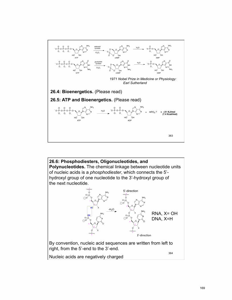
NH2 NH2 NH2
1971 Nobel Prize in Medicine or Physiology: Earl Sutherland
26.4: Bioenergetics. (Please read)
26.5: ATP and Bioenergetics. (Please read)
+ HPO4 2-O N
N
NN
NH2
HO OH
OPOO
OPO
OO
POO
O
ATP
H2O O N
N
NN
NH2
HO OH
OPOO
OPO
OO
ADP
+ ~31 KJ/mol (7.4 Kcal/mol)
364
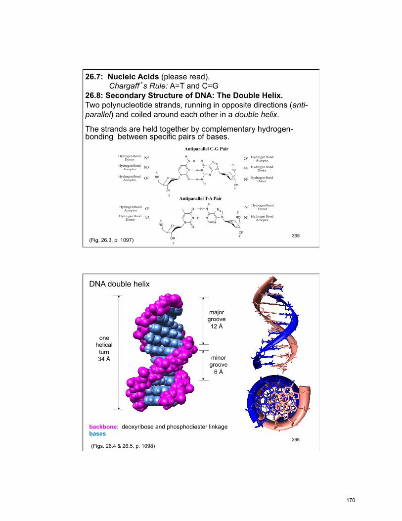
26.6: Phosphodiesters, Oligonucleotides, and Polynucleotides. The chemical linkage between nucleotide units of nucleic acids is a phosphodiester, which connects the 5’-hydroxyl group of one nucleotide to the 3’-hydroxyl group of the next nucleotide.
By convention, nucleic acid sequences are written from left to right, from the 5’-end to the 3’-end.
Nucleic acids are negatively charged
RNA, X= OH DNA, X=H
5’-direction
3’-‐direc)on
OO
O X
N
POO
OO
HO X
N
PO
OO
3'
3'
5'
5'
3'
5'
OH
-H2O
OO
O X
N
POO
OO
O X
PO
OO
3'
3'
5'
5'
3'
5'
N
NH2
O
N
O
NH2
N
N
N
NH2
N
N
N
N
NH2
169
365
26.7: Nucleic Acids (please read). Chargaff’s Rule: A=T and C=G
26.8: Secondary Structure of DNA: The Double Helix. Two polynucleotide strands, running in opposite directions (anti-parallel) and coiled around each other in a double helix.
The strands are held together by complementary hydrogen-bonding between specific pairs of bases.
O
OR
RON
N
N
O
HH
O
OR
RO
NN N
NO
NH
H
HHydrogen Bond
Donor
Hydrogen BondDonor
Hydrogen BondAcceptor
Hydrogen BondAcceptor
3'
5'
Hydrogen BondAcceptor
Hydrogen BondDonor
5'
O2
N4 O6
3'
Antiparallel C-G Pair
N3 N1
N2
O
OR
RO NN
O
OO
OR
RON
N N
NN
H
HH
5'
3'
3'
5'
Hydrogen BondDonorN6
Hydrogen BondAcceptor
Hydrogen BondAcceptor O4
Hydrogen BondDonor
Antiparallel T-A Pair
N1N3
(Fig. 26.3, p. 1097)
366
DNA double helix
one helical turn 34 Å
major groove 12 Å
minor groove
6 Å
backbone: deoxyribose and phosphodiester linkage bases
(Figs. 26.4 & 26.5, p. 1098)
170

367
26.9: Tertiary Structure of DNA: Supercoils. Each cell contains about two meters of DNA. DNA is “packaged” by coiling around a core of proteins known as histones. The DNA-histone assembly is called a nucleosome. Histones are rich is lysine and arginine residues.
Pdb code 1kx5
368
26.10: Replication of DNA. The Central Dogma (F. Crick):
DNA replication DNA transcription mRNA translation Protein (genome) (transcriptome) (proteome)
Expression and transfer of genetic information: Replication: process by which DNA is copied with very high fidelity.
Transcription: process by which the DNA genetic code is read and transferred to messenger RNA (mRNA). This is an intermediate step in protein expression
Translation: The process by which the genetic code is converted to a protein, the end product of gene expression.
The DNA sequence codes for the mRNA sequence, which codes for the protein sequence
“It has not escaped our attention that the specific pairing we have postulated immediately suggests a possible copying mechanism for the genetic material.” Watson & Crick
171

369
DNA is replicated by the coordinated efforts of multiple proteins and enzymes. For replication, DNA must be unknotted, uncoiled and the double helix unwound. Topoisomerase: Enzyme that unknots and uncoils DNA
Helicase: Protein that unwinds the DNA double helix.
DNA polymerase: Enzyme that replicates DNA using each strand as a template for the newly synthesized strand.
DNA ligase: enzyme that catalyzes the formation of the phosphodiester bond between pieces of DNA. DNA replication is semi-conservative: Each new strand of DNA contains one parental (old, template) strand and one daughter (newly synthesized) strand
370
Unwinding of DNA by helicases expose the DNA bases (replication fork) so that replication can take place. Helicase hydrolyzes ATP in order to break the hydrogen bonds between DNA strands.
DNA replication
(Fig. 26.8, p. 1100)
http://www.hhmi.org/biointeractive/dna-replication-advanced-detail
172

371
DNA Polymerase: the new strand is replicated from the 5’→ 3’ (start from the 3’-end of the template)
DNA polymerases are Mg2+ ion dependent
The deoxynucleotide 5’-triphosphate (dNTP) is the reagent for nucleotide incorporation
3’-hydroxyl group of the growing DNA strand acts as a nucleophile and attacks the α-phosphorus atom of the dNTP.
dNTP
(Fig 26.9, p. 1101)
G
O
O
OPO
-O
A
O
O
OT
O
OH
O P OO
C
O
OH
O
O-PO-
OO PO-
OO-
Mg2+
5'
3'
5'templatestrand(old)
(new)
372
Replication of the leading strand occurs continuously in the 5’→ 3’ direction of the new strand.
Replication of the lagging strand occurs discontinuously. Short DNA fragments are initially synthesized and then ligated together. DNA ligase catalyzes the formation of the phosphodiester bond between pieces of DNA.
(Fig. 26.8, p. 1100)
173

373
26.11 Ribonucleic Acid RNA contains ribose rather than 2-deoxyribose and uracil rather than thymine. RNA usually exist as a single strand.
There are three four major kinds of RNA: messenger RNA (mRNA): ribosomal RNA (rRNA) transfer RNA (tRNA) microRNA (miRNA)
DNA is found in the cell nucleus and mitochondria; RNA is more disperse in the cell.
374
Transcription: only one of the DNA strands is copied (coding or antisense strand). An RNA polymerase replicates the DNA sequence into a complementary sequence of mRNA (template or sense strand). mRNAs are transported from the nucleus to the cytoplasm, where they acts as the template for protein biosynthesis (translation). A three base segment of mRNA (codon) codes for an amino acid.The reading frame of the codons is defined by the start and stop codons.
(Table 26.4, p. 1103)
174

375
5'-cap 5'-UTR start stop 3'-UTRCoding sequence
The mRNA is positioned in the ribosome through complementary pairing of the 5’-untranslated region of mRNA with a rRNA.
Transfer RNA (tRNA): t-RNAs carries an amino acid on the 3’-terminal hydroxyl (A) (aminoacyl t-RNA) and the ribosome catalyzes amide bond formation. Ribosome: large assembly of proteins and rRNAs that catalyzes protein and peptide biosynthesis using specific, complementary, anti-parallel pairing interactions between mRNA and the anti-codon loop of specific tRNA’s.
376
Although single-stranded, there are complementary sequences within tRNA that give it a defined conformation.
The three base codon sequence of mRNA are complementary to the “anti-codon” loops of the appropriate tRNA. The base- pairing between the mRNA and the tRNA positions the tRNAs for amino acid transfer to the growing peptide chain.
aminoacyl t-RNA
TψC loop
D loop
variable loop
(Fig. 26.11, p. 1104)
175

377
26.12: Protein Biosynthesis. Ribosomal protein synthesis
P site U G U AA U C U CG U U
3'5'
A site A CU
OO
HN
SCH3
OHC
U G U AA U C U CG U U
3'5'
A CU
OO
HN
SCH3
OHC
U AA
OO
H2N
OH
U G U AA U C U CG U U3'5'
U AA
OO
HN
OHONH
CH3S
A CU
OH
OHC
U G U AA U C U CG U U3'5'
U AA
OO
HN
OHONH
CH3S
A CU
OH
OHC
U G U AA U C U CG U U3'5'
U AA
OO
HN
OHONH
CH3S
A CU
OHOHC
E site U G U AA U C U CG U U3'5'
U AA
OO
HN
OHONH
CH3S
OHC
G AC
O O
CH3H2N
U G U AA U C U CG U U3'5'
O
HNOH
OHN
G AC
O O
CH3HN
SCH3
U AA
OH
OJHC
U G U AA U C U CG U U3'5'
O
HNOH
OHN
G AC
O O
CH3HN
SCH3
U AA
OH
OHC
U G U AA U C U CG U U3'5'
O
HNOH
OHN
G AC
O O
CH3HN
SCH3
U AA
OH
OHC
(Fig. 26.12, p. 1105)
https://www.hhmi.org/biointeractive/translation-advanced-detail
378
26.13: AIDS. (please read) 26.14: DNA Sequencing. Maxam-Gilbert: relies on reagents that react with a specific DNA
base that can subsequent give rise to a sequence specific cleavage of DNA
Sanger: Enzymatic replication of the DNA fragment to be sequenced with a DNA polymerase, Mg+2, and dideoxynucleotides triphosphate (ddNTP) that truncates DNA replication
Restriction endonucleases: Bacterial enzymes that cleave DNA at specific sequences
5’-d(G-A-A-T-T-C)-3’!3’-d(C-T-T-A-A-G)-5’!!!5’-d(G-G-A-T-C-C)-3’!3’-d(C-C-T-A-G-G)-5’!
EcoR I BAM HI
5'
3'
3'
5'
5'
3'
3'
5'
OH 2-O3PO
2-O3PO OH
restrictionenzyme
176
379
Sanger Sequencing: key reagent: dideoxynucleotides triphosphates (ddNTP)
N
NN
N
NH2
OOPOO-
OPO
O-OP
O-O
O-
NH
NN
N
O
OOPOO-
OPO
O-OP
O-O
O-
ddATP
NH2
ddGTP
OOPOO-
OPO
O-OP
O-O
O-OOPO
O-
OPO
O-OP
O-O
O-
ddTTP ddCTP
N
NH
O
ON
N
NH2
O
When a ddNTP is incorporated elongation of the primer is terminated The ddNTP is specifically incorporated opposite its complementary nucleotide base
DNA polymerase
3'
5'-32P3'
5'
primer
Templateunknown sequence
Mg2+, dNTP's+ one ddATP
O
N3
HO NNH
O
OOHO N
N
N
NH
O
S
O OHNN
H2N
OOHO N
N
NH2
O
ddIAZT
(-)-3TC
Anti-Viral Nucleosides
d4C
380
Sanger Sequencing
Larger fragments
Smaller fragments
GTAACGTAATCACAG
ddA ddG ddC ddT
CATTGCATTAGTGTC
5'32P
3'
5'
3'
32P-5' 3'
primer template
http://www.hhmi.org/biointeractive/sanger-method-dna-sequencing
177
381
26.15: The Human Genome Project. (please read) 26.16: DNA Profiling and Polymerase Chain Reaction (PCR). method for amplifying DNA using a DNA polymerase, dNTPs and cycling the temperature.
Heat stable DNA Polymerases (from archaea): Taq: thermophilic bacteria (hot springs)- no proof reading Pfu: geothermic vent bacteria- proof reading
Mg 2+ two Primer DNA strands (synthetic, large excess) one sense primer and one antisense primer one Template DNA strand (double strand) dNTP’s
1 x 2 = 2 x 2 = 4 x 2 = 8 x 2 = 16 x 2 = 32 x 2 = 64 x 2 = 128 x 2 = 256 x 2 = 512 x 2 = 1,024 x 2 = 2,048 x 2 = 4,096 x 2 = 8,192 x 2 = 16,384 x 2 = 32,768 x 2 = 65,536 x 2 = 131,072 x 2 = 262,144 x 2 = 524,288 x 2 = 1,048,576
In principle, over one million copies per original, can be obtained after just twenty cycles
KARY B. MULLIS, 1993 Nobel Prize in Chemistry for his invention of the polymerase chain reaction (PCR) method.
382
Polymerase Chain Reaction
For a PCR animation go to: http://www.youtube.com/watch?v=ZmqqRPISg0g
5'
3'
3'
5'
95 °C
denaturation
5' 3'
3' 5'
anneal (+) and (-) primers
55 - 68 °C5' 3'
3' 5'
3'3'
72 °CTaq, Mg 2+, dNTPs
extension
5'
3'
3'
5'
5'
3'
3'
5'
95 °C
denaturation
2nd cycle
5'
3'
3'
5'
5'
3'
3'
5'
amplification of DNA
2 copies of DNA
repeat temperature cycles
(Fig. 26.14, p. 1109-10)
178


