
This Provisional PDF corresponds to the article as it appeared upon acceptance. Fully formattedPDF and full text (HTML) versions will be made available soon.
Cannabinoid receptor type 2 (CB2)-selective N-aryl-oxadiazolyl-propionamides:synthesis, radiolabelling, molecular modelling and biological evaluation
Organic and Medicinal Chemistry Letters 2012, 2:32 doi:10.1186/2191-2858-2-32
Thomas Rühl ([email protected])Winnie Deuther-Conrad ([email protected])
Steffen Fischer ([email protected])Robert Günther ([email protected])
Lothar Hennig ([email protected])Harald Krautscheid ([email protected])
Peter Brust ([email protected])
ISSN 2191-2858
Article type Original article
Submission date 11 June 2012
Acceptance date 21 September 2012
Publication date 15 October 2012
Article URL http://www.orgmedchemlett.com/content/2/1/32
This peer-reviewed article can be downloaded, printed and distributed freely for any purposes (seecopyright notice below).
Articles in Organic and Medicinal Chemistry Letters are listed in PubMed and archived at PubMedCentral.
For information about publishing your research in Organic and Medicinal Chemistry Letters go to
http://www.orgmedchemlett.com/authors/instructions/
For information about other SpringerOpen publications go to
http://www.springeropen.com
Organic and MedicinalChemistry Letters
© 2012 Rühl et al.This is an open access article distributed under the terms of the Creative Commons Attribution License (http://creativecommons.org/licenses/by/2.0),
which permits unrestricted use, distribution, and reproduction in any medium, provided the original work is properly cited.
Cannabinoid receptor type 2 (CB2)-selective N-aryl-
oxadiazolyl-propionamides: synthesis, radiolabelling,
molecular modelling and biological evaluation
Thomas Rühl1
Email: [email protected]
Winnie Deuther-Conrad1
Email: [email protected]
Steffen Fischer1
Email: [email protected]
Robert Günther1
Email: [email protected]
Lothar Hennig2
Email: [email protected]
Harald Krautscheid3
Email: [email protected]
Peter Brust1*
* Corresponding author
Email: [email protected]
1 Department of Neuroradiopharmaceuticals, Institute of Radiopharmacy, Research
Site Leipzig, Helmholtz-Zentrum Dresden-Rossendorf e.V., Permoserstr. 15, Leipzig
04318, Germany
2 Institute of Organic Chemistry, Faculty of Chemistry and Mineralogy, Universität
Leipzig, Johannisallee 29, Leipzig 04103, Germany
3 Institute of Inorganic Chemistry, Faculty of Chemistry and Mineralogy, Universität
Leipzig, Johannisallee 29, Leipzig 04103, Germany
Abstract
Background
The endocannabinoid system is involved in many physiological and pathological processes. Two
receptors (cannabinoid receptor type 1 (CB1) and type 2 (CB2)) are known so far. Many
unwanted psychotic side effects of inhibitors of this system can be addressed to the interaction
with CB1. While CB1 is one of the most abundant neuroreceptors, CB2 is expressed in the brain

only at very low levels. Thus, highly potent and selective compounds for CB2 are desired. N-
aryl-((hetero)aromatic)-oxadiazolyl-propionamides represent a promising class of such selective
ligands for the human CB2. Here, a library of various derivatives is studied for suitable routes for
labelling with 18
F. Such 18
F-labelled compounds can then be employed as CB2-selective
radiotracers for molecular imaging studies employing positron emission tomography (PET).
Results
By varying the N-arylamide substructure, we explored the binding pocket of the human CB2
receptor and identified 9-ethyl-9H-carbazole amide as the group with optimal size. Radioligand
replacement experiments revealed that the modification of the (hetero)aromatic moiety in 3-
position of the 1,2,4-oxadiazoles shows only moderate impact on affinity to CB2 but high impact
on selectivity towards CB2 with respect to CB1. Further, we could show by autoradiography
studies that the most promising compounds bind selectively on CB2 receptors in mouse spleen
tissue. Molecular docking studies based on a novel three-dimensional structural model of the
human CB2 receptor in its activated form indicate that the compounds bind with the N-arylamide
substructure in the binding pocket. 18
F labelling at the (hetero)aromatic moiety at the opposite
site of the compounds via radiochemistry was carried out.
Conclusions
The synthesized CB2-selective compounds have high affinity towards CB2 and good selectivity
against CB1. The introduction of labelling groups at the (hetero)aromatic moiety shows only
moderate impact on CB2 affinity, indicating the introduction of potential labelling groups at this
position as a promising approach to develop CB2-selective ligands suitable for molecular
imaging with PET. The high affinity for human CB2 and selectivity against human CB1 of the
herein presented compounds renders them as suitable candidates for molecular imaging studies.
Keywords
Cannabinoid receptors, Molecular modelling, Autoradiography, 18
F labelling, Molecular
imaging, PET, Neuroimaging
Background
The role of the endocannabinoid system in specific CNS disorders is related to the regulation of
the temporal dynamics of neurotransmitter release by the retrograde cannabinoid signalling
network [1]. Mediated by the G protein-coupled cannabinoid receptors [2], the release of
endogenous or the administration of exogenous ligands affects both the long-term synaptic
plasticity as well as the short-term regulation of synaptic transmission [1,3]. Two types of
specific cannabinoid receptors have been cloned so far, termed cannabinoid receptor type 1
(CB1) and type 2 (CB2) [2,4]. The existence of additional cannabinoid-binding receptors has
been suggested [5,6]. In contrast to classical neurotransmitters, endocannabinoids function as
retrograde synaptic messengers which are released from postsynaptic neurons, diffuse across
synapses, activate CB1 on presynaptic axons and eventually suppress neurotransmitter release

[7]. In addition, endocannabinoids and their receptors control the decision about survival or
death of neuronal cells [8], such that the pharmacological manipulation of this system might
provide either neuroprotective or pro-apoptotic effects. A therapeutic role of cannabinoids has
also been suggested for mood disorders [9], traumatic brain injury [10,11] and tumour treatment
[12,13]. The development of CB2-selective anticancer agents is regarded to be advantageous in
light of unwanted central effects exerted by binding of those agents to CB1 [14].
CB1 is abundantly expressed in the central nervous system and has been thoroughly investigated
on cellular [15] and functional levels [16,17] in the brain. In contrast to this, only few and, to
some extent, ambiguous data are available regarding CB2 expression and function in the brain.
Initial reports stated that CB2 is mainly expressed peripherally in the cells and organs of the
immune system such as the B lymphocyte-enriched marginal zone of the spleen or the cortex of
lymph nodes [4,18]. However, more recent studies have proven structural and functional CB2
expression in primed glial (as reviewed in [5]) or neural progenitor cells [19]. Moreover, CB2
mRNA was detected in mouse [20] and rat cerebellum [21]. It could be shown in
immunostaining studies that CB2 protein is abundant under basal conditions in neuronal and
glial processes in the cerebellum and hippocampus of mouse and rat [22-24]. As reviewed
recently by Atwood and Mackie [25], a growing number of reports substantiate a neuronal
expression of CB2 through immunohistochemical data. Thus, it has been described that the
activation of microglia is paralleled by an increase in CB2 immunoreactivity in the injured brain
[26]. For instance, significantly increased CB2 expression levels were observed in severe
Alzheimer's disease [27]. This indicates that CB2 expressed in the cerebellum might be involved
in the pathogenesis of various neurodegenerative disorders and therefore might represent an
interesting target for the diagnostics and therapy of such diseases [14,28]. Consequently, detailed
investigation of the functional changes of both receptor subtypes CB1 and CB2 is needed to
achieve a better understanding of the endocannabinoid system and the effects of potential
cannabinoid therapeutics in the normal and diseased human brain [29].
Despite numerous in vitro approaches to directly identify CB2 in the brain, a non-invasive and
quantitative analysis of these receptors in vivo has not been reported to date, probably due to the
lack of ligands applicable for molecular imaging approaches such as positron emission
tomography (PET). Recently, in a microPET study in a rat model, Evens et al. [30] observed
specific binding of the type 2 cannabinoid receptor PET tracer [11
C]NE40. In this model, hCB2
receptors were locally overexpressed in the brain after stereotactic injection of an adeno-
associated viral vector (AAV2/7) encoding hCB2R with a point mutation (Asp80Asn) or
hCB2(D80N) in the right striatum, yet the quantitative analysis of these receptors in native brain
tissue remains a challenge.
Although CB2 has been already mentioned as a valuable biomarker of, e.g. neuroinflammation
[30], the quantitative imaging of this target requires PET radioligands with superior affinity and
specificity towards the low-abundance cerebral CB2 [31]. The [11
C]-methoxy-labelled potent
inverse CB2 agonist Sch225336 [32] showed only poor brain uptake in mice under baseline
conditions. Various 18
F-fluoroethoxy- [33] and [11
C]-methoxy-labelled [34] 2-oxoquinoline
derivatives were suggested as CB2 PET radioligands, too, yet unfavourable brain-to-plasma
ratios reflect the need for further improvement(s) regarding CB2 imaging in the healthy brain as
recently reviewed [35].

Methods
Here, we extend the synthesis of various CB2-binding [36] N-aryl-((hetero)aromatic-
oxadiazolyl-propionamides as potential PET imaging compounds. Guided by molecular docking
studies employing a novel comparative model of the human CB2 receptor, the promising site for 18
F labelling was selected. In vitro binding experiments using human CB2-transfected Chinese
hamster ovary (CHO) cells were performed. The obtained affinity data are in good agreement
with predicted binding strength and could be verified by autoradiographic studies on mouse
spleen slices.
Results and discussion
Synthesis of N-aryl-((hetero)aromatic)-oxadiazolyl-propionamides
The substituted 1,2,4-oxadiazolyl-propionamides synthesized in this study are compiled in
Scheme 1.
Scheme 1 N-aryl-((hetero)aromatic)-oxadiazolyl-propionamides synthesized in this study
As depicted in Scheme 2, the two-step synthesis starts from a proper substituted nitrile and
hydroxylamine yielding the hydroxy-amidin derivative. Following the route described by Cheng
at al. [36], the products were dried after dilution with methylene chloride, and succinic acid
anhydride was added to give the 3-phenyl-subsituted 1,2,4-oxadiazole acids. To support the
formation of the oxadiazole acids, 0.1 eq. KF was added except for the compound with R1 = Br
and R2 = F. In this case, the free 3-phenyl-substituted 1,2,4-oxadiazol-5-yl propanoic acid was
protected, employing (diazomethyl)trimethylsilane, 2-cyano-4-fluoro-N′-hydroxybenzimidamide.
Subsequently, Br was substituted with CN by addition of 4.2 eq. KCN. Deprotection with LiOH
yielded the product. In a final step, the so obtained differently substituted 3-(hetero)aromatic-
1,2,4-oxadiazol-5-yl-propanoic acids were coupled to various arylamides using standard
(dimethylamino)-pyridine (DMAP)-catalysed carbodiimide chemistry. The obtained products
were isolated either by semi-preparative high-performance liquid chromatography (HPLC) or
crystallization (details can be found in the „Experimental‟ section).
Scheme 2 Synthesis of N-aryl-((hetero)aromatic)-oxadiazolyl-propionamides
To suggest potential radiolabelling schemes of the compounds suitable for molecular imaging
studies by PET, several strategies were evaluated, which are given in Scheme 3. For instance, 5e
was synthesized by methylation of 4e with methyl iodide. This approach opens the way to
introduce, for example, 11
C or 18
F by N-alkylation into the final compound. A further approach is
the use of prosthetic groups, such as the introduction of fluorine-substituted alkyl chains via
hydroxyl groups. In this study, 6k was synthesized from 6j by adding 1-fluoro-2-iodoethane. 18
F
can then be introduced into the molecule by using 18
F-labelled alkylating agents. Another
approach for the synthesis of 18
F-labelled compounds is the replacement of fluorine with 18
F via
[2.2.2] cryptand chemistry, which is shown in this study as an example in the „Radiochemistry‟
section.

Scheme 3 Two strategies to introduce fluorine into the molecule, allowing labelling with 18
F
Figure 1 shows the single-crystal X-ray structure of compound 6f. As expected, the amide bond
of this molecule (C5-N3 in Figure 1) is in trans configuration. Though several bonds in 6f are
freely rotatable, the structure is planar. Thus, the dihedrals of the planes defined by the ring
systems are 8.16(9)° between rings A and B and 11.2(1)° between rings B and C.
Figure 1 X-ray crystal structure of 6f (50% thermal ellipsoids). The planar ring systems have
been noted by A, B and C and are highlighted in grey
In vitro binding studies
The specific binding of [3H]CP55,940 towards cell membranes obtained from CHO cells stably
transfected with human CB1 (hCB1-CHO) or CB2 (hCB2-CHO) hCB-CHO accounted for 40%
to 50% of total binding. Non-specific binding was related mainly to binding to the glass fibre
(data not shown). For the binding of [3H]CP55,940 towards hCB1-CHO and hCB2-CHO cell
membranes, KD values of 2.43 ± 1.89 nM (n = 3) and 1.48 ± 0.88 nM (n = 4) were determined in
competitive binding experiments, which correspond with recently published data [37,38]. Kinetic
analysis of the binding of [3H]CP55,940 towards hCB2-CHO cell membranes revealed the rate
constants kon = 0.266 × 10⁹ M
−1 min
−1 and koff = 0.196 min
−1. The accordingly estimated KD
value of 0.73 nM is consistent with the data obtained by saturation assay. The inhibition
constants (Ki) for each of the human CB receptors are compiled together with reference
compounds CP55,940, SR144528 and SR141716A in Table 1. In Figure 2, the inhibition curves
of 6f and 6h on the hCB2-CHO cell obtained in the radioligand displacement experiments with
[3H]CP55,940 are given as examples.
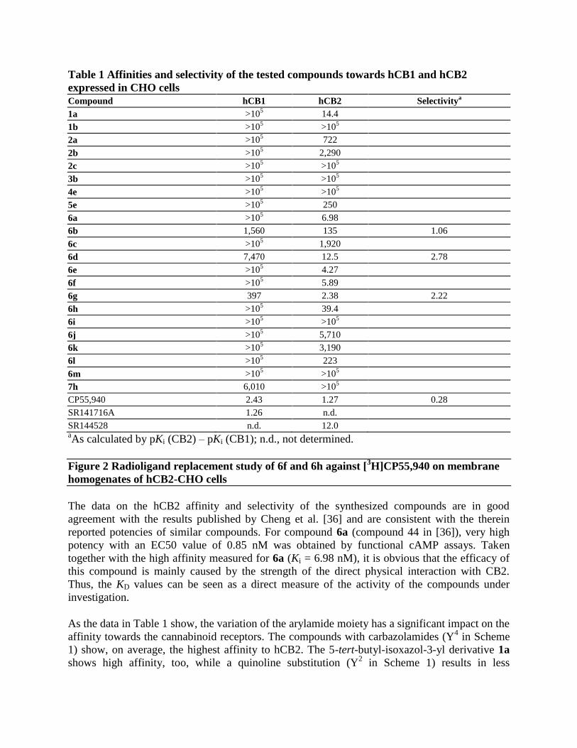
Table 1 Affinities and selectivity of the tested compounds towards hCB1 and hCB2
expressed in CHO cells Compound hCB1 hCB2 Selectivity
a
1a >105 14.4
1b >105 >10
5
2a >105 722
2b >105 2,290
2c >105 >10
5
3b >105 >10
5
4e >105 >10
5
5e >105 250
6a >105 6.98
6b 1,560 135 1.06
6c >105 1,920
6d 7,470 12.5 2.78
6e >105 4.27
6f >105 5.89
6g 397 2.38 2.22
6h >105 39.4
6i >105 >10
5
6j >105 5,710
6k >105 3,190
6l >105 223
6m >105 >10
5
7h 6,010 >105
CP55,940 2.43 1.27 0.28
SR141716A 1.26 n.d.
SR144528 n.d. 12.0 aAs calculated by pKi (CB2) – pKi (CB1); n.d., not determined.
Figure 2 Radioligand replacement study of 6f and 6h against [3H]CP55,940 on membrane
homogenates of hCB2-CHO cells
The data on the hCB2 affinity and selectivity of the synthesized compounds are in good
agreement with the results published by Cheng et al. [36] and are consistent with the therein
reported potencies of similar compounds. For compound 6a (compound 44 in [36]), very high
potency with an EC50 value of 0.85 nM was obtained by functional cAMP assays. Taken
together with the high affinity measured for 6a (Ki = 6.98 nM), it is obvious that the efficacy of
this compound is mainly caused by the strength of the direct physical interaction with CB2.
Thus, the KD values can be seen as a direct measure of the activity of the compounds under
investigation.
As the data in Table 1 show, the variation of the arylamide moiety has a significant impact on the
affinity towards the cannabinoid receptors. The compounds with carbazolamides (Y4 in Scheme
1) show, on average, the highest affinity to hCB2. The 5-tert-butyl-isoxazol-3-yl derivative 1a
shows high affinity, too, while a quinoline substitution (Y2 in Scheme 1) results in less

favourable properties. The approximately 100-fold lower hCB2 affinity of the 3-substituted
quinoline 2a is in good agreement with published efficacy data, where a 6-substituted quinoline
shows an approximately 50-fold lower [36] efficacy in comparison to 6a. The situation is
completely different for CB1. Here, replacement of the carbazole group with a smaller moiety
results in a strong loss of affinity towards the hCB1 receptor. For compounds 2c, 2b and 2a,
binding on the hCB1 receptor could not be observed. Some affinity towards hCB1 could be
observed for 7h, which bears a benzyl-substituted indole at the left-hand side. Remarkably, the
affinity towards hCB2 is lost in comparison to 6h despite the identical 2,4-diflourophenyl
moieties at the 3-position of the oxadiazole ring. While the majority of the compounds bind to
hCB2, those with an unsubstituted nitrogen in the ring system (aryles Y1, Y
2, Y
4a in Scheme 1)
show no binding to hCB1 at all. In general, the affinity of all compounds towards hCB1 is much
lower than that towards hCB2. This might indicate a different binding mode of these two
compounds at hCB2.
Comparing the compounds with the highest affinity (6g) and the best specificity for hCB2 (6e),
the cause of this seems to be encoded in the functional group at para-position of the phenyl
group. In concordance with published data [36], a fluorine atom at this position increases the
affinity and efficacy towards hCB2. Depending on its electronegativity, a second substituent at
ortho-position shows a significant impact on the affinity as shown in Figure 3. While the
introduction of -Br (6e) or -CN (6a) leads to a higher affinity, the -OCH3 (6l) or -OCH2CH2F
(6k) substituents decrease the affinity. Interestingly, replacement of the methoxy group with a
hydroxyl moiety (6j) results in a further loss of affinity.
Figure 3 Influence of substitution at ring C (cf. Figure 1) on affinity towards hCB2. The pKi
values were calculated according to pKi = −log (Ki). High pKi values represent high affinity
Autoradiographic studies
For further investigation of the CB2 affinity of the test compounds, a preliminary
autoradiographic investigation of the displacement potential of reference and test compounds on
mouse spleen tissue pre-incubated with [3H]CP55,940 has been performed. Figure 4 shows
representative autoradiograms of coronal mouse spleen sections incubated with 6 nM
[3H]CP55,940 in the presence of 1 μM of test compounds 6a, 6e and 6h. For comparison, the
autoradiograms for CP55,940, the CB1-selective antagonist SR141716A and the CB2-selective
antagonist SR144528 are shown in Figure 4b. As visible in Figure 4a, the distribution of
[3H]CP55,940 was heterogeneous within the coronal spleen sections, showing a pattern similar to
that reported previously [18,39]. Accordingly, high-density (HD) and low-density (LD) regions
were assumed to reflect binding sites in the white or the red pulp, respectively.
Figure 4 Autoradiogram of representative coronal sections of CD-1 mouse spleen tissue. (a)
Incubated with 6 nM [3H]CP55,940. HD binding and LD binding represent the white and the red
pulp, respectively (cf. text). (b) Incubation with 1 μM selective antagonists for CB2 (SR144528)
and CB1 (SR141716A). (c) Compounds 6e, 6a and 6h compete at 1 μM with the binding of
[3H]CP55,940 on CB2 in good agreement with the determined binding data: 6e > 6a > 6h
To estimate the displacement efficacy of the reference and test compounds, the intensity of the
radioligand binding in the HD regions was chosen to assess total binding of [3H]CP55,904.
Specific binding of [3H]CP55,904 was calculated by subtracting the intensity of the homogenous
binding of [3H]CP55,904 determined in the presence of 1 μM CP55,940 from the total binding
values determined in the absence or presence of 1 μM compounds. Accordingly, a high degree of
specific binding of [3H]CP55,904 in mouse spleen has been identified, which accounts for
approximately 90% of total [3H]CP55,940 binding. The density of specific binding sites of 6 nM
[3H]CP55,904 in mouse spleen, estimated by converting the photostimulated luminescence per
square millimetre values to femtomoles per milligram wet weight using [3H] standards,
resembles, with 150 ± 25 fmol/mg wet weight, the values reported for the binding of 10 nM
[3H]CP55,904 in rat spleen (59 to 108 fmol/mg wet weight) [18]. The assignment of the
[3H]CP55,904 binding sites in mouse spleen to either CB1 or CB2 has been obtained by co-
incubating the tissue with [3H]CP55,904 and either the CB1-selective SR141716A or the CB2-
selective SR144,528 antagonist. As visible in the panels of Figure 4, co-incubation with 1 μM
SR144528 displaced 78% of the specific binding of 6 nM [3H]CP55,904, while nearly no
displacement has been detected in the presence of 1 μM SR141716A. In the latter, 94% of the
specific CP55,940 binding remained. Thus, the basal binding of [3H]CP55,904 in mouse spleen
was identified to reflect mainly CB2 binding, and the experimental conditions in this
autoradiographic study are suitable to assess the CB2 binding potential of the test compounds.
At 1 μM, 6a, 6e and 6h displaced 83%, 71% and 66% of the specific binding of 6 nM
[3H]CP55,904, respectively, and these data correlate to the rank order of CB2 affinity obtained
on hCB2-CHO cells (Ki = 4.27, 6.98 and 39.4 nM, respectively). However, the displacement
potential of the three compounds in mouse spleen is slightly lower than that calculated according
to the Gaddum equation, based on which a fractional occupancy of 98%, 97% and 83%,
respectively, of [3H]CP55,904 binding sites has been estimated (cf. „Experimental‟ section). This
difference between experimental and calculated data might reflect the previously reported non-
significant variances between affinity values obtained on native tissue by in vitro
autoradiography and in radioligand displacement studies using transfected cells [40].
Alternatively, as shown for the binding of WIN55,212-2 towards [3H]CP55,940-labelled mouse
or human CB2, species differences might also explain the slightly distinct affinity of the studied
compounds [41].
Molecular modelling
In order to gain more insight into the binding of the herein synthesized compounds towards
hCB2, we performed molecular modelling studies. As no experimentally determined structures
of cannabinoid receptors are available yet, we created a three-dimensional (3D) structure model
of this G protein-coupled receptor (GPCR) by comparative modelling. Based on the results
obtained by a sequence search with the BLAST server, the 3D structure of the human beta1-
adrenergic receptor (bAR1) [PDB:2Y00] [42] was identified as the structure with the highest
sequence similarity to the hCB2 sequence. However, during the course of this study, a GPCR
structure with a bound agonist was published. In comparison to the bAR1 structure [PDB:2Y00],
which is crystalized with a bound inverse agonist, this novel structure is more opened and thus
represents a better description of a GPCR in an active state. Thus, the X-ray structure of the
human A2A adenosine receptor with UK-432097 ([PDB:3QAK]) [43] was chosen as the

template for comparative modelling. The co-crystalized ligand of this structure was kept in place
during the building process of the receptor model to ensure a state ready to host an agonist.
Figure 5 shows the constructed 3D model of the human CB2 with the proposed binding pocket.
In agreement with mutagenesis data compiled by Poso and Huffman [44], residues known to be
involved in the binding of CP55,940 are accessible in our model.
Figure 5 Model of the human cannabinoid receptor type 2. (a) Side view with the proposed
transmembrane region shaded in grey. The protein is shown with the N-terminus at the top. (b)
Top view from the extracellular space. The extracellular loops and the transmembrane helices are
labelled to guide the reader. Only the residues forming the binding pocket are shown. The
binding pocket identified by the Site Finder module of MOE (Chemical Computing Group Inc.,
Montreal, Canada) is shown as a surface with colour-coded features: H bonding (magenta),
lipophilic (green), mild polar (blue)
The results of the docking of 6a, 6e and 6h are shown in Figure 6. The pose with the lowest
predicted binding energy is shown in green. In the majority of the predicted poses, the molecules
are bent and deviated significantly from a planar structure, which was observed for 6f in the
single crystal. However, the bound conformation of a ligand is not necessarily identical to that of
the free molecule in solution or in a packed crystal. Moreover, due to thermal effects, several
differently populated conformations might exist. Typically, molecular docking procedures
deliver multiple suggestions for the binding geometry of a ligand in a protein-ligand complex.
Identifying the native pose out of these suggestions is often challenging, especially, if no
experimentally determined reference structure is available. By taking into account the computed
binding energy, it is possible to weight the suggested poses employing Boltzmann statistics.
Figure 6 Predicted binding modes of 6e, 6a, 6h to human CB2 model after docking with
GOLD. All compounds bind with their carbazole moiety (ring A, cf. Figure 1) in the binding
pocket indicated by black dots. The figure was created with the software MOE (Chemical
Computing Group Inc., Montreal, Canada) using the module PostDock [45]: The visibility of the
surfaces is proportional to their binding energies based on Boltzman statistics. The surfaces are
colour-coded with respect to the rmsd difference to the ligand with the lowest binding energy
(from yellow to blue). As reference, the best-docked ligand is shown with cyan sticks
Figure 6 is illustrated by encoding the energy in the visibility of the molecule according to the
method implemented in the program PostDock [45]. While for 6h only one representative pose
was predicted, for 6a and 6e, two and three lower populated binding modes were found,
respectively. This is illustrated by the blue shades in Figure 6, which represent the less
favourable poses of these compounds. Nevertheless, in all cases, the three compounds bind
primarily with their arylamide moiety inside the binding pocket. A detailed analysis of the
binding site revealed a hydrophobic microdomain at the bottom of the pocket. The comparison of
6a with 5e indicates that a substituent at the N atom of the ring system increases the affinity
towards CB2. If such a substituent is missing like in 4e, no binding at hCB2 could be observed
(data not shown). However, a replacement with a longer or bulkier moiety reduces the binding
affinity too, as it is visible from the data obtained for 7h. As illustrated in Figure 7, the preferred
binding of 7h is with the arylamide moiety outside the binding pocket, which, together with the

determined binding data, substantiates a different binding mode of this compound. Thus, the 9-
ethyl-9H-carbazole group seems to have an optimal size for fitting into the CB2 binding pocket.
Figure 7 Comparison of the predicted binding modes of 7h and 6h to the human CB2
model. 6h (cyan) binds with the carbazole moiety in the binding pocket. For 7h (orange), the
preferred binding is with ring A pointing outside of the cavity. This figure was created using
MOE (Chemical Computing Group Inc., Montreal, Canada)
The low nanomolar affinity of 6a (Ki = 6.98 nM), 6e (Ki = 4.27 nM) and 6h (Ki = 39.4 nM)
towards the human CB2 and the more than 1,000-fold selectivity over hCB1 (Ki > 1 μM) make
these fluorine-substituted analogues the currently most promising candidates for further
development of PET imaging agents within the series investigated. A proposed labelling at p-
position of the phenyl group at the right-hand site seems to be favourable, e.g. the replacement of
fluorine by 18
F would allow applying these compounds for molecular imaging of CB2 receptors.
Radiochemistry
The labelling with 18
F was performed exemplarily on 6g, yielding 18
F-6e, since 6e has been
identified as highly selective for hCB2 with a high affinity of Ki = 4.27 nM (Scheme 4). 18
F was
introduced by microwave-assisted nucleophilic substitution of the 4-NO2 moiety employing
[2.2.2] cryptand (Kryptofix®, VWR International GmbH, Dresden, Germany). The electron-
withdrawing effect (−I effect) of the Br substituent in meta-position to the leaving group -NO2
enables the nucleophilic attack.
Scheme 4 Radiochemistry
However, this electron-withdrawing effect might not be strong enough as the reaction delivered
only poor yields (3% radiochemical yield). An introduction of nitrogen into the aromatic ring
should facilitate the nucleophilic substitution. Thus, the pyridine equivalent 6c was labelled with 18
F under comparable conditions. Though not supported by a further electron-withdrawing
substituent, the radiochemical yield is significantly higher with 28%. Unfortunately, compounds
with a pyridine moiety as ring C show only low affinity towards hCB2, as visible in Table 1
(compounds 1b, 2b, 2c, 3b, 6b, 6c).
Experimental
Chemistry
All reagents were commercially obtaineda and used without further purification unless otherwise
stated. Anhydrous solvents were transferred via an oven-dried syringe or cannula. Flasks were
oven-dried under vacuum and cooled under a constant stream of nitrogen. 1H,
13C and
19F
nuclear magnetic resonance (NMR) spectra were recorded on VARIAN GEMINI 2000 (200
MHz for 1H NMR, 50 MHz for
13C NMR, 188 MHz for
19F NMR; Palo Alto, CA, USA),
VARIAN „MERCURY plus‟ (300 MHz for 1H NMR, 75 MHz for
13C NMR, 228 MHz for
19F
NMR) and VARIAN „MERCURY plus‟ and BRUKER DRX-400 (400 MHz for 1H NMR, 100
MHz for 13
C NMR, 367 MHz for 19
F NMR; Billerica, MA, USA). The chemical shifts are
reported relative to the residual solvent peak, which was used as an internal reference (chemical
shifts in δ values, J in Hz). The following abbreviations were used to describe peak splitting
patterns when appropriate: br = broad, s = singlet, d = doublet, t = triplet, q = quartet, m =
multiplet, dd = doublet of doublet, ddd = triplet of doublet, pt = pseudo-triplet. High-resolution
mass spectrometry (MS) was performed on a Bruker Daltonics APEX II FT-ICR, and low-
resolved mass spectra (using electrospray ionization (ESI)) were recorded on a Bruker
ESQUIRE. Reactions involving moisture-sensitive reactants were performed in oven-dried
glassware under an atmosphere of nitrogen, reactants being added via a syringe. Flash column
chromatography was performed on silica gel (VWR, 60 Å, 40 to 63 μm, VWR GmbH,
Darmstadt, Germany) and analytical thin-layer chromatography (TLC) on pre-coated silica gel
plates (Roth, 60 Å, F254, 0.25 mm, Carl Roth GmbH & Co. KG, Karlsruhe, Germany). HPLC
was performed on silica gel (ReproSil-Pur 120 ODS3, C18, end-capped (endc.)) from JASCO
(Jasco Labor- & Datentechnik GmbH, Groß-Umstadt, Germany) with a HPLC from Nordantec
GmbH (Bremerhaven, Germany). All compounds were obtained with 95% purity.
3-(3-(2-Bromo-4-fluorophenyl)-1,2,4-oxadiazol-5-yl)propanoic acid (8) and 2-
bromo-4-fluorobenzamide (9)
To a mixture of deoxygenated hydroxylamine hydrochloride (517 mg, 7.4 mmol, 1.0 eq.) and
sodium carbonate (394 mg, 3.7 mmol, 0.5 eq.), methanol/water (4:1, 35 mL) was added via a
cannula, and the reaction mixture was stirred for 10 min. 2-Bromo-4-fluorobenzonitrile (1,487
mg, 7.4 mmol, 1.0 eq.) was added, and the reaction mixture was stirred under diffuse light for 18
h at 80°C. The reaction mixture was diluted with dichloromethane (DCM), washed with water
and evaporated to dryness after concentration of the solvent in vacuo. To the deoxygenated
crude, intermediate succinic acid anhydride (638 mg, 6.4 mmol, 0.9 eq.) was added, and the
reaction mixture was stirred under diffuse light for 30 min at 140°C. The crude material was
purified by column chromatography on silica gel (eluting with petrol spirit or petrol ether/ethyl
acetate/acetic acid 1:1:0.01) and by crystallization at room temperature (DCM/methanol/water)
to give 8 (566 mg, 1.8 mmol, 24% yield) and 9 (559 mg, 2.6 mmol, 35% yield). 8: 1H NMR (400
MHz, CDCl3): δ (ppm) = 7.84 (dd, J = 6 Hz, J = 9 Hz, 1H, CH), 7.47 (dd, J = 3 Hz, J = 8 Hz,
1H, CH), 7.14 (ddd, J = 3 Hz, J = 9 Hz, 1H, CH), 3.29 (t, J = 7 Hz, 2H, CH2), 3.01 (t, J = 7 Hz,
2H, CH2). 19
F NMR (376 MHz, CDCl3): δ (ppm) = −107.89 (dd, J = 8 Hz, J = 14 Hz). 13
C NMR
(100 MHz, CDCl3): δ (ppm) = 177.7, 176.6, 167.2, 163.4 (d, J = 256 Hz), 133.3 (d, J = 9 Hz),
124.3 (d, J = 4 Hz), 122.8 (d, J = 10 Hz), 121.7 (d, J = 25 Hz), 114.9 (d, J = 22 Hz), 30.1, 21.7.
High-resolution mass spectrometry (HRMS) (ESI, negative ion) m/z: 312.9, 314.9 [M-H]−. 9:
1H
NMR (400 MHz, CD3OD): δ (ppm) = 7.51 (dd, J = 6 Hz, J = 8 Hz, 1H, CH), 7.47 (dd, J = 3 Hz,
J = 9 Hz, 1H, CH), 7.19 (pt, J = 2 Hz, J = 8 Hz, 1H, CH). 19
F NMR (376 MHz, CD3OD): δ
(ppm) = −111.64 (dd, J = 8 Hz, J = 15 Hz). 13
C NMR (101 MHz, CD3OD): δ (ppm) = 172.4,
164.2 (d, J = 252 Hz), 136.2 (d, J = 4 Hz), 131.5 (d, J = 9 Hz), 121.5 (d, J = 25 Hz), 121.0 (d, J =
10 Hz), 115.7 (d, J = 22 Hz). HRMS (ESI, positive ion) m/z: 217.96086 [M+H]+.

3-(3-(2-Bromo-4-nitrophenyl)-1,2,4-oxadiazol-5-yl)propanoic acid (10) and 2-
bromo-4-nitrobenzamide (11)
The propanoic acid 10 was synthesized similar to the synthesis of 8 but with 2-bromo-4-
nitrobenzonitrile as the reacting agent. The mixture was diluted with ethyl acetate, and potassium
fluoride (2 mg, 34 μmol, 0.1 eq.) was added during the reaction with succinic acid anhydride.
The reaction mixture was stirred under diffuse light for 30 min at 130°C. For the purification,
petrol spirit or petrol ether/ethyl acetate/acetic acid (1:1:0.005) was used, resulting in 10 (24 mg,
70 μmol, 16% yield) and 11 (22 mg, 90 μmol, 20% yield). 10: 1H NMR (300 MHz,
CDCl3/CD3OD 1:1): δ (ppm) = 8.56 (d, J = 2 Hz, 1H, CH), 8.29 (dd, J = 2 Hz, J = 9 Hz, 1H,
CH), 7.89 (d, J = 9 Hz, 1H, CH). 13
C NMR (75 MHz, CDCl3/CD3OD 1:1): δ (ppm) = 1,178.9,
173.0, 166.0, 148.5, 133.5, 132.2, 128.5, 122.0, 121.7, 29.4, 21.3. HRMS (ESI, negative ion)
m/z: 339.95731 [M-H+]
−, 680.92128 [2M-H
+]−. 11:
1H NMR (300 MHz, CDCl3/CD3OD): δ
(ppm) = 8.50 (d, J = 2 Hz, 1H, CH), 8.28 (dd, J = 2 Hz, J = 8 Hz, 1H, CH), 7.68 (d, J = 8 Hz,
1H, CH). 13
C NMR (100 MHz, CDCl3/CD3OD): δ (ppm) = 171.4, 150.0, 145.7, 130.6, 129.1,
123.7, 120.7. HRMS (ESI, positive ion) m/z: 244.95569 [M+H]+.
3-(3-(2-Fluoro-4-nitrophenyl)-1,2,4-oxadiazol-5-yl)propanoic acid (12) and 2-
fluoro-4-nitrobenzamide (13)
Compounds 12 and 13 were prepared using 2-fluoro-4-nitrobenzonitrile (prepared as in the
synthesis for 10): 12: 807 mg, 2.9 mmol, 10% yield; 13: 93 mg, 0.5 mmol, 2% yield. 12: 1H
NMR (300 MHz, CDCl3): δ (ppm) = 8.26 (dd, J = 7 Hz, J = 8 Hz, 1H, CH), 8.06 to 8.18 (m, 2H,
2xCH), 3.32 (t, J = 7 Hz, 2H, CH2), 3.03 (t, J = 7 Hz, 2H, CH2). 19
F NMR (282 MHz, CDCl3): δ
(ppm) = −103.63 (dd, J = 7 Hz, J = 10 Hz). 13
C NMR (75 MHz, CDCl3): δ (ppm) = 178.8,
176.0, 164.1 (d, J = 6 Hz), 160.5 (d, J = 263 Hz), 150.4 (d, J = 8 Hz), 131.8 (d, J = 3 Hz), 121.4
(d, J = 13 Hz), 119.5 (d, J = 4 Hz), 112.8 (d, J = 26 Hz), 30.7, 22.5. HRMS (ESI, negative ion)
m/z: 280.03753 [M-H+]
−, 316.01425 [M+Cl
−]−, 561.08202 [2M-H
+]
−. 13:
1H NMR (400 MHz,
CD3OD): δ (ppm) = 8.18 to 8.07 (m, 2H, 2xCH), 7.97 (dd, J = 7 Hz, J = 8 Hz, 2H, CH2). 19
F
NMR (376 MHz, CD3OD): δ (ppm) = −112.07 (t, J = 8 Hz). 13
C NMR (101 MHz, CD3OD): δ
(ppm) = 166.9, 160.8 (d, J = 254 Hz), 151.5 (d, J = 9 Hz), 132.8 (d, J = 3 Hz), 130.1 (d, J = 15
Hz), 120.5 (d, J = 4 Hz), 113.1 (d, J = 29 Hz). HRMS (ESI, positive ion) m/z: 185.03583
[M+H]+, 207.01776 [M+Na]
+, 391.04643 [2M+Na]
+.
3-(3-(2-Bromo-4-methoxyphenyl)-1,2,4-oxadiazol-5-yl)propanoic acid (14) and 2-
bromo-4-methoxybenzamide (15)
Compounds 14 and 15 were prepared using 2-bromo-4-methoxybenzonitrile (prepared as in the
synthesis of 10): 14: 2539 mg, 7.8 mmol, 33% yield; 15: impure. 14: 1H NMR (300 MHz,
CD3OD): δ (ppm) = 7.74 (d, J = 9 Hz, 1H, CH), 7.31 (d, J = 2 Hz, 1H, CH), 7.04 (dd, J = 3 Hz,
J = 9 Hz, 1H, CH), 3.86 (s, 3H, CH3), 3.24 (t, J = 7 Hz, 2H, CH2), 2.92 (t, J = 7 Hz, 2H, CH2).
Impurity: δ (ppm) = 2.56 (s, 1.0H). 13
C NMR (76 MHz, CD3OD): δ (ppm) = 180.2, 175.1, 168.8,
163.2, 133.9, 123.6, 121.5, 120.6, 114.4, 56.3, 31.1, 22.9. Impurity: δ (ppm) = 176.2, 29.9.
HRMS (ESI, negative ion) m/z: 324.98282 [M-H+]
−, 650.97272 [2M-H
+]
−. 15:
1H NMR (300
MHz, CD3OD): δ (ppm) = 7.44 (d, J = 9 Hz, 1H, CH), 7.19 (d, J = 3 Hz, 1H, CH), 7.00 (dd, J =

3 Hz, J = 9 Hz, 1H, CH), 3.83 (s, 3H, CH3). Impurity: δ (ppm) = 3.67 (s, 14.4H), 2.59 (s, 16H),
2.57 (s, 10H). HRMS (ESI, positive ion) m/z: 324.98282 [M-H+]−, 229.98094 [M+H]
+.
3-(3-(2,4-Difluorophenyl)-1,2,4-oxadiazol-5-yl)propanoic acid (16)
Carboxylic acid 16 was prepared using 2,4-difluorobenzonitrile (prepared as in the synthesis for
10): 5684 mg, 22.4 mmol, 64% yield. 16: 1H NMR (300 MHz, CDCl3/CD3OD 1:1): δ (ppm) =
8.04 (dd, J = 8 Hz, J = 16 Hz, 1H, CH), 7.14 to 6.96 (m, 2H, 2xCH), 3.24 (t, J = 7 Hz, 2H,
CH2), 2.92 (t, J = 7 Hz, 2H, CH2). 19
F NMR (282 MHz, CDCl3/CD3OD 1:1): δ (ppm) = −104.91
to −105.25 (m, 1xF), −105.85 to −106.23 (m, 1xF). 13
C NMR (75 MHz, CDCl3/CD3OD 1:1): δ
(ppm) = 179.5, 174.2, 165.4 (d, J = 12 Hz, J = 254 Hz), 165.0 (d, J = 6 Hz), 161.9 (d, J = 12
Hz, J = 260 Hz), 132.8 (dd, J = 4 Hz, J = 10 Hz), 112.6 (dd, J = 4 Hz, J = 22 Hz), 112.1 (dd, J
= 4 Hz, J = 13 Hz), 105.6 (pt, J = 26 Hz), 30.7, 22.5. HRMS (ESI, positive ion) m/z: 255.05735
[M+H]+, 277.03917 [M+Na]
+, 531.09038 [2M+Na]
+.
3-(3-(2-Cyano-4-fluorophenyl)-1,2,4-oxadiazol-5-yl)propanoic acid (17)
Lithium hydroxide monohydrate (50 mg, 1.200 mmol, 5.0 eq.) in water (1.2 mL) was added to
24 (66 mg, 241 μmol, 1.0 eq.) in methanol (9.5 mL) and stirred for 2.5 h at room temperature.
Ethyl acetate (38 mL) and HCl solution (13 mL, 6 mol/L) were added, and the phases were
mixed and separated. The aqueous HCl layer was washed with ethyl acetate, and the solvent was
concentrated in vacuo. This material was used without further purification; 17: 58 mg, 221 μmol,
92% yield. 17: 1H NMR (300 MHz, CDCl3, 50°C): δ (ppm) = 8.51 (s broad, 1H, COOH), 8.15
(dd, J = 5 Hz, J = 9 Hz, 1H, CH), 7.53 (dd, J = 3 Hz, J = 8 Hz, 1H, CH), 7.42 (ddd, J = 3 Hz, J
= 9 Hz, 1H, CH), 3.30 (t, J = 7 Hz, 2H, CH2), 3.01 (t, J = 7 Hz, 2H, CH2). 19
F NMR (282 MHz,
CDCl3, 50°C): δ (ppm) = −106.90 (dd, J = 8 Hz, J = 13 Hz). 13
C NMR (75 MHz, CDCl3, 50°C):
δ (ppm) = 178.9, 175.8, 165.6, 163.3 (d, J = 256 Hz), 132.3 (d, J = 9 Hz), 125.7 (d, J = 4 Hz),
121.8 (d, J = 25 Hz), 120.6 (d, J = 22 Hz), 116.0 (m), 113.3 (d, J = 9 Hz), 52.2, 30.0, 21.9. MS
(ESI, positive ion) m/z: 284 [M+Na]+. MS (ESI, negative ion) m/z: 260 [M-H
+]
−.
3-(3-(2-Cyano-4-nitrophenyl)-1,2,4-oxadiazol-5-yl)propanoic acid (18)
The propanoic acid 18 was prepared using 26 (prepared as for the synthesis of 17): 119 mg, 412
μmol, 99% yield. 18: 1H NMR (300 MHz, CDCl3): δ (ppm) = 8.74 (d, J = 2 Hz, 1H, CH), 8.58
(dd, J = 2 Hz, J = 9 Hz, 1H, CH), 8.38 (d, J = 9 Hz, 1H, CH), 3.27 (t, J = 7 Hz, hidden by
solvent, CH2), 2.93 (t, J = 7 Hz, 2H, CH2). 13
C NMR (76 MHz, CDCl3): δ (ppm) = 182.1, 174.9,
166.5, 150.4, 135.4, 132.7, 131.1, 129.1, 116.8, 113.4, 30.9, 23.0. MS (ESI, negative ion) m/z:
287 [M-H+]−, 575[2M-H
+]
−.
3-(3-(4-Fluoro-2-hydroxyphenyl)-1,2,4-oxadiazol-5-yl)propanoic acid (19)
To a mixture of deoxygenated tribromoborane (7,528 mg, 30.1 mmol, 8.0 eq.) in dry DCM (130
mL), a mixture of 20 (1,000 mg, 3.8 mmol, 1.0 eq.) in dry DCM (60 mL) was added via a
cannula at −70°C. The reaction mixture was stirred for 1 h at −70°C and overnight at room
temperature. The reaction mixture was neutralized with saturated aqueous NaHCO3 solution at
1°C and washed with ethyl acetate and evaporated to dryness after concentration of the solvent in

vacuo. The crude material was purified by column chromatography on silica gel (eluting with
DCM/MeOH = 9:1 and DCM/MeOH = 1:1). Water was added and the product was additionally
purified by washing with DCM to give 19 (188 mg, 746 μmol, 20%). 19: 1H NMR (400 MHz,
CD3OD): δH (ppm) = 7.97 (pdd, J = 7 Hz, J = 9 Hz, 1H, CH), 6.79 to 6.71 (m, 2 H, 2xCH), 3.27
(t, J = 7 Hz, 2H, CH2), 2.94 (t, J = 7 Hz, 2H, CH2). 19
F NMR (376 MHz, CD3OD): δF (ppm) =
−109.06 to −109.20 (m). 13
C NMR (101 MHz, CD3OD): δC (ppm) = 180.3, 175.1, 167.5, 166.9
(d, J = 250 Hz), 160.0 (d, J = 13 Hz), 131.6 (d, J = 11 Hz), 109.3 (d, J = 3 Hz), 108.5 (d, J = 23
Hz), 105.1 (d, J = 25 Hz), 31.0, 22.9. HR ESI -MS: C11H9FN2O4, m/z = 251.04731 [M-H+]−,
503.10122 [2M-H+]−.
3-(3-(4-Fluoro-2-methoxyphenyl)-1,2,4-oxadiazol-5-yl)propanoic acid (20)
Carboxylic acid 20 was prepared using 4-fluoro-2-methoxybenzonitrile (prepared as in the
synthesis for 8): 1,250 mg, 4.7 mmol, 15% yield. 20: 1H NMR (400 MHz, CD3OD): δ (ppm) =
7.99 (dd, J = 7 Hz, J = 9 Hz, 1H, CH), 6.97 (dd, J = 2 Hz, J = 11 Hz, 1H, CH), 6.82 (ddd, J = 2
Hz, J = 8 Hz, J = 11 Hz, 1H, CH), 3.93 (s, 3H, CH3), 3.21 (t, J = 7 Hz, 2H, CH2), 2.82 (t, J = 7
Hz, 2H, CH2). Impurity: δ (ppm) = 3.16 (q, J = 7 Hz, 6.5H), 2.96 (s, 4.6H), 1.28 (t, J = 7 Hz,
10.2H). 19
F NMR (376 MHz, CD3OD): δ (ppm) = −108.48 (td, J = 7 Hz, J = 11 Hz, J = 15 Hz). 13
C NMR (101 MHz, CD3OD): δ (ppm) = 180.3, 177.0 (broad), 167.1, 166.9 (d, J = 250 Hz),
161.3 (d, J = 11 Hz), 133.8 (d, J = 11 Hz), 113.2 (d, J = 3 Hz), 108.3 (d, J = 22 Hz), 101.1 (d, J
= 27 Hz), 56.7, 33.4 (broad), 22.1 (broad). Impurity: δ (ppm) = 177.6 (broad), 47.5, 23.8, 9.1.
HRMS (ESI, negative ion) m/z: 265.06292 [M-H+]
−, 531.13261 [2M-H
+]−.
3-(3-(6-Bromopyridin-3-yl)-1,2,4-oxadiazol-5-yl)propanoic acid (21)
Compound 21 was prepared using 6-bromonicotinonitrile (prepared as in the synthesis for 8):
164 mg, 550 μmol, 10% yield. 21: 1H NMR (300 MHz, CDCl3/CD3OD 1:1, 30°C): δ (ppm) =
8.95 (dd, J = 1 Hz, J = 2 Hz, CH), 8.22 (dd, J = 2 Hz, J = 8 Hz, 1H, CH), 7.68 (dd, J = 1 Hz, J
= 8 Hz, 1H, CH), 3.24 (t, J = 7 Hz, 2H, CH2), 2.92 (t, J = 7 Hz, 2H, CH2). 13
C NMR (76 MHz,
CDCl3/CD3OD 1:1, 30°C): δ (ppm) = 180.6, 174.1, 166.1, 149.2, 145.0, 137.9, 129.3, 123.3,
30.6, 22.5. HRMS (ESI, positive ion) m/z: 297.98160 [M+H]+, 319.96382 [M+Na]
+, 616.93871
[2M+Na]+.
3-(3-(6-Fluoropyridin-3-yl)-1,2,4-oxadiazol-5-yl)propanoic acid (22)
Compound 22 was prepared using 6-fluoronicotinonitrile (prepared as in synthesis for 10): 197
mg, 830 μmol, 5% yield. 22: 1H NMR (400 MHz, CDCl3/CD3OD 1:1): δ (ppm) = 8.87 to 8.83
(m, 1 H, CH), 8.48 (ddd, J = 2 Hz, J = 7 Hz, J = 8 Hz, 1H, CH), 7.15 (ddd, J = 0.4 Hz, J = 3
Hz, J = 9 Hz, 1H, CH), 3.24 (t, J = 7 Hz, 2H, CH2), 2.93 (t, J = 7 Hz, 2H, CH2). 19
F NMR (376
MHz, CDCl3/CD3OD 1:1): δ (ppm) = −65.88 (d, J = 6 Hz). 13
C NMR (101 MHz, CDCl3/CD3OD
1:1): δ (ppm) = 180.5, 174.2, 166.0, 165.6 (d, J = 244 Hz), 147.4 (d, J = 15 Hz), 141.2 (d, J = 9
Hz), 122.2 (d, J = 5 Hz), 110.9 (d, J = 37 Hz), 30.6, 22.5. HRMS (ESI, negative ion) m/z:
236.04779 [M-H+]−, 272.02442 [M-Cl
−]
−, 473.10191 [2M-H
+]−.
Methyl 3-(3-(2-bromo-4-fluorophenyl)-1,2,4-oxadiazol-5-yl)propanoate (23)
(Diazomethyl)trimethylsilane (2 M in hexane, 1.3 mL, 2.6 mmol, 1.5 eq.) was added dropwise to
deoxygenated 8 (547 mg, 1.7 mmol, 1.0 eq.) in dry toluene/methanol (9:1, 20 mL) at 0°C, and
the reaction mixture was stirred for 30 min at room temperature. The crude product was purified
by column chromatography on silica gel (eluting with petrol spirit or petrol ether/ethyl acetate
2:1) after concentration of the solvent in vacuo to give 23 (560 mg, 1.7 mmol, 98% yield). 23: 1H
NMR (400 MHz, CDCl3): δ (ppm) = 7.84 (dd, J = 6 Hz, J = 9 Hz, 1H, CH), 7.47 (dd, J = 3 Hz,
J = 8 Hz, 1H, CH), 7.14 (ddd, J = 3 Hz, J = 9 Hz, 1H, CH), 3.73 (s, 3H, CH3), 3.29 (t, J = 7 Hz,
2H, CH2), 2.94 (t, J = 7 Hz, 2H, CH2). 19
F NMR (376 MHz, CDCl3): δ (ppm) = −108.04 (dd, J =
8 Hz, J = 14 Hz). 13
C NMR (101 MHz, CDCl3): δ (ppm) = 178.0, 171.6, 167.2, 163.3 (d, J = 256
Hz), 133.3 (d, J = 9 Hz), 124.5 (d, J = 4 Hz), 122.7 (d, J = 10 Hz), 121.6 (d, J = 25 Hz), 114.8
(d, J = 21 Hz), 52.1, 30.2, 22.0. MS (ESI, positive ion) m/z: 351 [M+Na]+.
Methyl 3-(3-(2-cyano-4-fluorophenyl)-1,2,4-oxadiazol-5-yl)propanoate (24)
Copper(I) chloride (12 mg, 122 μmol, 0.4 eq.) was added to deoxygenated 10 (100 mg, 304
μmol, 1.0 eq.) in dry dimethylacetamide (2.2 mL). Potassium cyanide (79 mg, 1.215 mmol, 4.0
eq.) was added after 15 min, and the reaction mixture was stirred for 14 h at 130°C. Saturated
ammonium chloride (2.2 mL), ethyl acetate (8.7 mL) and water (7.6 mL) were added, and the
phases were mixed and separated. The aqueous layer was washed with ethyl acetate. The crude
product was purified by column chromatography on silica gel (eluting with petrol spirit or petrol
ether/ethyl acetate 2:1) after concentration of the solvent in vacuo to give 24 (23 mg, 83 μmol,
27% yield). 24: 1H NMR (300 MHz, CDCl3): δ (ppm) = 8.15 (dd, J = 5 Hz, J = 9 Hz, 1H, CH),
7.53 (dd, J = 3 Hz, J = 8 Hz, 1H, CH), 7.42 (ddd, J = 3 Hz, J = 9 Hz, 1H, CH), 3.73 (s, 3H,
CH3), 3.03 (t, J = 7 Hz, 2H, CH2), 2.96 (t, J = 7 Hz, 2H, CH2). 19
F NMR (282 MHz, CDCl3): δ
(ppm) = −106.85 (ddd, J = 5 Hz, J = 8 Hz). 13
C NMR (100 MHz, CDCl3): δ (ppm) = 179.1,
171.6, 165.5, 163.2 (d, J = 256 Hz), 132.3 (d, J = 9 Hz), 125.6 (d, J = 4 Hz), 121.8 (d, J = 25
Hz), 120.6 (d, J = 21 Hz), 116.2 (d, J = 3 Hz), 113.0 (d, J = 10 Hz), 52.2, 30.1, 22.0. MS (ESI,
positive ion) m/z: 276 [M+H]+, 298 [M+Na]
+.
Methyl 3-(3-(2-bromo-4-nitrophenyl)-1,2,4-oxadiazol-5-yl)propanoate (25)
Compound 25 was prepared using 10 (prepared as in the synthesis of 23): 230 mg, 646 μmol,
98% yield. 25: 1H NMR (400 MHz, CDCl3): δ (ppm) = 8.58 (d, J = 2 Hz, 1H, CH), 8.25 (dd, J =
2 Hz, J = 9 Hz, 1H, CH), 8.06 (d, J = 9 Hz, 1H, CH), 3.73 (s, 3H, CH3), 3.32 (t, J = 7 Hz, 2H,
CH2), 2.96 (t, J = 7 Hz, 2H, CH2). 13
C NMR (101 MHz, CDCl3): δ (ppm) = 178.7, 171.5, 166.5,
148.9, 133.9, 132.7, 129.2, 122.7, 122.1, 52.2, 30.1, 22.0. MS (ESI, positive ion) m/z: 378
[M+Na]+.
Methyl 3-(3-(2-cyano-4-nitrophenyl)-1,2,4-oxadiazol-5-yl)propanoate (26)
The carboxylic acid 26 was prepared using 10 (prepared as for the synthesis of 24): 83 mg, 275
μmol, 43% yield. 26: 1H NMR (300 MHz, CDCl3): δ (ppm) = 8.68 (d, J = 2 Hz, 1H, CH), 8.54
(dd, J = 2 Hz, J = 9 Hz, 1H, CH), 8.39 (d, J = 9 Hz, 1H, CH), 3.73 (s, 3H, CH3), 3.34 (t, J = 7
Hz, 2H, CH2), 2.98 (t, J = 7 Hz, 2H, CH2). 13
C NMR (75 MHz, CDCl3): δ (ppm) = 179.9, 171.5,

164.9, 148.7, 134.3, 131.3, 129.7, 127.4, 115.4, 112.7, 52.2, 30.0, 22.0. MS (ESI, positive ion)
m/z: 303 [M+H]+, 325 [M+Na]
+.
General amide coupling method A
The coupling reaction was initiated by dropwise addition of N-ethyl-N′-(3-
dimethylaminopropyl)carbodiimide (EDAC; 50 mg, 322 μmol, 1.1 eq.) in dry DCM (2.5 mL) to
a mixture of 1.0 eq. of the deoxygenated propanoic acid derivate and 1.1 eq. amine in dry DCM
(2.5 mL) at 0°C. The reaction mixture was stirred for 1 h at 0°C followed by stirring for 1 h at
room temperature. A tip of spatula of 4-DMAP was added at 0°C, and the reaction mixture was
stirred for 1 h at room temperature. EDAC (1.1 eq.) in dry DCM (2.5 mL) was added at 0°C, and
the reaction mixture was again stirred for 14 h at room temperature followed by evaporation to
dryness after concentration of the solvent in vacuo.
3-(3-(6-Bromopyridin-3-yl)-1,2,4-oxadiazol-5-yl)-N-(5-tert-butylisoxazol-3-
yl)propanamide (1a)
Compound 1a was prepared using 14 and 5-tert-butylisoxazol-3-amine according to method A.
The crude material was purified by column chromatography on silica gel (eluting with petrol
spirit/ethyl acetate 2:1) and by HPLC (RP18, ReproSil-Pur 120 ODS3, endc., methanol/water =
85:15) and by crystallization at room temperature (methanol), giving 1a (19 mg, 44 μmol, 27%
yield). 1a: 1H NMR (400 MHz, CDCl3): δ (ppm) = 10.20 (s broad, 1H, NH), 9.01 (d, J = 2 Hz,
1H, CH), 8.16 (dd, J = 2 Hz, J = 8 Hz, 1H, CH), 7.59 (d, J = 8 Hz, 1H, CH), 6.69 (s, 1H, CH),
3.39 (t, J = 7 Hz, 2H, CH2), 3.11 (t, J = 7 Hz, 2H, CH2), 1.31 (s, 9H, 3xCH3). 13
C NMR (76
MHz, CDCl3): δ (ppm) = 182.2, 179.5, 168.9, 165.8, 158.0, 149.1, 145.0, 137.0, 128.5, 122.5,
93.6, 33.2, 32.5, 28.7, 22.1. HRMS (ESI, positive ion): C17H18BrN5O3, m/z = 420.06635 [M+H]+,
839.12559 [2M+H]+.
3-(3-(6-Fluoropyridin-3-yl)-1,2,4-oxadiazol-5-yl)-N-(5-tert-butylisoxazol-3-
yl)propanamide (1b)
Compound 1b was synthesized similar to 1a with 22 (30 mg, 127 μmol, 1.0 eq.) as carboxylic
acid component. Before evaporating, water (5.0 mL) was added, and the reaction mixture was
washed with ethyl acetate. The crude material was purified as described for 1a, giving 1b (14
mg, 38 μmol, 30% yield). 1b: 1H NMR (300 MHz, CD3OD): δH (ppm) = 8.85 (d, 1H, CH), 8.52
(ddd, J = 2 Hz, J = 8 Hz, J = 9 Hz, 1H, CH), 7.26 to 7.19 (m, 1H, CH), 6.54 (s, 1H, CH), 3.34 (t,
J = 7 Hz, 2H, CH2), 3.06 (t, J = 7 Hz, 2H, CH2), 1.32 (s, 9H, 3xCH3). 19
F NMR (282 MHz,
CD3OD): δF (ppm) = −67.33 (d, J = 7 Hz). HR ESI +MS: C17H18FN5O2, m/z = 360.14680
[M+H]+, 382.12896 [M+Na]
+, 719.28664 [2M+H]
+, 741.26859 [2M+Na]
+.
3-(3-(2-Cyano-4-fluorophenyl)-1,2,4-oxadiazol-5-yl)-N-(quinolin-3-
yl)propanamide (2a)
Compound 2a was prepared following general amide coupling method A with 17 and quinoline-
3-amine. The crude material was purified by column chromatography on silica gel (eluting with
petrol spirit/ethyl acetate 1:1, ethyl acetate and DCM/methanol 1:1) and by crystallization at
room temperature (acetonitrile), giving pure 2a (25 mg, 64 μmol, 51% yield). 2a: 1H NMR (300
MHz, CDCl3/CD3OD = 1:1, 50°C): δH (ppm) = 8.83 (d, J = 3 Hz, 1H, CH), 8.65 (d, J = 2 Hz,
1H, CH), 8.14 (dd, J = 5 Hz, J = 9 Hz, 1H, CH), 7.94 (pd, J = 8 Hz, 1H, CH), 7.77 (d, J = 1 Hz, J
= 8 Hz, 1H, CH), 7.64 to 7.42 (m, 4H, CH), 3.41 (t, J = 7 Hz, 2H, CH2), 3.11 (t, J = 7 Hz, 2H,
CH2). 19
F NMR (282 MHz, CDCl3, 50°C): δF (ppm) = −107.89 (td, J = 5 Hz, J = 8 Hz, J = 16
Hz). APT NMR (75 MHz, CDCl3/CD3OD = 1:1, 50°C): δC (ppm) = 180.6, 171.2, 166.3, 164.1,
(d, J = 255 Hz), 145.1, 144.9, 133.1, 133.0 (d, J = 9 Hz), 129.12, 129.08, 128.6, 128.3, 127.9,
127.6, 126.3 (J = 4 Hz), 125.2, 122.4 (d, J = 26 Hz), 121.5 (d, J = 22 Hz), 116.8 (d, J = 3 Hz),
113.4 (d, J = 9 Hz), 32.9, 22.5. HR ESI +MS: C21H14FN5O2, m/z = 388.12051 [M+H]+,
410.10243 [M+Na]+, 775.23385 [2M+H]
+, 797.21557 [2M+Na]
+.
3-(3-(6-Fluoropyridin-3-yl)-1,2,4-oxadiazol-5-yl)-N-(quinolin-3-yl)propanamide
(2b)
The synthesis of 2b followed the procedure described for 2a with 21 as reactant. The crude
material was purified under the same conditions as 2a, resulting in 20 mg of 2b (55 μmol, 44%
yield). 2b: 1 H NMR (400 MHz, CDCl3/CD3OD = 1:1): δH (ppm) = 8.86 to 8.79 (m, 2H, 2xCH),
8.68 (d, J = 2 Hz, 1H, CH), 8.46 (ddd, J = 2 Hz, J = 8 Hz, J = 8 Hz, 1H, CH), 7.93 (d, J = 8 Hz,
1H, CH), 7.77 (dd, J = 2 Hz, J = 8 Hz, 1H, CH), 7.65 to 7.58 (m, 1H, CH), 7.55 to 7.48 (m, 1H,
CH), 7.12 (dd, J = 3 Hz, J = 9 Hz, 1H, CH), 3.34 (t, J = 7 Hz, 2H, CH2), 3.11 (t, J = 7 Hz, 2H,
CH2). 19
F NMR (376 MHz, CDCl3/CD3OD = 1:1): δF (ppm) = −66.00 (d, J = 7 Hz). 13
C NMR
(101 MHz, CDCl3/CD3OD = 1:1): δC (ppm) = 180.7, 171.3, 166.1, 165.6 (d, J = 244 Hz), 147.5
(d, J = 15 Hz), 145.0, 144.6, 141.2 (d, J = 9 Hz), 133.2, 129.19, 129.16, 128.44, 128.41, 128.1,
125.1, 122.3 (d, J = 5 Hz), 110.9 (d, J = 37 Hz), 32.9, 22.5. HR ESI +MS: C19H14FN5O2, m/z =
364.12066 [M+H]+, 386.10258 [M+Na]
+, 727.23464 [2M+H]
+, 749.21527 [2M+Na]
+.
3-(3-(6-Bromopyridin-3-yl)-1,2,4-oxadiazol-5-yl)-N-(quinolin-3-yl)propanamide
(2c)
Compound 2c was synthesized by method A, coupling 21 (50 mg, 168 μmol, 1.0 eq.) to
quinolin-3-amine. Before evaporating to dryness, water (5.0 mL) was added to the reaction
mixture and subsequently washed with ethyl acetate. The crude material was purified as
described for 2a, giving in 2c (33 mg, 77 μmol, 46% yield). 2c: 1H NMR (400 MHz,
CDCl3/CD3OD = 1:1): δH (ppm) = 8.94 (dd, J = 1 Hz, J = 2 Hz, 1H, CH), 8.78 (d, J = 2 Hz, 1H,
CH), 8.70 (d, J = 2 Hz, 1H, CH), 8.18 (dd, J = 2 Hz, J = 8 Hz, 1H, CH), 7.93 (d, J = 8 Hz, 1H,
CH), 7.76 (d, J = 1 Hz, J = 8 Hz, 1H, CH), 7.63 (dd, J = 1 Hz, J = 8 Hz, 1H, CH), 7.63 to 7.56
(m, 1H, CH), 7.53 to 7.47 (m, 1H, CH), 3.38 (t, J = 7 Hz, 2H, CH2), 3.10 (t, J = 7 Hz, 2H, CH2). 13
C NMR (101 MHz, CDCl3/CD3OD = 1:1): δC (ppm) = 180.5, 170.8, 166.0, 149.1, 144.9, 144.7,
144.3, 137.7, 132.9, 129.1, 129.0, 128.9, 128.3, 128.2, 127.8, 124.8, 123.1, 32.6, 22.4. HR ESI
+MS: C19H14BrN5O2, m/z = 424.04075 [M+H]+, 446.02267 [M+Na]
+, 847.07404 [2M+H]
+,
869.05547 [2M+Na]+.

3-(3-(6-Fluoropyridin-3-yl)-1,2,4-oxadiazol-5-yl)-N-(1-ethyl-1H-indol-5-yl)-
propanamide (3b)
Compound 3b was synthesized by coupling deoxygenated 21 (50 mg, 211 μmol, 1.0 eq.) to 1-
ethyl-1H-indol-5-amine (37 mg, 232 μmol, 1.1 eq.) via method A. The crude material was
purified by column chromatography on silica gel (eluting with petrol spirit/ethyl acetate 1:1) and
by crystallization at room temperature (DCM/petrol spirit), giving 3b (61 mg, 162 μmol, 77%
yield). 3b: 1H NMR (300 MHz, CDCl3): δH (ppm) = 8.91 (d, J = 2 Hz, 1H, CH), 8.41 (dt, J = 2
Hz, J = 8 Hz, 1H, CH), 7.76 (s, 1H, CH), 7.61 (s broad, 1H, NH), 7.24 (d, J = 10 Hz, 1H, CH),
7.21 (d, J = 2 Hz, 1H, CH), 7.10 (d, J = 3 Hz, 1H, CH), 7.02 (dd, J = 3 Hz, J = 9 Hz, 1H, CH),
6.41 (d, J = 3 Hz, 1H, CH), 4.12 (q, J = 7 Hz, 2H, CH2), 3.38 (t, J = 7 Hz, 2H, CH2), 2.97 (t, J =
7 Hz, 2H, CH2), 1.43 (t, J = 7 Hz, 3H, CH3). 19
F NMR (282 MHz, CDCl3): δF (ppm) = −64.21 (d,
J = 5 Hz). 13
C NMR (76 MHz, CDCl3): δC (ppm) = 179.8, 168.4, 165.6, 165.0 (d, J = 244 Hz),
147.4 (d, J = 16 Hz), 140.2 (d, J = 9 Hz), 133.5, 129.7, 128.8, 128.1, 121.5 (d, J = 5 Hz), 115.9,
113.2, 110.1 (d, J = 38 Hz), 109.5, 41.2, 33.0, 22.5, 15.6. HR ESI +MS: C20H18FN5O2, m/z =
380.15202 [M+H]+, 402.13403 [M+Na]
+, 759.29638 [2M+H]
+, 781.27720 [2M+Na]
+.
3-(3-(2-Bromo-4-fluorophenyl)-1,2,4-oxadiazol-5-yl)-N-(9H-carbazol-3-
yl)propanamide (4e)
Compound 4e was prepared using 8 and 9H-carbazol-3-amine via coupling method A. The crude
product was purified by column chromatography on silica gel (eluting with petrol spirit/ethyl
acetate 1:1, ethyl acetate and DCM/methanol 1:1) and by HPLC (RP18, ReproSil-Pur 120 ODS3,
endc., methanol/water = 85:15) and by crystallization at room temperature (chloroform), giving
60 mg of 4e (125 μmol, 64% yield). 4e: 1H NMR (300 MHz, CD3OD): δ (ppm) = 8.26 (d, J = 2
Hz, 1H, CH), 7.96 (d, J = 8 Hz, 1H, CH), 7.82 (dd, J = 6 Hz, J = 9 Hz, 1H, CH), 7.55 (dd, J = 3
Hz, J = 9 Hz, 1H, CH), 7.49 to 7.29 (m, 4H, 4xCH), 7.21 (td, J = 2 Hz, J = 8 Hz, 1H, CH), 7.11
(t, J = 7 Hz, 1H, CH), 3.38 (t, J = 7 Hz, 2H, CH2), 3.04 (t, J = 7 Hz, 2H, CH2). 19
F NMR (282
MHz, CD3OD): δ (ppm) = −110.02 (td, J = 6 Hz, J = 8 Hz). 13
C NMR (76 MHz, CD3OD): δ
(ppm) = 180.8, 171.4, 168.5, 164.8 (d, J = 254 Hz), 142.1, 138.7, 134.6 (d, J = 9 Hz), 131.1,
126.8, 126.1 (d, J = 4 Hz), 124.3, 124.2, 123.8 (d, J = 10 Hz), 122.4 (d, J = 25 Hz), 121.0,
120.7, 119.7, 116.0 (d, J = 22 Hz), 113.6, 111.8, 111.6, 33.4, 23.2. HR ESI +MS:
C23H16BrFN4O2, m/z = 501.03343 [M+Na]+, 979.07777 [2M+Na]
+.
3-(3-(2-Bromo-4-fluorophenyl)-1,2,4-oxadiazol-5-yl)-N-(9-methyl-9H-carbazol-3-
yl)propanamide (5e)
Compound 5e was synthesized by adding iodomethane (8 mg, 56 μmol, 1.3 eq.) in dry
dimethylformamide (0.5 mL) dropwise to deoxygenated 4e (21 mg, 43 μmol, 1.0 eq.) and
potassium carbonate (59 mg, 430 μmol, 10.0 eq.). The reaction mixture was stirred for 20 h at
room temperature followed by evaporation to dryness after concentration of the solvent in vacuo.
The crude product was purified by hot filtration in methanol and by HPLC (RP18, ReproSil-Pur
120 ODS3, endc., acetonitrile/water = 85:15) with subsequent crystallization in the fridge
(methanol) to give 5e (8 mg, 17 μmol, 39% yield). 5e: 1H NMR (300 MHz, CDCl3/CD3OD 1:1):
δ (ppm) = 8.30 (d, J = 2 Hz, 1H, CH), 8.00 (d, J = 8 Hz, 1H, CH), 7.81 (d, J = 9 Hz, 1H, CH),
7.57 to 7.35 (m, hidden by solvent, 4xCH), 7.33 (d, J = 9 Hz, 1H, CH), 7.20 to 7.11 (m, 2 H,
2xCH), 3.81 (s, 3H, CH3), 3.40 (t, J = 7 Hz, 2H, CH2), 3.03 (t, J = 7 Hz, 2H, CH2). 19
F NMR
(282 MHz, CDCl3/CD3OD 1:1): δ (ppm) = −108.76 (td, J = 6 Hz, J = 8 Hz). HR ESI +MS:
C23H16BrFN4O2, m/z = 515.04893 [M+Na]+, 1,007.10847 [2M+Na]
+.
3-(3-(2-Cyano-4-nitrophenyl)-1,2,4-oxadiazol-5-yl)-N-(9-ethyl-9H-carbazol-3-
yl)propanamide (6a)
For the synthesis of 6a, 17 was coupled to 9-ethyl-9H-carbazol-3-amine as described in method
A. After synthesis, water (5.0 mL) was added, and the reaction mixture was washed with ethyl
acetate and evaporated to dryness after concentration of the solvent in vacuo. The crude product
was purified by column chromatography on silica gel (eluting with petrol spirit/ethyl acetate
(2:1) + five drops of triethylamine and DCM/methanol (1:1) + five drops of triethylamine) and
by crystallization at room temperature (DCM/methanol), giving pure 6a (27 mg, 60 μmol, 50%
yield). 6a: 1H NMR (300 MHz, CDCl3/CD3OD = 1:1, 50°C): δH (ppm) = 8.28 (d, J = 2 Hz, 1H,
CH), 8.13 (dd, J = 5 Hz, J = 9 Hz, 1H, CH), 7.99 (d, J = 8 Hz, 1H, CH), 7.54 (dd, J = 2 Hz, J = 8
Hz, 1H, CH), 7.52 to 7.37 (m, 3H, 3xCH), 7.35 (d, J = 8 Hz, 1H, CH), 7.31 (d, J = 9 Hz, 1H,
CH), 7.13 (pt, J = 7 Hz, 1H, CH), 4.31 (q, J = 7 Hz, 2H, CH2), 3.40 (t, J = 7 Hz, 2H, CH2), 3.04
(t, J = 7 Hz, 2H, CH2), 1.36 (t, J = 7 Hz, 3H, CH3). 19
F NMR (282 MHz, CDCl3/CD3OD = 1:1,
50°C): δF (ppm) = −107.74 (ps). 13
C NMR (75 MHz, CDCl3/CD3OD = 1:1, 50°C): δC (ppm) =
179.6, 169.2, 165.1, 162.9 (d, J = 255 Hz), 140.1, 136.9, 131.9 (d, J = 9 Hz), 129.5, 125.32,
125.26 (d, J = 4 Hz), 122.5, 122.4, 121.3 (d, J = 26 Hz), 120.3 (d, J = 22 Hz), 119.9, 119.2,
118.2, 115.8, 112.4, 112.2, 108.1, 107.9, 37.0, 32.0, 21.9, 12.9. HR ESI +MS: C26H20FN5O2, m/z
= 496.13901 [M+Na]+, 969.28932 [2M+Na]
+.
3-(3-(6-Fluoropyridin-3-yl)-1,2,4-oxadiazol-5-yl)-N-(9-ethyl-9H-carbazol-3-
yl)propanamide (6b)
Compound 6b was synthesized as 6a by coupling deoxygenated 22 with 9-ethyl-9H-carbazol-3-
amine following method A. The crude material was purified by column chromatography on silica
gel (eluting with petrol spirit/ethyl acetate 1:1 and DCM/methanol 1:1) and by crystallization at
room temperature (DCM/methanol), giving 6b (42 mg, 98 μmol, 47% yield). 6b: 1H NMR (400
MHz, CDCl3/CD3OD = 1:1): δH (ppm) = 8.83 (d, J = 2 Hz, 1H, CH), 8.47 to 8.38 (m, 1H, CH),
8.28 (d, J = 2 Hz, 1H, CH), 7.98 (d, J = 8 Hz, 1H, CH), 7.47 (dd, J = 2 Hz, J = 9 Hz, 1H, CH),
7.44 to 7.33 (m, 2H, 2xCH), 7.30 (d, J = 9 Hz, 1H, CH), 7.13 (t, J = 7 Hz, 1H, CH), 7.07 (dd, J =
2 Hz, J = 8 Hz, 1H, CH), 4.30 (q, J = 7 Hz, 2H, CH2), 3.36 (t, J = 7 Hz, 2 H, CH2), 3.03 (t, J = 7
Hz, 2H, CH2), 1.34 (t, J = 7 Hz, 3H, CH3). 19
F NMR (282 MHz, CDCl3/CD3OD = 1:1): δF (ppm)
= −65.79 (d, J = 7 Hz). 13
C NMR (101 MHz, CDCl3/CD3OD = 1:1): δC (ppm) = 180.7, 170.3,
165.9, 165.5 (d, J = 244 Hz), 147.4 (d, J = 15 Hz), 141.1 (d, J = 9 Hz), 141.0, 137.8, 130.5,
126.4, 123.5, 123.3, 122.2 (d, J = 5 Hz), 120.9, 120.1, 119.2, 113.3, 110.8 (J = 37 Hz), 109.1,
109.0, 38.0, 32.8, 22.8, 14.0. HR ESI +MS: C24H20FN5O2, m/z = 452.14902 [M+Na]+, 859.32705
[2M+H]+, 881.30861 [2M+Na]
+.
3-(3-(6-Bromopyridin-3-yl)-1,2,4-oxadiazol-5-yl)-N-(9-ethyl-9H-carbazol-3-
yl)propanamide (6c)
Compound 6c was prepared as described for 6b with 21 as carboxylic acid reactant. The crude
material was purified as described for 6b, giving 6c (42 mg, 86 μmol, 51% yield). 6c: 1H NMR
(300 MHz, CDCl3/CD3OD = 1:1): δH (ppm) = 8.93 (dd, J = 1 Hz, J = 2 Hz, 1H, CH), 8.28 (d, J =
2 Hz, 1H, CH), 8.16 (dd, J = 2 Hz, J = 8 Hz, 1H, CH), 7.97 (d, J = 8 Hz, 1H, CH), 7.60 (dd, J =
1 Hz, J = 8 Hz, 1H, CH), 7.47 (dd, J = 2 Hz, J = 9 Hz, 1H, CH), 7.44 to 7.33 (m, 2H, 2xCH),
7.30 (d, J = 9 Hz, 1H, CH), 7.17 to 7.09 (m, 1H, CH), 4.30 (q, J = 7 Hz, 2H, CH2), 3.36 (t, J = 7
Hz, 2H, CH2), 3.03 (t, J = 7 Hz, 2H, CH2), 1.35 (t, J = 7 Hz, 3H, CH3). 13
C NMR (76 MHz,
CDCl3/CD3OD = 1:1): δC (ppm) = 180.8, 170.2, 166.0, 149.1, 144.9, 141.0 137.77, 137.75,
130.5, 129.2, 126.4, 123.4, 123.3, 120.9, 120.0, 119.2, 113.3, 109.1, 109.0, 38.0, 32.8, 22.8, 14.0.
HR ESI +MS: C24H20BrN5O2, m/z = 490.08770 [M+H]+, 512.06951 [M+Na]
+, 979.16767
[2M+H]+, 1,001.14935 [2M+Na]
+.
3-(3-(2-Fluoro-4-nitrophenyl)-1,2,4-oxadiazol-5-yl)-N-(9-ethyl-9H-carbazol-3-
yl)propanamide (6d)
Compound 6d was synthesized as 6a using 12. The crude product was purified by crystallization
in the fridge (DCM), resulting in 69 mg of pure compound (145 μmol, 81% yield). 6d: 1H NMR
(400 MHz, CD3CN, 70°C): δ (ppm) = 8.39 (s broad, 1H, NH), 8.34 to 8.24 (m, 2H, 2xCH), 8.18
to 8.10 (m, 2H, 2xCH), 8.07 (d, J = 8 Hz, 1H, CH), 7.57 to 7.43 (m, 4H, 4xCH), 7.20 (t, J = 7
Hz, 1H, CH), 4.41 (q, J = 7 Hz, 2H, CH2), 3.40 (t, J = 7 Hz, 2H, CH2), 3.02 (t, J = 7 Hz, 2H,
CH2), 1.39 (t, J = 7 Hz, 3H, CH3). 19
F NMR (376 MHz, CD3CN, 90°C): δ (ppm) = −106.52 (s,
broad). HR ESI +MS: C25H20FN5O4, m/z = 496.13901 [M+Na]+, 969.28932 [2M+Na]
+.
3-(3-(2-Bromo-4-fluorophenyl)-1,2,4-oxadiazol-5-yl)-N-(9-ethyl-9H-carbazol-3-
yl)propanamide (6e)
Compound 6e was prepared as described for 6a using 8 as carboxylic acid. The crude product
was purified by column chromatography on silica gel (eluting with DCM/methanol 100:1) and
by crystallization at room temperature (DCM/methanol) to give 6e (112 mg, 221 μmol, 76%
yield) 6e: 1H NMR (300 MHz, CDCl3): δ (ppm) = 8.26 (d, J = 2 Hz, 1H, CH), 8.02 (d, J = 8 Hz,
1H, CH), 7.83 (dd, J = 6 Hz, J = 9 Hz, 1H, CH), 7.69 (s broad, 1H, NH), 7.54 to 7.42 (m, 3H,
3xCH), 7.38 (d, J = 8 Hz, 1H, CH), 7.31 (d, J = 9 Hz, 1H, CH), 7.20 (pt, J = 8 Hz, 1H, CH),
7.12 (ddd, J = 2 Hz, J = 9 Hz, 1H, CH), 4.32 (q, J = 7 Hz, 2H, CH2), 3.45 (t, J = 7 Hz, 2H,
CH2), 3.02 (t, J = 7 Hz, 2H, CH2), 1.40 (t, J = 7 Hz, 3H, CH3). 19
F NMR (282 MHz, CDCl3): δ
(ppm) = −107.95 (dd, J = 8 Hz, J = 14 Hz). 13
C NMR (101 MHz, CDCl3): δ (ppm) = 178.6,
168.4, 167.2, 163.3 (d, J = 255 Hz), 140.4, 137.3, 133.3 (d, J = 9 Hz), 129.2, 125.9, 124.5 (d, J
= 4 Hz), 123.0, 122.8 (d, J = 10 Hz), 122.7, 121.6 (d, J = 25 Hz), 120.6, 119.5, 118.8, 114.9 (d,
J = 21 Hz), 113.0, 108.6, 108.5, 37.6, 33.1, 22.4, 13.78. HR ESI +MS: C25H20BrFN4O2, m/z =
507.08249 [M+H]+, 529.06444 [M+Na]
+, 1013.15637 [2M+H]
+, 1035.13966 [2M+Na]
+.
3-(3-(2-Cyano-4-nitrophenyl)-1,2,4-oxadiazol-5-yl)-N-(9-ethyl-9H-carbazol-3-
yl)propanamide (6f)
Compound 6f was synthesized as 6a using 18. The crude product was purified by column
chromatography on silica gel (eluting with petrol spirit/ethyl acetate (2:1) + five drops of
triethylamine and DCM/methanol (1:1) + five drops of triethylamine) and by crystallization at
room temperature (DCM/methanol and acetonitrile), resulting in 6f (25 mg, 51 μmol, 25%). 6f: 1H NMR (300 MHz, CD3CN, 70°C): δH (ppm) = 8.71 (d, J = 2 Hz, 1H, CH), 8.54 (dd, J = 2 Hz,
J = 9 Hz, 1H, CH), 8.38 (d, J = 9 Hz, 1H, CH), 8.32 (ps, 1H, CH), 8.07 (pd, J = 8 Hz, 1H, CH),
7.54 (dd, J = 2 Hz, J = 9 Hz, 1H), 7.51 to 7.43 (m, 3H, 3xCH), 7.20 (pt, J = 7 Hz, 1H, CH), 4.41
(q, J = 7 Hz, 2H, CH2), 3.43 (t, J = 7 Hz, 2H, CH2), 3.04 (t, J = 7 Hz, 2H, CH2), 1.39 (t, J = 7 Hz,
3H CH3). HR ESI +MS: C26H20N6O4, m/z = 503.14351 [M+Na]+, 983.29765 [2M+Na]
+.
3-(3-(2-Bromo-4-nitrophenyl)-1,2,4-oxadiazol-5-yl)-N-(9-ethyl-9H-carbazol-3-
yl)propanamide (6g)
Compound 6g was prepared as 6a but with 10 as the reactant. The crude product was purified as
described for 6e, yielding 6g (106 mg, 198 μmol, 68%). 6g: 1H NMR (300 MHz, CDCl3, 50°C):
δH (ppm) = 8.57 (d, J = 2 Hz, 1H, CH), 8.36 to 8.14 (m, 2H, 2xCH), 8.03 (pd, J = 8 Hz, 2H,
2xCH), 7.62 to 7.13 (m, hidden by solvent), 4.35 (q, J = 7 Hz, 2H, CH2), 3.49 (ps broad, 2H,
CH2), 3.04 (ps broad, 2H, CH2), 1.43 (t, J = 7 Hz, 3H, CH3). HR ESI +MS: C25H20BrN5O4, m/z =
556.05940 [M+Na]+, 1,067.14721 [2M+H]
+, 1,089.12816 [2M+Na]
+.
3-(3-(2,4-Difluorophenyl)-1,2,4-oxadiazol-5-yl)-N-(9-ethyl-9H-carbazol-3-
yl)propanamide (6h)
Compound 6h was synthesized similar to the procedure described for 6a using 16 as carboxylic
acid. The crude product was purified by column chromatography on silica gel (eluting with
petrol spirit/ethyl acetate 2:1, petrol spirit/ethyl acetate 2:1 and DCM/methanol 1:1) and by
crystallization at room temperature (DCM/methanol 1:1), yielding 6h (39 mg, 88 μmol, 49%).
6h: 1H NMR (400 MHz, CDCl3/CD3OD = 1:1): δH (ppm) = 8.29 (d, J = 2 Hz, 1H, CH), 8.04
(pdd, J = 8 Hz, J = 16 Hz, 1H, CH), 8.00 (d, J = 8 Hz, 1H, CH), 7.49 (dd, J = 2 Hz, J = 9 Hz, 1H,
CH), 7.46 to 7.36 (m, 2H, 2xCH), 7.33 (d, J = 8 Hz, 1H, CH), 7.14 (pt, J = 6 Hz, 1H, CH), 7.06
to 6.97 (m, 2H, 2xCH), 4.33 (q, J = 7 Hz, 2H, CH2), 3.38 (t, J = 7 Hz, 2H, CH2), 3.03 (t, J = 7
Hz, 2H, CH2), 1.37 (t, J = 7 Hz, 3H, CH3). 19
F NMR (376 MHz, CDCl3/CD3OD = 1:1): δF (ppm)
= −105.08 (pdd, J = 7 Hz, J = 14 Hz, 1xF), −105.89 to −106.04 (m, 1xF). 13
C NMR (101 MHz,
CDCl3/CD3OD = 1:1): δC (ppm) = 179.8, 170.4, 165.4 (dd, J = 12 Hz, J = 254 Hz), 165.0 (d, J =
6 Hz), 161.9 (dd, J = 12 Hz, J = 260 Hz), 141.1, 137.9, 132.7 (dd, J = 4 Hz, J = 10 Hz), 130.5,
126.4, 123.5, 123.4, 120.9, 120.2, 119.2, 113.4, 112.6 (dd, J = 4 Hz, J = 22 Hz), 112.1 (dd, J = 4
Hz, J = 13 Hz), 109.2, 109.0, 105.6 (t, J = 26 Hz), 38.0, 32.9, 22.8, 14.0. HR ESI +MS:
C25H20F2N4O2, m/z = 447.16279 [M+H]+, 469.14455 [M+Na]
+, 893.31771 [2M+H]
+, 915.29946
[M+Na]+.
3-(3-(4-Amino-2-fluorophenyl)-1,2,4-oxadiazol-5-yl)-N-(9-ethyl-9H-carbazol-3-
yl)propanamide (6i)
Tin(II) chloride (40 mg, 211 μmol, 5.0 eq.) was added to deoxygenated 6d (20 mg, 42 μmol, 1.0
eq.) in ethanol (1.0 mL), and the reaction mixture was stirred for 30 min at reflux. To the
reaction mixture, ethanol (1.0 mL) and tin(II) chloride (40 mg, 211 μmol, 5.0 eq.) were added,
and the reaction mixture was stirred for 30 min at reflux. Saturated sodium carbonate solution
(2.0 mL) was added at 0°C. The precipitate was filtered up, washed with water, diluted in
chloroform/methanol (1:1), filtered and evaporated to dryness after concentration of the solvent
in vacuo. The crude material was purified by column chromatography on silica gel (eluting with
chloroform/methanol 100:1) and by crystallization in the fridge (acetonitrile) to give 6i (12 mg,
26 μmol, 62% yield) 6i: 1H NMR (300 MHz, CD3CN, 65°C): δ (ppm) = 8.38 (s broad, 1H, NH),
8.32 (s, 1H, CH), 8.07 (d, J = 8 Hz, 1H, CH), 7.74 (t, J = 8 Hz, 1H, CH), 7.60 to 7.36 (m, 4H,
4xCH), 7.19 (t, J = 7 Hz, 1H, CH), 6.61 to 6.41 (m, 2H, 2xCH), 4.71 (s broad, 2 H, NH2), 4.39
(q, J = 7 Hz, 2H, CH2), 3.31 (t, J = 7 Hz, 2H, CH2), 2.96 (t, J = 7 Hz, 2H, CH2), 1.38 (t, J = 7
Hz, 3H, CH3). 19
F NMR (282 MHz, CD3CN, 65°C): δ (ppm) = −110.20 (dd, J = 9 Hz, J = 13
Hz). HRMS (ESI, positive ion) m/z: 466.16522 [M+Na]+, 909.34029 [2M+Na]
+.
3-(3-(4-Fluoro-2-hydroxyphenyl)-1,2,4-oxadiazol-5-yl)-N-(9-ethyl-9H-carbazol-3-
yl)propanamide (6j)
Compound 6j was prepared using 19 as described in the synthesis of 6a. The crude material was
purified as described for 6h, giving 38 mg of 6j (85 μmol, 23% yield). 6j: 1H NMR (300 MHz,
CD3CN): δ (ppm) = 8.55 (s, broad, 1H, NH), 8.35 (d, J = 1 Hz, 1H, CH), 8.06 (pd, J = 6 Hz, 1H,
CH), 8.00 (dd, J = 5 Hz, J = 7 Hz, 1H, CH), 7.55 to 7.42 (m, 4H, 4xCH), 7.18 (td, J = 1 Hz, J =
5 Hz, 1H, CH), 6.84 to 6.74 (m, 2H, 2xCH), 4.38 (q, J = 5 Hz, 2H, CH2), 3.37 (t, J = 5 Hz, 2H,
CH2), 3.02 (t, J = 5 Hz, 2H, CH2), 1.35 (t, J = 5 Hz, 3H, CH3). 19
F NMR (282 MHz, CD3CN): δ
(ppm) = −108.31 to −108.44 (m). HR ESI +MS: C25H21FN4O3, m/z = 445.16724 [M+H]+,
467.14897 [M+Na]+, 889.32803 [2M+H]
+, 911.30937 [2M+Na]
+.
3-(3-(4-Fluoro-2-(2-fluoroethoxy)phenyl)-1,2,4-oxadiazol-5-yl)-N-(9-ethyl-9H-
carbazol-3-yl)-propanamide (6k)
Potassium carbonate (14 mg, 101 μmol, 2.0 eq.) was added to deoxygenated 6j (22 mg, 50 μmol,
1.0 eq.) in dry dimethylformamide (0.5 mL), and the reaction mixture was stirred for 5 min. To
the reaction mixture, 1-fluoro-2-iodoethane (18 mg, 101 μmol, 2.0 eq.) was added, and the
reaction mixture was stirred for 20 h at 50°C. The reaction mixture was added to ice water,
washed with ethyl acetate and evaporated to dryness after concentration of the solvent in vacuo.
The crude material was purified by column chromatography on silica gel (eluting with petrol
spirit or petrol ether/ethyl acetate 1:1) and by crystallization at room temperature
(DCM/methanol) to give 6k (11 mg, 22 μmol, 43% yield). 6k: 1H NMR (300 MHz,
CDCl3/CD3OD 1:1): δ (ppm) = 8.52 (s, broad, 1H, NH), 8.36 (d, J = 2 Hz, 1H, CH), 8.06 (pd, J
= 8 Hz, 1H, CH), 7.94 (dd, J = 7 Hz, J = 9 Hz, 1H, CH), 7.58 to 7.39 (m, 4H, 4xCH), 7.18 (pt, J
= 7 Hz, 1H, CH), 6.93 (dd, J = 2 Hz, J = 11 Hz, 1H, CH), 6.86 (dt, J = 2 Hz, J = 8 Hz, 1H,
CH), 4.88 to 4.78/4.71 to 4.61 (dm, J = 48 Hz, 1 H/1H, CH2), (m, 1H, CH2), 4.47 to 4.32/4.31 to
4.22 (m, 2H, 1H/1H, 2xCH2), 3.31 (t, J = 7 Hz, 2H, CH2), 2.98 (t, J = 7 Hz, 2H, CH2), 1.35 (t, J

= 7 Hz, 3H, CH3). 19
F NMR (282 MHz, CDCl3/CD3OD 1:1): δ (ppm) = −108.79 to −108.96 (m,
1F), −223.78 to −224.42 (m, 1F). HR ESI +MS: C27H24F2N4O3, m/z = 513.17095 [M+Na]+,
1,003.35233 [2M+Na]+.
3-(3-(4-Fluoro-2-methoxyphenyl)-1,2,4-oxadiazol-5-yl)-N-(9-ethyl-9H-carbazol-3-
yl)-propanamide (6 l)
Compound 6l was synthesized via coupling method A as described for 6a with 20 as carboxylic
acid. The crude material was purified following the procedure for 6e, yielding 6l (76 mg, 166
μmol, 57%). 6l: 1H NMR (400 MHz, CDCl3/CD3OD = 1:1): δH (ppm) = 8.29 (d, J = 2 Hz, 1H,
CH), 8.02 to 7.91 (m, 2H, 2xCH), 7.48 (dd, J = 2 Hz, J = 8 Hz, 1H, CH), 7.43 to 7.33 (m, 2H,
2xCH), 7.30 (d, J = 9 Hz, 1H, CH), 7.13 (pt, J = 7 Hz, 1H, CH), 6.79 (dd, J = 2 Hz, J = 11 Hz,
1H, CH), 6.72 (dd, J = 2 Hz, J = 8 Hz, 1H, CH), 4.29 (q, J = 7 Hz, 2 H, CH2), 3.88 (s, 3H, CH3),
3.35 (t, J = 7 Hz, 2H, CH2), 3.01 (t, J = 7 Hz, 2H, CH2), 1.34 (t, J = 7 Hz, 3H, CH3). 13
C NMR
(101 MHz, CDCl3/CD3OD = 1:1): δC (ppm) = 178.7, 170.5, 166.6, 166.2 (d, J = 251 Hz), 160.4
(d, J = 11 Hz), 141.1 137.8, 133.3 (d, J = 11 Hz), 130.5, 126.4, 123.5, 123.4, 120.9, 120.1, 119.2,
113.4, 112.3 (d, J = 3 Hz), 109.2, 109.0, 108.0 (d, J = 22 Hz), 100.5 (d, J = 26 Hz), 56.4, 38.0,
33.0, 22.8, 14.0. HR ESI +MS: C26H23FN4O3, m/z = 459.18247 [M+H]+, 917.35797 [2M+H]
+.
3-(3-(2-Bromo-4-methoxyphenyl)-1,2,4-oxadiazol-5-yl)-N-(9-ethyl-9H-carbazol-3-
yl)propanamide (6m)
Compound 6m was synthesized as 6a with 14 as carboxylic acid reactant. The crude material
was purified by column chromatography on silica gel (eluting with DCM/methanol 100:1 and
DCM/methanol 100:2) and by crystallization at room temperature (DCM/methanol), yielding 6m
(83 mg, 159 μmol, 54%). 6m: 1H NMR (400 MHz, CDCl3/CD3OD = 1:1): δH (ppm) = 8.29 (s,
1H, CH), 8.00 (d, J = 8 Hz, 1H, CH), 7.72 (d, J = 8 Hz, 1H, CH), 7.55 to 7.27 (m, hidden by
solvent, 4xCH), 7.22 (d, J = 2 Hz, 1H, CH), 7.15 (t, J = 7 Hz, 1H, CH), 6.92 (dd, J = 2 Hz, J = 9
Hz, 1H, CH), 4.32 (q, J = 7 Hz, 2H, CH2), 3.80 (s, 3H, CH3), 3.38 (t, J = 7 Hz, 2H, CH2), 3.02 (t,
J = 7 Hz, 2H, CH2), 1.36 (t, J = 7 Hz, 3H, CH3). 13
C NMR (101 MHz, CDCl3/CD3OD = 1:1): δC
(ppm) = 179.4, 170.4, 168.2, 162.4, 141.0, 137.8, 133.3, 130.5, 126.4, 123.4, 123.3, 123.2,
120.9, 120.7, 120.13, 120.09, 119.2, 113.9, 113.4, 109.1, 109.0, 56.0, 38.0, 32.9, 22.8, 14.0. HR
ESI +MS: C26H23BrN4O3, m/z = 519.10249 [M+H]+, 541.08500 [M+Na]
+, 1,037.19973
[2M+H]+, 1,059.18045 [2M+Na]
+.
3-(3-(2,4-Difluorophenyl)-1,2,4-oxadiazol-5-yl)-N-(1-benzyl-1H-indol-3-yl)-
propanamide (7h)
Compound 7h was prepared by coupling of 16 with 1-benzyl-1H-indol-3-amine hydrochloride
according to coupling method A. The crude product was purified by column chromatography on
silica gel (eluting with petrol spirit/ethyl acetate 2:1 and ethyl acetate), yielding 7h (71 mg, 155
μmol, 87%). For affinity data, the product (20 mg) was further purified by HPLC (RP18,
ReproSil-Pur 120 ODS3, endc., acetonitrile/0.057 M triethylammonium acetate = 65:35) and by
crystallization at room temperature (ethanol/water) to an aspic. The aspic was dried in a speed
vacuum to give 14.7 mg of pure solid product. 7h: 1H NMR (400 MHz, CDCl3): δH (ppm) = 8.07
to 7.98 (m, 1H, CH), 7.92 (s broad, 1H, NH), 7.78 (s, 1H, CH), 7.51 (d, J = 8 Hz, 1H, CH), 7.36

to 7.22 (m, hidden by solvent, 4xCH), 7.19 (pt, J = 7 Hz, 1H, CH), 7.16 to 7.10 (m, 2H, 2xCH),
7.07 (pt, J = 7 Hz, 1H, CH), 7.02 to 6.92 (m, 2H, 2xCH), 5.24 (s, 2H, CH2), 3.42 (t, J = 7 Hz,
2H, CH2), 3.06 (t, J = 7 Hz, 2H, CH2). 19
F NMR (376 MHz, CDCl3): δF (ppm) = −104.01 to
−104.26 (m, 1xF), −104.97 to −105.20 (m, 1xF). 13
C NMR (101 MHz, CDCl3): δC (ppm) =
178.8, 167.7, 164.7 (dd, J = 12 Hz, J = 254 Hz), 164.6 (d, J = 6 Hz), 161.3 (dd, J = 12 Hz, J =
260 Hz), 137.4, 134.1, 132.1 (dd, J = 4 Hz, J = 10 Hz), 128.9, 127.8, 127.0, 122.6, 121.1, 119.9,
119.3, 116.8, 114.1, 112.1 (dd, J = 4 Hz, J = 22 Hz), 111.6 (dd, J = 4 Hz, J = 13 Hz), 110.1,
105.3 (t, J = 25 Hz), 50.3, 32.6, 22.6. HR ESI +MS: C26H20F2N4O2, m/z = 481.14447 [M+Na]+,
939.29960 [2M+Na]+.
X-ray structural analysis
The single-crystal X-ray diffraction data of compound 6f were collected on a diffractometer
IPDS-2T (Stoe & Cie GmbH, Darmstadt, Germany) at 180 K using Mo-Kα radiation (l = 71.073
pm). The data reduction was performed using the STOE X-AREA software package (2006, Stoe
& Cie, Darmstadt, Germany). The structure was solved by direct methods using the program
SHELXS-97 and refined using SHELXL-97 [46]. All non-H atoms were refined anisotropically,
and hydrogen (H) atoms were added geometrically using a riding model. Graphical visualization
of the structure was performed using the program Diamond 3.2e (DIAMOND 3.2e, K.
Brandenburg, 2010, Crystal Impact GbR, Bonn, Germany). The crystal structure has been
deposited at the Cambridge Crystallographic Data Centre and allocated the deposition number
CCDC 841516.
Cell culture
CHO cell lines stably transfected with human CB1 (hCB1-CHO; obtained from Euroscreen,
Gosselies, Belgium) or human CB2 (hCB2-CHO; obtained from Paul L. Prather, Department of
Pharmacology and Toxicology, College of Medicine, University of Arkansas for Medical
Sciences, USA) were cultured in Ham's F12 medium or Dulbecco's modified Eagle's medium
(DMEM) with 10% fetal calf serum, 100 IU/mL penicillin, 100 μg/mL streptomycin and 250 or
400 μg/mL of the selection antibiotic Geneticin (G418). The cells were cultured in a humidified
atmosphere with 5% CO2 at 37°C. Confluent cell layers of 175-cm2 flasks were scraped; cells
were harvested by centrifugation (800 rpm, 3 min), counted by trypan blue staining, suspended
in 200 μL of 50 mM tris(hydroxymethyl)aminomethane (TRIS; pH 7.4) at 4°C and stored at
−32°C until the binding experiment.
Determination of CB receptor affinity
Competitive binding experiments were performed with crude membrane homogenates obtained
from hCB1-CHO or hCB2-CHO cells as described above. Increasing concentrations of test
compounds (0.01 nM to 10 μM) diluted from 10 mM stock solutions in DMSO were incubated
with 30 to 50 μg of membrane protein (equivalent to 0.5 to 1 × 10⁶ hCB1-CHO or hCB2-CHO
cells) and 0.5 nM [3H]CP55,940 (6,438 GBq/mmol; PerkinElmer Life and Analytical Sciences,
Rodgau, Germany) in a final volume of 1 mL in a binding buffer (50 mM TRIS (pH 7.4) at
21°C, 0.1% bovine serum albumin (BSA), 5 mM MgCl2, 1 mM EDTA) for 90 min at room
temperature. Non-specific binding of [3H]CP55,940 was determined in the presence of 10 μM

CP55,940. Incubations were terminated by rapid filtration through a GF-B glass fibre filter pre-
incubated for 90 min at room temperature in freshly prepared 0.5% polyvinylpyrrolidone + 0.1%
Tween 20 solution using a 48-well cell harvester (Brandel, Gaithersburg, MD, USA).
Saturation binding of CP55,940 towards hCB1-CHO or hCB2-CHO cell membranes was
analysed in homologous competition experiments performed with increasing concentrations of
CP55,940 (0.01 nM to 10 μM) and one concentration of [3H]CP55,940 (0.5 nM). Kinetic
analysis of the binding of CP55,940 towards hCB2-CHO cell membranes was performed in one
association experiment using three concentrations of [3H]CP55,940 (0.207, 0.458 and 0.873 nM)
and incubation times of up to 120 min.
Binding data were analysed using GraphPad Prism version 2.01 (GraphPad Software, Inc., San
Diego, CA, USA) by non-linear regression to provide estimates of the half maximal inhibitory
concentration (IC50) values and the rate of association. KD values of [3H]CP55,940 from
saturation binding experiments were estimated according to Jeffries et al. [47] by KD = IC50 –
[radioligand] and from kinetic experiments according to Hulme and Trevethick [48]. The
corresponding inhibition constants (Ki) of test compounds were derived from the IC50 values
using the Cheng-Prusoff equation [49]. All binding experiments were performed in triplicates,
and data are given as mean values ± SD from independent experiments.
Animals
Animal experiments were performed under procedures approved by the State of Saxony Animal
Care and Use Committee and conducted in accordance with the German Law for the Protection
of Animals. Female CD-1 mice (10 to 12 weeks old, 20 to 25 g), obtained from the Medizinisch-
Experimentelles Zentrum der Universität Leipzig (Leipzig, Germany), were used for the
experiments.
Autoradiographic binding study
The animals were anesthetized and sacrificed, and their spleens were rapidly removed and frozen
in isopentane at −35°C. The spleens were brought to −15°C in a cryostat (Microm International
GmbH, Walldorf, Germany), and a series of 12-μm-thick coronal sections obtained from the
spleens of three animals were collected in parallel on microscopic glass slides. The sections were
dried at room temperature and stored at −35°C until they were processed for receptor binding
autoradiography.
For autoradiographic experiments, the slides were brought to room temperature, dried in a stream
of cold air, pre-incubated in a binding buffer (50 mM Tris–HCl (pH 7.4) at 21°C, 5% BSA) for
15 min at room temperature and dried again in a stream of cold air. To account for total binding,
samples were incubated for 2 h at room temperature with 6 nM [3H]CP55,940 (5,328
GBq/mmol, PerkinElmer Life and Analytical Sciences, Rodgau, Germany). Sections were
incubated in the presence of 1 μM CP55,940 (non-specific binding), SR144528 (CB2-selective
inverse agonist), SR141716A (CB1-selective antagonist) or a test compound in binding buffer.
Samples were washed twice for 30 min at 4°C in 50 mM Tris–HCl (pH 7.4) at 4°C, containing
1% BSA, dipped briefly in ice-cold deionized water (5 s), dried in a stream of cold air and

exposed along with [3H] standards (Amersham/GE Healthcare, Piscataway, NJ, USA) for 14
days to [3H]-sensitive screen plates (Fuji Photo Film, Co. Ltd., Tokyo, Japan).
The image plates were analysed using a BAS-1800II system bioimaging analyser (Fuji Photo
Film, Co. Ltd., Tokyo, Japan). Scan data were visualized and processed by computer-assisted
microdensitometry (Aida version 2.31, Raytest Isotopenmessgeräte GmbH, Straubenhardt,
Germany). Irregular regions of interest (ROIs) were drawn on selected areas of the spleen
labelled as red pulp (low-density binding) and white pulp (high-density binding) according to
Lynn and Herkenham [18] and Massi et al. [39]. The intensity of the radioligand binding was
assessed by measuring the background-corrected optical density in each ROI expressed as
photostimulated luminescence per square millimetre The theoretical fractional occupancy (f) of
[3H]CP55,940 and the reference and test compounds at CB2 was calculated using the Gaddum
equation: fA = [A] / ([A] + KD,A(1 + [B]/KD,B)) [50].
Receptor modelling
The receptor model was created employing the „Homology Modeling‟ module of the program
Molecular Operation Environment (MOE, version 2010.11, Chemical Computing Group Inc.
Montreal, Canada). For this purpose, the sequence of the human CB2 [Swiss-Prot:P34972] [4]
was subjected to a PDB search on the protein structure database implemented in MOE 2010.11
with the Z cut-off set to 2.0 while leaving the other parameters at their default values. The search
suggested the human adenosine receptor A2a (hAA2R) as the template for the comparative
modelling procedure. Since the recently published structure of the agonist-bound hAA2 receptor
[43] is not included in the MOE protein database, the hAA2R structure [PDB:3EML] [51] was
chosen as reference for the sequence alignment. The alignment of the human CB2 sequence with
that of hAA2R is given as Additional files 1, 2, and 3. After downloading the 3D structure of
[PDB:3QAK] [43] from the RCSB database, the structure was superimposed on the crystal
structure 3eml. After removal of all lipid molecules, ions and water molecules, the residues
between 209 and 222, which belong to lysozyme T4, were removed from the structure. In order
to improve the modelling accuracy, the co-crystalized agonist UK-432097 of [PDB:3QAK]
remained in the system during the modelling procedure. Out of ten independently generated
models employing the default parameters of the Homology Modeling module, a consensus
model was computed. To remove clashes and optimize the overall geometry of the protein, the
system was subjected to an energy minimization employing the implemented AMBER89 force
field using the default values. The quality of the model was verified by running the Protein
Report module of MOE. After removal of the template ligand UK-432097, the apo receptor was
energy-minimized to convergence, giving the final hCB2 model structure.
Docking studies
The small compounds were built in MOE with subsequent geometry optimization based on the
MMFF force field as incorporated in the program package with a distance-dependent dielectric
constant. The residues of the proposed binding pocket were defined based on the Site Finder
module of MOE. The docking site included residue Y1905.39
(numbering scheme according to
Ballesteros and Weinstein [52]), which is known to be involved in ligand binding (cf. [53] and
references cited therein). For the molecular docking studies, the program GOLD [54] (version 5)

with parameters set to their default values was employed. All single bonds of the small ligands
were allowed to rotate freely during the docking simulation. The side chains of the residues
composing the binding pocket were treated as flexible, and the atoms of the protein backbone
were kept at fixed positions. This approach is considered to represent the main characteristics of
an induced fit [55]. The predicted poses were clustered by the GOLD software with a cut-off of
1.0 Å (heavy atoms only), giving representative geometries of the small molecules. For each of
the compounds, all final poses were subjected to a further analysis with the program PostDock
[45].
Radiochemistry
The radiolabelling procedure was initiated by addition of 18
F-K2.2.2 and K2CO3 to 2 mg of
precursor in dry N,N-dimethylformamide under diffuse light and argon atmosphere (6g: 293
MBq [18
F]fluoride, K2.2.2. (7.4 μmol/GBq) and K2CO3 (3.2 μmol/GBq); 6c: 303.2 MBq
[18
F]fluoride, K2.2.2. (7.4 μmol/GBq) and K2CO3 (3.2 μmol/GBq)). The reaction mixtures were
subjected to microwave radiation in closed vessels employing a Discover microwave synthesizer
(CEM GmbH, Kamp-Lintfort, Germany) with the following scheme: heating to 60°C (30 s, 45 to
75 W with subsequent cooling to 20°C followed by three radiation cycles (100°C (60 s, 150 W);
20°C). Subsequently, 5 eq. SnCl2 was added and the reaction mixture was subjected to three
heating cycles (120°C (60 s, 150 W); 20°C). The yields of the labelled compounds were
determined by radio-TLC (EE/petrol ether = 2:1).
Conclusions
Various N-aryl-3-((hetero)aromatic-oxadiazolyl-propionamides have been synthesized. Among
those, 9-ethyl-9H-carbazole-substituted compounds showed the highest affinity towards CB2 and
good selectivity against CB1 receptors. The study revealed strong relationships between the
structure of the CB2 ligands and their CB2 affinity and selectivity. The data of this study indicate
that a modification of the compounds at the amide moiety shows a stronger impact on binding
and specificity of the compounds toward CB2 than variation at the (hetero)aromatic ring located
at the 3-position of the oxadiazoles. Thus, introduction of functional groups for subsequent 18
F
labelling at this moiety is a promising strategy for the creation of CB2-selective radioligands
suitable for (neuro)imaging by PET. Such imaging studies are currently in process at our
laboratory.
Endnotes
aVendors of chemicals (details can be obtained from the authors upon request): ABCR GmbH
(Karlsruhe, Germany), Activate Scientific GmbH (Prien, Germany), Alfa Aesar Johnson Matthey
Forschungschemikalien GmbH (Karlsruhe, Germany), Apollo Scientific LTD (Cheshire, UK),
AppliChem GmbH (Darmstadt, Germany), Carl Roth GmbH & Co. KG (Karlsruhe, Germany),
Fisher Scientific GmbH (Schwerte, Germany), Green Chempharm, Inc. (Bardonia, NY, USA),
Hande Sciences (Suzhou, China), Oakwood Products Inc. (West Columbia, SC, USA), Princeton
BioMolecular Research (Munich, Germany), Sigma-Aldrich Chemie GmbH (Munich, Germany),
VWR International GmbH (Darmstadt, Germany).

Competing interests
The authors declare that they have no competing interests.
Disclaimer
Some of the syntheses described here have partially been disclosed in the patent DE 10 2010 063
974 A1.
Acknowledgements
This work has been supported by the DFG (project BR 1360/12-1). The authors wish to thank
Bernhard Wünsch from Westfälische Wilhelms-Universität Münster for the fruitful discussions.
The authors are grateful to Claudia Birkenmeyer, Ramona Oehme and Aniko Kretschmar from
Universität Leipzig for the mass spectrometry and the HRMS data.
References
1. Fowler CJ, Rojo ML, Rodriguez-Gaztelumendi A (2010) Modulation of the endocannabinoid
system: neuroprotection or neurotoxicity? Exp Neurol 224(1):37–47
2. Matsuda LA, Lolait SJ, Brownstein MJ, Young AC, Bonner TI (1990) Structure of a
cannabinoid receptor and functional expression of the cloned cDNA. Nature 346(6284):561–564
3. Alger B (2002) Retrograde signaling in the regulation of synaptic transmission: focus on
endocannabinoids. Prog Neurobiol 68(4):247–286
4. Munro S, Thomas KL, Abu-Shaar M (1993) Molecular characterization of a peripheral
receptor for cannabinoids. Nature 365(6441):61–65
5. Stella N (2004) Cannabinoid signaling in glial cells. Glia 48(4):267–277
6. Howlett AC, Barth F, Bonner TI, Cabral G, Casellas P, Devane WA, Felder CC, Herkenham
M, Mackie K, Martin BR, Mechoulam R, Pertwee RG (2002) International Union of
Pharmacology. XXVII Classification of cannabinoid receptors Pharmacol Rev 54(2):161–202
7. Wilson RI, Nicoll RA (2002) Endocannabinoid signaling in the brain. Science 296(5568):678–
682
8. Guzmán M, Sánchez C, Galve-Roperh I (2002) Cannabinoids and cell fate. Pharmacol Ther
95(2):175–184
9. Witkin JM, Tzavara ET, Davis RJ, Li X, Nomikos GG (2005) A therapeutic role for
cannabinoid CB1 receptor antagonists in major depressive disorders. Trends Pharmacol Sci
26(12):609–617
10. Shohami E, Novikov M, Bass R (1995) Long-term effect of HU-211, a novel non-
competitive NMDA antagonist, on motor and memory functions after closed head injury in the
rat. Brain Res 674(1):55–62
11. Maas A, Murray G, Henney H, Kassem N, Legrand V, Mangelus M, Muizelaar J, Stocchetti
N, Knoller N (2006) Efficacy and safety of dexanabinol in severe traumatic brain injury: results
of a phase III randomised, placebo-controlled, clinical trial. Lancet Neurol 5(1):38–45
12. Alexander A, Smith PF, Rosengren RJ (2009) Cannabinoids in the treatment of cancer.
Cancer Lett 285(1):6–12
13. Oesch S, Gertsch J (2009) Cannabinoid receptor ligands as potential anticancer agents - high
hopes for new therapies? J Pharm Pharmacol 61(7):839–853
14. Pertwee RG, Thomas A (2010) Therapeutic applications for agents that act at CB1 and CB2
receptors. In: Reggio PH (ed) The cannabinoid receptors. Humana, New York, pp 361–392
15. Mackie K (2006) Cannabinoid receptors as therapeutic targets. Annu Rev Pharmacol Toxicol
46:101–122
16. Turu G, Hunyady L (2010) Signal transduction of the CB1 cannabinoid receptor. J Mol
Endocrinol 44(2):75–85
17. Ludányi A, Hu SS-J, Yamazaki M, Tanimura A, Piomelli D, Watanabe M, Kano M,
Sakimura K, Maglóczky Z, Mackie K, Freund TF, Katona I (2011) Complementary synaptic
distribution of enzymes responsible for synthesis and inactivation of the endocannabinoid 2-
arachidonoylglycerol in the human hippocampus. Neuroscience 174:50–63
18. Lynn AB, Herkenham M (1994) Localization of cannabinoid receptors and nonsaturable
high-density cannabinoid binding-sites in peripheral-tissues of the rat - implications for receptor-
mediated immune modulation by cannabinoids. J Pharmacol Exp Ther 268(3):1612–1623
19. Palazuelos J, Aguado T, Egia A, Mechoulam R, Guzmán M, Galve-Roperh I (2006) Non-
psychoactive CB2 cannabinoid agonists stimulate neural progenitor proliferation. FASEB J
20(13):2405–2407
20. Skaper S, Buriani A, DalToso R, Petrelli L, Romanello S, Facci L, Leon A (1996) The
ALIAmide palmitoylethanolamide and cannabinoids, but not anandamide, are protective in a
delayed postglutamate paradigm of excitotoxic death in cerebellar granule neurons. P Natl Acad
Sci USA 93(9):3984–3989
21. Van Sickle MD, Duncan M, Kingsley PJ, Mouihate A, Urbani P, Mackie K, Stella N,
Makriyannis A, Piomelli D, Davison JS, Marnett LJ, Di Marzo V, Pittman QJ, Patel KD,
Sharkey KA (2005) Identification and functional characterization of brainstem cannabinoid CB2
receptors. Science 310(5746):329–332
22. Gong J-P, Onaivi ES, Ishiguro H, Liu Q-R, Tagliaferro PA, Brusco A, Uhl GR (2006)
Cannabinoid CB2 receptors: immunohistochemical localization in rat brain. Brain Res
1071(1):10–23
23. Onaivi ES, Ishiguro H, Gong J-P, Patel S, Meozzi PA, Myers L, Perchuk A, Mora Z,
Tagliaferro PA, Gardner E, Brusco A, Akinshola BE, Hope B, Lujilde J, Inada T, Iwasaki S,
Macharia D, Teasenfitz L, Arinami T, Uhl GR (2008) Brain neuronal CB2 cannabinoid receptors
in drug abuse and depression: from mice to human subjects. PLoS One 3(2):e1640
24. Suárez J, Bermúdez-Silva FJ, Mackie K, Ledent C, Zimmer A, Cravatt BF, de Fonseca FR
(2008) Immunohistochemical description of the endogenous cannabinoid system in the rat
cerebellum and functionally related nuclei. J Comp Neurol 509(4):400–421
25. Atwood BK, Mackie K (2010) CB2: a cannabinoid receptor with an identity crisis. Br J
Pharmacol 160(3):467–479
26. Ashton JC, Wright JL, McPartland JM, Tyndall JDA (2008) Cannabinoid CB1 and CB2
receptor ligand specificity and the development of CB2-selective agonists. Curr Med Chem
15(14):1428–1443
27. Halleskog C, Mulder J, Dahlström J, Mackie K, Hortobágyi T, Tanila H, Kumar Puli L,
Färber K, Harkany T, Schulte G (2011) WNT signaling in activated microglia is
proinflammatory. Glia 59(1):119–131
28. Scotter EL, Abood ME, Glass M (2010) The endocannabinoid system as a target for the
treatment of neurodegenerative disease. Br J Pharmacol 160(3):480–498
29. Graham ES, Ashton JC, Glass M (2009) Cannabinoid receptors: a brief history and "what's
hot". Front Biosci 14:944–957
30. Evens N, Vandeputte C, Coolen C, Janssen P, Sciot R, Baekelandt V, Verbruggen AM,
Debyser Z, Van Laere K, Bormans GM (2011) Preclinical evaluation of [11
C]NE40, a type 2
cannabinoid receptor PET tracer. Nuclear Med Biol 39(3):389–399
31. Roche M, Finn DP (2010) Brain CB2 receptors: implications for neuropsychiatric disorders.
Pharmaceuticals 3(8):2517–2553
32. Lavey BJ, Kozlowski JA, Hipkin RW, Gonsiorek W, Lundell DJ, Piwinski JJ, Narula S,
Lunn CA (2005) Triaryl bis-sulfones as a new class of cannabinoid CB2 receptor inhibitors:
identification of a lead and initial SAR studies. Bioorg Med Chem Lett 15(3):783–786
33. Evens N, Muccioli GG, Houbrechts N, Lambert DM, Verbruggen AM, Van Laere K,
Bormans GM (2009) Synthesis and biological evaluation of carbon-11- and fluorine-18-labeled
2-oxoquinoline derivatives for type 2 cannabinoid receptor positron emission tomography
imaging. Nucl Med Biol 36(4):455–465
34. Gao M, Wang M, Miller KD, Hutchins GD, Zheng Q-H (2010) Synthesis and in vitro
biological evaluation of carbon-11-labeled quinoline derivatives as new candidate PET
radioligands for cannabinoid CB2 receptor imaging. Bioorg Med Chem 18(6):2099–2106
35. Evens N, Bormans GM (2010) Non-invasive imaging of the type 2 cannabinoid receptor,
focus on positron emission tomography. Curr Top Med Chem 10(15):1527–1543
36. Cheng Y, Albrecht BK, Brown J, Buchanan JL, Buckner WH, DiMauro EF, Emkey R,
Fremeau RT, Harmange J-C, Hoffman BJ, Huang L, Huang M, Lee JH, Lin F-F, Martin MW,
Nguyen HQ, Patel VF, Tomlinson SA, White RD, Xia X, Hitchcock SA (2008) Discovery and
optimization of a novel series of N-arylamide oxadiazoles as potent, highly selective and orally
bioavailable cannabinoid receptor 2 (CB2) agonists. J Med Chem 51(16):5019–5034
37. Shoemaker JL, Joseph BK, Ruckle MB, Mayeux PR, Prather PL (2005) The
endocannabinoid noladin ether acts as a full agonist at human CB2 cannabinoid receptors. J
Pharmacol Exp Ther 314(2):868–875
38. Gullapalli S, Amrutkar D, Gupta S, Kandadi MR, Kumar H, Gandhi M, Karande V,
Narayanan S (2010) Characterization of active and inactive states of CB1 receptor and the
differential binding state modulation by cannabinoid agonists, antagonists and inverse agonists.
Neuropharmacology 58(8):1215–1219
39. Massi P, Patrini G, Rubino T, Fuzio D, Parolaro D (1997) Changes in rat spleen cannabinoid
receptors after chronic CP-55,940: an autoradiographic study. Pharmacol Biochem Behav
58(1):73–78
40. McPartland JM, Glass M, Pertwee RG (2007) Meta-analysis of cannabinoid ligand binding
affinity and receptor distribution: interspecies differences. Br J Pharmacol 152(5):583–593
41. Shire D, Calandra B, Rinaldi-Carmona M, Oustric D, Pessegue B, Bonnin-Cabanne O, LeFur
G, Caput D, Ferrara P (1996) Molecular cloning, expression and function of the murine CB2
peripheral cannabinoid receptor. Biochim Biophys Acta 1307(2):132–136
42. Warne T, Moukhametzianov R, Baker JG, Nehmé R, Edwards PC, Leslie AGW, Schertler
GFX, Tate CG (2011) The structural basis for agonist and partial agonist action on a β(1)-
adrenergic receptor. Nature 469(7329):241–244
43. Xu F, Wu H, Katritch V, Han GW, Jacobson KA, Gao Z-G, Cherezov V, Stevens RC (2011)
Structure of an agonist-bound human A2A adenosine receptor. Science 332(6027):322–327
44. Poso A, Huffman JW (2008) Targeting the cannabinoid CB2 receptor: modelling and
structural determinants of CB2 selective ligands. Br J Pharmacol 153(2):335–346
45. Wiley EA, Deslongchamps G (2006) PostDock: a novel visualization tool for the analysis of
molecular docking. Comput Visual Sci 12(1):1–7
46. Sheldrick GM (2008) A short history of SHELX. Acta Crystallogr, A, Found Crystallogr
64(Pt 1):112–122
47. Jeffries WB, Waugh D, Abel PW (1997) Analysis of data from “cold saturation” radioligand
binding experiments. Methods Mol Biol 73:331–342
48. Hulme EC, Trevethick MA (2010) Ligand binding assays at equilibrium: validation and
interpretation. Br J Pharmacol 161(6):1219–1237
49. Cheng Y, Prusoff WH (1973) Relationship between the inhibition constant (Ki) and the
concentration of inhibitor which causes 50 per cent inhibition (IC50) of an enzymatic reaction.
Biochem Pharmacol 22(23):3099–3108
50. Foreman JC, Johansen T (2003) Textbook of receptor pharmacology, 2nd edn. CRC, Boca
Raton
51. Jaakola V-P, Griffith MT, Hanson MA, Cherezov V, Chien EYT, Lane JR, Ijzerman AP,
Stevens RC (2008) The 2.6 Ǻngström crystal structure of a human A2A adenosine receptor bound
to an antagonist. Science 322:1211–1217
52. Ballesteros JA, Weinstein H (1995) Integrated methods for the construction of three-
dimensional models and computational probing of structure-function relations in G protein-
coupled receptors. In: Sealfon SC (ed) Receptor molecular biology. Methods in neurosciences,
vol 25. Elsevier, San Diego, pp 366–428
53. Abood ME (2005) Molecular biology of cannabinoid receptors. Handb Exp Pharmacol
168:81–115
54. Verdonk ML, Cole JC, Hartshorn MJ, Murray CW, Taylor RD (2003) Improved protein–
ligand docking using GOLD. Proteins 52(4):609–623
55. Gunasekaran K, Nussinov R (2007) How different are structurally flexible and rigid binding
sites? Sequence and structural features discriminating proteins that do and do not undergo
conformational change upon ligand binding. J Mol Biol 365(1):257–273
Additional files
Additional_file_1 as CIF
Additional file 1 Contains crystal data and structure refinement for compound X-ray structural
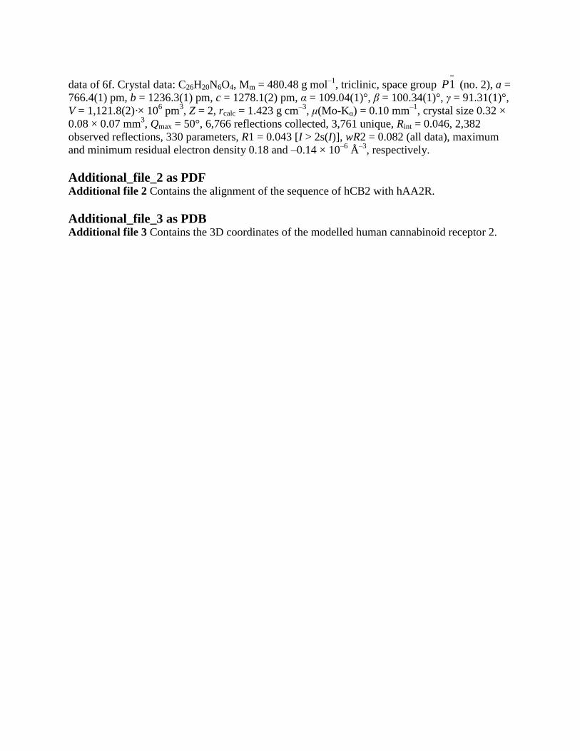
data of 6f. Crystal data: C26H20N6O4, Mm = 480.48 g mol–1
, triclinic, space group 1P (no. 2), a =
766.4(1) pm, b = 1236.3(1) pm, c = 1278.1(2) pm, α = 109.04(1)°, β = 100.34(1)°, γ = 91.31(1)°,
V = 1,121.8(2)·× 106 pm
3, Z = 2, rcalc = 1.423 g cm
–3, μ(Mo-Kα) = 0.10 mm
–1, crystal size 0.32 ×
0.08 × 0.07 mm3, Qmax = 50°, 6,766 reflections collected, 3,761 unique, Rint = 0.046, 2,382
observed reflections, 330 parameters, R1 = 0.043 [I > 2s(I)], wR2 = 0.082 (all data), maximum
and minimum residual electron density 0.18 and –0.14 × 10⁻⁶
Å–3
, respectively.
Additional_file_2 as PDF
Additional file 2 Contains the alignment of the sequence of hCB2 with hAA2R.
Additional_file_3 as PDB
Additional file 3 Contains the 3D coordinates of the modelled human cannabinoid receptor 2.
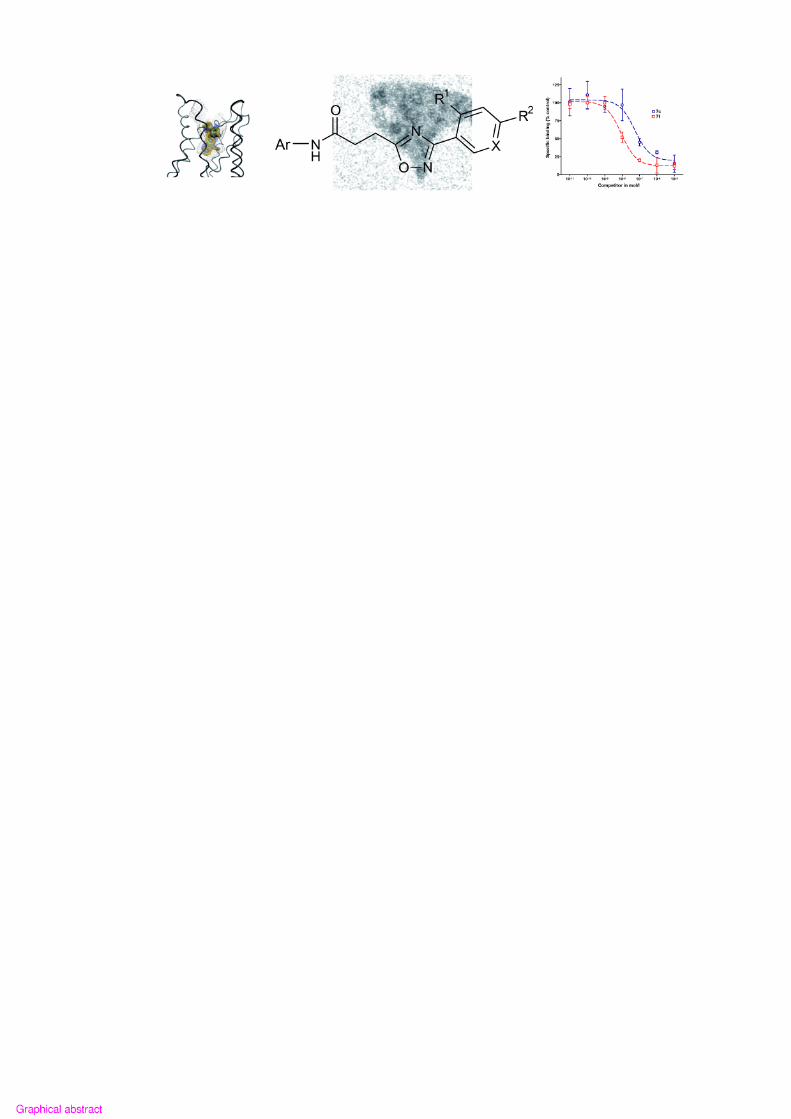

X
N
ON
NH
O
Ar
R1
R2
NO
N
N
N
N
R3
Ar
Y1
Y2
Y3
Y4a
Y5
Y4b
Y4c
R3 = H
R3 = CH2CH3
R3 = CH3
Compound
1a
1b
2a
2b
2c
3b
4e
5e
6a
6b
6c
6d
6e
6f
6g
6h
6i
6j
6k
6l
6m
7h
Ar
Y1
Y1
Y2
Y2
Y2
Y3
Y4a
Y4b
Y4c
Y4c
Y4c
Y4c
Y4c
Y4c
Y4c
Y4c
Y4c
Y4c
Y4c
Y4c
Y4c
Y5
R1
CN
CN
CN
CN
H
CN
Br
Br
CN
CN
H
F
Br
CN
Br
F
F
OH
OCH2CH2F
OCH3
Br
F
R2
F
F
F
F
Br
F
F
F
F
F
Br
NO2
F
NO2
NO2
F
NH2
F
F
F
OCH3
F
X
CH
N
CH
N
N
N
CH
CH
CH
N
N
CH
CH
CH
CH
CH
CH
CH
CH
CH
CH
CH
Scheme 1

R2
NHO
H2NX
R1
X
R1
R2
NC
1HOOC
O N
NX
R1
R2
O N
NX
R1
R2O
NH
Ar
Ar-NH2
2 3a or 3b
4
5
6
1.1 eq. EDAC, 1.1 eq. aryl-amine,DCM, 1h, 0 °C and 1h, rt
washed with H2O, evaporate to dryness1.0 eq. Benzonitrile, 24 h, 80 °C (diffuse light) DCM,�1.0 eq. Hydroxylamine, 0.5 eq. Na2CO3, MeOH/H2O = 4/1, 10 min, rt1
2
0.1 eq. KF, 3.0 eq. Succinic acid anhydride, 30 min,130° - 140 °C (diffuse light)(R1 = Br, R2 = F, X= C):
0.8/0.9 eq. succinic acid anhydride, 30 min,130° -140 °C (diffuse light)
3a3b
DMAP (catalyst), DCM, 1h, rt1.1 eq. EDAC, DCM, 1h, 0° C and 1d, rt
456
Scheme 2
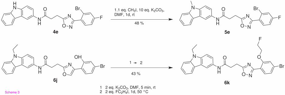
HN
O N
N
Br
F
O
NH
N
O
N Br
O
NH
N
O N
N
Br
F
O
NH
N
O N
N Br
O
NH
4e
6j6k
5e
1.1 eq. CH3I, 10 eq. K2CO3,DMF, 1d, rt
1
OH O
F
2
1 2 eq. K2CO3, DMF, 5 min, rt2 2 eq. FC2H4I, 1d, 50 °C
48 %
43 %
Scheme 3

N
O N
N
Br
NO2
O
NH
N
O N
N
N
Br
O
NH
N
O N
N
Br
18F
O
NH
N
O N
N
N
18F
O
NH
6g
6c[18F]6b
[18F]6e
2 mg 1, 1.5 mL DMFmw (pulse): (20) 65°C, 6x30 s,75 W, 3x60 s, 150 W, diffuse light
2 mg 1, 1.5 mL DMFmw (pulse): (20) 80° - 120° C75 W, 3x60 s, 75 W
Scheme 4

Figure 1
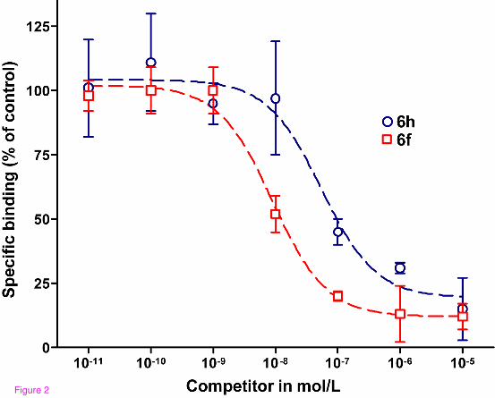
Figure 2

Figure 3

Figure 4
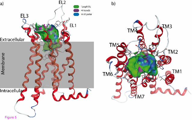
Figure 5

Figure 6
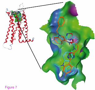
Figure 7

Additional files provided with this submission:
Additional file 1: 1467561248736151_add1.cif, 15Khttp://www.orgmedchemlett.com/imedia/1578328952824069/supp1.cifAdditional file 2: 1467561248736151_add2.pdf, 64Khttp://www.orgmedchemlett.com/imedia/7921352898240690/supp2.pdfAdditional file 3: 1467561248736151_add3.pdb, 179Khttp://www.orgmedchemlett.com/imedia/6755281548240690/supp3.pdb

![Principles of Organic Medicinal Chemistry Chapter 3 Physico Chemical Properties of Organic Medicinal Agents[1]](https://static.documents.pub/doc/80x56/563dbb63550346aa9aacb8cc/principles-of-organic-medicinal-chemistry-chapter-3-physico-chemical-properties.jpg)


