
See discussions, stats, and author profiles for this publication at: https://www.researchgate.net/publication/221870969
Organization of the Human Inferior Parietal Lobule Based on Receptor
Architectonics
Article in Cerebral Cortex · February 2012
DOI: 10.1093/cercor/bhs048 · Source: PubMed
CITATIONS
163READS
651
6 authors, including:
Some of the authors of this publication are also working on these related projects:
microscopical brain models View project
other publications View project
Svenja Caspers
Heinrich-Heine-Universität Düsseldorf
134 PUBLICATIONS 6,607 CITATIONS
SEE PROFILE
Axel Schleicher
Forschungszentrum Jülich
254 PUBLICATIONS 19,747 CITATIONS
SEE PROFILE
Mareike Bacha-Trams
Aalto University
10 PUBLICATIONS 254 CITATIONS
SEE PROFILE
Nicola Palomero-Gallagher
Forschungszentrum Jülich
82 PUBLICATIONS 7,214 CITATIONS
SEE PROFILE
All content following this page was uploaded by Svenja Caspers on 26 May 2014.
The user has requested enhancement of the downloaded file.

Cerebral Cortex
doi:10.1093/cercor/bhs048
Organization of the Human Inferior Parietal Lobule Based on Receptor Architectonics
Svenja Caspers1, Axel Schleicher2, Mareike Bacha-Trams1,3, Nicola Palomero-Gallagher1, Katrin Amunts1,4,5 and Karl Zilles1,2,4
1Institute of Neuroscience and Medicine (INM-1, INM-2), Research Centre Julich, 52425 Julich, Germany, 2C. and O. Vogt Institute
for Brain Research, Heinrich-Heine-University Dusseldorf, 40001 Dusseldorf, Germany, 3Max-Planck-Institute for Human Cognitive
and Brain Sciences, 04103 Leipzig, Germany, 4JARA-BRAIN, Julich-Aachen Research Alliance, 52425 Julich, Germany and5Department of Psychiatry, Psychotherapy, and Psychosomatics, RWTH Aachen University, 52072 Aachen, Germany
Address correspondence to Dr Svenja Caspers, Institut fur Neurowissenschaften und Medizin, INM-2, Forschungszentrum Julich, 52425 Julich,
Germany. Email: [email protected].
Human inferior parietal lobule (IPL) plays a key role in variouscognitive functions. Its functional diversity, including attention,language, and action processing, is reflected by its structuralsegregation into 7 cytoarchitectonically distinct areas, each withcharacteristic connectivity patterns. We hypothesized that com-monalities of the cytoarchitectonic, connectional, and functionaldiversity of the IPL should be reflected by a correlated transmitterreceptor--based organization. Since the function of a cortical arearequires a well-tuned receptor balance, the densities of 15 differentreceptors were measured in each IPL area. A hierarchical clusteranalysis of the receptor balance revealed a tripartite segregation ofthe IPL into a rostral, middle, and caudal group. Comparison withother cortical areas showed strong similarities with Broca’s regionfor all 3 groups, with the superior parietal cortex for the middle, andwith extrastriate visual areas for the caudal group. Notably, caudal-most area PGp has a receptor fingerprint very similar to that ofventral extrastriate visual cortex. We therefore propose a neworganizational model of the human IPL, consisting of 3 clusters,which corresponds to its known cytoarchitectonic, connectional,and functional diversity at the molecular level. This might reflecta general organizational principle of human IPL, beyond specificfunctional domains.
Keywords: architecture, cerebral cortex, inferior parietal lobe, structuralsegregation, transmitter receptors
Introduction
The human inferior parietal lobule (IPL) comprises the supra-
marginal gyrus rostrally and the angular gyrus caudally. Brodmann
(1909) subdivided the human IPL into 2 cytoarchitectonical
areas: BA 40 rostrally and BA 39 caudally. Electrophysiological
studies in macaques and functional neuroimaging in humans
suggest, however, a functionally much more heterogeneous IPL
than Brodmann’s map suggests. The cytoarchitectonic analysis of
von Economo and Koskinas (1925) hinted at a more detailed
parcellation. They defined several subtypes within the 2 main IPL
areas (termed PF and PG) but could not establish them as unique.
In monkeys, rostral IPL is involved in sensorimotor inte-
gration and contains mirror neurons (Fogassi et al. 2005),
whereas caudal IPL was found to participate in spatial attention,
visuomotor, and auditory processes (Mountcastle et al. 1975;
Hyvarinen 1982; Pandya and Seltzer 1982; Seltzer and Pandya
1984; Rozzi et al. 2008). A comparable functional segregation
was found in humans: Rostral human IPL seems to be involved in
motor planning and action-related functions and is part of the
human mirror neuron system (Iacoboni 2005; Rizzolatti 2005;
Keysers and Gazzola 2009; Caspers et al. 2010). The left caudal
IPL is active during language-related tasks with focus on
semantic and phonological issues (Price 2000; Vigneau et al.
2006), while the right caudal IPL was found to be involved in
spatial and nonspatial attention as well as motor preparation
(Fink et al. 2001; Corbetta et al. 2008).
This functional segregation found a structural correlation in
recent observations. In monkeys, 4 areas were identified on the
lateral surface of the IPL and 2 areas on the caudal part of the
parietal operculum within the Sylvian fissure (Pandya and Seltzer
1982; Gregoriou et al. 2006). In humans, a similar parcellation
could be established. Seven cytoarchitectonically distinct areas
were recently described, 5 of which are located on the lateral
surface, whereas the remaining 2 areas are located on the caudal
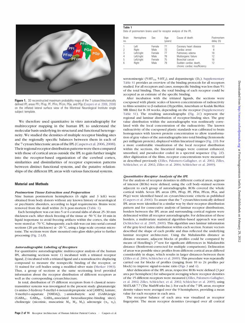
parietal operculum (Caspers et al. 2006, 2008) (Fig. 1).
The functional and architectonical diversity of the IPL are
also reflected by differential connectivity patterns of the areas.
The fiber tracts between the IPL and other cortical areas
change from rostral to caudal, as demonstrated in a recent
diffusion tensor imaging study (Caspers, Eickhoff, et al. 2011):
Whereas rostral IPL areas show strong connections with
inferior frontal, motor, premotor, and somatosensory areas,
caudal IPL areas are more strongly connected with posterior
parietal, higher visual, and temporal areas. Areas in the middle
of the IPL are connected with the targets of both rostral and
caudal IPL areas. A comparable differential connectivity pattern
was found by means of connectivity-based parcellation of the
IPL (Mars et al. 2011). This pattern strikingly resembles that
found in tracer studies in macaques (Cavada and Goldman-
Rakic 1989a, 1989b; Andersen et al. 1990; Rozzi et al. 2006).
Thus, the structural, functional, and connectivity data favor the
concept of a highly segregated brain region.
Mapping the regional and laminar distribution patterns of
different receptors in the cerebral cortex proved to be a
powerful tool for detecting functionally meaningful cortical
parcellations (Zilles and Palomero-Gallagher 2001; Zilles,
Palomero-Gallagher, et al. 2002; Zilles, Schleicher, et al. 2002;
Zilles and Amunts 2009). Not only primary motor, premotor,
and primary somatosensory cortices (Geyer et al. 1997, 1998)
but also higher order areas such as Broca’s region (Amunts et al.
2010), the striate and extrastriate visual cortex (Eickhoff et al.
2007, 2008) as well as the superior parietal lobule (Scheperjans,
Grefkes, et al. 2005; Scheperjans, Palomero-Gallagher, et al.
2005), the cingulate cortex (Palomero-Gallagher et al. 2009), and
the superior temporal gyrus (Morosan et al. 2005) have been
subdivided into distinct receptor-architectonical entities. More-
over, it has been demonstrated that cortical areas with similar
receptor expression patterns are nodes in the same functionally
distinct neural network (Zilles, Palomero-Gallagher, et al. 2002;
Zilles and Amunts 2009).
� The Authors 2012. Published by Oxford University Press.
This is an Open Access article distributed under the terms of the Creative Commons Attribution Non-Commercial License (http://creativecommons.org/licenses/by-nc/3.0), which permits
unrestricted non-commercial use, distribution, and reproduction in any medium, provided the original work is properly cited.
Cerebral Cortex Advance Access published February 28, 2012 at Forschungszentrum
Juelich, Zentralbibliothek on February 29, 2012
http://cercor.oxfordjournals.org/D
ownloaded from
We therefore used quantitative in vitro autoradiography for
multireceptor mapping in the human IPL to understand the
molecular basis underlying its structural and functional heteroge-
neity. We studied the densities of multiple receptor binding sites
and the regionally specific balances between them in each of
the7cytoarchitectonic areas of the IPL (Caspers et al. 2006, 2008).
Their regional receptor distributionpatternswere thencompared
with those of cortical areas outside the IPL to gain further insight
into the receptor-based organization of the cerebral cortex,
similarities and dissimilarities of receptor expression patterns
between distinct functional systems, and the putative relation-
ships of the different IPL areas with various functional systems.
Material and Methods
Postmortem Tissue Extraction and PreparationNine human postmortem hemispheres (6 right and 3 left) were
obtained from body donors without any known history of neurological
or psychiatric disorders, according to legal requirements. Brains were
removed from the skull within 24 h post-mortem (Table 1).
Each hemisphere was cut into 5 or 6 coronal slabs of about 25--30 mm
thickness each. After shock freezing of the tissue at –50 �C for 10 min in
liquid isopentane to avoid freezing artifacts within the cortex, the slabs
were stored at –70 �C. Subsequently, each slab was cut into serial coronal
sections (20 lm thickness) at –20 �C, using a large-scale cryostat micro-
tome. The sections were thaw mounted onto glass slides prior to further
processing (Fig. 2A,B).
Autoradiographic Labeling of ReceptorsFor quantitative autoradiographic multireceptor analysis of the human
IPL, alternating sections were 1) incubated with a tritiated receptor
ligand, 2) incubated with a tritiated ligand and a nonradioactive displacing
compound to measure the nonspecific binding of the receptor, or
3) stained for cell bodies using a modified silver stain (Merker 1983).
Thus, a group of sections at the same sectioning level provided
information about the receptor distribution of different receptors as
well as the corresponding cytoarchitecture.
In total, distribution of 15 different receptors from 6 classical neuro-
transmitter systems was investigated in the present study: glutamatergic
(a-amino-3-hydroxy-5-methyl-4-isoxazolepropionic acid [AMPA], kainate,
N-methyl-D-aspartate [NMDA]), c-aminobutyric acid (GABA)ergic
(GABAA-, GABAB-, GABAA-associated benzodiazepine--binding sites),
cholinergic (nicotinic, muscarinic M1, M2, M3), adrenergic (a1, a2),
serotoninergic (5-HT1A, 5-HT2), and dopaminergic (D1). Supplementary
Table S1 provides an overview of the binding protocols for all receptors
studied. For all receptors and cases, nonspecific binding was less than 5%
of the total binding. Thus, the total binding of each receptor could be
accepted as an estimate of the specific binding.
After incubation with the tritiated ligands, the sections were
coexposed with plastic scales of known concentrations of radioactivity
to films sensitive to b-radiation (Hyperfilm, Amersham or Kodak BioMax
MR films) for 8--18 weeks, depending on the receptor (Supplementary
Table S1). The resulting autoradiographs (Fig. 2C) represent the
regional and laminar distribution of receptor-binding sites. The gray
value distribution within the autoradiographs was nonlinearly corre-
lated with the local concentration of the radioactivity. The known
radioactivity of the coexposed plastic standards was calibrated to brain
homogenates with known protein concentration to allow transforma-
tion of gray values of the autoradiographs into total binding (femtomole
per milligram protein), displayed within linearized images (Fig. 2D). For
a more comfortable visualization of the local receptor distribution
within the sections, the linearized images were contrast enhanced,
smoothed, and pseudocolor coded in a spectral sequence (Fig. 2E).
After digitization of the films, receptor concentrations were measured
as described previously (Zilles, Palomero-Gallagher, et al. 2002; Zilles,
Schleicher, et al. 2002; Zilles et al. 2004; Schleicher et al. 2009).
Quantitative Receptor Analysis of the IPLFor the analysis of receptor densities in different cortical areas, regions
of interest (ROIs) were defined, using the cell body--stained sections
adjacent to each group of autoradiographs. ROIs covered the whole
cortical width. Seven IPL areas (PFt, PFop, PF, PFm, PFcm, PGa, and
PGp) were identified based on cytoarchitectonic criteria as published
(Caspers et al. 2006). To assure that the 7 cytoarchitectonically defined
IPL areas were identified in a similar way by their receptor distribution
pattern and for consecutive analysis of laminar receptor distributions,
borders between the IPL areas and surrounding cortical regions were
delineated within all receptor autoradiographs. For delineation of these
borders, a multivariate statistical algorithm--based approach was used
(Schleicher et al. 2005, 2009). This algorithm uses laminar information
of the gray-level index distribution within each section. Feature vectors
described the shape of each profile and thus reflected the underlying
laminar receptor architecture. Using the Mahalanobis distance as
distance measure, adjacent blocks of profiles could be compared by
means of Hotelling’s T2-test for significant differences in Mahalanobis
distance (Bonferroni-corrected for multiple comparisons). Delineation
of areas was possible since profiles from different cortical areas differed
considerably in shape, which results in larger distances between them
(Zilles et al. 2004; Schleicher et al. 2009). This procedure was repeatedly
carried out for blocks of profiles (ranging from 10 to 24 profiles per
block) to improve signal-to-noise ratio (Figs 3 and 4).
After delineation of the IPL areas, respective ROIs were defined (3 per
area per hemisphere) for subsequent averaging where receptor densities
of the 15 different receptors were measured (Zilles, Palomero-Gallagher,
et al. 2002; Zilles, Schleicher, et al. 2002; Schleicher et al. 2009), using
MATLAB 7.7 (The MathWorks Inc.). For each of the 7 IPL areas, receptor
density values were averaged over the 9 hemispheres, providing a mean
value for each receptor in each area.
The receptor balance of each area was visualized as receptor
fingerprint. The mean receptor densities (averaged over all cortical
Figure 1. 3D reconstructed maximum probability maps of the 7 cytoarchitectonicallydefined IPL areas PFt, PFop, PF, PFm, PFcm, PGa, and PGp (Caspers et al. 2006, 2008)on the inflated lateral surface view of the Montreal Neurological Institute singlesubject template.
Table 1Data of postmortem brains used for receptor analysis of the IPL
Brainno.
Hemisphere Sex Age(years)
Cause of death Postmortemdelay (h)
1 Left Female 77 Coronary heart disease 102 Right Male 72 Cardiac arrest 83 Left/right Female 77 Pulmonary edema 184 Left/right Male 78 Multiorganic failure 125 Left/right Female 75 Bronchial cancer 166 Right Male 79 Sudden cardiac death,
chronic cardiac insufficiency12
Page 2 of 14 Receptor Architecture of Human Inferior Parietal Cortex d Caspers et al.
at Forschungszentrum Juelich, Z
entralbibliothek on February 29, 2012http://cercor.oxfordjournals.org/
Dow
nloaded from

layers) for each receptor type (averaged over hemispheres) were
registered in a polar plot, which represent the characteristic receptor
fingerprint of each area. These fingerprints could consecutively be
compared with regard to their size and shape by using a unified scaling
for each receptor for all areas (Zilles, Palomero-Gallagher, et al. 2002;
Zilles, Schleicher, et al. 2002). This allows a direct comparison of
different cortical areas to reveal similarities and differences in their
receptor distribution pattern.
For comparison with other cortical areas, additional ROIs were
defined based on published cytoarchitectonic and macroanatomical
criteria. Cortical areas were chosen to optimally categorize the IPL
areas in relation to other cortical areas. Therefore, ROIs within primary
as well as higher order association cortices were defined: primary
motor cortex (M1; Geyer et al. 1996); primary somatosensory areas 3b
and 1 (S1_3b, S1_1; Geyer et al. 1999, 2000); primary and secondary
visual cortex (V1, V2; Amunts et al. 2000); ventral extrastriate visual
cortex lateral to V1 and V2 (mainly V3v, V4v; Rottschy et al. 2007);
primary and secondary auditory cortex (A1, A2; Morosan et al. 2001);
Broca’s area (area 44; Amunts et al. 1999, 2010); and posterior superior
parietal lobule (area 7A; Scheperjans, Hermann, et al. 2008; Scheperjans,
Eickhoff, et al. 2008). ROIs within Broca’s area and the superior parietal
lobule were chosen representatively. It was shown that both these
regions could be parcellated into several subdivisions based on their
receptor architecture. But the architecture within these subdivisions
was very similar to each other, especially as compared with other
cortical areas (Scheperjans, Grefkes, et al. 2005; Scheperjans, Palomero-
Gallagher, et al. 2005; Amunts et al. 2010). Thus, including more
subdivisions within the present analysis would not add substantial new
information for a basic functional classification of the IPL areas.
Statistical AnalysisThe mean density values of all 15 receptors studied were combined
into a feature vector for each area. Since absolute receptor concen-
trations differed considerably between receptor types, all values were
z-transformed across areas prior to any further analysis. The trans-
formation enabled analyses where all receptors had equal weight.
Similarities and differences between receptor distribution patterns of
areas were analyzed by means of a hierarchical cluster analysis (MATLAB
7.7, Statistics Toolbox, The MathWorks Inc.), using Euclidean distances in
combination with the Ward linkage method. Euclidean distances between
feature vectors became smaller the more similar the areas were.
In addition, areal feature vectors were further analyzed by means
of a multidimensional scaling (MDS; Systat 12) to detect similar and
dissimilar groups of areas. MDS resulted in a 2D display of the 15-
dimensional receptor feature vectors. To identify those receptors,
which accounted most for separation into different clusters, a multi-
variate canonical discriminant analysis was performed (Systat 12).
All these analyses were carried out on the mean receptor densities of
all IPL areas. The hierarchical cluster analysis was also conducted for
the comparison of IPL with other cortical areas.
Results
Receptor Mapping of IPL Areas
The measurement of the receptor density of each area from the
cortical surface to the cortex/white matter border demonstrates
the quantitative laminar-specific distribution of the receptors.
Figure 2. Quantitative in vitro receptor autoradiography. (A) Right human hemisphere prior to sectioning into 6 slabs (white lines) for further processing. (B) Blockface of a frozenslab on the cryotome with the labeled ROI in the present study (IPL). The mirror on the left side provides a lateral view of the tissue slab. (C) Autoradiograph of the GABABreceptor of the same slab, ROI marked by a box. (D) Scaled autoradiograph (same as in C) with gray values reflecting the receptor concentrations, calculated from coexposedplastic scales of known radioactivity concentrations. (E) Pseudocolor-coded autoradiograph (same as in C). The colors indicate receptor concentrations, from black for low to redfor high concentrations (for concentrations in femtomole per milligram protein, see color bar). IPS: intraparietal sulcus.
Cerebral Cortex Page 3 of 14
at Forschungszentrum Juelich, Z
entralbibliothek on February 29, 2012http://cercor.oxfordjournals.org/
Dow
nloaded from

Figure 3. Parcellation of IPL based on receptor distribution patterns. (A) Part of a receptor autoradiograph (NMDA receptor) of the IPL (border region between areas PF and PFmas shown in Figure 5A for whole IPL). The autoradiograph of the cortical ribbon (upper left) was covered by traverses running perpendicular to the cortical layers (upper middle)and pseudocolor coded for visualization purposes only (upper right). Results of the algorithmic parcellation are shown below: the left graph shows the significant maxima ofvarying block sizes (ranging from 10 to 24); it indicates a consistently occurring border between 2 cortical areas at profile location 33. Right next to it, a line plot shows theMahalanobis distances between neighboring blocks of profiles; it confirms the location of the maximal distance, and thus, the maximal dissimilarity between adjacent profiles atprofile location 33, which defines an architectural border. The border is also labeled in the autoradiographs above. The graph on the right side of (A) shows the laminar distribution(with standard deviations) of the NMDA receptor throughout the cortical width (0% at the transition from the pial surface to layer I; 100% at the transition from layer VI to thewhite matter) in areas PF and PFm. The profiles differ between both areas. (B) Parcellation of the same part of the cortex by 3 other receptors (kainate, a2, and GABAB). Figuresand graphs of (B) show the results of the mapping procedure comparable to (A).
Page 4 of 14 Receptor Architecture of Human Inferior Parietal Cortex d Caspers et al.
at Forschungszentrum Juelich, Z
entralbibliothek on February 29, 2012http://cercor.oxfordjournals.org/
Dow
nloaded from

Figure 4. Algorithm-based detection of areal borders in receptor and corresponding cytoarchitectonic sections. (A) Cytoarchitectonic border between area PFm and areas withinthe intraparietal sulcus (IPS), sectioning level (red line), and schematic drawing of the IPL within this section with all detected borders (black thick lines) depicted on the left.Corresponding gray level index image and traverses covering the cortical ribbon beneath with detected border indicated by a white bold line at profile position no. 47. (B) Sameborder on corresponding sections of kainate, GABAA, and a1 receptors. For each receptor, the linearized autoradiograph, superimposed with traverses covering the ROI, andpseudocolor coded for visualization purposes. Position of the border indicated by white bold lines and in the graphs at the bottom at the respective profile position (same type ofgraphs as in Fig. 3). Area PFm differs from intraparietal areas by means of higher concentrations of kainate in middle and lower layers, higher concentrations of a1 in infragranularlayers and of GABAA in supragranular layers. Note the close resemblance of the position of the border in cyto- and receptor sections. cs: central sulcus, ips: intraparietal sulcus,poc: postcentral sulcus, sts: superior temporal sulcus.
Cerebral Cortex Page 5 of 14
at Forschungszentrum Juelich, Z
entralbibliothek on February 29, 2012http://cercor.oxfordjournals.org/
Dow
nloaded from

The differences between the density profiles were used for the
statistically testable and observer-independent definition of areal
borders (for details, see Fig. 3 and Schleicher et al. 2005). As an
example for the multireceptor mapping of the IPL, the receptor-
architectonically defined border for different receptor types
between areas PF and PFm is shown in Figure 3.
The receptor-based parcellation approach (Zilles, Palomero-
Gallagher, et al. 2002; Zilles, Schleicher, et al. 2002; Morosan
et al. 2005; Zilles and Amunts 2009) led to the identification of
the same 7 IPL areas as previously identified by cytoarchitec-
tonic criteria (Caspers et al. 2006, 2008): areas PFt, PFop, PF,
PFm, PFcm, PGa, and PGp. The precise match between receptor
and cytoarchitectonic mapping can be demonstrated by com-
paring receptor architectonic with corresponding (neighboring)
cytoarchitectonic sections of the same brain (Fig. 4).
Differences in laminar patterns largely contribute (in addition
to differences in the absolute concentration within the cortex) to
the regional segregation of the IPL into 7 receptor-architectonic
areas. The border regions between neighboring IPL areas are
shown in Figures 5 and 6.
It has already been noted that not all receptors show each
border (Zilles, Palomero-Gallagher, et al. 2002; Zilles, Schleicher,
et al. 2002) and that borders are not equally clear pronounced
by all receptor types. However, if a border has been detected
by several or all receptor types, it has the same spatial position
(Figs 3--6). Differences between the rostral-most IPL areas PFop,
PFt, and PF were most prominently indicated by the kainate,
NMDA, GABAA, and a1 receptors. Here, PF showed higher
concentrations for the kainate, NMDA, and a1 receptors and
lower concentrations for the GABAA receptor as compared with
PFt and PFop (Fig. 5).
Figure 6 displays the border regions between the more
caudal IPL areas. Area PFm could be distinguished from area PF
most clearly by the NMDA and GABAB receptors, whereby PFm
Figure 5. Receptor distribution patterns in areas PF, PFop, and PFt illustrated for 14 of the 15 receptors studied. Pseudocolor-coded autoradiographs show the borders betweenthe IPL areas (white lines). The color bar beneath each autoradiograph indicates receptor concentrations by the different colors, from black for low to red for high concentrations(in femtomole per milligram protein). Note that the scaling is different for each receptor.
Page 6 of 14 Receptor Architecture of Human Inferior Parietal Cortex d Caspers et al.
at Forschungszentrum Juelich, Z
entralbibliothek on February 29, 2012http://cercor.oxfordjournals.org/
Dow
nloaded from

had lower concentrations in the supragranular layers than area
PF. Conversely, area PFm showed higher concentrations of
kainate and a2 receptors in the supragranular layers than area
PF (Fig. 6A).
Area PFcm most prominently differed from area PF with
regard to the AMPA, kainate, GABAA, and the D1 receptors,
whereby PFcm showed considerably lower concentrations
than PF (Fig. 6B).
Caudal-most areas PGa and PGp were best delineated by the
5-HT1A, 5-HT2, a1, and D1 receptors. PGp showed higher
concentrations of 5-HT1A and a1 receptors in the infragranular
layers and of the D1 receptor in the supragranular layers than
PGa. Concentrations of 5-HT2 receptors were higher in supra-
granular layers of PGa as compared with PGp (Fig. 6C).
Quantitative Analysis of Mean Receptor Densities
Receptor Fingerprints of the IPL Areas
The receptor densities of each IPL area and each receptor type
are displayed in Table 2. Highest mean densities (averaged
over all cortical layers) are found for the NMDA, GABAA, GABAB,
and benzodiazepine-binding sites, lowest densities are reached
by the D1, nicotinic, and M2 receptors. Maximal or minimal
receptor densities of each receptor type are found in different
areas of the IPL. Thus, each area has a specific balance between
the different receptor types.
The area-specific balances between the 15 receptors can be
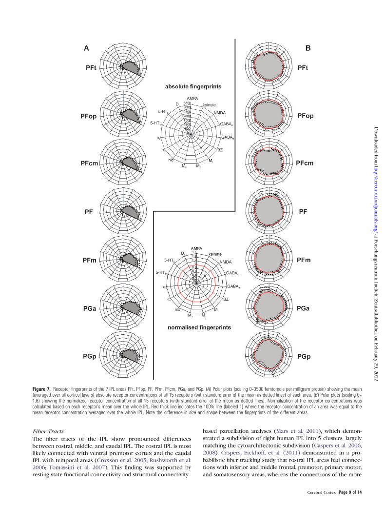
visualized as ‘‘receptor fingerprints’’ (Fig. 7). Comparing the
shapes of the fingerprints revealed a rostrocaudal gradient:
The fingerprints based on absolute receptor concentrations
(Fig. 7A) showed higher concentrations of the benzodiazepine
binding sites in the rostral (Fig. 7, upper part) as compared with
more caudal IPL areas (Fig. 7, lower part). Fingerprints based
on normalized receptor concentrations (Fig. 7B) additionally
showed lower AMPA, GABAA, a2, and D1 receptor concentrations
and higher kainate, 5-HT1A, and 5-HT2 receptor concentrations
in the rostral as compared with the caudal IPL areas. Caudal-
most area PGp is characterized by high concentrations of the M2
receptor, whereas area PGa shows exceptionally high concen-
trations of the nicotinic receptor.
Figure 6. Receptor distribution patterns of areas PF, PFm, PFcm, PGa, and PGp for those receptors, which showed most prominent differences between the areas. (A)Delineation of areas PF and PFm (same level as in Fig. 3). (B) Delineation of areas PF and PFcm. (C) Delineation of areas PGa and PGp. For other conventions, see Figure 4.
Cerebral Cortex Page 7 of 14
at Forschungszentrum Juelich, Z
entralbibliothek on February 29, 2012http://cercor.oxfordjournals.org/
Dow
nloaded from

Molecular Organization of the IPL
For a comprehensive analysis of similarities between the
receptor fingerprints of the different IPL areas, we performed
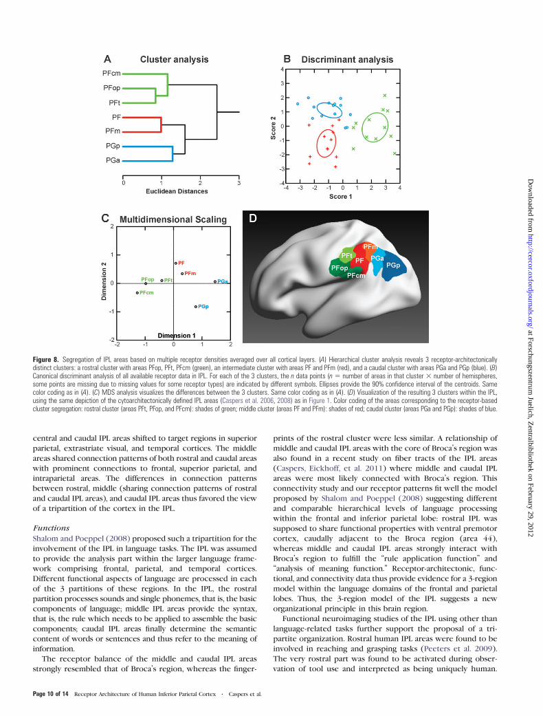
a hierarchical cluster analysis (Fig. 8A). Three groups with
similar receptor distributions within each group were identi-
fied: a rostroventral group with areas PFt, PFop, and PFcm;
a middle group of areas PF and PFm; and a caudal group
consisting of areas PGa and PGp. Furthermore, it became
apparent that the PG areas were more similar to each other as
compared with the rest of the IPL. This result reflects a clear
architectural distinction between rostral and caudal IPL.
A consecutive canonical discriminant analysis with 2 discrim-
inating dimensions (x- and y-axes in Fig. 8B) revealed a most
pronounced distinction between the clusters within the first
dimension (x-axis; 72% explained variance), complemented by
the distinction within the second dimension (y-axis; 28%
explained variance). Ranking the coefficients of the canonical
discriminant analysis revealed those receptors, which contri-
buted most to the distinction between the clusters in both
dimensions. The kainate and 5-HT2 receptors contributed most
to the distinction in both dimensions (absolute values of the
coefficients: kainate: 2.22 [score 1] and 1.91 [score 2]; 5-HT2:
2.84 [score 1] and 1.58 [score 2]). The M1 and a2 receptors
provided additional criteria for this segregation within the first
dimension (absolute values of coefficients: M1: 1.10; a2: 1.05),whereas the GABAA, nicotinic, and D1 receptors were re-
sponsible for distinction between the clusters within the second
dimension (absolute values of coefficients: GABAA: 1.02; nico-
tinic: 1.10; D1: 1.18).
MDS analysis (Fig. 8C) of the receptor densities highlights an
inhomogeneity within the caudal cluster of areas PGa and PGp:
the receptor organization of PGp seems to be more different
(higher distance) from all the other IPL areas. This dissimilarity
was not revealed by our previous cytoarchitectonic analysis of
the IPL (Caspers et al. 2006, 2008). Based on the present results,
area PGp might be reclassified as not being a typical parietal
cortex. It might provide a transition to adjoining visual cortex,
which can be underpinned by comparison with the receptor
architecture of other cortical areas (see next paragraph).
Comparison with Other Cortical Areas
We compared the IPL fingerprints with those of primary,
secondary, and higher order sensory areas and the motor
cortex to study the functional aspect of the receptor-based IPL
segregation (Fig. 9).
Using a hierarchical cluster analysis, the fingerprints of the
primary and secondary auditory and visual as well as primary
somatosensory and motor cortices differed considerably from
the cluster containing the IPL areas. The 7 IPL areas formed the
same rostral, middle, and caudal subclusters as already found in
the first cluster analysis of the IPL areas alone (Fig. 8A--C). The
middle and caudal clusters of IPL areas are more similar to area
44 of Broca’s area than the rostral cluster comprising PFcm,
PFop, and PFt. The fingerprints of the centrally positioned areas
PF and PFm are similar to the fingerprint of the superior
parietal lobule. The caudally positioned areas PGa and PGp are
similar to the higher ventral extrastriate area hOC3v (V3v),
particularly for area PGp.
This result suggests again a potential role of area PGp as
a higher visual area, linking occipital and parietal cortex.
Discussion
Functional performance of a cortical area depends on a well-
tuned and area-specific balance between numerous receptor
types (Barnes and Sharp 1999; Goldman-Rakic et al. 2000;
Gibbs and Summers 2002; Bergson et al. 2003; Bredt and
Nicoll 2003; Friedman et al. 2004). Based on the similarities in
receptor fingerprints of the 7 IPL areas, we propose a new
organizational model of the IPL (Fig. 8D), comprising
a rostroventral (areas PFt, PFop, and PFcm), an intermediate
(areas PF and PFm), and a caudal group (areas PGa and PGp).
The molecular structure of caudal-most IPL area PGp argues
for a reclassification of this area as transition area between
parietal and visual areas.
The 3-Region Model of Human IPL
It has been shown repeatedly that receptor distributions are
not only related to functional network properties of cortical
areas (Barnes and Sharp 1999; Goldman-Rakic et al. 2000; Gibbs
and Summers 2002; Bergson et al. 2003; Bredt and Nicoll 2003;
Friedman et al. 2004) but also to their connectivity pattern
(Rakic et al. 1988). The 3-region model of human IPL as
revealed by multireceptor distribution could thus provide the
molecular basis for the structural, functional, and connectivity
components within a common organizational framework.
Table 2Mean receptor densities (averaged over all cortical layers) in femtomole per milligram protein (±SD) of IPL areas
Receptor IPL areas
PFop PFt PFcm PF PFm PGa PGp
AMPA 358.09 ± 62.73 363,65 ± 51.11 324.84 ± 65.24 424.66 ± 65.09 398.26 ± 48,09 437.99 ± 64.62 464.30 ± 85.84Kainate 457.37 ± 77.49 541.48 ± 91.61 521,42 ± 131.47 659.84 ± 92.94 596.46 ± 97.96 587.77 ± 122.92 487.41 ± 104.19NMDA 1142.85 ± 84.49 1077.73 ± 107.60 1114.54 ± 129.36 1158.25 ± 97.03 1240.96 ± 79.20 1223.37 ± 73.86 1116.08 ± 124.75GABAA 1588.22 ± 131.31 1675.23 ± 126.70 1460.58 ± 164.06 1508.03 ± 124.92 1539.34 ± 102.92 1939.48 ± 157.07 1832.05 ± 244.97GABAB 2195.31 ± 213.24 2285.14 ± 152.90 2033.04 ± 280.58 2192.64 ± 208.12 2200.67 ± 260.67 2664.09 ± 121.44 2297.44 ± 248.66BZ 2828.36 ± 250.41 2797.80 ± 280.24 2845.69 ± 410.11 2716.50 ± 281.80 2489.03 ± 344.75 2445.60 ± 293.86 2378.00 ± 331.185-HT1A 439.85 ± 73.24 405.46 ± 60.78 414.99 ± 103.43 361.44 ± 49.54 335.93 ± 47.62 313.00 ± 46.05 328.45 ± 65.465-HT2 433.94 ± 41.13 434.17 ± 48.31 434.28 ± 53.21 441.92 ± 49.34 424.76 ± 59.87 412.24 ± 70.71 382.79 ± 59.43M1 535.57 ± 73.88 470.10 ± 61.39 497.67 ± 92.89 459.90 ± 69.93 489.62 ± 80.42 456.47 ± 67.90 452.02 ± 61.00M2 159.29 ± 14.33 161.32 ± 12.12 138.68 ± 19.31 170.53 ± 25.40 173.50 ± 26.22 158.87 ± 29.98 201.75 ± 48.54M3 902.58 ± 220.18 850.92 ± 178.10 660.20 ± 143.45 821.06 ± 105.22 775.76 ± 107.59 736.76 ± 96.82 741.39 ± 131.85nic 44.93 ± 10.09 48.10 ± 8.83 38.29 ± 8.85 58.91 ± 13.44 60.39 ± 11.46 68.50 ± 11.51 46.07 ± 11.59a1 362.04 ± 36.82 365.44 ± 34.94 335.63 ± 36.52 372.21 ± 57.03 393.38 ± 49.38 343.41 ± 50.17 356.91 ± 40.37a2 304.26 ± 80.51 335.93 ± 88.95 297.52 ± 95.33 370.43 ± 118.34 303.08 ± 84.95 343.25 ± 86.58 327.10 ± 64.74D1 89.11 ± 10.07 81.54 ± 11.86 86.46 ± 12.34 100.46 ± 15.63 105.61 ± 16.27 132.44 ± 18.00 105.14 ± 16.55
Note: Fifteen different receptors were measured in 9 hemispheres. SD: standard deviation.
Page 8 of 14 Receptor Architecture of Human Inferior Parietal Cortex d Caspers et al.
at Forschungszentrum Juelich, Z
entralbibliothek on February 29, 2012http://cercor.oxfordjournals.org/
Dow
nloaded from
Fiber Tracts
The fiber tracts of the IPL show pronounced differences
between rostral, middle, and caudal IPL. The rostral IPL is most
likely connected with ventral premotor cortex and the caudal
IPL with temporal areas (Croxson et al. 2005; Rushworth et al.
2006; Tomassini et al. 2007). This finding was supported by
resting-state functional connectivity and structural connectivity--
based parcellation analyses (Mars et al. 2011), which demon-
strated a subdivision of right human IPL into 5 clusters, largely
matching the cytoarchitectonic subdivision (Caspers et al. 2006,
2008). Caspers, Eickhoff, et al. (2011) demonstrated in a pro-
babilistic fiber tracking study that rostral IPL areas had connec-
tions with inferior and middle frontal, premotor, primary motor,
and somatosensory areas, whereas the connections of the more
Figure 7. Receptor fingerprints of the 7 IPL areas PFt, PFop, PF, PFm, PFcm, PGa, and PGp. (A) Polar plots (scaling 0--3500 femtomole per milligram protein) showing the mean(averaged over all cortical layers) absolute receptor concentrations of all 15 receptors (with standard error of the mean as dotted lines) of each area. (B) Polar plots (scaling 0--1.6) showing the normalized receptor concentration of all 15 receptors (with standard error of the mean as dotted lines). Normalization of the receptor concentrations wascalculated based on each receptor’s mean over the whole IPL. Red thick line indicates the 100% line (labeled 1) where the receptor concentration of an area was equal to themean receptor concentration averaged over the whole IPL. Note the difference in size and shape between the fingerprints of the different areas.
Cerebral Cortex Page 9 of 14
at Forschungszentrum Juelich, Z
entralbibliothek on February 29, 2012http://cercor.oxfordjournals.org/
Dow
nloaded from
central and caudal IPL areas shifted to target regions in superior
parietal, extrastriate visual, and temporal cortices. The middle
areas shared connection patterns of both rostral and caudal areas
with prominent connections to frontal, superior parietal, and
intraparietal areas. The differences in connection patterns
between rostral, middle (sharing connection patterns of rostral
and caudal IPL areas), and caudal IPL areas thus favored the view
of a tripartition of the cortex in the IPL.
Functions
Shalom and Poeppel (2008) proposed such a tripartition for the
involvement of the IPL in language tasks. The IPL was assumed
to provide the analysis part within the larger language frame-
work comprising frontal, parietal, and temporal cortices.
Different functional aspects of language are processed in each
of the 3 partitions of these regions. In the IPL, the rostral
partition processes sounds and single phonemes, that is, the basic
components of language; middle IPL areas provide the syntax,
that is, the rule which needs to be applied to assemble the basic
components; caudal IPL areas finally determine the semantic
content of words or sentences and thus refer to the meaning of
information.
The receptor balance of the middle and caudal IPL areas
strongly resembled that of Broca’s region, whereas the finger-
prints of the rostral cluster were less similar. A relationship of
middle and caudal IPL areas with the core of Broca’s region was
also found in a recent study on fiber tracts of the IPL areas
(Caspers, Eickhoff, et al. 2011) where middle and caudal IPL
areas were most likely connected with Broca’s region. This
connectivity study and our receptor patterns fit well the model
proposed by Shalom and Poeppel (2008) suggesting different
and comparable hierarchical levels of language processing
within the frontal and inferior parietal lobe: rostral IPL was
supposed to share functional properties with ventral premotor
cortex, caudally adjacent to the Broca region (area 44),
whereas middle and caudal IPL areas strongly interact with
Broca’s region to fulfill the ‘‘rule application function’’ and
‘‘analysis of meaning function.’’ Receptor-architectonic, func-
tional, and connectivity data thus provide evidence for a 3-region
model within the language domains of the frontal and parietal
lobes. Thus, the 3-region model of the IPL suggests a new
organizational principle in this brain region.
Functional neuroimaging studies of the IPL using other than
language-related tasks further support the proposal of a tri-
partite organization. Rostral human IPL areas were found to be
involved in reaching and grasping tasks (Peeters et al. 2009).
The very rostral part was found to be activated during obser-
vation of tool use and interpreted as being uniquely human.
Figure 8. Segregation of IPL areas based on multiple receptor densities averaged over all cortical layers. (A) Hierarchical cluster analysis reveals 3 receptor-architectonicallydistinct clusters: a rostral cluster with areas PFop, PFt, PFcm (green), an intermediate cluster with areas PF and PFm (red), and a caudal cluster with areas PGa and PGp (blue). (B)Canonical discriminant analysis of all available receptor data in IPL. For each of the 3 clusters, the n data points (n 5 number of areas in that cluster 3 number of hemispheres,some points are missing due to missing values for some receptor types) are indicated by different symbols. Ellipses provide the 90% confidence interval of the centroids. Samecolor coding as in (A). (C) MDS analysis visualizes the differences between the 3 clusters. Same color coding as in (A). (D) Visualization of the resulting 3 clusters within the IPL,using the same depiction of the cytoarchitectonically defined IPL areas (Caspers et al. 2006, 2008) as in Figure 1. Color coding of the areas corresponding to the receptor-basedcluster segregation: rostral cluster (areas PFt, PFop, and PFcm): shades of green; middle cluster (areas PF and PFm): shades of red; caudal cluster (areas PGa and PGp): shades of blue.
Page 10 of 14 Receptor Architecture of Human Inferior Parietal Cortex d Caspers et al.
at Forschungszentrum Juelich, Z
entralbibliothek on February 29, 2012http://cercor.oxfordjournals.org/
Dow
nloaded from

Recent meta-analyses demonstrated that rostral IPL area PFt
participates in the action observation and imitation network
(Molenberghs et al. 2009; Van Overwalle and Baetens 2009;
Caspers et al. 2010). Data from studies in macaques also point
to the relevance of rostral-most IPL together with ventral
premotor area 6 for the mirror neuron system (Rizzolatti 2005;
Petrides and Pandya 2009). This fits the interaction within the
language network (Shalom and Poeppel 2008) as described
above. The middle IPL areas PF and PFm are activated by
nonspatial attention tasks, especially when reevaluating con-
flicting choice options (Vossel et al. 2006; Boorman et al. 2009;
Mevorach et al. 2009; Caspers, Heim, et al. 2011) as well as
spatial attention and reorienting tasks (Rushworth et al. 2001;
Corbetta et al. 2008). Together with intraparietal areas, middle
IPL contributes to rule change during visually guided attention
(Corbetta and Shulman 2002). Caudal areas PGa and PGp were
most prominently implicated in language-related processing
with special focus on semantic and phonological processing,
partially found in both hemispheres (Price 2000; Hickok and
Poeppel 2004; Marangolo et al. 2006; Vigneau et al. 2006). These
areas have also consistently been found during moral decision
making, being particularly concerned with egocentric and allo-
centric perspective taking (for review: Raine and Yang 2006).
The involvement of the IPL within different functional
domains could thus be summarized as follows: Rostral IPL deals
with tool, action, or sound. Middle IPL areas provide rules for
word differentiation as well as visually guided attention and
nonspatial attention processes. Caudal IPL is involved in
decoding the meaning of words, scenes, or personal morally
relevant interactions. Thus, the same IPL areas are involved in
different tasks, which should have a functional commonality
representing the role of the IPL areas on a more abstract level.
It already seems plausible to assume that the hierarchical
3-region model of language functions in the IPL is a starting
point for searching analogous commonalities in other func-
tional domains.
The present study provides evidence for a general 3-region
model of the IPL on a molecular basis regarding the receptor
balance of different neurotransmitter systems. The relevance of
the receptor balance of an area for its involvement in different
functional networks has been repeatedly stressed (Barnes and
Sharp 1999; Goldman-Rakic et al. 2000; Gibbs and Summers
2002; Bergson et al. 2003; Bredt and Nicoll 2003; Friedman et al.
2004). It can thus be assumed that not the distribution pattern
of a single receptor, but the interplay between different
receptors of different neurotransmitter systems as displayed in
the receptor fingerprints of each IPL area (Fig. 7) might set the
molecular basis for the role, which is played by 3 different parts
of the IPL across various functional domains.
The Role of Area PGp
The present findings additionally provide new insights into the
potential role of area PGp. Its receptor distribution was
different from the other IPL areas and showed most pro-
nounced similarities with higher extrastriate visual areas,
particularly V3v. This is further promoted by connectivity
analyses, which showed consistent connections between PGp
and extrastriate visual areas (Caspers, Eickhoff, et al. 2011). It
might thus be hypothesized that area PGp might serve as
linking hub between occipital and parietal cortex for trans-
formation of visual input to visual associations.
The cytoarchitectonic analysis of the IPL areas (Caspers et al.
2006) did not show a comparable difference. The cytoarchi-
tecture of area PGp resembled that of the other IPL areas. It
could be clearly demarcated from areas of the occipital cortex
where the layers are dominated by large pyramidal cells as
described by von Economo and Koskinas (1925). Area PGp is,
therefore, clearly different from this ‘‘occipital type’’ of cortical
architecture at the cytoarchitectonical level.
The probabilistic fiber tracking with area PGp as seed region
shows connections to extrastriate visual areas (Caspers, Eickhoff,
et al. 2011). The same situation was demonstrated in macaques
for area Opt (Cavada and Goldman-Rakic 1989a, 1989b; Andersen
et al. 1990; Rozzi et al. 2006), which favored the view that visual
input to the IPL arrives via this caudal-most area.
The visual system has classically been subdivided into a
ventral and dorsal visual stream, processing either ‘‘what’’ or
‘‘where’’ information, respectively (Ungerleider and Mishkin
1982; Ungerleider and Haxby 1994). The role of the dorsal
visual stream within this framework was nevertheless not fully
elucidated, fostering the notion of not only processing ‘‘where’’
but ‘‘how’’ information (Goodale and Milner 1992; Milner and
Goodale 1995; Kravitz et al. 2011). It was furthermore suggested
that the 2 systems are not fully separated from each other but
rather interact to fulfill the task of providing the information on
how an action should be executed (Pisella et al. 2006; Kravitz
et al. 2011). The interaction is supposed to involve a ventrodorsal
pathway, which involves caudal IPL. This region interacts with
medially located areas of the superior parietal lobule, the pos-
terior cingulate and retrosplenial cortex, and the parahippo-
campal gyrus. It might provide information about peripersonal
Figure 9. Receptor distributions of IPL areas compared with those of other corticalareas. The hierarchical cluster analysis that shows the same tripartition of the IPLareas as shown in Figure 8 but additionally reveals similarities of the intermediatecluster (areas PF and PFm, red) with superior parietal areas (SPLs) and of the caudalcluster (areas PGa and PGp, blue) with extrastriate visual areas. The IPL areas aremost similar to each other and similar to higher order areas (Broca_44, SPL, and V3v)but are most dissimilar to primary and secondary areas (A1/A2, M1, S1, and V1/V2).Note the close resemblance of area PGp with extrastriate visual area V3v. A1/A2:primary/secondary auditory cortex, Broca_44: area 44 of Broca’s region, M1: primarymotor cortex, S1_3b: area 3b of primary somatosensory cortex, S1_1: area 1 ofprimary somatosensory cortex, SPL: superior parietal lobule, V1/V2: primary/secondary visual cortex, V3v: ventral extrastriate visual cortex.
Cerebral Cortex Page 11 of 14
at Forschungszentrum Juelich, Z
entralbibliothek on February 29, 2012http://cercor.oxfordjournals.org/
Dow
nloaded from

space with regard to egocentric or allocentric perspectives
(Pisella et al. 2006; Rushworth et al. 2006; Kravitz et al. 2011).
This corresponds to the findings of activation within caudal IPL
during moral decision making where additional activation
clusters were found in posterior cingulate cortex in addition
to ventral and medial prefrontal cortex (Raine and Yang 2006).
Here, the simultaneous activation of caudal IPL with posterior
cingulate cortex was especially found during personal versus
impersonal and utilitarian versus nonutilitarian moral judgments
(Greene et al. 2004). Both these decisions involve allocentric
versus egocentric perspectives to come to the respective moral
judgment.
The results of the present study support the idea of area PGp
serving as higher visual processing hub within the IPL. The
similarity between the receptor balances of area PGp with that
of ventral extrastriate visual area hOC3v (V3v) supports the
idea that area PGp is key region in the ventrodorsal visual
stream (Pisella et al. 2006), since it receives input from an area
of that visual stream.
Conclusions and Outlook
Based on the regionally specific multireceptor balances (receptor
fingerprints), a 3-region model of human IPL is proposed
(Fig. 8D). A hierarchical cluster analysis of the receptor finger-
prints between the IPL areas and visual-, motor-, auditory-, and
language-related cortical areas shows the highest similarity of all
IPL areas with area 44 of Broca’s region, of the areas in the
middle of IPL with the superior parietal cortex, and for the most
caudal areas with the extrastriate visual cortex. Notably, PGp has
a receptor fingerprint very similar to that of ventral extrastriate
area hOC3v (V3v). Since receptor fingerprints covary with the
cytoarchitecture, function, and connectivity of each IPL area, the
present study provides a molecular perspective of the organiza-
tional principles behind the regional and functional segregation
of the IPL.
As a link to the function of an area, the receptor-based
delineation of cortical areas poses an additional question: Do
the receptor density patterns always follow the cytoarchitec-
tonic boundaries? In our study, we independently mapped
receptor profiles of each receptor and defined receptor-based
borders within the respective sections. Additionally, we
measured the cytoarchitectonic profiles of the same brain
within alternate cell body--stained sections. The borders of
both approaches did precisely coincide. It has to be noted that
not all receptors showed every border of the IPL or other
cortical areas (Zilles, Palomero-Gallagher, et al. 2002; Amunts
et al. 2010). Vice versa, some receptors might show additional
borders, which would hint at further subdivisions of cortical
areas on a molecular level. Taking an independent mapping
approach for all receptors will allow providing complete brain
maps for each receptor in future studies, each revealing an
individual view on the molecular architecture of the cortex.
Supplementary Material
Supplementary material can be found at: http://www.cercor.
oxfordjournals.org/
Funding
This project was supported by grants of the Initiative and
Networking Fund of the Helmholtz Association within the
Helmholtz Alliance on Systems Biology (Human Brain Model to
K.Z.); the Helmholtz Alliance for Mental Health in an Aging
Society (HelMA to K.A. and K.Z.); and the German Ministry for
Education and Research (01GW0771 and 01GW0623 to K.A.).
The funders had no role in study design, data collection and
analysis, decision to publish, or preparation of the manuscript.
Notes
Conflict of Interest : None declared.
References
Amunts K, Lenzen M, Friederici AD, Schleicher A, Morosan P, Palomero-
Gallagher N, Zilles K. 2010. Broca’s region: novel organizational
principles and multiple receptor mapping. PLoS Biol. 8(9):e1000489.
Amunts K, Malikovic A, Mohlberg H, Schormann T, Zilles K. 2000.
Brodmann’s areas 17 and 18 brought into stereotaxic space—where
and how variable? Neuroimage. 11:66--84.
Amunts K, Schleicher A, Burgel U, Mohlberg H, Uylings HBM, Zilles K.
1999. Broca’s region revisited: cytoarchitecture and intersubject
variability. J Comp Neurol. 412:319--341.
Andersen RA, Asanuma C, Essick G, Siegel RM. 1990. Corticocortical
connections of anatomically and physiologically defined subdivi-
sions within the inferior parietal lobule. J Comp Neurol. 296:65--113.
Barnes NM, Sharp T. 1999. A review of central 5-HT receptors and their
function. Neuropharmacology. 38(8):1083--1152.
Bergson C, Levenson R, Goldman-Rakic PS, Lidow MS. 2003. Dopamine
receptor-interacting proteins: the Ca(2+) connection in dopamine
signalling. Trends Pharmacol Sci. 24:486--492.
Boorman ED, Behrens TE, Woolrich MW, Rushworth MF. 2009. How
green is the grass on the other side? Frontopolar cortex and the
evidence in favor of alternative courses of action. Neuron.
62(5):733--743.
Bredt DS, Nicoll RA. 2003. AMPA receptor trafficking at excitatory
synapses. Neuron. 40:361--379.
Brodmann K. 1909. Vergleichende Lokalisationslehre der Großhirnrinde.
Leipzig (Germany): Barth.
Caspers S, Eickhoff SB, Geyer S, Scheperjans F, Mohlberg H, Zilles K,
Amunts K. 2008. The human inferior parietal lobule in stereotaxic
space. Brain Struct Funct. 212:481--495.
Caspers S, Eickhoff SB, Rick T, von Kapri A, Kuhlen T, Huang R, Shah NJ,
Zilles K. 2011. Probabilistic fibre tract analysis of cytoarchitectoni-
cally defined human inferior parietal lobule areas reveals similarities
to macaques. Neuroimage. 58(2):362--380.
Caspers S, Geyer S, Schleicher A, Mohlberg H, Amunts K, Zilles K. 2006.
The human inferior parietal cortex: cytoarchitectonic parcellation
and interindividual variability. Neuroimage. 33(2):430--448.
Caspers S, Heim S, Lucas MG, Stephan E, Fischer L, Amunts K, Zilles K.
2011. Moral concepts set decision strategies to abstract values. PLoS
One. 6(4):e18451.
Caspers S, Zilles K, Laird AR, Eickhoff SB. 2010. ALE meta-analysis of
action observation and imitation in the human brain. Neuroimage.
50:1148--1167.
Cavada C, Goldman-Rakic PS. 1989a. Posterior parietal cortex in rhesus
monkey: I. Parcellation of areas based on distinctive limbic and
sensory corticocortical connections. J Comp Neurol. 287:393--421.
Cavada C, Goldman-Rakic PS. 1989b. Posterior parietal cortex in rhesus
monkey: II. Evidence of segregated corticocortical networks linking
sensory and limbic areas with the frontal lobe. J Comp Neurol.
287:422--445.
Corbetta M, Patel G, Shulman GL. 2008. The reorienting system of the
human brain: from environment to theory of mind. Neuron.
58:306--324.
Corbetta M, Shulman GL. 2002. Control of goal-directed and stimulus-
driven attention in the brain. Nat Rev Neurosci. 3(3):201--215.
Croxson PL, Johansen-Berg H, Behrens TE, Robson MD, Pinsk MA,
Gross CG, Richter W, Richter MC, Kastner S, Rushworth. 2005.
Quantitative investigation of connections of the prefrontal cortex in
the human and macaque using probabilistic diffusion tractography.
J Neurosci. 25:8854--8866.
Page 12 of 14 Receptor Architecture of Human Inferior Parietal Cortex d Caspers et al.
at Forschungszentrum Juelich, Z
entralbibliothek on February 29, 2012http://cercor.oxfordjournals.org/
Dow
nloaded from

Eickhoff SB, Rottschy C, Kujovic M, Palomero-Gallagher N, Zilles K.
2008. Organizational principles of human visual cortex revealed by
receptor mapping. Cereb Cortex. 18(11):2637--2645.
Eickhoff SB, Rottschy C, Zilles K. 2007. Laminar distribution and co-
distribution of neurotransmitter receptors in early human visual
cortex. Brain Struct Funct. 212:255--267.
Fink GR, Marshall JC, Weiss PH, Zilles K. 2001. The neural basis of
vertical and horizontal line bisection judgements: an fMRI study of
normal volunteers. Neuroimage. 14:59--67.
Fogassi L, Ferrari PF, Gesierich B, Rozzi S, Chersi F, Rizzolatti G. 2005.
Parietal lobe: from action organization to intention understanding.
Science. 308:662--667.
Friedman JI, Stewart DG, Gorman JM. 2004. Potential noradrenergic
targets for cognitive enhancement in schizophrenia. CNS Spectr.
9:350--355.
Geyer S, Ledberg A, Schleicher A, Kinomura S, Schormann T, Burgel U,
Klingberg T, Larsson J, Zilles K, Roland PE. 1996. Two different areas
within the primary motor cortex of man. Nature. 382:805--807.
Geyer S, Matelli M, Luppino G, Schleicher A, Jansen Y, Palomero-
Gallagher N, Zilles K. 1998. Receptor autoradiographic mapping of
the mesial motor and premotor cortex of the macaque monkey.
J Comp Neurol. 397:231--250.
Geyer S, Schleicher A, Zilles K. 1997. The somatosensory cortex of
human: cytoarchitecture and regional distributions of receptor-
binding sites. Neuroimage. 6:27--45.
Geyer S, Schleicher A, Zilles K. 1999. Areas 3a, 3b, and 1 of human
primary somatosensory cortex: 1. Microstructural organization and
interindividual variability. Neuroimage. 10(1):63--83.
Geyer S, Schormann T, Mohlberg H, Zilles K. 2000. Areas 3a, 3b, and 1 of
human primary somatosensory cortex. Part 2: spatial normalization
to standard anatomical space. Neuroimage. 11:684--696.
Gibbs ME, Summers RJ. 2002. Role of adrenoceptor subtypes in memory
consolidation. Prog Neurobiol. 67(5):345--391.
Goldman-Rakic PS, Muly EC III, Williams GV. 2000. D(1) receptors in
prefrontal cells and circuits. Brain Res Brain Res Rev. 31:295--301.
Goodale MA, Milner AD. 1992. Separate visual pathways for perception
and action. Trends Neurosci. 15:20--25.
Greene JD, Nystrom LE, Engell AD, Darley JM, Cohen JD. 2004. The
neural bases of cognitive conflict and control in moral judgment.
Neuron. 44:389--400.
Gregoriou GG, Borra E, Matelli M, Luppino G. 2006. Architectonic
organization of the inferior parietal convexity of the macaque
monkey. J Comp Neurol. 496:422--451.
Hickok G, Poeppel D. 2004. Dorsal and ventral streams: a framework for
understanding aspects of the functional anatomy of language.
Cognition. 92:67--99.
Hyvarinen J. 1982. Posterior parietal lobe of the primate brain. Physiol
Rev. 62:1060--1129.
Iacoboni M. 2005. Neural mechanisms of imitation. Curr Opin Neuro-
biol. 15:632--637.
Keysers C, Gazzola V. 2009. Expanding the mirror: vicarious activity for
actions, emotions, and sensations. Curr Opin Neurobiol. 19:666--671.
Kravitz DJ, Kadharbatcha SS, Baker CI, Mishkin M. 2011. A new neural
framework for visuospatial processing. Nat Rev Neurosci.
12:217--230.
Marangolo P, Piras F, Galati G, Burani C. 2006. Functional anatomy of
derivational morphology. Cortex. 42:1093--1106.
Mars RB, Jbabdi S, Sallet J, O’Reilly JX, Croxson PL, Olivier E,
Noonan MP, Bergmann C, Mitchell AS, Baxter MG, et al. 2011.
Diffusion-weighted imaging tractography-based parcellation of the
human parietal cortex and comparison with human and macaque
resting-state functional connectivity. J Neurosci. 31(11):4087--4100.
Merker B. 1983. Silver staining of cell bodies by means of physical
development. J Neurosci Methods. 9:235--241.
Mevorach C, Humphreys GW, Shalev L. 2009. Reflexive and preparatory
selection and suppression of salient information in the right and left
posterior parietal cortex. J Cogn Neurosci. 21:1204--1214.
Milner AD, Goodale MA. 1995. The visual brain in action. Oxford:
Oxford University Press.
Molenberghs P, Cunnington R, Mattingley JB. 2009. Is the mirror
neuron system involved in imitation? A short review and meta-
analysis. Neurosci Biobehav Rev. 33(7):975--980.
Morosan P, Rademacher J, Schleicher A, Amunts K, Schormann T,
Zilles K. 2001. Human primary auditory cortex: cytoarchitectonic
subdivisions and mapping into a spatial reference system. Neuro-
image. 13:684--701.
Morosan P, Schleicher A, Amunts K, Zilles K. 2005. Multimodal
architectonic mapping of human superior temporal gyrus. Anat
Embryol (Berl). 210(5--6):401--406.
Mountcastle VB, Lynch JC, Georgopoulos A, Sakata H, Acuna C. 1975.
Posterior parietal association cortex of the monkey: command
functions for operations within extrapersonal space. J Neurophysiol.
38:871--908.
Palomero-Gallagher N, Vogt BA, Schleicher A, Mayberg HS, Zilles K.
2009. Receptor architecture of human cingulate cortex: evaluation
of the four-region neurobiological model. Hum Brain Mapp.
30(8):2336--2355.
Pandya DN, Seltzer B. 1982. Intrinsic connections and architectonics of
posterior parietal cortex in the rhesus monkey. J Comp Neurol.
204:196--210.
Peeters R, Simone L, Nelissen K, Fabbri-Destro M, Vanduffel W,
Rizzolatti G, Orban GA. 2009. The representation of tool use in
humans and monkeys: common and uniquely human features.
J Neurosci. 29(37):11523--11539.
Petrides M, Pandya DN. 2009. Distinct parietal and temporal pathways
to the homologues of Broca’s area in the monkey. PLoS Biol.
7(8):e1000170.
Pisella L, Binkofski F, Lasek K, Toni I, Rossetti Y. 2006. No double-
dissociation between optic ataxia and visual agnosia: multiple sub-
streams for multiple visuo-manual integrations. Neuropsychologia.
44:2734--2748.
Price CJ. 2000. The anatomy of language: contributions from functional
neuroimaging. J Anat. 197:335--359.
Raine A, Yang Y. 2006. Neural foundations to moral reasoning and
antisocial behaviour. Soc Cogn Affect Neurosci. 1:203--213.
Rakic P, Goldman-Rakic PS, Gallager D. 1988. Quantitative autoradiog-
raphy of major neurotransmitter receptors in the monkey striate
and extrastriate cortex. J Neurosci. 8:3670--3690.
Rizzolatti G. 2005. The mirror neuron system and its function in
humans. Anat Embryol (Berl). 210:419--421.
Rottschy C, Eickhoff SB, Schleicher A, Mohlberg H, Kujovic M, Zilles K,
Amunts K. 2007. Ventral visual cortex in humans: cytoarchitectonic
mapping of two extrastriate areas. Hum Brain Mapp. 28:1045--1059.
Rozzi S, Calzavara R, Belmalih A, Borra E, Gregoriou GG, Matelli M,
Luppino G. 2006. Cortical connections of the inferior parietal
convexity of the macaque monkey. Cereb Cortex. 16(10):1389--1417.
Rozzi S, Ferrari PF, Bonini L, Rizzolatti G, Fogassi L. 2008. Functional
organization of inferior parietal lobule convexity in the macaque
monkey: electrophysiological characterization of motor, sensory
and mirror responses and their correlation with cytoarchitectonic
areas. Eur J Neurosci. 28(8):1569--1588.
Rushworth MF, Behrens TE, Johansen-Berg H. 2006. Connection
patterns distinguish 3 regions of human parietal cortex. Cereb
Cortex. 16:1418--1430.
Rushworth MF, Paus T, Sipila PK. 2001. Attention systems and the
organization of the human parietal cortex. J Neurosci. 21:5262--5271.
Scheperjans F, Eickhoff SB, Homke L, Mohlberg H, Hermann K,
Amunts K, Zilles K. 2008. Probabilistic maps, morphometry, and
variability of cytoarchitectonic areas in the human superior parietal
cortex. Cereb Cortex. 18(9):2141--2157.
Scheperjans F, Grefkes C, Palomero-Gallagher N, Schleicher A,
Zilles K. 2005. Subdivisions of human parietal area 5 revealed
by quantitative receptor autoradiography: a parietal region
between motor, somatosensory, and cingulated cortical areas.
Neuroimage. 25:975--992.
Scheperjans F, Hermann K, Eickhoff SB, Amunts K, Schleicher A,
Zilles K. 2008. Observer-independent cytoarchitectonic mapping of
the human superior parietal cortex. Cereb Cortex. 18(4):846--867.
Scheperjans F, Palomero-Gallagher N, Grefkes C, Schleicher A, Zilles K.
2005. Transmitter receptors reveal segregation of cortical areas in
Cerebral Cortex Page 13 of 14
at Forschungszentrum Juelich, Z
entralbibliothek on February 29, 2012http://cercor.oxfordjournals.org/
Dow
nloaded from
the human superior parietal cortex: relations to visual and
somatosensory regions. Neuroimage. 28:362--379.
Schleicher A, Morosan P, Amunts K, Zilles K. 2009. Quantitative
architectural analysis: a new approach to cortical mapping. J Autism
Dev Disord. 39(11):1568--1581.
Schleicher A, Palomero-Gallagher N, Morosan P, Eickhoff SB, Kowalski T,
Amunts K, Zilles K. 2005. Quantitative architectural analysis: a new
approach to cortical mapping. Anat Embryol (Berl). 210(5):373--386.
Seltzer B, Pandya DN. 1984. Further observations on parieto-temporal
connections in the rhesus monkey. Exp Brain Res. 55:301--312.
Shalom DB, Poeppel D. 2008. Functional anatomic models of language:
assembling the pieces. Neuroscientist. 14(1):119--127.
Tomassini V, Jbabdi S, Klein JC, Behrens TE, Pozzilli C, Matthews PM,
Rushworth MFS, Johansen-Berg H. 2007. Diffusion-weighted imag-
ing tractography-based parcellation of the human lateral premotor
cortex identifies dorsal and ventral subregions with anatomical and
functional specializations. J Neurosci. 27:10259--10269.
Ungerleider LG, Haxby JV. 1994. ‘What’ and ‘where’ in the human brain.
Curr Opin Neurobiol. 4(2):157--165.
Ungerleider LG, Mishkin M. 1982. Two cortical visual systems. In: Ingle DJ,
Goodale MA, Mansfield RJW, editors. Analysis of visual behaviour.
Cambridge (MA): MIT Press. p. 549--586.
Van Overwalle F, Baetens K. 2009. Understanding other’s actions and
goals by mirror and mentalizing systems: a meta-analysis. Neuro-
image. 48(3):564--584.
Vigneau M, Beaucousin V, Herve PY, Duffau H, Crivello F, Houde O,
Mazoyer B, Tzourio-Mazoyer N. 2006. Meta-analyzing left hemi-
sphere language areas: phonology, semantics, and sentence pro-
cessing. Neuroimage. 30:1414--1432.
von Economo K, Koskinas G. 1925. Die Cytoarchitektonik der
Hirnrinde des erwachsenen Menschen. Wien (Austria): Springer.
Vossel S, Thiel CM, Fink GR. 2006. Cue validity modulates the neural
correlates of covert endogenous orienting of attention in parietal
and frontal cortex. Neuroimage. 32:1257--1264.
Zilles K, Amunts K. 2009. Receptor mapping: architecture of the human
cerebral cortex. Curr Opin Neurol. 22(4):331--339.
Zilles K, Palomero-Gallagher N. 2001. Cyto-, myelo-, and receptor
architectonics of the human parietal cortex. Neuroimage. 14:S8--S20.
Zilles K, Palomero-Gallagher N, Grefkes C, Scheperjans F, Boy C,
Amunts K, Schleicher A. 2002. Architectonics of the human cerebral
cortex and transmitter receptor fingerprints: reconciling functional
neuroanatomy and neurochemistry. Eur Neuropsychopharmacol.
12:587--599.
Zilles K, Palomero-Gallagher N, Schleicher A. 2004. Transmitter
receptors and functional anatomy of the cerebral cortex. J Anat.
205:417--432.
Zilles K, Schleicher A, Palomero-Gallgher N, Amunts K. 2002.
Quantitative analysis of cyto- and receptor architecture of the
human brain. In: Toga A, Mazziotta J, editors. Brain mapping: the
methods. 2nd ed. San Diego (CA): Academic Press. p. 573--602.
Page 14 of 14 Receptor Architecture of Human Inferior Parietal Cortex d Caspers et al.
at Forschungszentrum Juelich, Z
entralbibliothek on February 29, 2012http://cercor.oxfordjournals.org/
Dow
nloaded from
View publication statsView publication stats


