

1/5/14
1
Posterior Segment Disease: Case Challenges
Steven Ferrucci, OD, FAAO Chief, Optometry Sepulveda VA
Professor, SCCO/MBKU
CHRPE
! Lesions are almost always stable in size, but
color may change. n Very rare instances of enlargement with time
! Typically asymptomatic, and found on routine exam, but large lesions have been shown to have VF defects
CHRPE
! Can also appear as multifocal CHRPE n From 3 to 30 lesions, 0.1 to 3.0 mm in size
! Benign, stationary and unilateral in 85% of the cases
! Often called bear tracks
Gardner’s Syndrome
! Multifocal CHRPE have been associated with Gardner’s Syndrome n Familial condition of colonic polyps that may be
precursor to colon cancer n However, these lesions are bilateral, have more
irregular borders, and are often scattered throughout the fundus
CHRPE
! Deferential includes nevi and choroidal melanoma n Nevi: nevi are rarely jet black and tend to
have more indistinct borders n Melanomas tend to be greater than 2mm in
thickness, where CHRPE are flat ! B-scan, serial photos and frequent
monitoring of assistance
Nevus
! Common, benign tumor of the posterior fundus ! Typically slate –gray or brown in color, with
somewhat indistinct borders n Often have overlying drusen, which signify
chronicity of lesion
! Vary in size from 1/3 DD to as much as 7 DD n Flat or minimally elevated, < 2mm

1/5/14
2
Nevus
! Very common, with prevalence ranging from 0.2% up to 32% of patients
! More common in Caucasian population ! Asymptomatic, and usually found on routine
exams ! Management consists of serial photography and
frequent follow-up, with ultrasound if needed for more suspicious lesions
Nevus
! TFSOM: To Find Small Ocular Melanomas n T: Thickness: lesions > 2 mm n F: Fluid: any subretinal fluid suggestive of RD n S: Symptoms of photopsia or vision loss n O: Orange pigment overlying the lesion n M: Margin touching the optic nerve head
! No factor= 3% risk of converting to melanoma in 5 yrs ! 1 factor=8% risk ! 2 or more factors =50% risk
Update
! Arch Ophth Aug 2009: Shields and Shields ! Suggests adding two new features that are
predictive for growth of nevi to melanoma n UH: Ultrasonic Hollowness
! 25% with hollowness progressed vs. 4% w/o
n H: Halo absence ! 7% w/o halo progressed vs 2% w/halo
! To Find Small Ocular Melanomas Using Helpful Hints
Retinal Plaques
! Several different types of plaques can often be visualized in the retinal vasculature
! Pt is typically elderly, has HTN, CAD, hypercholesterolemia/hyperlipidemia, and/or atherosclerotic disease
! Often totally asymptomatic and found on routine exam
Retinal Plaques
! May present with amarosis fugax, transient episodes of monocular blindness
! Rarely, may report transient ischemic attack (TIA) , which is above with hemiparesis, parasthesia or aphasia
! Three different types of plaques, but all share strong association to significant cardiovascular disease
Retinal Plaques
! Cholesterol (Hollenhorst) plaque n shiny yellow-orange in appearance n typically from the ipsilateral carotid artery n Rarely causes occlusion, unless multiple n Typically occurs at bifurcations n Mobile in nature

1/5/14
3
Retinal Plaques
! Calcific n Appears more whitish than HH n Classically within arteriole, not at bifurcation n Typically immobile n Often causes BRAO n Often from cardiac arethromas of heart valves
Retinal Plaques
! Fibrino-platelet n Appear as dull white to gray, long plugs n Typically within arterioles, not at bifurcations n May break-up and dissolve with time n May lead to BRAO or CRAO n Often associated with carotid disease or mitral
valve insufficiency
Retinal plaques
! Talc retinopathy n Represents an exogenous plaques as
opposed to others n Appears typically as multiple shiny yellow
plaques within capillaries in posterior pole n Typically smaller than other plaques n Typically seen in IV drug users n Rarely cause complications, but reported
cases of associated NV and occlusions
Retinal Plaques
! No direct management of plaques is needed
! Management is aimed at discovering source of embolus to decrease risk of other emboli, occlusion, or stroke
! Pts need referral to internist for complete physical
Retinal Plaques
! Examination should include n Complete physical, including cardiac risk
factors and BP evaluation n Carotid ultrasound n Stress echocardiogram n Fasting BS n Lipid profiles n Cardiac enzymes
Retinal Plaques
! After ruling out underlying etiology, see patient regularly, q 6 -12 mos, to evaluate for additional plaques or other disease associated with vascular disease n BRVO/CRVO n BRAO/CRAO n NTG

1/5/14
4
Retinal Plaques ! If carotid stenosis or coronary artery disease is
found treatment may include n Carotid endarterectomy n Angioplasty n Aspirin therapy n Other anti-coagulation therapy, such as coumadin
! Pts with cholesterol HH emboli have 15% mortality at 1 yr, 29% by year 3, and 54% by 7 years n Mostly from cardiac disease
SF CASE
! Really no consensus ! Symptomatic PVD without retinal break
n AOA:1-2 weeks n AAO: depending on symptoms, risk
factors and clinical finings: ! 1-6 weeks ! Then 6 mos to 1 year
n Cleveland Clinic: 4-6 Weeks n Others: if no heme or other issues, very low
risk so no need to see to back
PVD
! Floaters are typically most common symptom n Cobwebs n Files n Hairs
! Flashes n Indicative of traction on retina, but not
necessarily a tear or break
The Vitreous Humor ! Vitreous attached most firmly
at n Macula
! VMT n Vitreous base n Around optic nerve head
! Weiss’ Ring n Also, some traction
on blood vessels ! Vit heme
Physiologic Changes
! With age, liquifaction due to reduction in hyaluronic acid causes loss of support.
! This process is referred to as synchesis.
Physiologic Changes
! Vitreous shrinkage, contraction and collapse can cause traction.
! This process is referred to as syneresis.
1/5/14
5
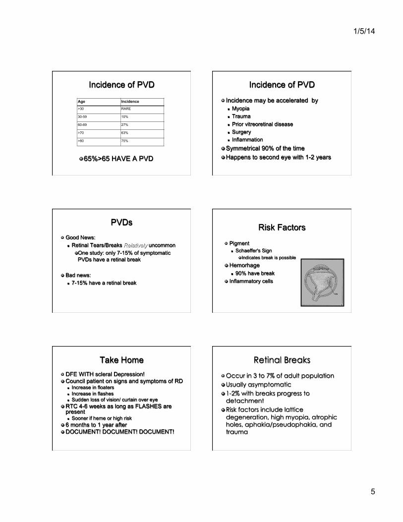
Incidence of PVD
Age Incidence
>30 RARE
30-59 10%
60-69 27%
>70 63%
>80 75%
! 65%>65 HAVE A PVD
Incidence of PVD
! Incidence may be accelerated by n Myopia n Trauma n Prior vitreoretinal disease n Surgery n Inflammation
! Symmetrical 90% of the time ! Happens to second eye with 1-2 years
PVDs
! Good News: n Retinal Tears/Breaks Relatively uncommon
! One study: only 7-15% of symptomatic PVDs have a retinal break
! Bad news: n 7-15% have a retinal break
Risk Factors
! Pigment n Schaeffer's Sign
! Indicates break is possible
! Hemorhage n 90% have break
! Inflammatory cells
Take Home ! DFE WITH scleral Depression! ! Council patient on signs and symptoms of RD
n Increase in floaters n Increase in flashes n Sudden loss of vision/ curtain over eye
! RTC 4-6 weeks as long as FLASHES are present n Sooner if heme or high risk
! 6 months to 1 year after ! DOCUMENT! DOCUMENT! DOCUMENT!
Retinal Breaks
! Occur in 3 to 7% of adult population ! Usually asymptomatic ! 1-2% with breaks progress to
detachment ! Risk factors include lattice
degeneration, high myopia, atrophic holes, aphakia/pseudophakia, and trauma

1/5/14
6
Procedure
! Laser treatment is used to seal the break by creating adhesion between the retinal tissue and underlying RPE
! Provides barrier to continued enlargement from vitreo-retinal traction and prevents accumulation of subretinal fluid
! Adhesion present 24 hours after surgery, and strengthens over several days
Procedure
! Topical or retrobulbar anesthesia ! Entire lesion should be enclosed by at
least 3 rows in a honeycomb pattern
Follow-up
! RTC 1-2 weeks after laser for symptomatic tears
! 3-4 weeks for asymptomatic ! If large or superior, RTC even sooner ! If enlargement or new subretinal fluid,
retreat with 1 week follow-up ! RTC 6-8 weeks after initial follow-up ! Yearly thereafter
Complications
! Few complications n inadequate burn intensity, causing
ineffective adhesion n possible CNVM n intraretinal hemorrhage n vitreous hemorrhage n ERM formation
Basic Guidelines for Treatment
Guidelines for management of retinal breaks and lattice degeneration(from: Weingeist TA. Sneed SR. Laser Surgery in Ophthalmology)
Retinal lesion
Lattice/ atrophic hole flap tear
Symptomatic Asymptomatic Symptomatic Asymptomatic
SRF No SRF Fellow SRF No SRF Phakic SRF or High Fellow Aphakia/ Eye traction myopia eye pseudophakia
treat consider treat consider follow treat follow treat treat treat treat
SRF=Subretinal fluid
RD
! Rule-of-thumb: n For macula off RD, want to get it repaired in
same amount of time it has been off n So if off for 4 days, best to try repair within 4 days!
! Macula on RD is emergency! n Same day referral to retinal specialist n Remind pt NPO until sees specialist in case same-
day surgery

1/5/14
7
Retinal Detachments
! Rhegmatogenous RD occur when liquefied vitreous fluid enters the sub-retinal space through a full-thickness retinal break.
! Occurs in 1/100,000 per yr ! Treatment options include scleral
buckle, pars planar vitrectomy, and pneumatic retinopexy
Scleral Buckle
! Works by altering the geometry and fluid dynamics of the eye causing closure of a retinal tear n Placed so that once the retina is flattened
the breaks will lie upon the area of indentation created by the buckle
! Most scleral buckles consist of solid silicone rubber n Silicone sponges and fascia lata also used
Advantages
! External procedure so avoids complications of intraocular surgery n Minimal cataract progression n Very low rate of endophthalmitis
! One of longest studied procedures ! Appropriate for almost all RDs
n Exception is giant retinal breaks, posterior retinal breaks
! Pos-op positioning may be easier, as tamponade is often not needed
! Success rate > 90%
Disadvantages
! Greater post-operative pain ! Extrusion of buckle ! Induced myopia ! Diplopia ! Increased intraocular
pressure
Pars Plana Vitrectomy ! Allows for direct relief of vitreous traction
associated with retinal breaks ! Good for many detachments that are not
amendable to SB n Giant retinal tears, posterior retinal breaks, breaks with
significant vitreous heme ! Fluid is drained, retina is flattened, and
endolaser photocoagulation is used to create choroidal adhesion
! Intraocular gas bubble or silicone oil can be used as tamponade
Advantages
! Less post-operative pain than SB ! Less induced myopia ! Removes floaters ! Enables small peripheral retinal holes
to be viewed and treated if needed ! Success rate 85-90%

1/5/14
8
Disadvantages
! Increased cataract formation n Preferred in pseudophakic patients
! Increased risk of iatrogenic retinal breaks
! Retinal or optic nerve damage from instruments
! Elevated intraocular pressure ! Risk of endophthalmitis
Pneumatic Retinopexy
! Intraocular gas bubble is used to provide temporary tamponade until retinal adhesion can occur, either by cryopexy or laser
! Indications are fairly limited n Ideal candidate is phakic patient with single superior
break < 1 clock hour
Advantages
! In office procedure n Lower cost
! Minimal post-operative pain with quicker recovery time
! Success rate 75-80%
Disadvantages
! Patient must be in strict head positioning for extended period of time
! Iatrogenic retinal breaks ! Intraocular pressure spikes from gas ! Cataract formation ! Fairly limited indications
Follow-up
! Monitor IOP ! Monitor inflammation ! Monitor for signs/symptoms of
endophthalmitis ! Make sure retina is flat with no new
tears or breaks
Retinal Detachments
! Many factors go into selecting which procedure is best for patient n Phakic/pseudophakic n Location of tear n Size of tear
! Experience of retinal surgeon is essential! n Do your homework!


