

Proof-of-Concept, Randomized, Controlled Clinical Trial of Bacillus-Calmette-Guerin for Treatment of Long-Term Type 1 Diabetes
CitationFaustman, Denise L., Limei Wang, Yoshiaki Okubo, Douglas Burger, Liqin Ban, Guotong Man, Hui Zheng, David Schoenfeld, Richard Pompei, Joseph Avruch, and David M. Nathan. 2012. Proof-of-concept, randomized, controlled clinical trial of bacillus-calmette-guerin for treatment of long-term type 1 diabetes. PLoS ONE 7(8): e41756.
Published Versiondoi:10.1371/journal.pone.0041756
Permanent linkhttp://nrs.harvard.edu/urn-3:HUL.InstRepos:10482572
Terms of UseThis article was downloaded from Harvard University’s DASH repository, and is made available under the terms and conditions applicable to Other Posted Material, as set forth at http://nrs.harvard.edu/urn-3:HUL.InstRepos:dash.current.terms-of-use#LAA
Share Your StoryThe Harvard community has made this article openly available.Please share how this access benefits you. Submit a story .
Accessibility

Proof-of-Concept, Randomized, Controlled Clinical Trialof Bacillus-Calmette-Guerin for Treatment of Long-TermType 1 DiabetesDenise L. Faustman1*, Limei Wang1, Yoshiaki Okubo1, Douglas Burger1, Liqin Ban1, Guotong Man1,
Hui Zheng2, David Schoenfeld2, Richard Pompei3, Joseph Avruch3, David M. Nathan3
1 The Immunobiology Laboratory, Massachusetts General Hospital and Harvard Medical School, Boston, Massachusetts, United States of America, 2 Department of
Biostatistics, Massachusetts General Hospital, Boston, Massachusetts, United States of America, 3 Diabetes Unit, Massachusetts General Hospital, Boston, Massachusetts,
United States of America
Abstract
Background: No targeted immunotherapies reverse type 1 diabetes in humans. However, in a rodent model of type 1diabetes, Bacillus Calmette-Guerin (BCG) reverses disease by restoring insulin secretion. Specifically, it stimulates innateimmunity by inducing the host to produce tumor necrosis factor (TNF), which, in turn, kills disease-causing autoimmunecells and restores pancreatic beta-cell function through regeneration.
Methodology/Principal Findings: Translating these findings to humans, we administered BCG, a generic vaccine, in a proof-of-principle, double-blind, placebo-controlled trial of adults with long-term type 1 diabetes (mean: 15.3 years) at one clinicalcenter in North America. Six subjects were randomly assigned to BCG or placebo and compared to self, healthy pairedcontrols (n = 6) or reference subjects with (n = 57) or without (n = 16) type 1 diabetes, depending upon the outcomemeasure. We monitored weekly blood samples for 20 weeks for insulin-autoreactive T cells, regulatory T cells (Tregs),glutamic acid decarboxylase (GAD) and other autoantibodies, and C-peptide, a marker of insulin secretion. BCG-treatedpatients and one placebo-treated patient who, after enrollment, unexpectedly developed acute Epstein-Barr virus infection,a known TNF inducer, exclusively showed increases in dead insulin-autoreactive T cells and induction of Tregs. C-peptidelevels (pmol/L) significantly rose transiently in two BCG-treated subjects (means: 3.49 pmol/L [95% CI 2.95–3.8], 2.57 [95% CI1.65–3.49]) and the EBV-infected subject (3.16 [95% CI 2.54–3.69]) vs.1.65 [95% CI 1.55–3.2] in reference diabetic subjects.BCG-treated subjects each had more than 50% of their C-peptide values above the 95th percentile of the reference subjects.The EBV-infected subject had 18% of C-peptide values above this level.
Conclusions/Significance: We conclude that BCG treatment or EBV infection transiently modified the autoimmunity thatunderlies type 1 diabetes by stimulating the host innate immune response. This suggests that BCG or other stimulators ofhost innate immunity may have value in the treatment of long-term diabetes.
Trial Registration: ClinicalTrials.gov NCT00607230
Citation: Faustman DL, Wang L, Okubo Y, Burger D, Ban L, et al. (2012) Proof-of-Concept, Randomized, Controlled Clinical Trial of Bacillus-Calmette-Guerin forTreatment of Long-Term Type 1 Diabetes. PLoS ONE 7(8): e41756. doi:10.1371/journal.pone.0041756
Editor: T. Mark Doherty, Statens Serum Institute, Denmark
Received May 3, 2012; Accepted June 25, 2012; Published August 8, 2012
Copyright: � 2012 Faustman et al. This is an open-access article distributed under the terms of the Creative Commons Attribution License, which permitsunrestricted use, distribution, and reproduction in any medium, provided the original author and source are credited.
Funding: The Iacocca Foundation and philanthropic dollars supported this study. The authors also reserve gratitude to the James B Pendleton Charitable Trust.Finally, the authors extend their appreciation to the Friends United for Juvenile Diabetes Research and Partnership for Cures. DMN was supported in part by theCharlton Fund for Innovative Diabetes Research. NIH support included #P30DK057521 to DLF. No drug company or for-profit resources supported this trial. Thefunders had no role in study design, data collection and analysis, decision to publish, or preparation of the manuscript. This study was funded by philanthropicgrants only.
Competing Interests: The authors have declared that no competing interests exist.
* E-mail: [email protected]
Introduction
Along-standinggoalof immunologyis todeveloptargeted immune
therapies that eliminate the predominant cause of type 1 diabetes: the
autoimmune T lymphocytes (T cells) that destroy the insulin-
secreting cells of the pancreas. Current immune treatments for type
1 diabetes, such as immunosuppressants and anti-cytokines, are non-
specific, killing or harming both the pathological T cells (i.e., insulin-
autoreactive cytotoxic T cells) and healthy cells.
Two decades of autoimmune disease research in animal
models, including the non-obese diabetic (NOD) mouse model
of type 1 diabetes, have uncovered overlapping genetic and
functional mechanisms of disease and led to the identification of
the cytokine tumor necrosis factor (TNF) as a potential novel
immunotherapy [1–7]. In the case of type 1 diabetes, the
rationale for administering TNF is that insulin-autoreactive T
cells bear several intracellular signaling defects that make them
selectively vulnerable to death upon exposure to TNF [4–7].
TNF destroys insulin-autoreactive T cells, but not healthy T
PLoS ONE | www.plosone.org 1 August 2012 | Volume 7 | Issue 8 | e41756

cells, in in vitro studies of human diabetic blood samples and in
the NOD mouse model. TNF exposure may also augment
production of beneficial regulatory T cells (Tregs), a subset of T
cells believed to suppress insulin-autoreactive T cells. Interven-
tions that have destroyed insulin-autoreactive T cells and
boosted beneficial types of T cells have led to regeneration of
insulin-producing islet cells in the pancreas of rodents with
autoimmune diabetes, resulting in restoration of normoglycemia,
even in advanced disease [7,8].
TNF treatment at high doses in humans is limited by its
systemic toxicity. An alternative approach is to test a safe, U.S.
Food and Drug Administration (FDA)-approved vaccine contain-
ing Mycobacterium bovis bacillus- Calmette-Guerin (BCG), which has
been known for over 20 years to induce TNF [9]. This avirulent
strain of Mycobacterium is different from that which causes
tuberculosis in humans (Mycobacterium tuberculosis).
The release of TNF after exposure to pathogens, such as BCG,
is an example of a first-line host defense commonly called the
innate immune response [9]. Similar results to those with TNF
administration have been achieved with BCG or its non-FDA
approved variant, complete Freund’s adjuvant (CFA), in rodent
models of autoimmune diabetes [7,8,10–12].
Nearly two decades ago, a single, low dose of BCG in humans
with late-stage pre-diabetes was initially found to successfully
induce a clinical remission in some patients [13], but when efficacy
was re-evaluated in expanded trials, it could not be observed a
year after vaccination. At the time, the mechanisms behind BCG’s
failure were not understood and specific biomarkers or knowledge
of TNF action and autoimmunity were unavailable. In recent
years, however, the mechanism of action underlying the thera-
peutic potential of BCG and TNF in autoimmune disease has been
further elucidated [1], supporting the hypothesis based on animal
data that BCG vaccination may be beneficial in type 1 diabetes,
especially if the mechanism of action of BCG trigger TNF can be
closely followed with sophisticated and early biomarkers of safety.
We conducted a proof-of-principle, double-blind, placebo-
controlled trial, in which we administered two low-dose BCG
vaccinations to patients with long-term type 1 diabetes. Here, we
report on the safety of two low-dose BCG vaccinations and their
effects on four serially studied biomarkers in long-term type 1
diabetes.
Frequent blood sampling for up to 5 months was conducted to
measure biomarkers of immune and pancreatic function, includ-
ing: (1) levels and viability of cytotoxic autoreactive T cells against
insulin, a known autoantigen in diabetes; (2) induction of
protective Tregs; (3) antibodies against the autoantigen glutamic
acid decarboxylase (GAD); and (4) levels of fasting C-peptide, a
marker of endogenous insulin production.
Methods
The protocol for this trial and supporting CONSORT checklist
are available as supporting information: see Checklist S1 and
Protocol S1.
Clinical Trial ParticipantsAll clinical trial participants were required to be adults, ages 18
to 50 years, with long-term diabetes treated continuously with
insulin from the time of diagnosis; have no demonstrable insulin
secretion (fasting and glucagon-stimulated C-peptide less than
0.2 pmol/L) as assessed by a standard C-peptide assay by an
outside vendor; be pancreatic GAD autoantibody positive; have a
normal complete blood count (CBC); and have a negative purified
protein derivative (PPD) test. Diabetic patients were excluded if
they were pregnant or not using acceptable birth control; had a
chronic infectious disease, including human immunodeficiency
virus (HIV); had a history of tuberculosis (TB) or current TB
infection; were currently receiving treatment with glucocorticoids,
chronic immunosuppressive medications or high dose aspirin
(.160 mg/day); or were currently living with an immunosup-
pressed individual. Also excluded were type 1 diabetics with keloid
formation or hemoglobin A1C (HbA1C) values greater than 8%.
Non-diabetic Matched ControlsHealthy, non-diabetic control subjects were included if they
were 18 to 45 years of age, with no history of autoimmune disease
or diabetes, no history of HIV, and no history of autoimmunity in
first-degree family members. These participants were paired
weekly/bi-weekly to the diabetic patients who were randomized
to BCG or placebo.
Reference Groups and SubjectsThe study also included several reference groups: a reference
group of type 1 diabetic individuals serially monitored for at least
20 weeks (n = 57) and a one-time serial studied reference group of
type 1 diabetics (n = 17) studied for one outcome measure (insulin-
autoreactive T cells) and matched in disease duration and age to
the diabetic clinical trial subjects. The clinical trial subjects were
compared to one or more of these groups, depending on the
outcome measure as shown in Figure 1. The criteria for inclusion
and exclusion of diabetic reference subjects were the same as those
for the clinical trial subjects as related to age of onset, duration of
diabetes and HbA1C values. The reference subjects studied for
insulin-autoreactive T cells were also matched for human
leukocyte antigen (HLA)-A2 status. The serial study of these
reference subjects was performed to expand the database of
autoreactive T cell variation and serially studied C-peptide values
in single subjects, i.e., these separate and sequential blood draws
defined the biological variation in assays in single cohorts and
distinguished this biological variation from variation possibly
attributable to BCG treatment in the randomized clinical trial
subjects also studied in a serial fashion.
EthicsThis study was approved by the Human Studies Committee at
Massachusetts General Hospital, Boston, MA and by the FDA. All
patients provided written informed consent.
Trial DesignThis was a proof-of-principle, double-blinded, placebo-con-
trolled clinical trial that also included a paired healthy control
population and reference subjects. All interventions were admin-
istered and clinical trial participants seen at one clinical center in
North America (Massachusetts General Hospital, Boston, MA,
USA) between 2009 and 2011. The FDA approved this protocol in
2007 and when funding was secured, the enrollment was launched
in 2009.
Intervention Population and Paired Healthy ControlsFor the double-blind, placebo-controlled portion of the study,
diabetic subjects were randomly assigned to BCG or placebo
(saline) vaccinations according to the randomization scheme
prepared by the Massachusetts General Hospital (MGH) research
pharmacy. The BCG injection was prepared by the research
pharmacy from lyophilized BCG (TheraCysH, Sanofi-Pasteur,
Toronto, Ontario, Canada), and all syringes (BCG and saline)
were prefilled by the pharmacy. Randomized patients received
BCG Transiently Reverses Long-Term Diabetes
PLoS ONE | www.plosone.org 2 August 2012 | Volume 7 | Issue 8 | e41756

Figure 1. CONSORT flow chart (A) and flow diagraph (B) with depicts of treatment concept, outcomes and subject comparisongroups for the study.doi:10.1371/journal.pone.0041756.g001
BCG Transiently Reverses Long-Term Diabetes
PLoS ONE | www.plosone.org 3 August 2012 | Volume 7 | Issue 8 | e41756

two 0.1 ml intradermal injections into the deltoid area containing
either low-dose BCG (1.6–3.26106 colony-forming units/injec-
tion) or saline placebo, administered four weeks apart (Week 0 and
Week 4). Weekly blood sampling was performed until Week 8,
followed by bi-weekly blood sampling until Week 12 and then a
final visit at Week 20. This frequent blood monitoring was
performed to validate outcomes and observe any early effects of
therapy. All subjects were seen in the morning and were required
to be fasting and normoglycemic prior to having their blood
drawn.
All injections were administered in the MGH diabetes clinic.
Staff who administered BCG or placebo injections were not the
same as those who examined the participants to grade any
reactions at the injection site. All blood was processed within two
hours of being drawn. All blood samples were blinded and
simultaneously sent to the laboratory for monitoring of T cell
response and for storage of serum for pancreas response tests
(ultrasensitive C-peptide assay and autoantibodies), which were
performed by outside vendors at the completion of the trial as
described in ‘‘Assay methods’’.
A group of paired healthy control participants, receiving neither
BCG nor placebo, had blood samples obtained at the same time as
diabetic subjects. Their samples were analyzed immediately for T
cells in a masked fashion on the same day as the samples from
diabetic subjects.
Masking and UnblindingThe MGH research pharmacy performed all masking of BCG
and saline vaccinations. All blood samples that were collected were
randomly coded prior to blinded submission to the MGH lab or
outside vendor lab for processing. Unblinding did not occur until
all samples were processed and all data were downloaded into the
central computers.
Primary Outcome MeasuresWe monitored the safety of BCG in advanced type 1 diabetes
and its action on immune and pancreas outcomes, including levels
of insulin-autoreactive T cells, Treg cells, autoantibodies (includ-
ing GAD), and C-peptide, an indicator of endogenous insulin
secretion.
T Cell Assay MethodsThe two cell-based assays (Treg cells and autoreactive T cells)
were performed through Week 12.Cell isolation. CD4 and CD8 T cells were isolated from
fresh human blood within 2 hours of venipuncture using
InvitrogenTM DynalH CD4 positive isolation kit and DynalHCD8 positive isolation kit (Life Technologies Corporation,
Carlsbad, CA, USA). This method is unique in yielding cells both
free of magnetic particles and free of an attached positive selection
antibody to either the CD4 protein or the CD8 protein. The blood
was drawn into BD VacutainerH tubes (BD, Franklin Lakes, NJ,
USA) containing acid citrate and dextrose or ethylenediaminetet-
raacetic acid (EDTA). The CD8 or CD4 cells extracted for these
studies were selected from fresh blood and were required, for
standardization purposes, to be greater than 98% pure, 95%
viable, and 85% yield for the validated T cell assays [4,14]
described below.
Use of controls. For all T cell assays in this study, a diabetic
blood sample was always drawn at the same time as blood from a
paired healthy control to allow assay standardization.
Detection of autoreactive CD8 T cells to a fragment of
insulin. Insulin-autoreactive T cells were assayed by flow
cytometry after fresh blood cell separations [14] to obtain high-
yield and highly pure and viable CD8 T cells for tetramer staining.
Tetramers are T cell detection reagents composed of the binding
region of specific HLA class I proteins with loaded peptides in the
exterior binding grooves. The tetramers, which are made
fluorescent, bind to specific T cells with specific reactivity to the
presented peptide fragment, thereby allowing for cell identifica-
tion. To detect autoreactive T cells to insulin, we used tetramers to
HLA-A2 *0210 insulin beta 10–18 with a fragment of HLVEA-
LYLV (Beckman Coulter #T02001) [15]. To further confirm the
specificity of insulin-autoreactive T cell detection, cell samples
were examined simultaneously with T cell reagents to detect
oncogene-specific human epidermal growth factor receptor-2
(HER-2) or Epstein-Barr virus (EBV)-specific T cells of acute
infection. For simultaneously studied healthy controls, the
following tetramer reagents were used: HLA*0201 Her-2/neu
with a sequence to KIFGSLAFL (Beckman Coulter #T02001), a
breast cancer peptide; HLA*0201 null without a non-specific
peptide fragment (Beckman Coulter #T01044); or an EBV
tetramer reagent HLA-A*0201 EBV with sequence of
GLCTLVAML (Beckman Coulter #T01010).
Tetramer reagent staining was conducted on the highly pure
CD8 T cells after 12 hours of culture at 26uC followed by 6 hours
at 37uC and/or 1 hour rest at 26uC followed by 12 hours at 37uC.
Cells were then stained with phycoerythrin-labeled class I
tetramers (Beckman Coulter, Fullerton, CA) and SYTOX green
dye (MBL International, Woburn, MA) and/or CD8 antibodies
(BD Biosciences, San Jose, CA). All CD8 T cells were stained at
4uC in the dark for 30 minutes and then washed twice in Hanks
balanced salt solution with 2% heat inactivated bovine serum. On
average, 100,000 highly pure CD8 T cells were analyzed to ensure
optimized data points on the Becton Dickinson FACSCalibur
using the Cell Quest acquisition program and allow the detection
of rare autoreactive T cells. All cells were analyzed while fresh to
prevent fixation artifacts and enable quantification of dead versus
viable cells. Prior to tetramer staining, cells were neither frozen nor
expanded. Calculations of insulin-positive T cells were reported as
the percentage of insulin-autoreactive T cells to the total numbers
of isolated pure CD8 T lymphocytes.
Note that all diabetic treated patients in the randomized portion
of the study were HLA-A2+ except for diabetic #iv. Although
diabetic #iv was HLA-A2 negative, the formal binding site for the
HLA-A2 insulin-autoreactive T cell reagent was HLA-A6802.
HLA-A6802 is a subtype of the HLA-A2 family and has an
identical binding cleft to HLA-A2 and other common subtypes
within the HLA-A2 family. Therefore, if diabetic subject #iv were
to have detectable insulin-autoreactive T cells, those cells would
stain positive for the insulin-autoreactive T cell reagent. Three
healthy controls in this study (Control #ii, Control #iii and
Control #v) were also HLA-A2+.
Reference diabetics were monitored over a three-year period for
the presence or absence of insulin-autoreactive T cells and
compared to their paired healthy reference controls.
Detection of Treg CD4+ cells. Treg cells were assayed by
flow cytometry after fresh blood cell separations as described
above and by Burger et al [14]. Two different methods of cell
detection were employed. Treg cells were detected as either CD4,
CD25bright with Foxp3 staining, or with CD4, CD25bright and
CD127lowantibody staining. Intracellular staining of Foxp3 was
performed with Human Treg FlowTM Kit (Biolegend, San Diego,
CA, USA), according to the manufacturer’s instructions. Isolated
CD4 positive cells were incubated briefly with CD4-PE-Cy5 (clone
RPA-T4) and CD25-PE (clone BC96) antibodies for 20 minutes at
room temperature. After washing, cells were fixed with Foxp3 Fix/
Perm solution (Biolegend) for 20 minutes at room temperature.
BCG Transiently Reverses Long-Term Diabetes
PLoS ONE | www.plosone.org 4 August 2012 | Volume 7 | Issue 8 | e41756

Cells were washed again and permeabilized with Foxp3 Perm
Buffer (Biolegend) for 15 minutes at room temperature. Cells were
then stained with Foxp3 Alexa FluorH 488 antibody (clone 259D,
Biolegend) for 30 minutes. Isotype controls were done for each
sample prior to flow cytometric analysis. For detection of Treg
cells, staining was performed with a CD4 antibody (clone RPA-
T4, BD Biosciences, San Jose, CA, USA), a CD25 antibody (clone
4E3, Miltenyi Biotech, Auburn, CA, USA) and an anti-human
CD127 antibody (clone hIL-7R-M21, BD Biosciences).
Flow cytometry for T cell assays. For the flow cytometry
studies, the flow gates were set ‘‘open’’ for inclusion of CD8 or
CD4 T cells of all sizes, but exclusion of the following: cell debris,
red blood cells, fragmented cells, and apoptotic bodies. The ‘‘open
gate’’ was chosen for the purified CD8 or CD4 T cells because T
cells undergoing cell death, especially by apoptosis, can display
changes in light scattering properties. The goal was to ensure
accuracy by analyzing high numbers of cells per sample and to
capture dying cells of all shapes. Cell viability was quantified by
either of two stains that fluorescently labeled dead cells, i.e., Sytox
(MBL international Co., Woburn, MA, USA) or propidium iodine
(PI). Purified CD8 cells form distinct scatter pictures on forward
versus side scatter highlighted the shrunken size of dead versus
viable cells.
With open gating and inclusion of all purified CD8 T cells in
each sample, some reference diabetics consistently displayed
insulin-autoreactive T cells. In contrast, some reference diabetics
consistently had undetectable insulin-autoreactive T cells com-
pared to healthy reference controls, which were simultaneously
studied at each monitoring time. The data were collected over the
multi-year time span. The signal for insulin-autoreactive T cells
was in the range of 0.06–0.09%. The healthy control background
signal is in the range of 0.04–0.05% [15]. The reverse was also
true: diabetics who initially lacked insulin-autoreactive T cells, on
repeat sampling, continued to lack those cells.
Serum Assay Methods for Pancreas MonitoringGAD autoantibody and fasting C-peptide levels were assayed by
radio-binding and ELISA assays in diabetic subjects to assess
whether the subjects had a pancreas response to the BCG
injection. For these serum assays, fresh human blood was collected
by venipuncture into red top tubes and allowed to clot. The serum
was then separated by centrifugation within 2 hours of venipunc-
ture. Serum was stored at 280uC until analysis. The C-peptide
assay was undertaken through week 20.
Detection of C-peptide secretion. Measurement of con-
necting peptide (C-peptide) co-secreted with insulin permits direct
estimation of any remaining insulin from the pancreas in contrast
with endogenous sources. The first, performed by the Mayo Clinic
(Rochester, MN, USA) utilizing the Roche Cobas C-peptide assay
(Roche Diagnostics, Indianapolis, IN, USA) for clinically detect-
able C-peptide, was used for eligibility and had a lower limit of
detection of 330–470 pmol/L. This insensitive but standard assay
was applied to fasting and glucagon-stimulated blood samples.
After screening negative for enrollment purposes, subjects’ serum
was stored and freezer-banked. For subsequent samples (baseline
through Week 20), the saved serum was sent to Sweden for
analysis of serial C-peptide levels by an ultrasensitive C-peptide
assay with a lower level of detection of 1.5 pmol/L and an assay
range up to 285 pmol/L (Mercodia AB, Uppsala, Sweden). For C-
peptide values of 1.5–37 pmol/L, the within-assay coefficient of
variation was 3.8%; for values of 38–60 pmol/L, it was 2.6%; and
for values of 143–285 pmol/L, it was 2.5%. The Mercodia
Ultrasensitive C-peptide ELISA kit, which is an FDA-listed
reagent and has a filed document registration, has been evaluated
for accuracy and is classified in the United States as a class one
device for ultrasensitive detection of C-peptide levels. This assay is
calibrated against the International Reference Reagent for C-
peptide, IRR C-peptide 84/510. All statistics on C-peptide levels
were performed using the lower level of detection of the assay, i.e.,
1.5 pmol/L.
Detection of GAD autoantibodies. GAD autoantibodies
provide evidence of diabetic autoimmunity since GAD proteins
are intracellular proteins specific to insulin secreting cells and are
released from T cell mediated beta cell destruction. The release of
intracellular GAD results in the immune response of autoanti-
bodies. Enrolled patients were required to be GAD autoantibody
positive. Prior to enrollment, a single serum sample for GAD
autoantibody was sent either to the Joslin Clinic in Boston, MA,
USA (Subject #vi, Subject #i, Subject #ii, Subject #iv) or to
Quest Diagnostics (Cambridge, MA, USA) (Subject #iii, Subject
#iv). After the first BCG or placebo injection, serum samples
collected from baseline to Week 20 were sent to Germany for
diabetic autoantibody panels [16] at the laboratories of Drs. Ezio
Bonifacio and Peter Achenbach of the Diabetes Research Institute
in Munich, Germany. The autoantibodies studied were GAD, IA-
2A (islet-specific protein tyrosine phosphatase), and ZnT8Carg-A
(pancreatic beta cell-specific zinc transporter) [17]. The GAD
assay sensitivity is 86%, specificity is 100%, and inter-assay
variation is 18%. For the IA-2 autoantibody assay, the sensitivity is
72%, the specificity is 100%, and the inter-assay variation is 16%.
For the ZnT8Carg-A assay, the sensitivity is 72%, the specificity is
99%, and the inter-assay variation is 17%.
Sample SizeSample size for the randomized population was determined in
conjunction with the FDA and with the intense use of serial
biomarker studies as outlined by the Institute of Medicine
guidelines for clinical trials [18]. A sample size of 6 randomized
patients was determined as appropriate for the intense serial blood
monitoring required in this proof-of-concept trial for the placebo
or BCG interventional limbs and an expanded population of
diabetics and non-diabetic controls for assay validation that is
referred to as reference subjects.
Statistical AnalysisRandomized participants were compared to self, healthy paired
controls, or reference subjects with or without type 1 diabetes,
depending on the outcome measure, according to the schema
depicted in Figure 1. None of the analyses compared the BCG-
treated to placebo-treated clinical trial subjects.
For each randomized patient, a linear regression model with
auto-correlated errors was used for statistical comparisons between
baseline and post-treatment periods in autoantibody levels over
the course of the study. This was the appropriate test for this
comparison because any change in autoantibodies should be
sustained over the monitoring period of this trial, i.e., the t 1/2 of B
cells that produce antibodies exceeds 60 days. P-values compared
the values of each person to their post-baseline values by two-sided
test based on a regression model with auto-correlated errors. For
C-peptide assays, a cut-off value of 1.5 pmol/L was used since this
value is the lower limit of detection of the ultra-sensitive assay used
in this study. C-peptide assays were performed by the outside
vendors in duplicate; figures are therefore presented as the means
+/2 the SE. For the comparison of EBV-infected or BCG-
injected patients to the long-term diabetic reference samples, the
Kolmogorov-Smirnov two-sample test was used to compare the
distribution of each patient with the reference samples. We applied
this method in a conservative fashion by overestimating the
BCG Transiently Reverses Long-Term Diabetes
PLoS ONE | www.plosone.org 5 August 2012 | Volume 7 | Issue 8 | e41756
variability of the clinical trial sample, as a more exact comparison
is difficult to obtain due to the low sampling frequency and small
numbers of measurements per patient in the reference group. P-
values of ,0.05 were considered statistically significant. SASHversion 9.2 was used for the statistical analysis.
For serum samples sent out to commercial sources for assay
performance, both published inter-assay and intra-assay variability
was considered for the statistical analysis of the clinical trial
samples. We also verified that the inter-assay variability was
consistent in the plate for the clinical samples by comparing the
pre-treatment values with all post-treatment values of the same
patient to self in the same plate. This self-comparison analysis was
performed for serum assays such as C-peptide or autoantibodies.
The area under the curve (AUC) was calculated for all treatment
and control groups, although the control group varied according
to the assay.
Results
Participant Enrollment and CharacteristicsA total of 85 participants were studied: 63 type 1 diabetics and
22 non-diabetic controls (Fig. 1, Fig. 2.). In the double-blinded,
placebo-controlled portion of the study, a total of six diabetic
subjects were randomly assigned to BCG or placebo vaccinations.
The randomized clinical trial subjects had disease for a mean
duration of 15.3 years (range 7–23 years) and mean age of 35 years
(range 26–47) (Fig. 2), and were paired to healthy controls (n = 6)
at each weekly blood drawing time for greater than 20 weeks of
study. In addition to these participants, 57 additional reference
subjects with long-term diabetes and 16 reference healthy subjects
served as reference subjects for both serial T cell assays and serum
sample comparisons. Diabetic reference patients had disease for a
mean duration 20 years (range 8–53 years) and mean age of 39
years (range 21–65) (Fig. 2). The intense serial monitoring of blood
samples of all clinical trial subjects resulted in a total of 1,012
blood samples from diabetic or comparison subjects to quantify
both T cell and pancreas changes. This serial study of biomarkers
and comparison groups for the subjects are depicted in Fig. 1. This
intensive study of novel T cell and pancreas biomarkers required
different comparison groups (Fig. 1) due to the lack of serial
normative data on the four parameters chosen to study BCG
efficacy in advanced type 1 diabetes. The objective of the trial was
to test safety of multi-dosing BCG in long-term diabetics. Four
monitored endpoints of efficacy were studied as markers of disease
activity: death of insulin autoreactive T cells, induction of Treg
cells, changes in autoantibodies and the restoration of endogenous
insulin secretion through C-peptide levels.
Epstein-Barr Virus (EBV) InfectionAt screening for clinical trial enrollment, and unbeknownst to
us, one diabetic clinical trial subject had an acute undiagnosed
case of EBV infection. This patient presented with cold/flu
symptoms at weeks 3–4 after the placebo injection (Fig. 3). The
presence of the new EBV infection in blood samples was detected
during our blinded laboratory protocols that required analysis of
EBV-reactive T cells (EBV-tetramer positive CD8 T cells) as a
control during the CD8 insulin-autoreactive T cell assays. Further
confirmation of this diagnosis of acute EBV infection was obtained
at the end of the trial with serology sent for commercial antibody
testing (Quest Diagnostics, Cambridge, MA, USA) (Fig. 3).
This placebo-treated EBV subject completed the five-month
trial and was subjected to the same types of statistical analyses and
outcome studies as other clinical trial subjects. The treatment team
and subject remained masked to treatment assignment.
The course of the EBV infection was reconstructed from serially
studied fresh T cell samples and by standard clinical laboratory
tests on stored serum samples (Fig. 3). To understand the precise
time course of the EBV infection, this diabetic’s serum was
screened for EBV VCA antibody (IgM), an antibody that is
typically positive days after infection onset to 3–6 weeks post-
infection. The serum was also tested for EBV Early Antigen D Ab,
an antibody that is typically positive only in the infection window
running from 1 month after infection to 2 months post-infection
(Fig. 3).
This placebo-treated diabetic subject was early antigen D
antibody-positive at the first baseline sample at week 0, had CD8
lymphocytosis over 12 weeks of study (Fig. 3) and demonstrated
mildly elevated liver enzyme levels early in the trial course, all
consistent with an acute EBV infection. As the EBV serologic
studies show, Subject #vi had an acute infection that lasted longer
than one month but did not exceed two months in duration. The
EBV tetramer positive cells became vividly positive at week 6 in
the T cell assay and were still vividly positive at week 8, although
declining slightly (Fig. 3). As a comparison, we include the EBV
positive data from a long-term diabetic that was not part of this
clinical trial, but who had a very distant past EBV infection, to
show the low numbers of EBV memory cells seen using the EBV
tetramer methods when infection is not acute (Fig. S1).
All other clinical trial subjects in this study were negative for
both acute and past EBV infections throughout the duration of T
cell monitoring during the trial (Fig. S1). EBV infections, like
BCG, trigger innate immunity by inducing secretion of host TNF
[9]. The patient’s EBV status and receipt of placebo saline
injections fortuitously enabled us to compare the serial T cell and
pancreas effects of EBV- and BCG-triggered innate immune
responses in the same study [9,19]. All other clinical trial subjects
in this study were negative for both acute and past EBV infections
through T cell monitoring during the trial (Fig. S1).
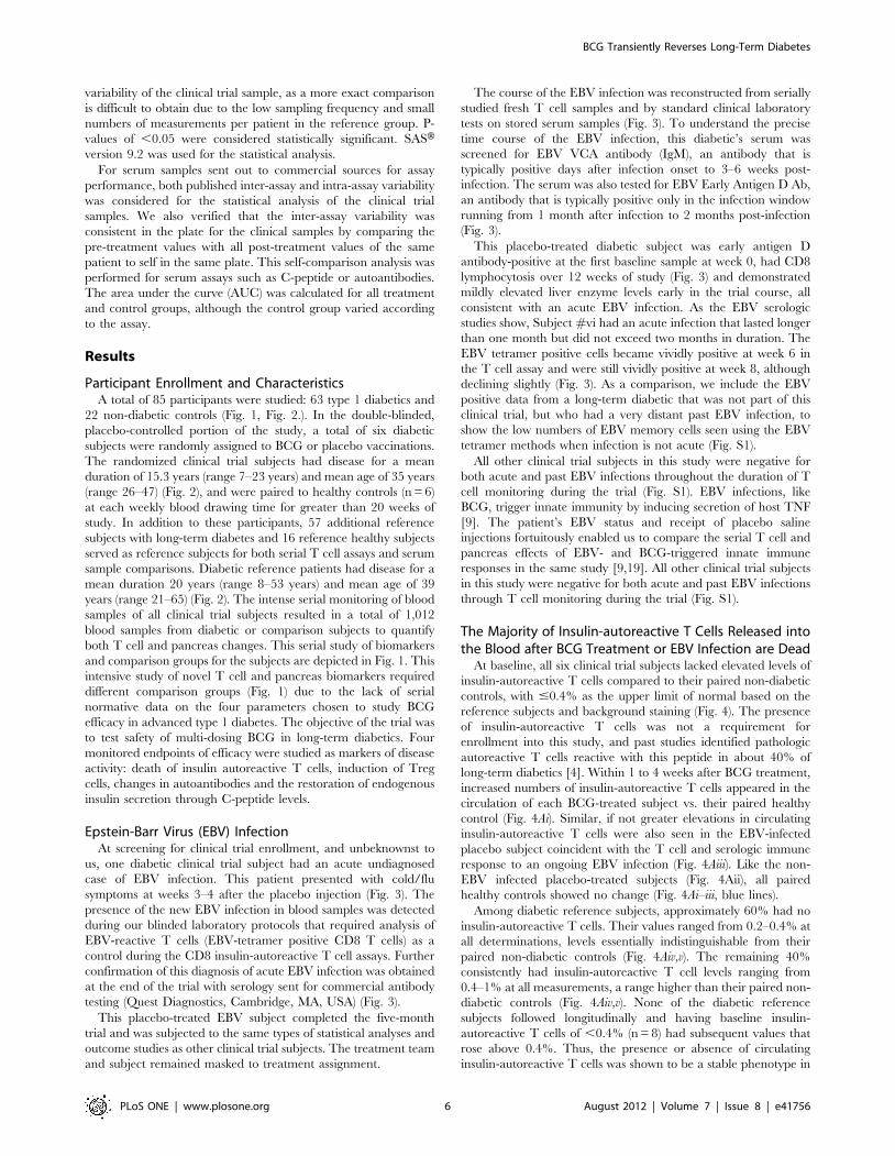
The Majority of Insulin-autoreactive T Cells Released intothe Blood after BCG Treatment or EBV Infection are Dead
At baseline, all six clinical trial subjects lacked elevated levels of
insulin-autoreactive T cells compared to their paired non-diabetic
controls, with #0.4% as the upper limit of normal based on the
reference subjects and background staining (Fig. 4). The presence
of insulin-autoreactive T cells was not a requirement for
enrollment into this study, and past studies identified pathologic
autoreactive T cells reactive with this peptide in about 40% of
long-term diabetics [4]. Within 1 to 4 weeks after BCG treatment,
increased numbers of insulin-autoreactive T cells appeared in the
circulation of each BCG-treated subject vs. their paired healthy
control (Fig. 4Ai). Similar, if not greater elevations in circulating
insulin-autoreactive T cells were also seen in the EBV-infected
placebo subject coincident with the T cell and serologic immune
response to an ongoing EBV infection (Fig. 4Aiii). Like the non-
EBV infected placebo-treated subjects (Fig. 4Aii), all paired
healthy controls showed no change (Fig. 4Ai–iii, blue lines).
Among diabetic reference subjects, approximately 60% had no
insulin-autoreactive T cells. Their values ranged from 0.2–0.4% at
all determinations, levels essentially indistinguishable from their
paired non-diabetic controls (Fig. 4Aiv,v). The remaining 40%
consistently had insulin-autoreactive T cell levels ranging from
0.4–1% at all measurements, a range higher than their paired non-
diabetic controls (Fig. 4Aiv,v). None of the diabetic reference
subjects followed longitudinally and having baseline insulin-
autoreactive T cells of ,0.4% (n = 8) had subsequent values that
rose above 0.4%. Thus, the presence or absence of circulating
insulin-autoreactive T cells was shown to be a stable phenotype in
BCG Transiently Reverses Long-Term Diabetes
PLoS ONE | www.plosone.org 6 August 2012 | Volume 7 | Issue 8 | e41756

Figure 2. Clinical characteristics of groups of clinical trial subjects and reference subjects.doi:10.1371/journal.pone.0041756.g002
Figure 3. Clinical laboratory studies reveal acute EBV infection in placebo-treated diabetic. (A) Weekly course of EBV infection fromserum of diabetic subject #vi (B) Positive Early Antigen D antibody versus negative values. (C) CD8 T-cell proliferative response. (D) Flow scatter plotsof appearance of EBV-reactive T-cells vs. paired control, week 6 to 8. All newly appearing EBV-reactive T-cells were viable.doi:10.1371/journal.pone.0041756.g003
BCG Transiently Reverses Long-Term Diabetes
PLoS ONE | www.plosone.org 7 August 2012 | Volume 7 | Issue 8 | e41756
serially studied and untreated type 1 diabetic subjects with these
monitoring methods.
The insulin-autoreactive T cells appearing in the circulation
after BCG or EBV infection were more likely dead than alive
compared to paired healthy controls (Fig. 4, Fig. S2, Fig. 5),
probably indicating not only the rapid release of pre-formed
insulin-autoreactive T cells after BCG treatment or EBV infection
but also their redundant death by TNF induction. Also unlike the
low affinity insulin-autoreactive T cells observed with routine
monitoring of diabetics, the TNF-targeted death of pathogenic
cells allowed the identification of both low affinity as well as newly
appearing, high affinity subsets of autoreactive T cells not
previously identified in the circulation (Fig. 5). For the three
BCG-treated subjects, the AUC representing the cumulative
concentrations of insulin-autoreactive T cells over the course of
study were 2.22, 0.71 and 1.03 compared to their paired healthy
control. The two non-EBV infected placebo-treated subjects’
AUCs were 0.57 and 0.07, while the EBV-infected subject had a
strikingly elevated AUC of 5.69, reflecting the large numbers of
dead insulin-autoreactive T cells being released into the circulation
after the EBV infection. The transient increases in the number of
insulin-autoreactive cells seen in the BCG-treated or EBV-infected
clinical trial subjects (Fig. 4Ai, iii) formed a pattern distinctly
different than the stable levels observed in the two other placebo-
treated subjects (Fig. 4Aii) and in reference diabetic subjects
(Fig. 4Aiv,v). Cytometric study of dead and living insulin-
autoreactive T cells revealed that the pathogenic T cells captured
in the blood had both the common low affinity insulin-
autoreactive cells as well as the treatment-specific release of high
affinity autoreactive T cells for the insulin peptide fragments
(Fig. 5). Routine monitoring of diabetics for insulin autoreactive T
cells by diverse studies only reveals low affinity insulin-autoreactive
T cells in diabetes subjects without treatment [4]. The TNF-
induced death in vivo of insulin-autoreactive T cells with BCG
vaccinations or acute EBV infection was confined to the
autoreactive T cells.
Regulatory T Cells are Induced by BCG and EBVThe EBV-infected subject and two BCG-treated subjects
appeared to exhibit increases in the numbers of Treg cells
compared to their paired healthy controls studied simultaneously
(Fig. 6Aii, iii, vi); the other two placebo-control subjects had stable
levels (Fig. 6Aiv, v). A similar trend for elevations in Tregs in
response to BCG or EBV was observed by measuring the AUC, a
measure of the total accumulation of Treg ratios. The three BCG-
treated subjects had cumulative Treg ratios of patients compared
to controls of 0.12, 0.42 and 0.30 compared to placebo treated
subject accumulations of 0.11 and 0.03. The EBV infected subject
had cumulative Tregs of 0.32.
GAD Autoantibody Levels Show Sustained Change afterBCG Treatment
At baseline, GAD autoantibodies, ranging from 60 to 650 units,
were present in all diabetic clinical trial subjects except one BCG-
treated subject (Fig. 6B). There was a statistically significant and
sustained change in GAD autoantibody levels in two of the three
BCG-treated subjects after injections, with one diabetic showing a
decrease and the other an increase relative to self-baseline
(p = 0.0001 and p = 0.0017, respectively (Fig. 6Bii,iii). In contrast,
none of the other diabetic subjects showed any variations from
their baseline values of GAD (Fig. 6Biv,v,vi). The other islet-specific
autoantibodies studied, tyrosine phosphatase IA-2A and beta cell-
specific zinc transporter (ZnT8A), were present in some of the
diabetic subjects at baseline (Fig. 7); only ZnT8A had statistically
significant decreases in one BCG treatment subject. A similar
trend for higher or lower acute elevations in GAD in response to
BCG was observed by measuring the AUC, a measure of the total
positive or negative accumulations of GAD autoantibody levels
Figure 4. Insulin-autoreactive T-cells released into the circulation are dead after BCG treatment or EBV infection. (A) Percentage ofinsulin-autoreactive T-cells of total CD8 T-cells over 12 weeks for BCG-treated (Ai, Bi), placebo-treated, (Aii, Bii) and EBV-infected clinical trial subjects(Aiii, Biii). Reference diabetics without or with insulin-autoreactive T-cells vs. reference healthy controls (Aiv,v). (B) Insulin-autoreactive T-cellsstratified by viability in clinical trial subjects. Red diamonds are long-term diabetics; blue diamonds paired healthy controls. Arrows are BCG orplacebo injection times.doi:10.1371/journal.pone.0041756.g004
BCG Transiently Reverses Long-Term Diabetes
PLoS ONE | www.plosone.org 8 August 2012 | Volume 7 | Issue 8 | e41756

BCG Transiently Reverses Long-Term Diabetes
PLoS ONE | www.plosone.org 9 August 2012 | Volume 7 | Issue 8 | e41756

over the course of the trial. The total raw levels of GAD
autoantibodies over the trial course were 0.00, 2379 and +433 for
the BCG-treated subjects and 2102 and 2116 for the placebo
treated subject. The EBV-subject accumulated GAD autoanti-
bodies of 245. Altered GAD autoantibody levels have been
documented to decrease after re-exposure of the immune system
to childhood BCG vaccinations and acutely increase or decrease
after islet transplantation although the clinical significance is
unknown [20–22].
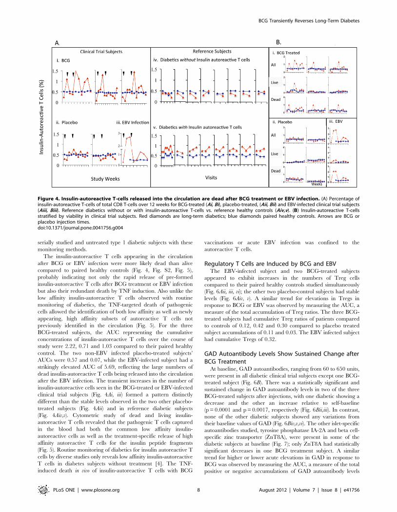
Fasting Insulin Secretion Temporarily Increased asMeasured by C-peptide after BCG and EBV Infection
At baseline as a recruitment requirement, none of the six
diabetic clinical trial subjects had detectable levels of fasting or
stimulated C-peptide using a relatively low sensitivity C-peptide
assay for screening in the standard clinic setting. Serum from all
clinical trial patients was saved for subsequent insulin secretion
studies with an ultrasensitive C-peptide assay. When the
baseline samples were re-assayed with the ultrasensitive assay,
all six clinical trial subjects had detectable C-peptide above the
lower range of sensitivity of the ultrasensitive assay (.1.5 pmol/
L) (Fig. 8).
Two of the three BCG-treated subjects and the EBV-infected
subject had transient increases in fasting C-peptide levels by
Week 20 compared to either their baseline or to the values in
41 reference diabetic subjects. Specifically, C-peptide levels
transiently and significantly rose with BCG administration in
Subject #i (mean concentration 3.49 pmol/L [95% CI 2.95–
3.8]), Subject #ii (2.57 pmol/L [95% CI 1.65–3.49]), as well as
in the EBV-infected placebo Subject #vi (3.16 pmol/L [95%
CI 2.54–3.69]) relative to 41 reference diabetic subjects
(mean = 1.65 pmol/L [95% CI 1.55–3.2]), using the Kolmo-
gorov-Smirnov two-sample test (Fig. 8). Subjects #i and #ii
each had more than 50% of their C-peptide values above the
95th percentile of the reference levels. Subject #vi had 18% of
C-peptide values above this level. Neither non-EBV infected
placebo-treated diabetic subject (iv and v) had C-peptide
fluctuations of statistical significance. The biologic stability of
low levels of fasting C-peptide levels with serial monitoring in
the ultra-sensitive assay is apparent in 41 reference diabetics
(Fig. 8D) and confirmed in 17 additional diabetic subjects
evaluated weekly for 12 weeks that were collected after the trial
completion to further confirm the stability of the ultrasensitive
C-peptide assay in serially studied long term diabetics with these
low levels (Fig. 9). AUC measurements of C-peptide, a measure
of cumulative changes of C-peptide levels over the 5-month
trial, were higher in the two BCG-treated and one EBV-
infected subject than in the non-EBV infected placebo clinical
trial subjects.
Other Clinical and Safety MonitoringThere were no significant changes in any of the clinical trial
patients in any safety monitoring parameters, including routine
chemistry and liver function tests, hematologic studies, or HbA1c
levels. Other than the expected vaccination scars associated with
BCG, no adverse effects occurred. None of the participants
dropped out of the clinical trial.
Discussion
The goals of the current trial were to determine whether
activation of the innate immune system could be accomplished
safely with repeated BCG vaccinations and whether this treatment
would ameliorate, for any time period, the advanced autoimmune
state of long-term type 1 diabetes. We found that repeated BCG
vaccination at low doses was safe and well tolerated. We also found
that BCG vaccination and an unexpected EBV infection in a
placebo-treated diabetic subject, both known triggers of innate
immunity, caused rapid increases in circulating insulin-autoreac-
tive T cells that were mostly dead. The rapid release of dead
insulin-autoreactive T cells supports the hypothesis, first demon-
strated in the NOD-mouse model of autoimmune diabetes, that
BCG ameliorates the advanced autoimmune process underlying
type 1 diabetes by stimulating TNF, which selectively kills only
disease-causing cells and, further, permits pancreas regeneration
[7,8] as evidenced by the transient increase in C-peptide secretion
we observed using an ultrasensitive C-peptide assay.
The response we observed in the placebo subject who
experienced an acute EBV infection provides evidence that
infectious agents other than Mycobacterium can activate innate
immunity in long-term diabetic subjects and modify the host’s
aberrant autoimmune response [9]. The subjects EBV status and
receipt of placebo saline injections fortuitously enabled us to
compare the serial T cell and pancreas effects of EBV- and BCG-
triggered innate immune responses in the same study [9,19]. EBV
infections, like BCG, are known to trigger innate immunity by
inducing a strong host TNF response [9,19], and the changes in
autoimmune cells and beta cell responses we observed in BCG-
treated subjects were similar or sometimes even larger in the EBV-
infected subject, suggesting that a larger dose of BCG might be
more effective. The transient increases in C-peptide, found after
both an acute EBV infection and with BCG vaccinated subjects,
suggests a positive impact of these immune perturbations on beta
cell function.
This study may offer mechanistic insights into ongoing clinical
trials of broad-spectrum immunosuppressive drugs, such as anti-
CD3 antibodies, in new-onset type 1 diabetes. The administration
of humanized anti-CD3 antibodies is associated with side effects,
including re-activation of EBV in recent-onset type 1 diabetes. as
reported to the FDA. Lowering the dose of anti-CD3 antibodies
reduced EBV reactivation in clinical studies, but also eliminated
efficacy. In another trial of anti-CD3 in new-onset diabetes, the
release of greater numbers of insulin-autoreactive-specific T cells
correlated with the simultaneous appearance in the circulation of
EBV-specific T cells. Taken together, findings from anti-CD3
trials and the trial reported in this paper demonstrate that EBV
Figure 5. Two-color flow pictures of the serial weekly blood monitoring of dead and live insulin autoreactive T cells in a controlsubject (left) and BCG-treated diabetic subject (right). After the first BCG treatment, predominantly dead insulin-autoreactive T cells appear inthe circulation of the diabetic compared to the simultaneously studied paired healthy control. For all recruited BCG-treated diabetic subjects, the startof the trial shows fresh blood samples with no insulin-autoreactive T-cells in these longterm diabetics, followed by dead insulin-autoreactive T-cellsthat persist through week 4, recurrent dead insulin-autoreactive cells released again after the second injection of BCG followed by the gradualdisappearance of the dead insulin-autoreactive T-cells by week 12 of monitoring. It should be noted that the newly released insulin-autoreactive cellsafter BCG are unique in representing both low affinity (*) autoreactive T-cells that can be observed in the routine monitoring of positive patients andhigh affinity(***) autoreactive T-cells that are never observed in routine monitoring of diabetic patients. In contrast to the serial monitoring of a BCGtreated subject, the serial studied fresh blood samples of the control subject reveal throughout the study the lack of either live or dead insulin-autoreactive T-cells.doi:10.1371/journal.pone.0041756.g005
BCG Transiently Reverses Long-Term Diabetes
PLoS ONE | www.plosone.org 10 August 2012 | Volume 7 | Issue 8 | e41756

infection or BCG vaccination marshals innate immunity charac-
terized by known elevations in TNF and that this leads to
potentially therapeutic benefits, especially death of insulin-
autoreactive T cells.
Drug development is facilitated by understanding drug mech-
anism and by development of biomarkers for monitoring early
responses to therapy. One previous uncontrolled study of a single
dose BCG vaccination reported possibly successful stabilization of
blood sugars in 65% of pre-diabetic patients [13]. Subsequent
controlled clinical studies of a single low-dose BCG vaccination in
new-onset diabetic children did not show a benefit when the
patients were re-studied, typically a year later [23–25]. The
Figure 6. TREG cells and GAD-autoantibodies change in response to BCG and EBV. (A) TREG cell ratios in BCG-treated, placebo, and EBV-infected clinical trial subjects by week vs. paired healthy controls. (B) GAD autoantibody levels vs. own baseline in BCG-treated placebo-treated, andEBV-infected clinical trial in each subject, by week. B is baseline prior to trial. Arrows are BCG or placebo injection times.doi:10.1371/journal.pone.0041756.g006
BCG Transiently Reverses Long-Term Diabetes
PLoS ONE | www.plosone.org 11 August 2012 | Volume 7 | Issue 8 | e41756

Figure 7. IA-2A and ZnT8 autoantibodies in clinical trial subjects by study week.doi:10.1371/journal.pone.0041756.g007
BCG Transiently Reverses Long-Term Diabetes
PLoS ONE | www.plosone.org 12 August 2012 | Volume 7 | Issue 8 | e41756

current trial is unique in now understanding the mechanism of
BCG and the development of closely linked bio-markers to track
mechanism. We additionally utilized multi-dosing of BCG
combined with frequent monitoring for disease-specific biomarkers
for up to 20 weeks to observe any TNF-driven immune effects.
Intensive monitoring uncovered alterations in disease-specific T
cells and changes in C-peptide secretion that suggest brief
functional improvement in the pancreas. Our findings are
consistent with trials showing BCG vaccination decreased disease
activity and prevented progression of brain lesions in advanced
multiple sclerosis, an autoimmune disease similarly sharing
autoreactive T cells vulnerable to TNF-triggered cell death
[26,27]. Recent findings also suggest repeat BCG administration,
but not single BCG vaccinations in childhood prevents diabetes
onset [28] and childhood BCG vaccinations prevent autoantibody
formation [20].
In the current study, BCG was expressly chosen as a treatment
for its induction of TNF, which has been shown to play a
therapeutic role in at least in four rodent models of five
autoimmune diseases [3,7,8,10,12,29,30] and in vitro [4]. In
contrast to the clinical utility of anti-TNF therapies in rheumatoid
arthritis but worsening of symptoms when anti-TNF is used in
most other autoimmune diseases [31–37], these experiments have
repeatedly shown that TNF or TNF-inducers protect against onset
and progression of many forms of autoimmunity. They also have
reversed autoimmune disease, ameliorated advanced autoimmune
disease, if administered in newly transplanted islet tissues, and/or
permitted regeneration of the end organs. In some of these diverse
rodent and human models of autoimmunity, the underlying
mechanism of TNF’s therapeutic effect has been traced to various
genetic and functional errors in the proteasome or proteasome-
activated transcriptional factor NFkB (nuclear factor-kB) signaling
pathway [1,17,38–55].
For a therapeutic and sustained amelioration of the autoim-
mune state, including a permanent elimination of insulin-
autoreactive T cells in diabetes, potentially leading to a sustained
return of C-peptide secretion, more frequent or higher dosing of
BCG will likely be required. Past human studies have established
that even modest levels of remaining C-peptide activity are
beneficial in the reduced incidence of retinopathy and nephrop-
athy as well as the avoidance of hypoglycemia [56]. Our findings
provide proof-of-principle evidence that insulin-autoreactive T
cells can be specifically targeted and eliminated, albeit briefly, in
vivo, even in long-standing disease with a transient restoration of C-
peptide. This paves the way for either higher doses or more
frequent BCG administered in future trials for patients with
advanced disease to maintain or restore C-peptide levels.
Supporting Information
Figure S1 Levels of EBV-specific memory T-cells inplacebo subject with latent EBV infection who was notpart of this trial (A) Negative levels of EBV-specific memory T-
Figure 8. Fasting C-peptide levels show transient increase in BCG-treated and EBV-infected clinical trial subjects. Fasting C-peptidefor (A) BCG-treated, (B) Placebo-treated, and (C) EBV-infected clinical trial subjects by week vs. (D) Reference diabetics, by visit. C-peptide levels aremeasured by ultrasensitive C-peptide assay in duplicate. Arrows are BCG or placebo injection times.doi:10.1371/journal.pone.0041756.g008
BCG Transiently Reverses Long-Term Diabetes
PLoS ONE | www.plosone.org 13 August 2012 | Volume 7 | Issue 8 | e41756
Figure 9. C-peptide levels remain stable and near the lower limit of an ultrasensitive assay in a longterm diabetic group (N = 17)sampled weekly for 12 weeks in a fasting state.doi:10.1371/journal.pone.0041756.g009
BCG Transiently Reverses Long-Term Diabetes
PLoS ONE | www.plosone.org 14 August 2012 | Volume 7 | Issue 8 | e41756
cells in clinical trial subjects, both BCG-treated and placebo-
treated clinical trial subjects.
(TIFF)
Figure S2 Flow cytometric methods used for theanalysis of purified CD8 T-cells for quantifying thenumbers of dead versus live cells. Fresh CD8 T-cells
cultured overnight can be demonstrated by forward versus side
scatter histograms on a flow cytometer to be either viable or dead
based on the placement on a side-scatter versus forward scatter
flow gate. The CD8 T cells can additionally be confirmed as dead
or alive based not only by the size of dying cells (scatter) but also by
staining with propidium iodide (PI), a reagent that stains dead
cells. With differential flow gating and/or staining with PI, the
dead cells are concentrated in the left upper quadrant and the
viable cells are concentrated in the right lower quadrant.
(TIFF)
Checklist S1 CONSORT Checklist.
(DOC)
Protocol S1 Trial Protocol.
(DOC)
Acknowledgments
We thank L. Murphy and M. Davis, PhD and D. Briscoe, MPH, for
providing formatting and editorial assistance.
Author Contributions
Conceived and designed the experiments: DLF JA DMN. Performed the
experiments: LW YO DB LB GM RP. Analyzed the data: HZ DS. Wrote
the paper: DLF JA DMN.
References
1. Kodama S, Davis M, Faustman DL (2005) The therapeutic potential of tumornecrosis factor for autoimmune disease: a mechanistically based hypothesis. Cell
Mol Life Sci 62: 1850–1862.
2. Satoh J, Seino H, Abo T (1989) Recombinant human tumor necrosis factor asuppresses autoimmune diabetes in nonobese diabetic mice. J Clin Invest 84:
1345–1348.
3. Grewal IS, Grewal KD, Wong FS, Picarella DE, Janeway CA, et al. (1996) Local
expression of transgene encoded TNF alpha in islets prevents autoimmunediabetes in non-obese diabetic (NOD) mice by preventing the development of
autoreactive islet specific T cells. J Exp Med 184: 1963–1974.
4. Ban L, Zhang J, Wang L, Kuhtreiber W, Burger D, et al. (2008) Selective deathof autoreactive T cells in human diabetes by TNF or TNF receptor 2 agonism.
Proc Natl Acad Sci U S A 105: 13644–13649.
5. Qin HY, Chaturvedi P, Singh B (2004) In vivo apoptosis of diabetogenic T cellsin NOD mice by IFN-c/TNF-a. Int Immunol 16: 1723–1732.
6. Christen U, Von Herrath MG (2002) Apoptosis of autoreactive CD8
lymphocytes as a potential mechanism for the abrogation of type 1 diabetes
by islet-specific TNF-alpha expression at a time when the autoimmune process isalready ongoing. Ann N Y Acad Sci 958: 166–169.
7. Ryu S, Kodama S, Ryu K, Schoenfeld DA, Faustman DL (2001) Reversal of
established autoimmune diabetes by restoration of endogenous beta cellfunction. J Clin Invest 108: 63–72.
8. Kodama S, Kuhtreiber W, Fujimura S, Dale EA, Faustman DL (2003) Islet
regeneration during the reversal of autoimmune diabetes in NOD mice. Science
302: 1223–1227.
9. Rahman MM, McFadden G (2006) Modulation of tumor necrosis factor bymicrobial pathogens. PLoS Pathog 2: e4.
10. Harada M, Kishimoto Y, Makino S (1990) Prevention of overt diabetes and
insulitis in NOD mice by a single BCG vaccination. Diabetes Res Clin Prac 8:85–89.
11. Sadelain MWJ, Hui-Yu Q, Lauzon J, Singh B (1990) Prevention of type I
diabetes in NOD mice by adjuvant immunotherapy. Diabetes 39: 583–589.
12. McInerney MF, Pek SB, Thomas DW (1991) Prevention of insulitis and diabetes
onset by treatment with complete Freund’s adjuvant in NOD mice. Diabetes 40:715–725.
13. Shehadeh N, Calcinaro F, Bradley BJ, Bruchlim I, Vardi P, et al. (1994) Effect of
adjuvant therapy on development of diabetes in mouse and man [seecomments]. Lancet 343: 706–707.
14. Burger DE, Wang L, Ban L, Okubo Y, Kuhtreiber WM, et al. (2011) Novel
automated blood separations validate whole cell biomarkers. PLoS One 6:e22430.
15. Pinkse GG, Tysma OH, Bergen CA, Kester MG, Ossendorp F, et al. (2005)
Autoreactive CD8 T cells associated with beta cell destruction in type 1 diabetes.
Proc Natl Acad Sci U S A 102: 18425–18430.
16. Achenbach P, Lampasona V, Landherr U, Koczwara K, Krause S, et al. (2009)Autoantibodies to zinc transporter 8 and SLC30A8 genotype stratify type 1
diabetes risk. Diabetologia 52: 1881–1888.
17. Castiblanco J, Anaya J-M (2008) The I kappa BL gene polymorphism influencesrisk of acquiring systemic lupus erythematosus and Sjogren’s syndrome. Human
Immunology 69: 45–51.
18. Trials CoSfS-N-PCR, Policy BoHS (2001) Small Clinical Trials: Issues and
Challenges: The National Academies Press.
19. Devergne O, Hatzivassiliou E, Izumi KM, Kaye KM, Kleijnen MF, et al. (1996)Association of TRAF1, TRAF2, and TRAF3 with an Epstein-Barr virus LMP1
domain important for B-lymphocyte transformation: role in NF-kappaBactivation. Mol Cell Biol 16: 7098–7108.
20. Sanjeevi CB, Das AK, Shtauvere-Brameus A (2002) BCG vaccination and
GAD65 and IA-2 autoantibodies in autoimmune diabetes in southern India.
Ann N Y Acad Sci 958: 293–296.
21. Braghi S, Bonifacio E, Secchi A, Di Carlo V, Pozza G, et al. (2000) Modulation
of humoral islet autoimmunity by pancreas allotransplantation influences
allograft outcome in patients with type 1 diabetes. Diabetes 49: 218–224.
22. Bosi E, Braghi S, Maffi P, Scirpoli M, Bertuzzi F, et al. (2001) Autoantibody
response to islet transplantation in type 1 diabetes. Diabetes 50: 2464–2471.
23. Pozzilli P (1997) BCG vaccine in insulin-dependent diabetes mellitus. IMDIABGroup. Lancet 349: 1520–1521.
24. Allen HF, Klingensmith GJ, Jensen P, Simoes E, Hayward A, et al. (1999) Effectof Bacillus Calmette-Guerin vaccination on new-onset type 1 diabetes. A
randomized clinical study. Diabetes Care 22: 1703–1707.
25. Elliott JF, Marlin KL, Couch RM (1998) Effect of bacille Calmette-Guerinvaccination on C-peptide secretion in children newly diagnosed with IDDM.
Diabetes Care 21: 1691–1693.
26. Ristori G, Buzzi MG, Sabatini U, Giugni E, Bastianello S, et al. (1999) Use ofBacille Calmette-Guerin (BCG) in multiple sclerosis. Neurology 53: 1588–1589.
27. Paolillo A, Buzzi MG, Giugni E, Sabatini U, Bastianello S, et al. (2003) Theeffect of Bacille Calmette-Guerin on the evolution of new enhancing lesions to
hypointense T1 lesions in relapsing remitting MS. Journal of Neurology 250:
247–248.
28. Khan M, Aydin M (2012) Effect of BCG vaccine in the prevention of type 1
diabetes mellitus. Contemp J Med 2: 1–8.
29. Sadelain MW, Qin HY, Sumoski W, Parfrey N, Singh B, et al. (1990) Preventionof diabetes in the BB rat by early immunotherapy using Freund’s adjuvant.
Journal of Autoimmunity 3: 671–680.
30. Okubo Y, Kanazawa Y, Oikawa Y, Miyazaki JI, Shimada A. (2006) Islet
hypertrophy observed in ‘‘reversed’’ diabetic NOD mouse after pancreatic beta
cell line administration Diabetes A 281: 1193.
31. Enayati PJ, Papadakis KA (2005) Association of Anti-tumor Necrosis Factor
Therapy With the Development of Multiple Sclerosis. J Clin Gastroenterol 39:303–306.
32. Sicotte NL, Voskuhl RR (2001) Onset of multiple sclerosis associated with anti-
TNF therapy. Neurology 57: 1885–1888.
33. Thomas CW Jr, Weinshenker BG, Sandborn WJ (2004) Demyelination during
anti-tumor necrosis factor alpha therapy with infliximab for Crohn’s disease.
Inflamm Bowel Dis 10: 28–31.
34. van Oosten BW, Barkhof F, Truyen L, Boringa JB, Bertelsmann FW, et al.
(1996) Increased MRI activity and immune activation in two multiple sclerosispatients treated with the monoclonal anti-tumor necrosis factor antibody cA2.
Neurology 47: 1531–1534.
35. Boulton JG, Bourne JT (2007) Unstable diabetes in a patient receiving anti-TNF-alpha for rheumatoid arthritis. Rheumatology 46: 178–179.
36. Ramos-Casals M, Brito-Zeron P, Soto MJ, Cuadrado MJ, Khamashta MA
(2008) Autoimmune diseases induced by TNF-targeted therapies. Best Practice& Research in Clinical Rheumatology 22: 847–861.
37. Ko JM, Gottlieb AB, Kerbleski JF (2009) Induction and exacerbation of psoriasiswith TNF-blockade therapy: A review and analysis of 127 cases. Journal of
Dermatological Treatment 20: 100–108.
38. Hayashi T, Faustman D (1999) NOD mice are defective in proteasomeproduction and activation of NF- kappaB. Mol Cell Biol 19: 8646–8659.
39. Hayashi T, Faustman DL (2001) Selected contribution: Association of gender-
related LMP2 inactivation with autoimmune pathogenesis. J Appl Physiol 91:2804–2815.
40. Hayashi T, Kodama S, Faustman DL (2000) Reply to ‘LMP2 expression andproteasome activity in NOD mice’. Nat Med 6: 1065–1066.
41. Guo D, Li M, Zhang Y, Yang P, Eckenrode S, et al. (2004) A functional variant
of SUMO4, a new I kappa B alpha modifier, is associated with type 1 diabetes.Nat Genet 36: 837–841.
42. Wong HK, Kammer GM, Dennis G, Tsokos GC (1999) Abnormal NF-kappa Bactivity in T lymphocytes from patients with systemic lupus erythematosus is
BCG Transiently Reverses Long-Term Diabetes
PLoS ONE | www.plosone.org 15 August 2012 | Volume 7 | Issue 8 | e41756

associated with decreased p65-RelA protein expression. J Immunol 163: 1682–
1689.43. Kessel A, Rosner I, Rozenbaum M, Zisman D, Sagiv A, et al. (2004) Increased
CD8+ T cell apoptosis in scleroderma is associated with low levels of NF-kappa
B. J Clin Immunol 24: 30–36.44. Valero R, Baron ML, Guerin S, Beliard S, Lelouard H, et al. (2002) A defective
NF-kappa B/RelB pathway in autoimmune-prone New Zealand black mice isassociated with inefficient expansion of thymocyte and dendritic cells. J Immunol
169: 185–192.
45. Zaiss DMW, Bekker CPJ, Grone A, Lie BA, Sijts AJAM (2011) ProteasomeImmunosubunits Protect against the Development of CD8 T Cell-Mediated
Autoimmune Diseases. Journal of Immunology 187: 2302–2309.46. Bohren KM, Nadkarni V, Song JH, Gabbay KH, Owerbach D (2004) A M55V
polymorphism in a novel SUMO gene (SUMO-4) differentially activates heatshock transcription factors and is associated with susceptibility to type I diabetes
mellitus. J Biol Chem 279: 27233–27238.
47. Vereecke L, Beyaert R, van Loo G (2009) The ubiquitin-editing enzyme A20(TNFAIP3) is a central regulator of immunopathology. Trends in Immunology
30: 383–391.48. Trynka G, Zhernakova A, Romanos J, Franke L, Hunt KA, et al. (2009) Coeliac
disease-associated risk variants in TNFAIP3 and REL implicate altered NF-
kappa B signalling. Gut 58: 1078–1083.49. Zhang G-L, Zou Y-F, Feng X-L, Shi H-J, Du X-F, et al. (2011) Association of
the NFKBIA gene polymorphisms with susceptibility to autoimmune andinflammatory diseases: a meta-analysis. Inflammation Research 60: 11–18.
50. Till A, Rosenstiel P, Krippner-Heidenreich A, Mascheretti-Croucher S,
Croucher PJ, et al. (2005) The Met-196 -. Arg variation of human tumornecrosis factor receptor 2 (TNFR2) affects TNF-alpha-induced apoptosis by
impaired NF-kappaB signaling and target gene expression. J Biol Chem 280:
5994–6004.51. Allcock RJN, de la Concha EG, Fernandez-Arquero M, Vigil P, Conejero L, et
al. (1999) Susceptibility to multiple sclerosis mediated by HLA-DRB1 isinfluenced by a second gene telomeric of the TNF cluster. Human Immunology
60: 1266–1273.
52. Allcock RJN, Christiansen FT, Price P (1999) The central MHC gene IKBLcarries a structural polymorphism that is associated with HLA-A3,B7,DR15.
Immunogenetics 49: 660–665.53. Miterski B, Bohringer S, Klein W, Sindern E, Haupts M, et al. (2002) Inhibitors
in the NFkappaB cascade comprise prime candidate genes predisposing tomultiple sclerosis, especially in selected combinations. Genes Immun 3: 211–219.
54. Wandinger KP, Sturzebecher CS, Bielekova B, Detore G, Rosenwald A, et al.
(2001) Complex immunomodulatory effects of interferon-beta in multiplesclerosis include the upregulation of T helper 1-associated marker genes. Annals
of Neurology 50: 349–357.55. Fu Y, Yan G, Shi L, Faustman D (1998) Antigen processing and autoimmunity.
Evaluation of mRNA abundance and function of HLA-linked genes. Annals of
the New York Academy of Sciences 842: 138–155.56. Steffes MW, Sibley S, Jackson M, Thomas W (2003) Beta-cell function and the
development of diabetes-related complications in the diabetes control andcomplications trial. Diabetes Care 26: 832–836.
BCG Transiently Reverses Long-Term Diabetes
PLoS ONE | www.plosone.org 16 August 2012 | Volume 7 | Issue 8 | e41756


