
Protocol
PepStarTM
Microarrays
Ready-to-use peptide microarrays for humoral immune response profiling and epitope mapping for 1 individual sample
Revision 3.3
Contact us:
Support: +49-30-6392-7878
Order per fax: +49-30-6392-7888
Or e-mail: [email protected]
www: www.jpt.com
JPT Peptide Technologies GmbH Volmerstrasse 5 (UTZ) 12489 Berlin GERMANY
Product Use & Liability
THE PRODUCTS ARE FOR EXPERIMENTAL LABORATORY USE ONLY AND NOT INTENDED FOR HUMAN OR HOUSEHOLD USE.
Only qualified personnel should handle these chemicals.
Furthermore, JPT Peptide Technologies stresses that missing hazard warnings do not mean that the relevant product is harmless. In regard to classification the products are only for research purposes. JPT Peptide Technologies cannot be made responsible for damages arising from misuse of any product.
JPT Peptide Technologies makes no warranty of any kind, expressed or implied, which extends beyond the description of the product in this brochure, except that the material will meet our described specifications at the time of delivery. JPT Peptide Technologies makes no guarantee of results and assumes no liability for injuries, damages or penalties resulting from product use, since the conditions of handling and use are beyond our control.

2 Rev. 3.3
Table of Contents
1 INTRODUCTION ............................................................................................................. 3
2 LIST OF COMPONENTS ................................................................................................. 4
3 STORAGE AND HANDLING ........................................................................................... 5
3.1 STORAGE OF PEPSTAR™ PEPTIDE MICROARRAY SLIDES ................................................. 5 3.2 HANDLING OF PEPSTAR™ PEPTIDE MICROARRAY SLIDES ................................................ 5
4 GENERAL CONSIDERATIONS ...................................................................................... 6
4.1 EXPERIMENTAL BASICS ................................................................................................... 6 4.2 ASSAY PRINCIPLE ........................................................................................................... 7 4.3 PEPSTAR
TM PEPTIDE MICROARRAY LAYOUT .................................................................... 8
5 EXPERIMENTAL PROTOCOLS .....................................................................................10
5.1 ADDITIONAL MATERIALS REQUIRED ................................................................................11 5.2 ADDITIONAL HARDWARE AND SOFTWARE .......................................................................12 5.3 INCUBATION PROCEDURE ...............................................................................................13 5.3.1 FULLY AUTOMATED MICROARRAY PROCESSING ............................................................13 5.3.2 WORKFLOW FOR MANUAL INCUBATION OF PEPSTAR
TM PEPTIDE MICROARRAYS* ............15
5.3.3 SAMPLE INCUBATION USING MICROARRAY-CHIP-SANDWICH ............................................16 5.3.4 SAMPLE INCUBATION USING INCUBATION TROUGH .........................................................20 5.3.5 DETECTION ANTIBODY INCUBATION AND POST PROCESSING ..........................................21 5.3.6 DATA ANALYSIS ...........................................................................................................22
6 PEPSTARTM MICROARRAY INCUBATION PROTOCOL ..............................................23
7 NOTES / TROUBLESHOOTING .....................................................................................24
8 RELATED PRODUCTS ..................................................................................................26

3 Rev. 3.3
1 Introduction
Antibody-antigen interactions are key events in immunology. Therefore, the
identification of epitopes or immunodominant regions in antigens represents an
important step in the characterization of antibodies. One of the most efficient ways to
identify such epitopes is incubation of a collection of antigen-derived peptides
displayed on glass slides (PepStar TM peptide microarrays) with antibodies of interest.
JPT Peptide Technologies’ PepStarTM peptide microarrays represent customized or
catalog peptide microarray slides covering individual or collections of proteins for
rapid screening of protein-peptide interactions. The peptides displayed on glass
slides are chemoselectively and covalently bound, enabling effective interaction with
binding partners. Immobilized sequences represent overlapping peptides derived
from single or multiple antigens as well as epitope and random peptide collections
allowing efficient profiling of humoral immune responses using patient samples and
protein-protein interaction studies. In addition, full proteome PepStarTM microarrays
are available covering overlapping peptide scans through complete microbial or viral
proteomes. Upon incubation with your protein or patient sample the binding event
can be detected by fluorescently labelled primary or secondary (2nd) antibody.
4 Rev. 3.3
2 List of Components
1. PepStar™ peptide microarray
Glass slide displaying peptides in three identical subarrays, see section 4.3 for
details
2. Blank slides engraved with “Blank-Slide”
One blank slide per PepStarTM peptide microarray
3. JPT Peptide Microarray Spacers
Vials containing 20 spacers each
Two spacers per PepStarTM peptide microarray are needed
4. Product Information
Relevant files for the specific peptide microarray (protocol and datasheet as pdf-
files, sequence info as gal-file and JPT's Microarray Feature Viewer as zipped
package).

5 Rev. 3.3
3 Storage and Handling
3.1 Storage of PepStar™ Peptide Microarray Slides
Optimal storage conditions for peptide microarray slides are in a cool (approx.
4°C / 39°F) and dry environment.
Peptide microarrays are stable for at least 6 months when stored at 4°C (39°F).
Do not freeze the peptide microarrays.
3.2 Handling of PepStar™ Peptide Microarray Slides
Always handle the delicate peptide microarrays with care.
Never touch the peptide microarray slide surface.
Always wear laboratory gloves when handling peptide microarray slides.
Please hold peptide microarray slides at the end, which carries the engraved data
label. This label provides a unique identification of the specific microarray.
Please take care when dispensing solutions onto the slide surface. Make sure not
to touch the surface with pipette-tips or dispensers.
Never whisk the surface of the peptide microarray slide with a cloth.
Never use chemicals other than described. Inappropriate chemicals may destroy
the chemical bonding of the peptides to the glass surface.
Avoid dust or other particles during each step of the experiment. Dust, particles
and resulting scratches will cause artifacts during final signal readout.
Please filter all solutions for the washing steps with minimum 2µm, preferably
0.4µm, particle filters before use.
PLEASE READ THE ENTIRE PROTOCOL BEFORE STARTING YOUR EXPERIMENTS!
CAREFULLY NOTE THE HANDLING AND STORAGE CONDITIONS OF JPT´s
PEPSTARTM PEPTIDE MICROARRAYS.
PLEASE CONTACT JPT PEPTIDE TECHNOLOGIES´ TECHNICAL SERVICE FOR
ASSISTANCE IF NECESSARY.

6 Rev. 3.3
4 General Considerations
4.1 Experimental Basics
JPT Peptide Technologies' PepStarTM peptide microarrays comprise synthetic
peptides, derived from antigens (principle of epitope detection see Figure 1) or other
sources that are chemoselectively and covalently immobilized to the glass surface.
An optimized hydrophilic linker moiety is inserted between the glass surface and the
peptide sequence to avoid false negatives caused by sterical hindrance. For
technical reasons all peptides contain a C-terminal glycine.
JPT’s PepStarTM peptide
microarrays are designed for
detecting potential biomarkers for
infectious diseases, autoimmune
diseases, cancer and allergies
and to elucidate protein-protein
interactions. Each spot in the
microarray represents a single
individual peptide.
After incubation of the peptide
microarray with an analyte, a
fluorescently labelled detection
molecule is used for signal readout.
Figure 1: General principle of epitope detection using overlapping peptide scans.
If not ordered otherwise all peptides are displayed in three identical subarrays on
each slide. PepStarTM slide surfaces are delivered in a pre-treated form minimizing
unspecific binding of your target sample. Therefore, usually no blocking step is
needed.

7 Rev. 3.3
4.2 Assay Principle
The most common application of JPT´s PepStarTM peptide microarrays is the epitope
mapping procedure (Figure 1). A schematic view of the assay principle is shown in
Figure 2.
Figure 2: Peptide microarray assay principle.
The peptide microarray is incubated using a primary antibody or patient sample – e.g.
serum, plasma or saliva – for a defined time. This enables the formation of stable
peptide-antibody interactions. Subsequent to this incubation, the fluorescently
labelled secondary antibody is applied. Bound to the peptide-bound primary
antibodies, the fluorescence label of the secondary antibody enables readout of
antibody interaction by microarray scanning systems. Each spot that shows an
interaction with the primary antibody will gain signal in the resulting scan-image.
It is crucial to perform control incubations in order to distinguish between real signals and false positives. To avoid false positive signals induced by peptide-secondary antibody interaction, JPT recommends performing regular control incubations using secondary antibodies only. In addition, JPT recommends performing control incubations applying unrelated primary antibodies to check for false positive signals induced by interaction of peptide with Fc-fragment of the primary antibody.
For seroscreening application JPT recommends checking the secondary antibody for selectivity and specificity. Signals induced by cross-reactivity of secondary antibodies directed against IgG towards IgM or IgE may result in false positive results.
PeptidePeptide
8 Rev. 3.3
4.3 PepStarTM Peptide Microarray Layout
Please refer to the .gal-file provided with the product documentation for identity and
location of the spots on the microarray surface. The microarray side carrying the
engraved label represents the surface displaying the attached peptides. The .gal-file
can be opened using microarray evaluation software-modules capable of evaluating
high-density microarray slides or for simple analysis by JPT’s Microarray Feature
Viewer. Since .gal-files are tab-separated text files, they can also be accessed by
text-processing software such as Microsoft Editor (Notepad) or Microsoft Excel. With
the .gal-file provided, evaluation can be performed using software modules like
GenePix, ArrayPro or similar programs, which align the .gal-file induced grid onto the
resulting image. Additionally, JPT´s Microarray feature viewer (part of data files sent
by customer support) may be applied for layout visualization, spot identification and
feature extraction using any conventional modern web browser.
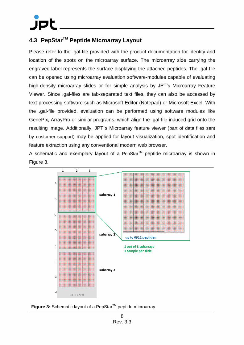
A schematic and exemplary layout of a PepStarTM peptide microarray is shown in
Figure 3.
Figure 3: Schematic layout of a PepStarTM peptide microarray.

9 Rev. 3.3
As shown in 3 the peptide microarray is printed in three identical subarrays. This
enables highly efficient intra-chip-reproducibility tests using scatter plots or
correlation functions.
For PepStarTM microarrays, each subarray is printed in individual blocks (see Figure
4). The number of blocks and the final layout will vary according to the final number
of peptides.
Figure 4: Exemplary view for a PepStarTM microarray slide with 3 subarrays (left) and a subarray consisting of 16 individual blocks (right).

10 Rev. 3.3
5 Experimental Protocols
Note: The following procedure is given as a guideline only. The optimal
experimental conditions will vary depending on the investigated sample and
instruments used and can, therefore, not be predetermined. The optimal
experimental conditions must be established by the user. No warranty or
guarantee of performance using this procedure with a target antibody or serum
can be made or is implied.
The PepStarTM peptide microarray is designed as a ready-to-use product to identify
epitopes, peptide binders or immunodominant regions in antigens.
Ordinarily, there is no need to perform blocking steps on the slide surface prior to
incubation with the target sample. However, in case of incubations with patient sera
or plasma, JPT recommends to include an additional blocking step prior to incubation
with patient sample.
Please refer to the .gal-files provided with the product documentation for identity and
location of the spots on the peptide microarray surface. The side of the slide
displaying the peptides is marked with the engraved lot number.
Note: For analysis of protein/protein-interaction no specific guideline can be
provided. Several factors such as buffer components, ion strength, pH-value,
temperature, washing conditions and more may influence the binding affinity
of the target protein to the immobilized peptides. JPT also recommends to
perform a direct labeling reaction of the protein of interest as well as several
independent incubations covering different conditions such as concentrations,
temperatures and washing procedures.
11 Rev. 3.3
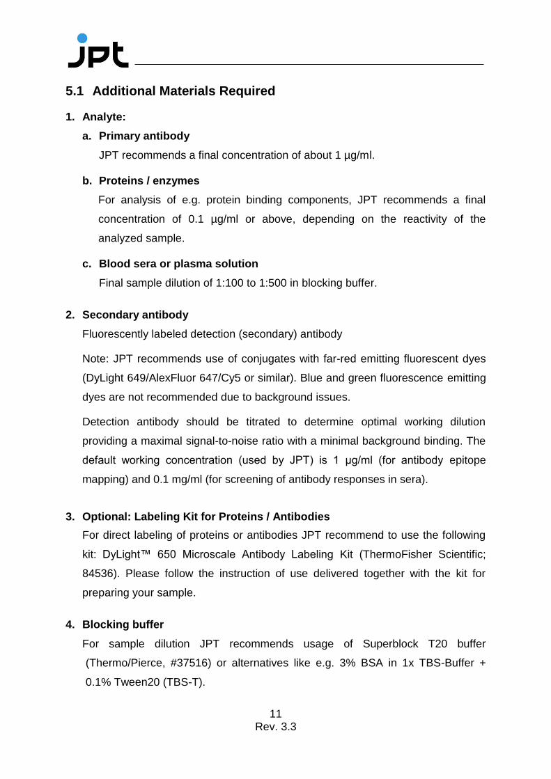
5.1 Additional Materials Required
1. Analyte:
a. Primary antibody
JPT recommends a final concentration of about 1 µg/ml.
b. Proteins / enzymes
For analysis of e.g. protein binding components, JPT recommends a final
concentration of 0.1 µg/ml or above, depending on the reactivity of the
analyzed sample.
c. Blood sera or plasma solution
Final sample dilution of 1:100 to 1:500 in blocking buffer.
2. Secondary antibody
Fluorescently labeled detection (secondary) antibody
Note: JPT recommends use of conjugates with far-red emitting fluorescent dyes
(DyLight 649/AlexFluor 647/Cy5 or similar). Blue and green fluorescence emitting
dyes are not recommended due to background issues.
Detection antibody should be titrated to determine optimal working dilution
providing a maximal signal-to-noise ratio with a minimal background binding. The
default working concentration (used by JPT) is 1 μg/ml (for antibody epitope
mapping) and 0.1 mg/ml (for screening of antibody responses in sera).
3. Optional: Labeling Kit for Proteins / Antibodies
For direct labeling of proteins or antibodies JPT recommend to use the following
kit: DyLight™ 650 Microscale Antibody Labeling Kit (ThermoFisher Scientific;
84536). Please follow the instruction of use delivered together with the kit for
preparing your sample.
4. Blocking buffer
For sample dilution JPT recommends usage of Superblock T20 buffer
(Thermo/Pierce, #37516) or alternatives like e.g. 3% BSA in 1x TBS-Buffer +
0.1% Tween20 (TBS-T).

12 Rev. 3.3
5. Washing buffer
1x TBS-Buffer + 0.1% Tween20 (TBS-T)
6. De-ionized water
For final washing steps of the microarrays
5.2 Additional Hardware and Software
1. Tweezers
For handling of PepStarTM Peptide Microarrays
2. Automated Incubation/Hybridization Station
Tecan Hybridization Station HS4X00
Note: Alternatively, incubation in a microarray-chip-sandwich format or in a
incubation trough can be used. Please refer to point Fehler! Verweisquelle
konnte nicht gefunden werden. for further details.
3. 4-Well Dish, Microscope Slide Staining Dish or 50 mL-Falcon Tubes
(Incubation trough)
For manual incubation and washing steps.
4. Rocking Platform
For all manual incubation and washing steps (Note: Do not shake Microarray-
Chip-Sandwiches).
5. Slide Centrifuge (optional)
Alternatively, the slides may by dried by a gentle stream of nitrogen.
6. Fluorescence Scanner/Imager
Capable of excitation of appropriate fluorophore moiety and with a resolution of at
least 10 µm per pixel.
7. Analysis Software
Allowing quantification of the image and the assignment of signal intensities to
individual peptides using the provided gal-file.

13 Rev. 3.3
5.3 Incubation Procedure
5.3.1 Fully Automated Microarray Processing
All PepStarTM peptide microarrays produced by JPT have an identical layout
concerning active area and spotted surface. Although the content of the microarrays
varies, the overall layout and dimensions are the same (see Figure 5).
Please check with the manufacturer of your microarray processing station for compatibility with the required liquids. Most microarray processing stations are sensible towards strong acids and organic solutions. Protocols have to be adapted to prevent permanent damage to your device.
All peptide microarrays produced by JPT are adjusted to fit into common fully
automated microarray processing systems. JPT recommends using Tecan HS4X00
Hybridisation systems. All PepStar TM microarrays are printed according to the layout
of the Single chamber option of Tecans HS4X00 Pro station.
Figure 5: Maximum area dimension on JPT peptide microarrays.
An exemplary protocol for the use of JPT’s PepStar TM peptide microarrays in
Tecan’s HS4X00 processing machine is shown Figure 6.

14 Rev. 3.3
Figure 6: Exemplary method for incubation of JPT’s PepStar TM microarrays in Tecan HS4X00 processing machines:
Ch.: 1 and 2: TBS buffer, 0.1% Tween20 Ch.: 5: 0.1x SSC buffer Step 1: Pre-wash and filling of incubation chambers Step 2-4: Blocking procedure Step 5-7: Incubation with primary antibody/sera Step 8-10: Incubation with secondary antibody Step 11: Final washing steps Step 12: Slide drying procedure

15 Rev. 3.3
5.3.2 Workflow for Manual Incubation of PepStarTM Peptide Microarrays*
* For the design of a PepStar experiment, the attached PepStarTM Microarray Incubation Protocol (see point 6) can be used.
Limited
amount of
primary antibody /
analyte?
Incubation in
Microarray-Chip- Sandwich
Final assay volume ~300 µL
Incubation in
Final assay volume ~2,0 mL
Assembly of a
Microarray-Chip- Sandwich
(for details see: 5.3.3)
I. Incubation With primary antibody / analyte diluted in Blocking Buffer
Insert Peptide Microarrays into
the Incubation Trough
(for details see: 5.3.4)
Apply ~300 µL sample dilution into
the Microarray- Chip-Sandwich
Apply ~2,0 mL sample dilution into
the Peptide- Microarray containing
Incubate @30° C (86°F) / 1-2 hrs
Fill 2 mL Wash Buffer into the Incubation Trough .
Carefully transfer the Chip - Sandwich into the buffer.
Use tweezers to disassemble the sandwich and remove the Incubation Spacers.
II. Disassembling
Replace Wash Buffer by
4 mL fresh Wash Buffer
Replace sample dilution by 4 mL
Wash Buffer III. Washing
Wash
5x 3 - 4 min with Wash Buffer
IV. IncubationReplace Wash Buffer by 2 mL freshly prepared
Detection Antibody diluted in Blocking Buffer
Incubate @30° C (86° F) / 45 min
Wash
5x 3 - 4 min with Wash Buffer
5x 3 - 4 min with de- ionized H2O
V. Washing
VI. Slide Drying
VII. Imaging
VIII. Data Analysis
Using microarray centrifuge / by blowing a gentle stream of
nitrogen on the microarray surface
Fluorescence scanning
Note: Scanning resolution = 10 µm pixel size !
Determination of signal intensities of each peptide spot.
Bioinformatic evaluation of data.
Yes! No!
Protect from light! Gentle shaking!
Protect from light! Store dried
microarrays in an ozone - free
environment!
Gentle shaking!
Gentle shaking!
Limited
amount of
primary antibody /
?
- -
µL
Incubation in
Incubation Trough
Final assay volume ~2,0 mL
Assembly of a
- Sandwich
I. Incubation analyte diluted in Blocking Buffer
Insert into
the
Apply ~300 L sample dilution into
- Sandwich
Apply ~2,0 mL sample dilution into
the Peptide- Microarray containing
Incubation Trough
° C (86°F) / 1-2 hrs
.
-
.
II. Disassembling
III. Washing
Wash
4 min with
IV. Incubation° C (86° F) / 45 min
-
- 2O
V. Washing
VI. Slide Drying
VII. Imaging
VIII. Data Analysis
µ
Yes! No!
-
Gentle shaking!
16 Rev. 3.3
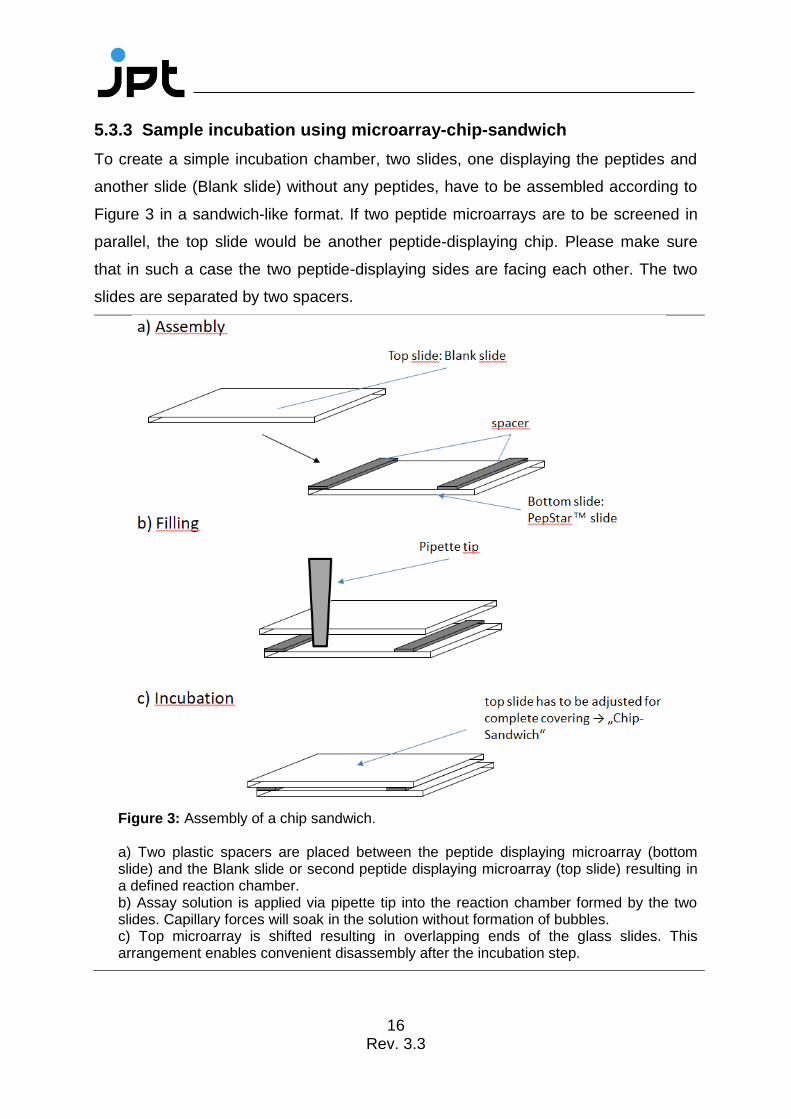
5.3.3 Sample incubation using microarray-chip-sandwich
To create a simple incubation chamber, two slides, one displaying the peptides and
another slide (Blank slide) without any peptides, have to be assembled according to
Figure 3 in a sandwich-like format. If two peptide microarrays are to be screened in
parallel, the top slide would be another peptide-displaying chip. Please make sure
that in such a case the two peptide-displaying sides are facing each other. The two
slides are separated by two spacers.
Figure 3: Assembly of a chip sandwich. a) Two plastic spacers are placed between the peptide displaying microarray (bottom slide) and the Blank slide or second peptide displaying microarray (top slide) resulting in a defined reaction chamber. b) Assay solution is applied via pipette tip into the reaction chamber formed by the two slides. Capillary forces will soak in the solution without formation of bubbles. c) Top microarray is shifted resulting in overlapping ends of the glass slides. This arrangement enables convenient disassembly after the incubation step.
17 Rev. 3.3
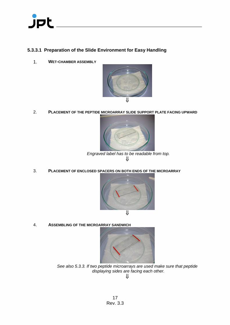
5.3.3.1 Preparation of the Slide Environment for Easy Handling
1. WET-CHAMBER ASSEMBLY
2. PLACEMENT OF THE PEPTIDE MICROARRAY SLIDE SUPPORT PLATE FACING UPWARD
Engraved label has to be readable from top.
3. PLACEMENT OF ENCLOSED SPACERS ON BOTH ENDS OF THE MICROARRAY
4. ASSEMBLING OF THE MICROARRAY SANDWICH
See also 5.3.3. If two peptide microarrays are used make sure that peptide
displaying sides are facing each other.

18 Rev. 3.3
5. PREPARATION OF FINAL ASSAY SOLUTION CONTAINING TARGET ANTIBODY/ANALYTE Approx. 300 µL if enclosed spacers are used.
6. PIPETTING OF THE COMPLETE VOLUME INTO MICROARRAY CHIP-SANDWICH
Capillary forces will suck the solution in between the two slides. Avoid air
bubbles within the sandwich.
Make sure not to touch the microarray with the pipette tip. Scratches and marks on the surface may destroy the deposited microarray and will cause artifacts!
7. ADJUSTMENT OF THE PEPTIDE MICROARRAY SANDWICH
8. CLOSING OF THE PETRI-DISH WITH A MATCHING COVER TO CREATE AN INCUBATION
CHAMBER.
JPT does not recommend use of fluorescently labeled primary or secondary antibodies in microarray sandwich-like incubations.
19 Rev. 3.3
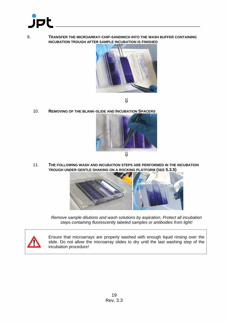
9. TRANSFER THE MICROARRAY-CHIP-SANDWICH INTO THE WASH BUFFER CONTAINING
INCUBATION TROUGH AFTER SAMPLE INCUBATION IS FINISHED
10. REMOVING OF THE BLANK-SLIDE AND INCUBATION SPACERS
11. THE FOLLOWING WASH AND INCUBATION STEPS ARE PERFORMED IN THE INCUBATION
TROUGH UNDER GENTLE SHAKING ON A ROCKING PLATFORM (SEE 5.3.5)
Remove sample dilutions and wash solutions by aspiration. Protect all incubation steps containing fluorescently labeled samples or antibodies from light!
Ensure that microarrays are properly washed with enough liquid rinsing over the slide. Do not allow the microarray slides to dry until the last washing step of the incubation procedure!

20 Rev. 3.3
5.3.4 Sample Incubation using Incubation trough
1. PLACEMENT OF THE PEPTIDE MICROARRAY SLIDES IN THE INCUBATION TROUGH
Engraved label has to be readable from top!
2. ADDITION OF THE SAMPLE DILUTION
Not directly on the microarray slide surface, but in one edge of the incubation trough!
3. CLOSE THE LID AND INCUBATE MICROARRAY SLIDES FOR THE APPROPRIATE TIME AT
THE DESIRED TEMPERATURE
Gentle shaking!
4. REMOVE SAMPLE DILUTIONS AND WASH BUFFERS BY ASPIRATION
Ensure that microarrays are properly washed with enough liquid rinsing over the slide. Do not allow the microarray slides to dry until the last washing step of the incubation procedure!

21 Rev. 3.3
5.3.5 Detection Antibody Incubation and Post Processing
Independent of the method used for the sample incubation, JPT recommends to incubate the microarray slides with the fluorescently labeled detection antibody by using a incubation trough!
1. AFTER SAMPLE INCUBATION, WASHING AND WASHBUFFER REMOVING, ADD
FLUORESCENTLY LABELED ANTIBODY
Not directly on the microarray slide surface, but in one edge of the incubation trough!
2. CLOSE THE LID AND INCUBATE THE MICROARRAY SLIDES FOR THE APPROPRIATE TIME AT
THE DESIRED TEMPERATURE
Gentle shaking! Protect all incubation steps containing fluorescently labeled samples or antibodies from light!
3. REMOVE LABELED ANTIBODY AND WASH BUFFERS BY ASPIRATION
4. PERFORM A FINAL WASH STEP WITH DEIONIZED WATER TO REMOVE ALL SALT RESIDUES
5. DRYING THE MICROARRAY SLIDES USING A MICROARRAY CENTRIFUGE OR BY BLOWING A
GENTLE STREAM OF A NITROGEN ON THE MICROARRAY SURFACE
6. PERFORM FLUORESCENCE SCANS OF MICROARRAY SLIDES ACCORDING TO SCANNER
TYPE AND LASER SETTINGS CORRESPONDING TO THE FLUORESCENCE LABEL OF THE
DETECTION ANTIBODY
Since fluorescence dyes are affected by direct light, ozone and other environmental conditions, please make sure to scan the slides immediately after incubation. If longer storage of incubated slides is required, please seal the slides using inert gas in a dark and dry microarray box.

22 Rev. 3.3
5.3.6 Data Analysis
For details about application and modification of .gal files, refer to the protocol:
" Microarray Feature Viewer Help " enclosed to the Microarray Feature Viewer.
1. Generation of a list containing signal intensities of each peptide spot by means of
microarray evaluation software.
2. Calculation of the mean value for the signal intensities of spots with identical
peptides (three identical spots per subarray).
3. The highest values indicate the spots displaying peptides recognized most
effectively by your antibody.
4. Create heatmap or bar-plot diagram for visualization and identification of major
binding sites (examples see Figure 8).
Figure 8: Microarray incubation using microarray-chip-sandwich. The numerical data were processed using JPT’s proprietary evaluation and visualization bioinformatics tools. Upper panel: visualization of results by heatmap diagram. The peptides are sorted on the x-axis according to their position in the scanned protein. Lower panel: for two regions of protein, the contribution of each individual residue to the epitope recognized was calculated using information from overlapping peptides.
23 Rev. 3.3
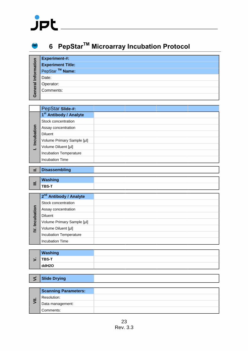
6 PepStarTM
Microarray Incubation Protocol
Gen
era
l In
form
ati
on
Experiment-#:
Experiment Title:
PepStar TM
Name:
Date:
Operator:
Comments:
PepStar Slide-#:
I. In
cu
bati
on
1st
Antibody / Analyte
Stock concentration
Assay concentration
Diluent
Volume Primary Sample [µl]
Volume Diluent [µl]
Incubation Temperature
Incubation Time
II.
Disassembling
III.
Washing
TBS-T
IV. In
cu
bati
on
2nd
Antibody / Analyte
Stock concentration
Assay concentration
Diluent
Volume Primary Sample [µl]
Volume Diluent [µl]
Incubation Temperature
Incubation Time
V.
Washing
TBS-T
ddH2O
VI.
Slide Drying
VII.
Scanning Parameters:
Resolution:
Data management:
Comments:

24 Rev. 3.3
7 Notes / Troubleshooting
Problem Cause Solution
Artifacts Dust particles and resulting scratches
Avoid dust or other particles during each step of the experiment
Use filtered buffers and solutions only
High background Nature of the sample
Sample / 2nd antibody concentration
Insufficient washing
Contaminated wash buffer
Direct fluorescently labeled proteins tend to induce back-ground signals via unspecific binding to the slide surface. Changing of buffer conditions in the incubation step can reduce background signals very efficiently
Additional washing steps can reduce non-specific binding
Variation of blocking buffers (initial blocking steps are not recommended by JPT)
Increased concentrations of
sample / 2nd antibody may
yield high background signals caused by unspecific binding to the slide surface
Adjustment of washing conditions
All buffers and solutions should be prepared freshly every day
Saturated peptide spots
Concentration of the 2nd antibody
Scanning conditions
Higher dilution rates of the 2nd antibody
Adjustment of scanning parameters
Unspecific signals Nature of the sample
Insufficient washing
Variation of blocking buffers
Adjustment of washing conditions

25 Rev. 3.3
Specificity of the 2nd antibody
Control incubations using labeled 2nd antibody alone should be performed in parallel to the actual experiment to ensure that found signals are not caused by non-specific binding of the 2nd antibody to the immobilized peptides
Little or no signals Incubation time
Bleaching effects
Scanning conditions
Warranty of sufficient incubation time
During the incubation step with fluorescently labeled 2nd antibody, protect the slides from light!
After application of secondary antibody keep slides in an ozone-free environment
Adjustment of scanning parameters

26 Rev. 3.3
8 Related Products For further information visit our homepage (www.jpt.com) or contact our customer
support team ([email protected]).
PepStarTM Multiwell: customized peptide microarrays displaying individually
synthesized peptides with capacity for 7 or 21 individual samples
PepSpotsTM: customized peptide arrays on cellulose membranes
Peptide ELISA: peptide coated microtiter plates


