

Respiratory Pathophysiology Respiratory Pathophysiology
• Montana Hospital Association• June 15, 2011 10 - Noon• © Irene Mueller, EdD, RHIA
http://media.healthday.com/images/editorial/respiratory.jpg11

ICD-10-CM/PCS RespiratoryICD-10-CM/PCS Respiratory
• ICD-10-CM– Chapter 10 – J00-J99– Chapter 18 – R04-R09
• ICD-10-PCS– 0B1-0BY– 090-09W (Sinus = part of Ear, Nose, Sinus)
22
Objectives Objectives • Coding-focused
Review of Respiratory system anatomy and physiology
• Focus on the medical knowledge requirements of ICD-10-CM/PCS coding
• Review of ICD-10-CM Ch10– Organization – Guidelines (minimal)
• COPD• Acute Respiratory
Failure• Influenza
– Excludes1 and Excludes2 notes
– Combination and Multiple coding (External causes)
33

S&S in Respiratory SystemS&S in Respiratory System
• Dyspnea / SOB• Orthopnea• Apnea• Tachypnea• Wheezing, Stridor,
Rales, Rhonchi• Coughing
– Sputum/mucus– Hemoptysis
• Nasal Discharge
• Chest Pain• Hypoxemia
– Barrel chest– Cyanosis– Clubbing
• Hiccups
44
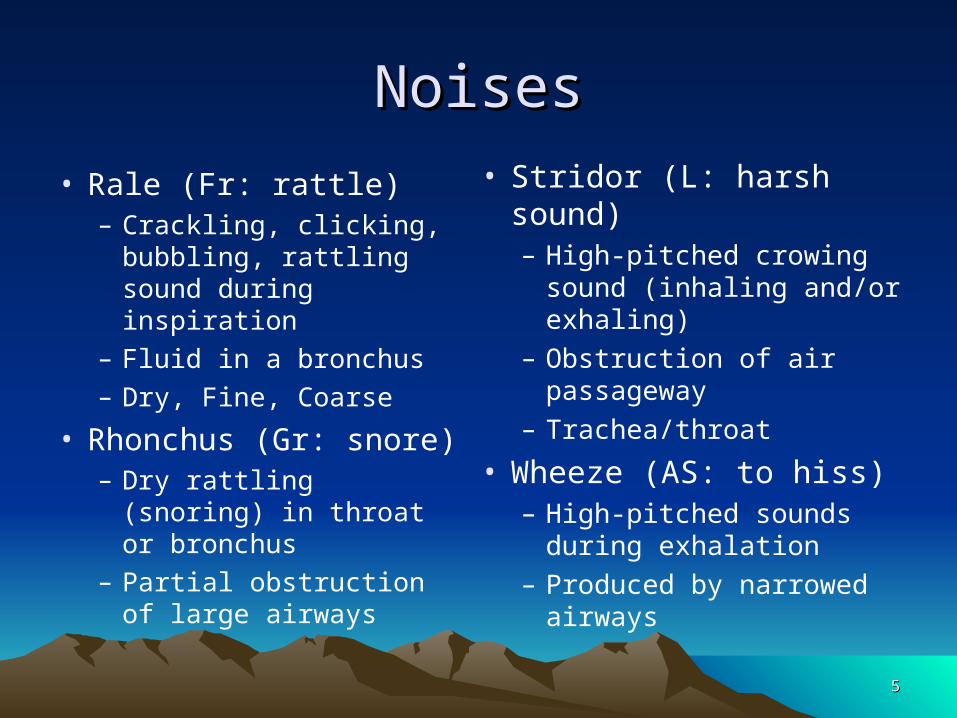
NoisesNoises
• Rale (Fr: rattle)– Crackling, clicking,
bubbling, rattling sound during inspiration
– Fluid in a bronchus– Dry, Fine, Coarse
• Rhonchus (Gr: snore)– Dry rattling (snoring) in
throat or bronchus– Partial obstruction of
large airways
• Stridor (L: harsh sound)– High-pitched crowing
sound (inhaling and/or exhaling)
– Obstruction of air passageway
– Trachea/throat
• Wheeze (AS: to hiss)– High-pitched sounds during
exhalation– Produced by narrowed
airways
55
Respiratory-related chest painRespiratory-related chest pain
• Asthma
• Bronchitis
• Costochondritis (MS chapter)
• Pneumothorax
• Pulmonary embolism (circ Chapter)
• Pulmonary HTN (circ Chapter)
66

Respiratory Respiratory Diseases/ConditionsDiseases/Conditions
• Requested– Asthma– COPD– Respiratory Chest pain
• Pleurisy
– Infections (Pneumonia)
• ICD-10-CM Ch 10– Many combination
codes– Multiple coding– Extensive use of
• Excludes1 notes• Excludes2 notes
77
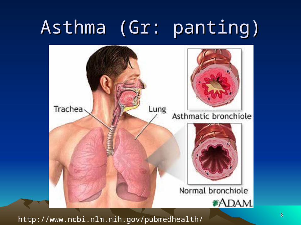
Asthma (Gr: panting)Asthma (Gr: panting)
88http://www.ncbi.nlm.nih.gov/pubmedhealth/PMH0001196/

Definition of AsthmaDefinition of Asthma
• AKA – bronchial asthma• Chronic condition (allergic/non-allergic)
– Affects 5-10% of children– Leading cause of childhood illness– Males 2 times more likely before puberty
• Exposure to allergen when hypersensitive– Bronchospasm - Muscular constriction of bronchi– Mucosal lining swells (edema)– Mucus thickens, can form plugs
99

Two types of AsthmaTwo types of Asthma
• Extrinsic or atopic asthma – type I IgE-mediated
hypersensitivity reaction to foreign antigens.
– Begins in childhood - respiratory tract mast cells are sensitized to a substance extrinsic to body. Clinical examples: pollen, food, animal dander
• Intrinsic or non-atopic asthma
• non-immune reaction Clinical examples: aspirin, virus, stress, exercise.
1010

AsthmaAsthma
• Death rate has increased 8%/yr since 1980s
• Genetic predisposition (100+ genes)– Gene/environment interaction
• Prenatal risk factors– Maternal smoking
• New-onset asthma in adults– Occupational basis
1111
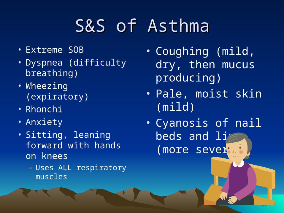
S&S of AsthmaS&S of Asthma• Extreme SOB• Dyspnea (difficulty
breathing)• Wheezing (expiratory)• Rhonchi• Anxiety• Sitting, leaning forward
with hands on knees– Uses ALL respiratory
muscles
• Coughing (mild, dry, then mucus producing)
• Pale, moist skin (mild)• Cyanosis of nail beds
and lips (more severe)
1212
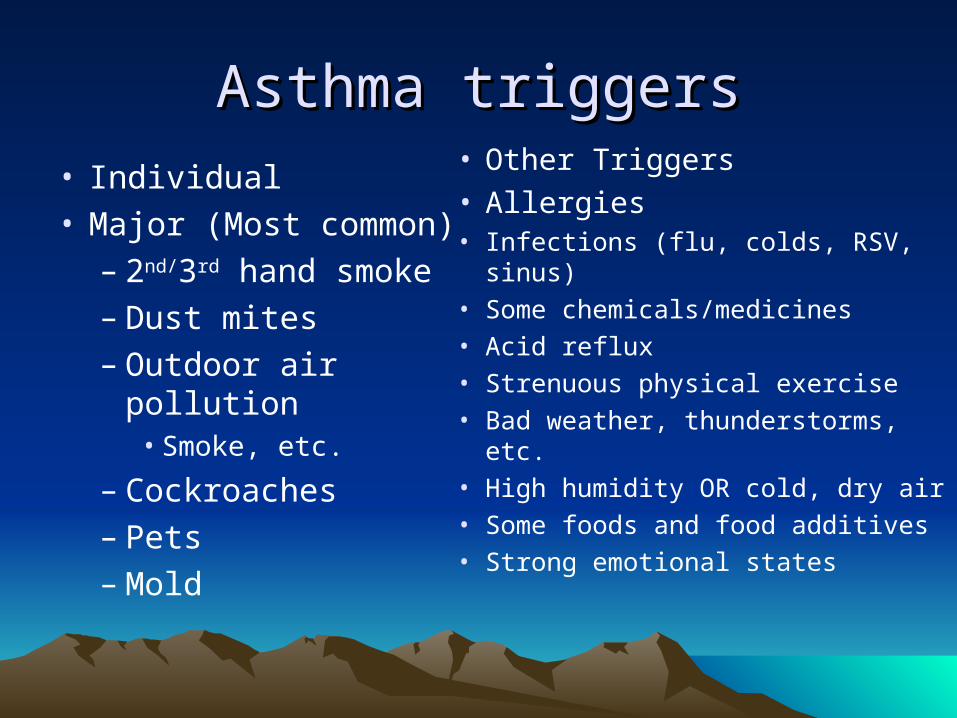
Asthma triggersAsthma triggers
• Individual• Major (Most common)
– 2nd/3rd hand smoke– Dust mites– Outdoor air pollution
• Smoke, etc.
– Cockroaches– Pets– Mold
• Other Triggers• Allergies• Infections (flu, colds, RSV, sinus)• Some chemicals/medicines• Acid reflux• Strenuous physical exercise• Bad weather, thunderstorms, etc.• High humidity OR cold, dry air• Some foods and food additives• Strong emotional states

Status AsthmaticusStatus Asthmaticus
• Life-threatening form of asthma
• Progressively worsening reactive airways
• Unresponsive to usual appropriate therapy
• Leads to pulmonary insufficiency
1414

Asthma diagnosisAsthma diagnosis
• PE• Chest x-rays (usu. Normal, except severe• Pulmonary functions studies• Allergy tests• CBC w/diff leukocyte count
– Inc. eosinophil count, elev. IgE level
• Peak flowmeter to monitor• Peak expiratory flow value indicates degree of
airway obstruction1515

Asthma txAsthma tx
• Avoidance of triggers
• Desensitization
• Education– Deep breathing, posture, relaxation techniques
• Medications– Bronchodilators– Anti-inflammatories– Mucolytics
1616
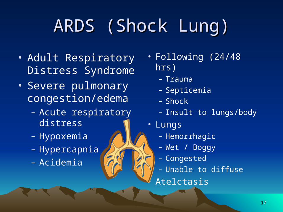
ARDS (Shock Lung)ARDS (Shock Lung)
• Adult Respiratory Distress Syndrome
• Severe pulmonary congestion/edema– Acute respiratory
distress– Hypoxemia– Hypercapnia– Acidemia
• Following (24/48 hrs)– Trauma– Septicemia– Shock– Insult to lungs/body
• Lungs– Hemorrhagic– Wet / Boggy– Congested– Unable to diffuse
• Atelctasis
1717
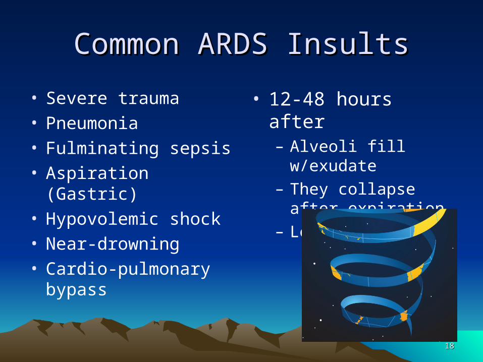
Common ARDS InsultsCommon ARDS Insults
• Severe trauma• Pneumonia• Fulminating sepsis• Aspiration (Gastric)• Hypovolemic shock• Near-drowning• Cardio-pulmonary
bypass
• 12-48 hours after– Alveoli fill w/exudate– They collapse after
expiration– Less gas exchange
1818

ARDS SymptomsARDS Symptoms
• Sudden and severe dyspnea
• Rapid, shallow respirations
• Inspirations– Intercostal &
suprasternal retractions (inward)
– Cyanosis or mottled skin
• Rales, Rhonchi, Wheezes may occur
• NO improvement with O2
1919
ARDS TxARDS Tx• NO cure• Supportive interventions only• Correct underlying cause is attempted• O2, suctioning• Mechanical ventilation• PEEP (Positive end-expiratory pressure)• IV - nutrition and cautious hydration• 60-75% of patients recover
2020
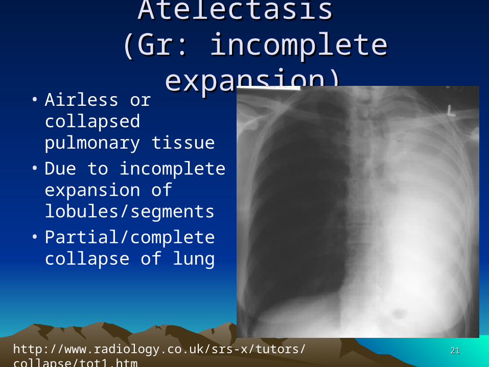
Atelectasis Atelectasis (Gr: incomplete expansion)(Gr: incomplete expansion)
• Airless or collapsed pulmonary tissue
• Due to incomplete expansion of lobules/segments
• Partial/complete collapse of lung
2121http://www.radiology.co.uk/srs-x/tutors/collapse/tot1.htm

Atelectasis S&SAtelectasis S&S
• Hypoxemia• Dyspnea
– Mild to severe
• Substernal retraction• Cyanosis• Diaphoresis• Tachycardia• Anxiety
• Chest x-ray may show mediastinal shift toward collapse
2222

Atelectasis EtiologyAtelectasis Etiology
• Obstruction in bronchial tree
• Mucus plug, FB, Cancer
• Plural effusion
• Lack of deep breathing following surgery
• Prolonged inactivity
• NB– Prematurity, hyaline membrane disease– Narcotics during labor (across placenta)– Mucus plug, Lack of surfactant
2323

Atelectasis Diagnosis/TxAtelectasis Diagnosis/Tx
• Diagnosis• Chest x-rays• H&P
– Deminished breath sounds, Dull percussion
• CT scan of chest• Bronchoscopy if FB
• Treatment• Preventative (surgery)
– Early ambulation– Deep breathing– Coughing
• Suctioning• Spirometry• Antibiotics (if
infection)
2424
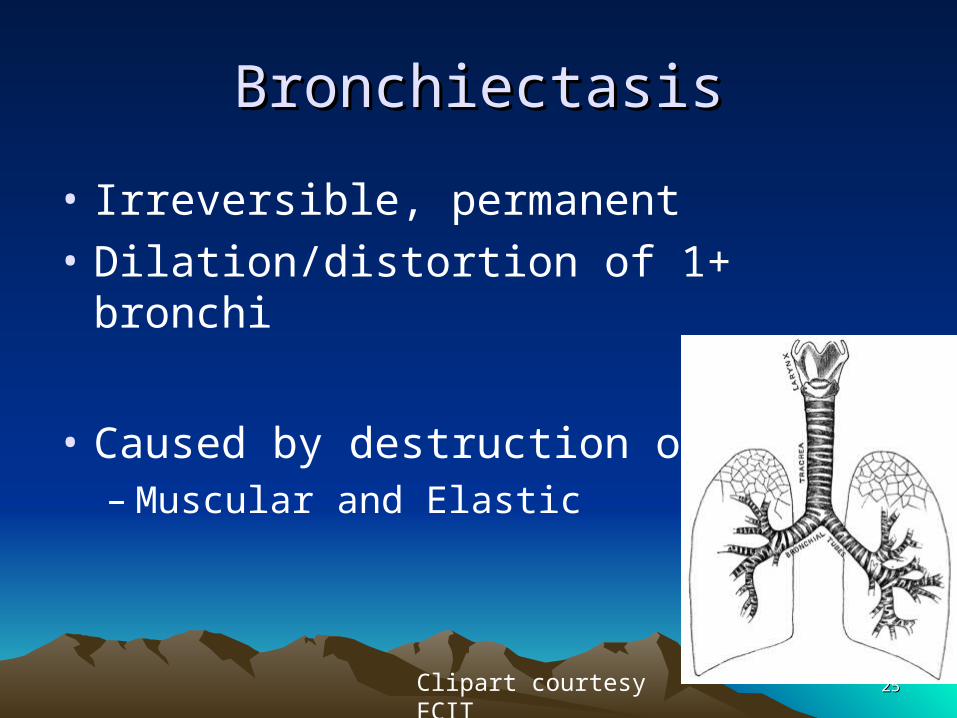
BronchiectasisBronchiectasis
• Irreversible, permanent
• Dilation/distortion of 1+ bronchi
• Caused by destruction of walls– Muscular and Elastic
2525Clipart courtesy FCIT

BronchiectasisBronchiectasis
• Develops over many years
• Usually bilateral• In lower lobes
• S&S– Chronic cough
– Purulent, foul-smelling sputum in large amounts (Classic sign)
– Hemoptysis
– Dyspnea
– Wheezing
– Fever
– General malaise
– Halitosis
2626
Bronchiectasis EtiologiesBronchiectasis Etiologies
• Repeated wall damage
• Recurrent airway infections
• Pneumonia, TB
• Corrosive gas inhalation
• Bronchial obstruction
• Complication of pertussis or measles
• Immune deficiency
2727
Bronchiectasis Dx and TxBronchiectasis Dx and Tx
• Diagnosis– Difficult in early stages– H&P– Chest x-rays– CT scan (high-
resolution)– Bronchoscopy– Sputum culture– PFTs
• Treatment– Antibiotics– Bronchodilators– Avoiding irritants
• Smoking• Pollution
– Surgery to remove affected part of lung
• When much hemoptysis
2828
Bronchitis, acuteBronchitis, acute
• Inflammation of mucosal lining of bronchi
• Cough – deep, persistent, productive
• Sputum – deep yellow to gray
• Other S&S– SOB, wheezing, slight temperature, rales– Pain in upper chest, can be increased w/cough
• Lasts about one week– cough can last 2-3 weeks
2929

Acute BronchitisAcute Bronchitis
• S&S worse in winter
• Cold, damp weather or pollution worsen
• Part of generalURI– Viral or bacterial nasopharyngeal infection– Allergens predisposing factor
• Diagnosis– Chest x-rays, PFTs, ABGs, sputum analysis
3030

Acute Bronchitis TxAcute Bronchitis Tx
• Usually viral, so symptoms are tx
• Aspirin, fluids, vaporizer/humidifier
• Bronchodilator inhaler
• Cough suppressant
• Anti-biotic IF 2ndary bacterial infection
• AVOID primary causative factors– Smoking, pollutants, recurrent resp. infections
3131
Bronchitis, ChronicBronchitis, Chronic
• Inflammation of mucosal lining of bronchi– Persists and worsens
• Mild – slight cough in mornings• Aggravated with URIs (colds, flu)• Obstructive/asthmatic symptoms appear• Dyspnea (coughing, SOB)• Diminished expansion of chest
– Rales and wheezing
• Constant, worse; in cold, damp, pollution3232

Chronic Bronchitis Dx & TxChronic Bronchitis Dx & Tx
• Diagnosis– H&P– R/O other diseases– Chest x-rays– PFTs– ABGs– Sputum analyses
• Guarded prognosis
• Treatment– Based on disease stage– Prompt tx of acute inf.
– Low-flow O2 tx
– Postural drainage– Percussion– Aerosolized
corticosteroids– NO smoking– Avoid crowds
3333

COPD (COLD)COPD (COLD)
• Chronic Obstructive Pulmonary Disease– Progressive, irreversible
• Signs and Symptoms
• Pathophysiology– Chronic Bronchitis– Emphysema
3434

COPDCOPD• Includes several
obstructive lung diseases– Asthma– Bronchiectasis– Chronic bronchitis– Cystic fibrosis
(genetic)– Emphysema
• Pneumoconiosis (occupational dust inhalation)– Fibrosis (stiff tissue)– Asbestosis
• most common – Libby, MT
– Anthracosis• black lung
– Silicosis • stone/metal dust
– Can affect family members of workers
3535

COPDCOPD
• Regardless of cause of obstruction
• Same consequences
• Inability to ventilate lungs easily =
• Ineffective exchange of gases =
• Diminished response to elevated CO2
3636
Costochondritis (M94.0)Costochondritis (M94.0)
• AKA chest wall pain, costosternal syndrome, costosternal chondrodynia
• Pain w/coughing, deep breathing, exertion
• Tietze syndrome =– costrochondritis + swelling
• inflammation of cartilage bet. ribs & sternum
• Can mimic AMI or other heart conditions
3737
Costrochondritis EtiologyCostrochondritis Etiology
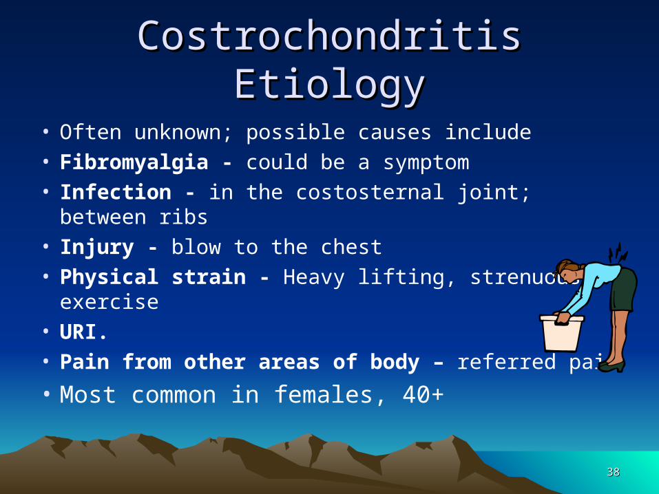
• Often unknown; possible causes include• Fibromyalgia - could be a symptom• Infection - in the costosternal joint; between ribs • Injury - blow to the chest• Physical strain - Heavy lifting, strenuous
exercise• URI. • Pain from other areas of body – referred pain
• Most common in females, 40+3838

Emphysema (Gr: blowing)Emphysema (Gr: blowing)
• Destructive alveolar wall changes
• Permanent enlargement of alveoli spaces
• Alveolar septa are
destroyed
• Interferes with
breathing and gas
exchange
3939http://imglib.lbl.gov/ImgLib/COLLECTIONS/LUNG_STRUCTURE/.tour/pores.html
Emphysema S&SEmphysema S&S
• Decreased area for gas exchange = dyspnea
• S&S onset is insidious (gradual)– Dyspnea, tachypnea,
wheezing
– Cough slight or not present
– Must use accessory muscles to force trapped air out
• Barrel chest• Pursed lips
• More females dx than males since 2000
• Most common cause of death from respiratory disease
• 4th leading cause of death in US
4040
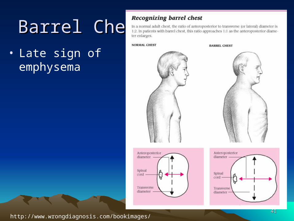
Barrel ChestBarrel Chest• Late sign of
emphysema
4141http://www.wrongdiagnosis.com/bookimages/8/2495.png

Emphysema EtiologyEmphysema Etiology
• Smoking is major risk factor (up to 85%)– Childhood 2nd-hand
• 3 major types– localized (distal acinar,
paraseptal)– centrilobular
(centriacinar)• Most common, usu.
caused by smoking
– panlobular (panacinar)
• Repeated respiratory tract infections
• Pollution– Ozone, sulfur dioxide,
nitrogen oxides, occupational
• Familial tendency (2%)– alpha1-antitrypsin enzyme
deficiency
• POOR prognosis
4242
Emphysema Dx and TxEmphysema Dx and Tx
• Diagnosis– H&P– PFT
• Increased tidal volume
• Increased residual vol.
• Decreased vital capacity
– Chest x-rays• Depressed diaphragm
• Translucent lungs
– Blood gases• Increased CO2
• Treatment– Avoid smoke and other
irritants– Avoid exposure to RTI– Flu vaccinations– Low flow O2– Meds– Pulmonary rehab– Surgery
• Experimental• Lung reduction• Lung transplantation
4343
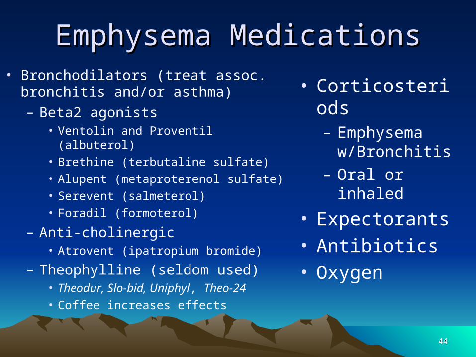
Emphysema MedicationsEmphysema Medications• Bronchodilators (treat assoc.
bronchitis and/or asthma)– Beta2 agonists
• Ventolin and Proventil (albuterol)• Brethine (terbutaline sulfate)• Alupent (metaproterenol sulfate)• Serevent (salmeterol)• Foradil (formoterol)
– Anti-cholinergic • Atrovent (ipatropium bromide)
– Theophylline (seldom used)• Theodur, Slo-bid, Uniphyl, Theo-24• Coffee increases effects
• Corticosteriods– Emphysema
w/Bronchitis– Oral or inhaled
• Expectorants• Antibiotics• Oxygen
4444
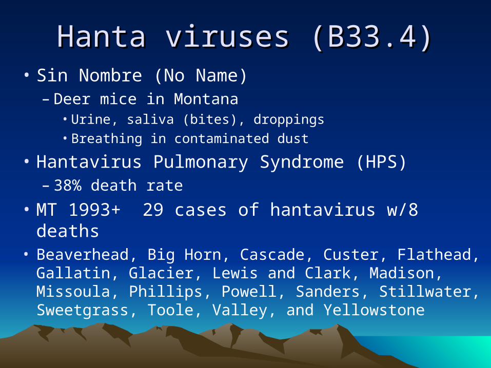
Hanta viruses (B33.4)Hanta viruses (B33.4)• Sin Nombre (No Name)
– Deer mice in Montana• Urine, saliva (bites), droppings• Breathing in contaminated dust
• Hantavirus Pulmonary Syndrome (HPS)– 38% death rate
• MT 1993+ 29 cases of hantavirus w/8 deaths• Beaverhead, Big Horn, Cascade, Custer, Flathead,
Gallatin, Glacier, Lewis and Clark, Madison, Missoula, Phillips, Powell, Sanders, Stillwater, Sweetgrass, Toole, Valley, and Yellowstone
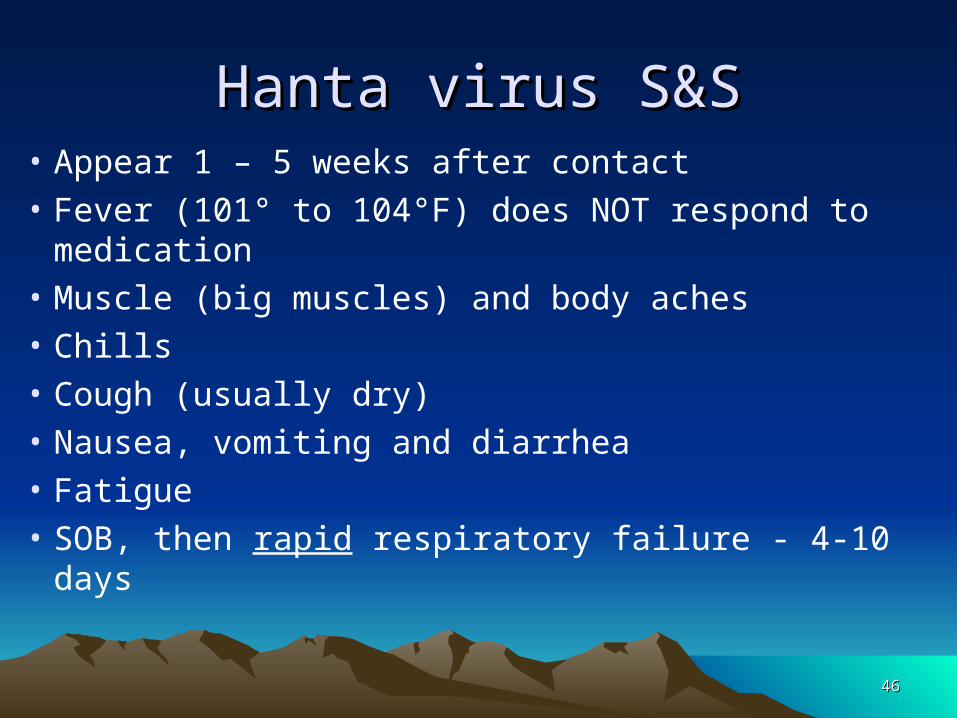
Hanta virus S&SHanta virus S&S• Appear 1 – 5 weeks after contact• Fever (101° to 104°F) does NOT respond to
medication• Muscle (big muscles) and body aches• Chills• Cough (usually dry)• Nausea, vomiting and diarrhea• Fatigue• SOB, then rapid respiratory failure - 4-10 days
4646

Hanta virus Dx & TxHanta virus Dx & Tx
• Diagnosis• H&P• Exposure to rodents
• Treatment• Supportive (ICU)
– Earlier the better
4747

InfluenzaInfluenza
• Acute, highly contagious, viral respiratory infection
• Spread by coughing
• Many strains– A, B, C– H0N1, H2N2, H3N2, etc.
• Patients may die w/in 48 hours
4848

4949
InfluenzaInfluenza• Three categories of virus
• Type A– Most serious– Infects Hmans and animals - including birds, pigs,
horses, whales, and seals
• Type B– Usually only in Humans
• Type C– Least serious– Usually only in Humans
5050
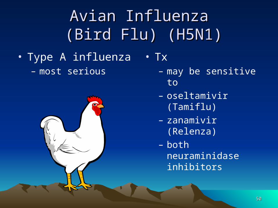
Avian InfluenzaAvian Influenza (Bird Flu) (H5N1) (Bird Flu) (H5N1)
• Type A influenza– most serious
• Tx – may be sensitive to – oseltamivir (Tamiflu)– zanamivir (Relenza)– both neuraminidase
inhibitors
5151
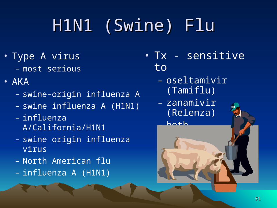
H1N1 (Swine) Flu H1N1 (Swine) Flu
• Type A virus– most serious
• AKA– swine-origin influenza A – swine influenza A (H1N1)– influenza A/California/H1N1– swine origin influenza virus– North American flu– influenza A (H1N1)
• Tx - sensitive to – oseltamivir (Tamiflu)– zanamivir (Relenza)– both neuraminidase
inhibitors

Influenza S&SInfluenza S&S
• Sudden fever• Chills• Headache, back and muscle pain
• Cough, runny nose, sore throat• Sneezing, N&V, hoarseness, diarrhea• Complications following flu
– Bronchopneumonia, neuritis, otitis media, pleurisy
5252

Influenza Dx & TxInfluenza Dx & Tx
• Diagnosis– Similar to cold– Duration of S&S – Epidemics in winter
and early spring– Severity of S&S
• Throat culture
• SymptomaticTreatment– Bedrest– Anagesics– Antipyretics– Antibiotics IF 2ndary inf.
• Staph, strep, pneumococcus
• Annual Vaccinations to prevent
5353

Pleural Effusion (Hydrothorax)Pleural Effusion (Hydrothorax)
• Fluid in the chest cavity• Due to
– CHF, TB, pneumonia
• Asymptomatic OR• Dyspnea and chest or pleuritic pain• Chest x-ray confirms dx• Tx
– Thoracentesis to drain– Underlying cause
5454
Pleurisy (Pleuritis)Pleurisy (Pleuritis)• Inflammation of pleural membranes
• Usually 2ndary to other diseases/infections
• Injury
• Tumor
• 2 types (Wet and Dry)– Increased pleural fluid compresses lung
w/dyspnea (wet)– Decreased pleural fluid; layers rub together (dry)
• Congested and edematous
5555

Pleurisy S&SPleurisy S&S
• Sharp, needlelike pain
• Increasing with coughing or inspiration
• Cough
• Fever/Chills
• Shallow rapid breathing
5656
Pleurisy Dx & TxPleurisy Dx & Tx
• Diagnosis– H&P– Pleural rub on
auscultation of lungs– X-rays
• May leave permanent adhesions, restricting lung expansion
• Treatment– Underlying cause– Antibiotics– Analgesics– Splinting chest– Deep breathing
exercises
5757

Break TimeBreak Time
Fluid Exchanges
5858


