
Hindawi Publishing CorporationGastroenterology Research and PracticeVolume 2012, Article ID 872716, 16 pagesdoi:10.1155/2012/872716
Review Article
Probiotics, Prebiotics, and Synbiotics: Gut and Beyond
Usha Vyas and Natarajan Ranganathan
Kibow Biotech Inc., Newtown Business Center, 4781 West Chester Pike, Newtown Square, PA 19073, USA
Correspondence should be addressed to Usha Vyas, [email protected]
Received 16 March 2012; Accepted 20 July 2012
Academic Editor: Maurizio Gabrielli
Copyright © 2012 U. Vyas and N. Ranganathan. This is an open access article distributed under the Creative CommonsAttribution License, which permits unrestricted use, distribution, and reproduction in any medium, provided the original work isproperly cited.
The human intestinal tract has been colonized by thousands of species of bacteria during the coevolution of man and microbes.Gut-borne microbes outnumber the total number of body tissue cells by a factor of ten. Recent metagenomic analysis of the humangut microbiota has revealed the presence of some 3.3 million genes, as compared to the mere 23 thousand genes present in thecells of the tissues in the entire human body. Evidence for various beneficial roles of the intestinal microbiota in human health anddisease is expanding rapidly. Perturbation of the intestinal microbiota may lead to chronic diseases such as autoimmune diseases,colon cancers, gastric ulcers, cardiovascular disease, functional bowel diseases, and obesity. Restoration of the gut microbiota maybe difficult to accomplish, but the use of probiotics has led to promising results in a large number of well-designed (clinical)studies. Microbiomics has spurred a dramatic increase in scientific, industrial, and public interest in probiotics and prebioticsas possible agents for gut microbiota management and control. Genomics and bioinformatics tools may allow us to establishmechanistic relationships among gut microbiota, health status, and the effects of drugs in the individual. This will hopefullyprovide perspectives for personalized gut microbiota management.
1. Introduction
Bacteria, unicellular eukaryotes, and other organisms inhabitthe human body in large numbers. The human gut is dom-inated by several bacterial phyla including Bacteroidetes,Firmicutes, and Actinobacteria. The term “microbiota,” “mi-croflora,” or “normal flora” is used to designate this vast hostof microbes which coexist with the host [1–3].
It is estimated that the human microbiota contains asmany as 1014 bacterial cells, a number that is 10 times greaterthan the number of human cells present in our bodies [4–6].Virtually every surface of the human body starting from theskin surface to the genitourinary tract, oral cavity, respiratorytract, ear, and the gastrointestinal tract is colonized heavilyby various species of bacteria [3, 7–9]. By far, the mostheavily colonized organ is the gastrointestinal tract (GIT)which houses a huge microbial ecosystem; the colon aloneis estimated to contain over 70% of all the microbes in thehuman body [4, 6].
The gut microbiota or microflora has a crucial role inhuman health and disease. The GIT is comprised of the entiredigestive system from the stomach to the anus. The colon
or the large intestine is the organ which is the preferredsite for bacterial colonization. The GIT is also rich in manymolecules which can be used as nutrients by microbes.Hence the GIT has the potential to be heavily colonized byvarious bacteria both harmful and beneficial. The mucosaof the gastrointestinal tract is continuously exposed to anenvironment that is rich in foreign substances, such asfood particles and antigens of microbial origin. Particularchanges in the intestinal ecosystem might contribute tothe development of certain illness. There is therefore aneed for an exhaustive review on the functions of thegut microbiota, occurrence of gut dysbiosis (alteration orimbalance of the microflora), how these intestinal bacteriatrigger development of disease once the normal flora of ahealthy individual is imbalanced, exploiting this intricateand interwoven ecosystem for understanding human health,development of biotherapeutics, and future perspectives.
The imbalanced gut bacteria have been studied in diseas-es such as inflammatory bowel disease, antibiotic-associateddiarrhea, colon cancer, hypercholesterolemia, and others.Lactic acid bacteria, belonging to the genus Lactobacillusand Bifidobacterium, have been shown to positively influence
2 Gastroenterology Research and Practice
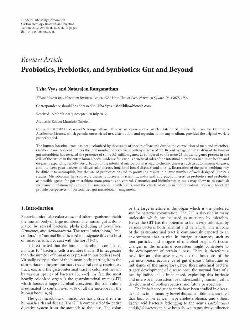
“the microbiota can be viewed as a metabolic organ exquisitely tuned to our
physiology that performs function we have not had to evolve on our own”
Backhed et al. 2004. PNAS 101:15718-15723
Amount of bacteria per gram of cellular component
Human body 1013
cells 23,000 genescells on the human body.
3.3 million genes
Nose
Mouth
Smallintestine
Skin
Lungs
Stomach
Colon
RectumUrinary/vaginaltract(s)
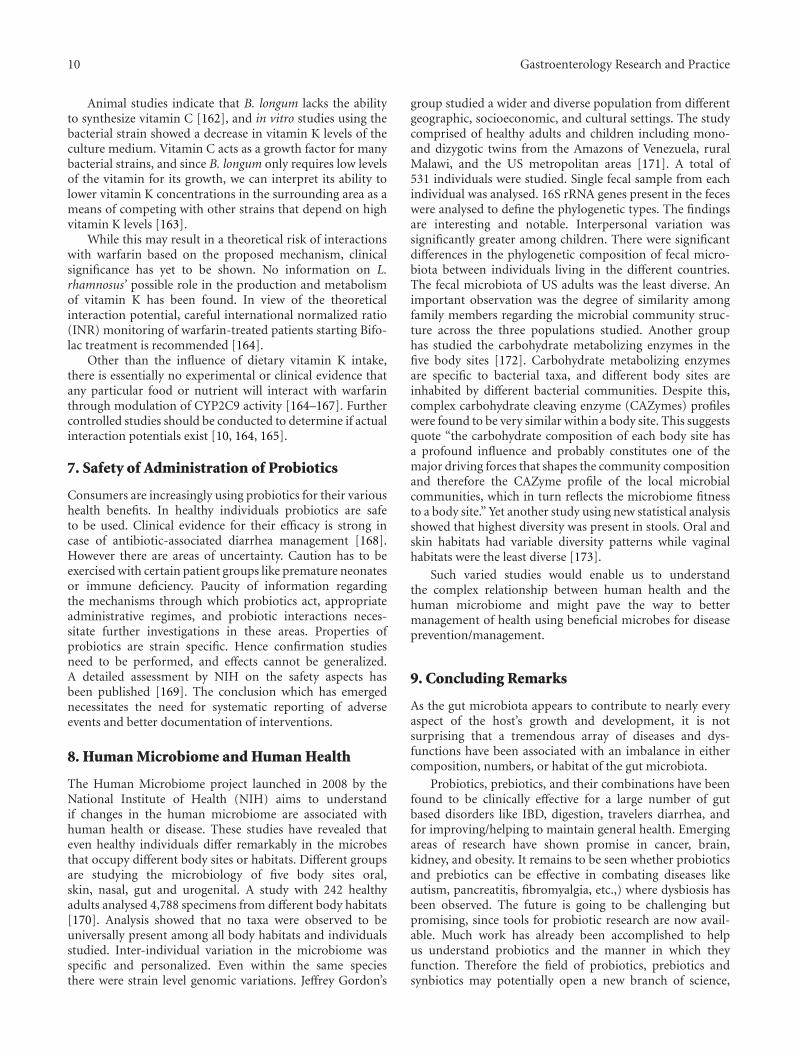
14 microbialNormal flora 10
1 to 102 cells• Stomach−10
3 cells• Duodenum−10
4 cells• Jejunum−10
4 to 107 cells• Ileum 10
10 to 1011 cells• Proximal colon 10
11 to 1012 cells• Transverse colon 10
>1012 cells• Distal colon
Nose
Mouth
mallestine
Skin
Lung
Stom
Colon
RectumUrinary/vaginal
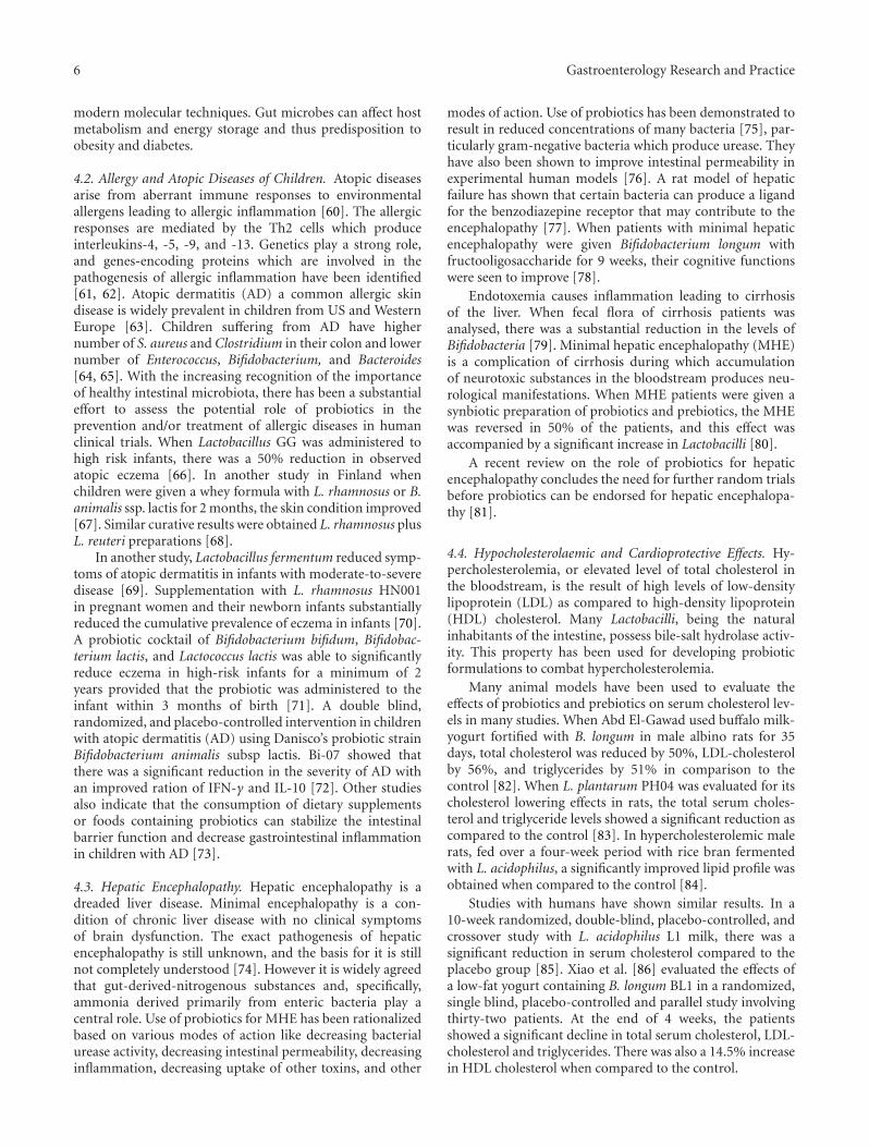
Figure 1: The Human Body and number of bacteria present in the total microflora.
health. Hence, re-establishing the balance by using thesebacteria (termed “probiotics”) for disease treatment and pre-vention should prove advantageous. Probiotics along withprebiotics and synbiotics have been used and studied invarious disease areas. Several studies have indicated that analtered gut microbiota is associated with several diseases thatare particularly prevalent in the 21st century.
N. Williams [10] has previously reviewed the pharmacol-ogy, uses, dosage, safety, drug interactions, and contraindica-tions of probiotics. The first part of this updated and currentreview will give an overview of the gut microbiota andits main characteristics and describe the major factors thatcould modulate gut microbiota composition. The secondpart describes various new diseases and reports on studiesin which probiotics, prebiotics, and synbiotics have beenused. The virtues of probiotics are already well recognizedfor general gut health, antibiotic-associated diarrhea, andimmunity. Application of these areas will not be examined.The last part of the review focuses on future potentialapplications of probiotics, prebiotics and synbiotics in newemerging areas of studies like autism and gut-brain connec-tion. Finally, the paper will conclude with a discussion on thefuture of this field. A short review article of this length cannotdo full justice to this field. A broad overview, composed ofexcerpts taken from various publications including reviewarticles, is presented here. For each of the important areas,we have included references to review articles for readerswishing to delve and analyze more deeply.
2. The Gut Microbiota and Functions
A newborn baby has a sterile gut that is colonized by bacteriafrom the mother and from the baby’s surroundings or envi-ronment [11]. An adult human has 10 times more bacterial
cells on, and in, the entire body as compared to the totalhuman cells (Figure 1). The human microbiome is highlycomplex and diverse. Its composition and number variesfrom the nose and mouth to the distal colon and rectum. Thecomposition and complexity of the gut microbiota changeswhen the baby is weaned to solid foods. Dietary changes inadulthood are also greatly responsible for the compositionof gut microbiota. Development of 16S ribosomal RNA(rRNA) gene-sequence-based metagenomic methods has ledto major advances in defining the total microbial populationof the gut [12]. This technique has been used to showthat 90% of the bacteria belong to two phyla, namely, theBacteroidetes and Firmicutes [13].
The gut microbiota plays an important role in themaintenance of health. These are summarized below.
2.1. Structure and Histological Function. The intestinal struc-ture and function is ensured by the microbiota presentwithin. The intestinal mucus layer is a balance of mucin se-cretion and degradation. This mucin layer creates an obstacleto proinflammatory compounds and uptake of antigens [14].Evidence indicates that butyrate induces secretion of mucin,antimicrobial peptides, and other factors. This reinforces thedefense barrier in the colon [15].
Secondly, the gut microflora has a role in the develop-ment of cell and tissue. Butyrate, a short chain fatty acidthat is secreted by these colonic microbes, regulates cellgrowth and differentiation, inhibits transformation of cellgrowth and helps in reverting the cells from a neoplastic toa nonneoplastic phenotype [16]. The development of themicrovasculature of the intestinal villi is dependent on theindigenous microbes. This has been demonstrated in studiesusing germ-free mice and its subsequent colonization by B.thetaiotamicron by Jeffrey Gordon’s group [17]. This signifies
Gastroenterology Research and Practice 3
SCFAs
AMPs
↑ BacteroidetesB. fragilis (PSA)
Gm-PGGm-LPS
ImmunocompetenceTolerance
DC tolerization
E. coli
Cellular immunityLymphoid organogenesis
Mucosal immunity
↑ IgA
↑ Lapactivation
inactivation
B. thetaiotaomicron
PeristalsisGlycosylation
Barriermaintenance
Bifidobacterium spp.Clostridium spp.
SCFAmetabolism
Lipidmetabolism
Conjugation oflinoleic acid
Xenobiotics metabolismDrug disposition
GIT surface maturationGIT functional maturation
Normalization ofHPA stress response
B. infantis
Angiogenesis
O. formigenes
Lactobacillus spp.
↓ Oxalateexcretion
Behavior
Nutrition
NFκB
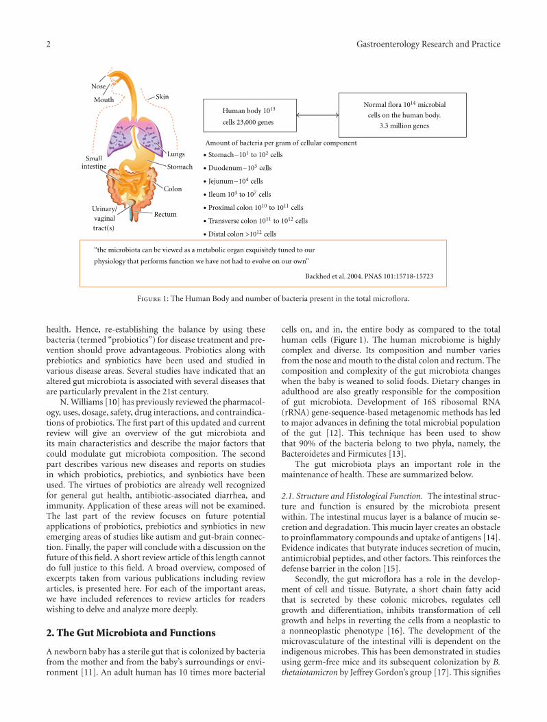
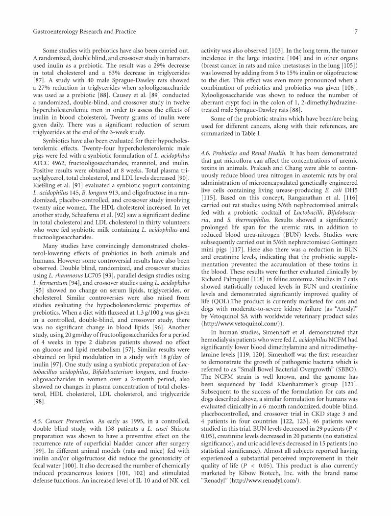
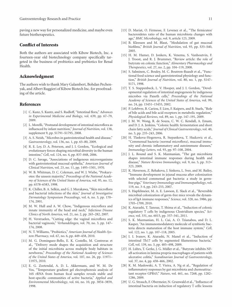
Figure 2: The complex web of gut microbiota contributions to host physiology. Different gut microflora components can affect many aspectsof normal host development, while the microbiota as a whole often exhibits functional redundancy. Members of the microbiota are shownin gray, with their components or products of their metabolism. Their effects on the host at the cellular or organ level are shown in white.Black ellipses represent the affected host phenotypes. Only some examples of microbial members/components contributing to any givenphenotype are shown. AMP: antimicrobial peptides; DC: dendritic cells; Gm−: gram negative; HPA: hypothalamus-pituitary adrenal; Iap:intestinal alkaline phosphatase; PG: peptidoglycan; PSA: polysaccharide A. Extracted from: Phys Rev 2010 Sekirov et al.
the importance of the gut microbes in the development ofthe structure and morphology of the gut (Figure 2).
2.2. Metabolic Functions. The gut bacteria are known toproduce a large number of vitamins like the B group ofvitamins, synthesize amino acids, and carry out biotransfor-mation of the bile. Biotransformation of bile by microbialenzymes is important for the metabolism of glucose andcholesterol [18]. Importantly, the microbiome provides themuch needed biochemical pathways for the fermentation ofnondigestible substrates like fibers and endogenous mucus.Fermentation or metabolism of these nondigestible sub-strates leads to the growth of these microbes and the pro-duction of short chain fatty acids and gases [19]. The majorshort-chain fatty acids produced are acetate, propionate,and butyrate. Other bacterial end products include lactate,ethanol, succinate, formate, valerate, caproate, isobutyrate,2-methyl-butyrate, and isovalerate. Bacterial fermentationtakes place in the cecum and colon, where the short-chainfatty acids are absorbed, stimulating the absorption of saltsand water. These short-chain fatty acids have a protectiveeffect on the intestinal epithelium [19]. The colonic bacteriaprefer butyrate as the sole source of energy, and most of it iscompletely metabolized. The principal short chain fatty acid
produced in the colon is acetate, and it serves as a substratefor biosynthesis of cholesterol. Thus the gut microbiotaperforms various metabolic acitivities which are essential forthe host’s metabolism (Figure 2).
2.3. Protective Functions. Many of the commensal organ-isms produce antimicrobial compounds and compete fornutrients and sites of attachment in the gut lining, therebypreventing colonization by pathogens. This helps reduce theproduction of lipopolysaccharides and peptidoglycans whichcan all be detrimental to the host [20]. The development ofthe immune system is also governed by the nature of theindigenous microflora [21]. Germ free animals have fewerdendritic cells, and evidence shows that bacterial systemshave a role to play in development of B cells [22, 23]. Thedevelopment of regulatory T cells, T helper type 1 and 2 cells,and T helper 17 cells is also dependent on the signals given bythe intestinal bacteria [24–26]. Short-chain fatty acids, suchas butyrate, have been shown to inhibit NF-kB in patientswith ulcerative colitis thus exerting immunomodulatoryeffects [27, 28].
These concepts illustrate a dynamic relationship betweenthe immune system and the microbiota. The intestinalmucosa averts threats by signaling to the innate immune

4 Gastroenterology Research and Practice
system through toll-like receptors. These recognize and bindto specific microbial macromolecules, like lipopolysaccha-ride, flagellin, peptidoglycan, and N-formylated peptides. Inthe intestinal mucosa, the activation of toll-like receptorsinitiates nuclear factor-kB pathways, mitogen-activated pro-tein kinase, and caspase-dependent signaling cascades. Theselead to the production and release of protective peptides,cytokines, chemokines, and phagocytes. The result can be aprotective response to commensal bacteria, an inflammatoryresponse to pathogenic organisms, or a trigger of apoptosis.Therefore, commensal bacteria of the gastrointestinal tractplay active roles in the development and homeostasis of theimmune system, as shown in Figure 2.
3. Dysbiosis and Modulating ofthe Gut Microbiota
Normal physiology of the host depends on the signals givenby the intestinal microbes. The intestinal lumen consistingof gastric acid, digestive enzymes, and IgA constitutes thefirst line of defense and is lethal to invading and ingestedpathogenic bacteria. The indigenous microbes degradeintraluminal antigens and inhibit the pathogenic microbesfrom adherence and colonization. They also are necessaryfor the induction of regulatory T cells [29]. Any changesto this microbial ecosystem could cause an imbalance ordysregulation of the microbiota (dysbiosis) often associatedwith various disease states ranging from the most commonIBD [30, 31] and IBS [32] to the more unexpected activationof chronic human immunodeficiency virus (HIV) infection[33] and generation of atopy [34–36] (Figure 3).
It is therefore important to reestablish the bacterialhomeostasis which may have been disturbed by any or severalfactors. One of the ways to favorably alter the intestinalmicrobiota is through the use of prebiotics, probiotics, andsynbiotics (a combination of both prebiotics and probi-otics given together). These agents can favorably influencemicrobial interactions with the immune system and gutepithelium.
A prebiotic is a selectively fermented ingredient thatresults in specific changes in the composition and/or activityof the gastrointestinal microbiota, thus conferring healthbenefit(s) upon the host. Prebiotics are generally oligomersmade up of 4 to 10 monomeric hexose units.
Probiotics, according to the currently adopted definitionby FAO/WHO [37, 38], are “Live microorganisms whichwhen administered in adequate amounts confer a health ben-efit on the host.” The International Scientific Associationfor Probiotics and Prebiotics (ISAPP with Glenn Gibson,Todd Klaenhammer, and Mary Ellen Sanders on its board ofdirectors) and the International Probiotic Association (IPA,an association of over 150 probiotic business organizationsmanufacturing and distributing probiotics) are two groupswhich are working with these beneficial microbes.
Synbiotics is a combination of probiotics and prebioticsadministered together.
Common, well-known beneficial bacteria which have along-standing association with health include lactic acid pro-ducing genera such as the Bifidobacteria or Lactobacilli. These
bacteria can be introduced into the gut and/or encouragedto multiply either through ingestion by the individual ofappropriate probiotic strains or through the provision ofprebiotic growth substrates also known as soluble fibers.
That probiotics and prebiotics are becoming increasinglypopular is evidenced by rapidly expanding research supportand an ever widening choice of products. Probiotics and pre-biotics are available commercially in many forms, includingfoods, dietary supplements, and clinical therapeutics withoral or non-oral delivery.
To be a candidate for commercialization, a probioticmust retain its properties during large-scale industrial prepa-ration. Naturally, it should also remain viable and stableduring storage and use. For most applications, the probioticshould be able to survive in the intestinal ecosystem andthe host animal should gain beneficially from its presence.Clearly, the organisms used should be “generally regarded assafe”-GRAS as per USFDA regulations or well documentedin the literature.
Prebiotics must provide selective stimulation of thegrowth or activity of beneficial native bacteria. Since prebi-otics are non-viable, stability is not a concern, but safe con-sumption levels must be established. A detailed guideline forprobiotics and prebiotics has been published by the WorldGastroenterology Organization [39].
4. Clinical Applications of Various Probiotics,Prebiotics and Synbiotics
4.1. Gut Microbiota and Obesity. The metabolic equilibriumof the host is maintained by the gut microbes [40, 41].One study in adult population with type 2 diabetes [42]has shown that their gut microbiota differs from that ofnon-diabetic adults, and that health may potentially improvewhen the gut microflora is modified by the administration ofprobiotics and prebiotics. In spite of these findings, and therelationship between diabetes and abdominal fat, few studieshave been aimed at finding correlations between the compo-sition of the microbiota and the occurrence of inflammationand metabolic alterations in individuals with obesity [42,43]. A study in patients with diabetes mellitus showed thatthese individuals had a lower number of Faecalibacteriumprausnitzii and an increase in with inflammatory markers[43]. Obesity was found to be associated with large changesin the abundances of different bacteria from different taxa[44].
The Bifidobacteria population (and most other organ-isms in the group of Firmicutes) is slightly lower inindividuals with obesity than in lean people [45]. A similarfinding was reported in patients with type 2 diabetesmellitus in comparison with nondiabetic patients [46]. Thesefindings suggest that Bifidobacteria may play a part inthe development of obesity and its related comorbidities.When prebiotics like inulin-type fructans were fed to mice,these were used as energy substrates by bacteria [47–49]. The number of Bifidobacteria increased significantly,and there was an inverse correlation with the levels oflipopolysaccharide, glucose tolerance and development of fat
Gastroenterology Research and Practice 5
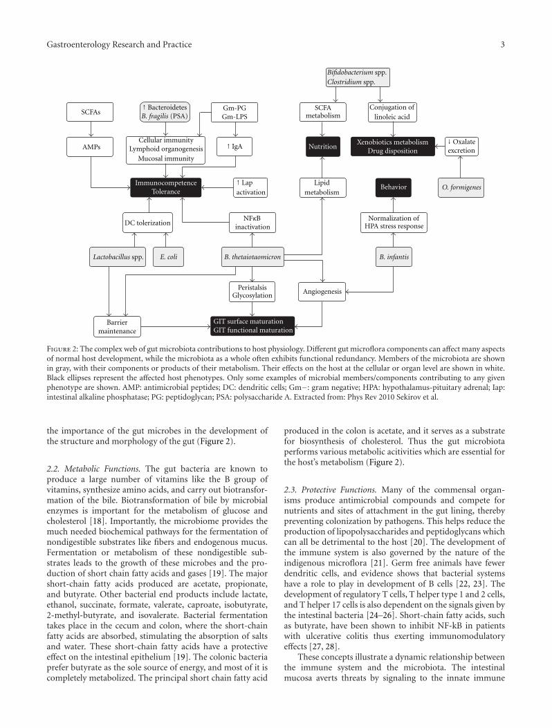
Gut-brain hypothesis1. Autism↑ C. bolteae/clostridia spores
mechanism unknown2. Mood: depression, anxiety
Asthma/atopyHygiene hypothesis:Exagergated innate immune responseUpregulation of regulatory T cellsafter capture of Ags by DCs↓↑ Clostridia
Hypertension/
ischemicheartdisease
Biliary disease Altered enterohepatic circulation of bile
Diet high in red meat and animal fatLow SCFA/butyrateHigh fecal fatsLow vitamin absorption↑ 7α dehydroxylating bacteria:cholic acid → deoxycholic acid (cocarcinogen)Low in H2S metabolizing bacteria
Altered xenobiotic/drug metabolism
e.g., paracetamol metabolism:↑ predose urinary p-cresol sulfate leads to ↓ postdose urinaryacetaminophen sulfate: acetaminophen glucuronide.Bacterially mediated p-cresol generation and competitiveo-sulfonation of p-cresol reduces the effective systemic capacityto sulfonate acetaminophen.
Peripheral vascular disease
Result of metabolic syndrome-
metabolism
Obesity/metabolic syndrome
↓ Bacteroidetes and ↑ Actinobacteria in obeseAltered energy/lipid metabolismHigher relative abundance of glycoside hydrolases,carbohydrate-binding modules,glycosyltransferases, polysaccharide lyases, and carbohydrateesterases in the BacteroidetesTLR mediated
Inflammatory bowel disease
Hygiene hypothesisAltered immune response: TLR signalingLess microbial diversityActivation of specific species e.g., Escherichia
Bifidobacteria, gram
altered lipid deposition/
Colon cancer
+ve organisms
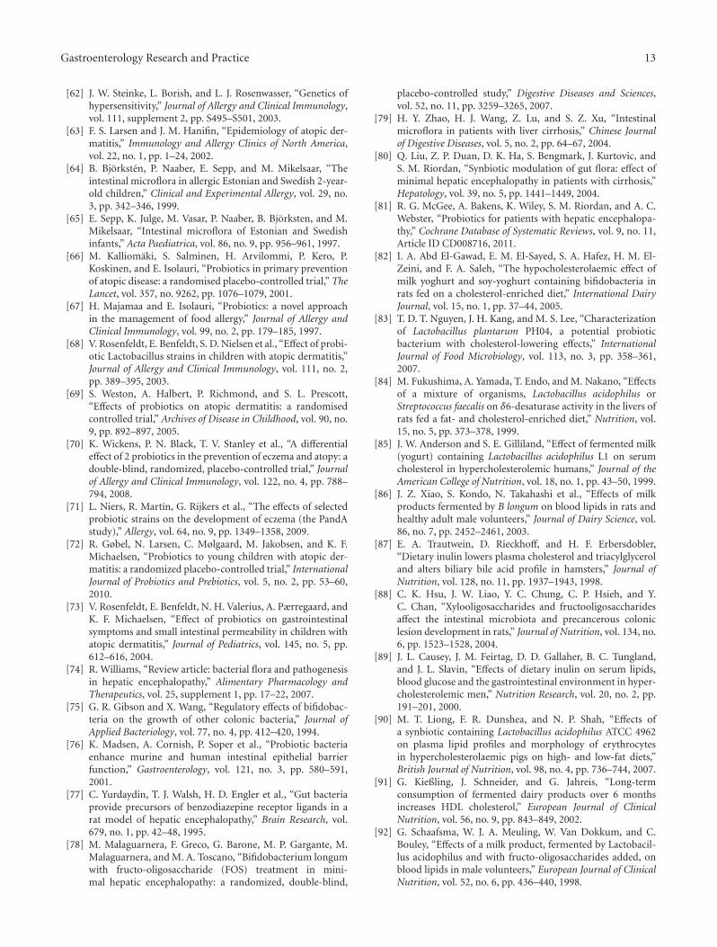
Figure 3: Diseases influenced by gut microbial metabolism. The variety of systemic diseases that are directly influenced by gut microbialmetabolism and its influence on other mammalian pathways, such as the innate immune system, are shown. Specifically highlighted arethe metabolic pathways involved in drug metabolism and obesity that are directly influenced by the gut microbial content. Ags, antigens; C.bolteae, Clostridium bolteae; DCs; dendritic cells; SCFA, short-chain fatty acid; TLR, toll-like receptor. Kinross et al. Genome Medicine 20113:14.
mass [47, 48]. Moreover the prebiotic approach preventedthe overexpression of several host genes that are related toadiposity and inflammation.
Studies have been carried out using probiotics to pro-mote specific changes in the gut microbiota. Angiopoetin-related protein 4 (Angptl4), a lipoprotein lipase inhibitorwhich inhibits the uptake of fatty acids from circulatingtriglyceride-rich lipoproteins in white adipose and muscletissues was found to be increased in mice fed with ahigh fat diet supplemented with L. paracasei [50]. Obeseindividuals when administered with Lactobacillus acidophilusNCFM and Lactobacillus gasseri SBT2055 showed a decrease
in fat mass and the risk of type 2 diabetes mellitus andinsulin resistance [51, 52]. In the active group which con-sumed L. gasseri, abdominal, visceral, and subcutaneous fatareas decreased significantly. Body weight also decreasedsignificantly. In the L. acidophilus NCFM study the insulinsensitivity was preserved, but there was no effect on thesystemic inflammatory response. Clinical trials using pre-biotics like arabinoxylan [53–55] and inulin-type fructans[56–58] have shown positive results in diabetic, overweight,and obese populations. A review article [59] discusses thetight relationship which exists between mammalian gutcomposition and functions and the host metabolism using
6 Gastroenterology Research and Practice
modern molecular techniques. Gut microbes can affect hostmetabolism and energy storage and thus predisposition toobesity and diabetes.
4.2. Allergy and Atopic Diseases of Children. Atopic diseasesarise from aberrant immune responses to environmentalallergens leading to allergic inflammation [60]. The allergicresponses are mediated by the Th2 cells which produceinterleukins-4, -5, -9, and -13. Genetics play a strong role,and genes-encoding proteins which are involved in thepathogenesis of allergic inflammation have been identified[61, 62]. Atopic dermatitis (AD) a common allergic skindisease is widely prevalent in children from US and WesternEurope [63]. Children suffering from AD have highernumber of S. aureus and Clostridium in their colon and lowernumber of Enterococcus, Bifidobacterium, and Bacteroides[64, 65]. With the increasing recognition of the importanceof healthy intestinal microbiota, there has been a substantialeffort to assess the potential role of probiotics in theprevention and/or treatment of allergic diseases in humanclinical trials. When Lactobacillus GG was administered tohigh risk infants, there was a 50% reduction in observedatopic eczema [66]. In another study in Finland whenchildren were given a whey formula with L. rhamnosus or B.animalis ssp. lactis for 2 months, the skin condition improved[67]. Similar curative results were obtained L. rhamnosus plusL. reuteri preparations [68].
In another study, Lactobacillus fermentum reduced symp-toms of atopic dermatitis in infants with moderate-to-severedisease [69]. Supplementation with L. rhamnosus HN001in pregnant women and their newborn infants substantiallyreduced the cumulative prevalence of eczema in infants [70].A probiotic cocktail of Bifidobacterium bifidum, Bifidobac-terium lactis, and Lactococcus lactis was able to significantlyreduce eczema in high-risk infants for a minimum of 2years provided that the probiotic was administered to theinfant within 3 months of birth [71]. A double blind,randomized, and placebo-controlled intervention in childrenwith atopic dermatitis (AD) using Danisco’s probiotic strainBifidobacterium animalis subsp lactis. Bi-07 showed thatthere was a significant reduction in the severity of AD withan improved ration of IFN-γ and IL-10 [72]. Other studiesalso indicate that the consumption of dietary supplementsor foods containing probiotics can stabilize the intestinalbarrier function and decrease gastrointestinal inflammationin children with AD [73].
4.3. Hepatic Encephalopathy. Hepatic encephalopathy is adreaded liver disease. Minimal encephalopathy is a con-dition of chronic liver disease with no clinical symptomsof brain dysfunction. The exact pathogenesis of hepaticencephalopathy is still unknown, and the basis for it is stillnot completely understood [74]. However it is widely agreedthat gut-derived-nitrogenous substances and, specifically,ammonia derived primarily from enteric bacteria play acentral role. Use of probiotics for MHE has been rationalizedbased on various modes of action like decreasing bacterialurease activity, decreasing intestinal permeability, decreasinginflammation, decreasing uptake of other toxins, and other
modes of action. Use of probiotics has been demonstrated toresult in reduced concentrations of many bacteria [75], par-ticularly gram-negative bacteria which produce urease. Theyhave also been shown to improve intestinal permeability inexperimental human models [76]. A rat model of hepaticfailure has shown that certain bacteria can produce a ligandfor the benzodiazepine receptor that may contribute to theencephalopathy [77]. When patients with minimal hepaticencephalopathy were given Bifidobacterium longum withfructooligosaccharide for 9 weeks, their cognitive functionswere seen to improve [78].
Endotoxemia causes inflammation leading to cirrhosisof the liver. When fecal flora of cirrhosis patients wasanalysed, there was a substantial reduction in the levels ofBifidobacteria [79]. Minimal hepatic encephalopathy (MHE)is a complication of cirrhosis during which accumulationof neurotoxic substances in the bloodstream produces neu-rological manifestations. When MHE patients were given asynbiotic preparation of probiotics and prebiotics, the MHEwas reversed in 50% of the patients, and this effect wasaccompanied by a significant increase in Lactobacilli [80].
A recent review on the role of probiotics for hepaticencephalopathy concludes the need for further random trialsbefore probiotics can be endorsed for hepatic encephalopa-thy [81].
4.4. Hypocholesterolaemic and Cardioprotective Effects. Hy-percholesterolemia, or elevated level of total cholesterol inthe bloodstream, is the result of high levels of low-densitylipoprotein (LDL) as compared to high-density lipoprotein(HDL) cholesterol. Many Lactobacilli, being the naturalinhabitants of the intestine, possess bile-salt hydrolase activ-ity. This property has been used for developing probioticformulations to combat hypercholesterolemia.
Many animal models have been used to evaluate theeffects of probiotics and prebiotics on serum cholesterol lev-els in many studies. When Abd El-Gawad used buffalo milk-yogurt fortified with B. longum in male albino rats for 35days, total cholesterol was reduced by 50%, LDL-cholesterolby 56%, and triglycerides by 51% in comparison to thecontrol [82]. When L. plantarum PH04 was evaluated for itscholesterol lowering effects in rats, the total serum choles-terol and triglyceride levels showed a significant reduction ascompared to the control [83]. In hypercholesterolemic malerats, fed over a four-week period with rice bran fermentedwith L. acidophilus, a significantly improved lipid profile wasobtained when compared to the control [84].
Studies with humans have shown similar results. In a10-week randomized, double-blind, placebo-controlled, andcrossover study with L. acidophilus L1 milk, there was asignificant reduction in serum cholesterol compared to theplacebo group [85]. Xiao et al. [86] evaluated the effects ofa low-fat yogurt containing B. longum BL1 in a randomized,single blind, placebo-controlled and parallel study involvingthirty-two patients. At the end of 4 weeks, the patientsshowed a significant decline in total serum cholesterol, LDL-cholesterol and triglycerides. There was also a 14.5% increasein HDL cholesterol when compared to the control.
Gastroenterology Research and Practice 7
Some studies with prebiotics have also been carried out.A randomized, double blind, and crossover study in hamstersused inulin as a prebiotic. The result was a 29% decreasein total cholesterol and a 63% decrease in triglycerides[87]. A study with 40 male Sprague-Dawley rats showeda 27% reduction in triglycerides when xylooligosaccharidewas used as a prebiotic [88]. Causey et al. [89] conducteda randomized, double-blind, and crossover study in twelvehypercholesterolemic men in order to assess the effects ofinulin in blood cholesterol. Twenty grams of inulin weregiven daily. There was a significant reduction of serumtriglycerides at the end of the 3-week study.
Synbiotics have also been evaluated for their hypocholes-terolemic effects. Twenty-four hypercholesterolemic malepigs were fed with a synbiotic formulation of L. acidophilusATCC 4962, fructooligosaccharides, mannitol, and inulin.Positive results were obtained at 8 weeks. Total plasma tri-acylglycerol, total cholesterol, and LDL levels decreased [90].Kießling et al. [91] evaluated a synbiotic yogurt containingL. acidophilus 145, B. longum 913, and oligofructose in a ran-domized, placebo-controlled, and crossover study involvingtwenty-nine women. The HDL cholesterol increased. In yetanother study, Schaafsma et al. [92] saw a significant declinein total cholesterol and LDL cholesterol in thirty volunteerswho were fed synbiotic milk containing L. acidophilus andfructooligosaccharides.
Many studies have convincingly demonstrated choles-terol-lowering effects of probiotics in both animals andhumans. However some controversial results have also beenobserved. Double blind, randomized, and crossover studiesusing L. rhamnosus LC705 [93], parallel design studies usingL. fermentum [94], and crossover studies using L. acidophilus[95] showed no change on serum lipids, triglycerides, orcholesterol. Similar controversies were also raised fromstudies evaluating the hypocholesterolemic properties ofprebiotics. When a diet with flaxseed at 1.3 g/100 g was givenin a controlled, double-blind, and crossover study, therewas no significant change in blood lipids [96]. Anotherstudy, using 20 gm/day of fructooligosaccharides for a periodof 4 weeks in type 2 diabetes patients showed no effecton glucose and lipid metabolism [57]. Similar results wereobtained on lipid modulation in a study with 18 g/day ofinulin [97]. One study using a synbiotic preparation of Lac-tobacillus acidophilus, Bifidobacterium longum, and fructo-oligosaccharides in women over a 2-month period, alsoshowed no changes in plasma concentration of total choles-terol, HDL cholesterol, LDL cholesterol, and triglyceride[98].
4.5. Cancer Prevention. As early as 1995, in a controlled,double blind study, with 138 patients a L. casei Shirotapreparation was shown to have a preventive effect on therecurrence rate of superficial bladder cancer after surgery[99]. In different animal models (rats and mice) fed withinulin and/or oligofructose did reduce the genotoxicity offecal water [100]. It also decreased the number of chemicallyinduced precancerous lesions [101, 102] and stimulateddefense functions. An increased level of IL-10 and of NK-cell
activity was also observed [103]. In the long term, the tumorincidence in the large intestine [104] and in other organs(breast cancer in rats and mice, metastases in the lung [105])was lowered by adding from 5 to 15% inulin or oligofructoseto the diet. This effect was even more pronounced when acombination of prebiotics and probiotics was given [106].Xylooligosaccharide was shown to reduce the number ofaberrant crypt foci in the colon of 1, 2-dimethylhydrazine-treated male Sprague-Dawley rats [88].
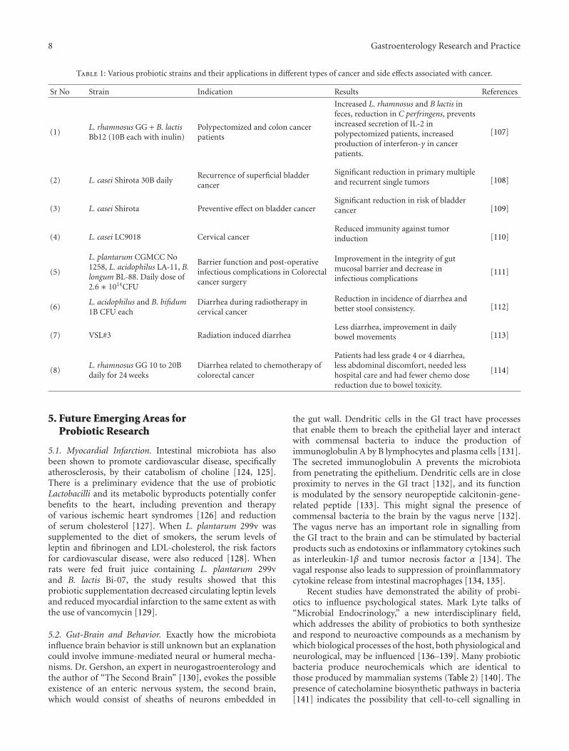
Some of the probiotic strains which have been/are beingused for different cancers, along with their references, aresummarized in Table 1.
4.6. Probiotics and Renal Health. It has been demonstratedthat gut microflora can affect the concentrations of uremictoxins in animals. Prakash and Chang were able to contin-uously reduce blood urea nitrogen in azotemic rats by oraladministration of microencapsulated genetically engineeredlive cells containing living urease-producing E. coli DH5[115]. Based on this concept, Ranganathan et al. [116]carried out rat studies using 5/6th nephrectomised animalsfed with a probiotic cocktail of Lactobacilli, Bifidobacte-ria, and S. thermophilus. Results showed a significantlyprolonged life span for the uremic rats, in addition toreduced blood urea-nitrogen (BUN) levels. Studies weresubsequently carried out in 5/6th nephrectomised Gottingenmini pigs [117]. Here also there was a reduction in BUNand creatinine levels, indicating that the probiotic supple-mentation prevented the accumulation of these toxins inthe blood. These results were further evaluated clinically byRichard Palmquist [118] in feline azotemia. Studies in 7 catsshowed statistically reduced levels in BUN and creatininelevels and demonstrated significantly improved quality oflife (QOL).The product is currently marketed for cats anddogs with moderate-to-severe kidney failure (as “Azodyl”by Vetoquinol SA with worldwide veterinary product sales(http://www.vetoquinol.com/)).
In human studies, Simenhoff et al. demonstrated thathemodialysis patients who were fed L. acidophilus NCFM hadsignificantly lower blood dimethylamine and nitrodimethy-lamine levels [119, 120]. Simenhoff was the first researcherto demonstrate the growth of pathogenic bacteria which isreferred to as “Small Bowel Bacterial Overgrowth” (SBBO).The NCFM strain is well known, and the genome hasbeen sequenced by Todd Klaenhammer’s group [121].Subsequent to the success of the formulation for cats anddogs described above, a similar formulation for humans wasevaluated clinically in a 6-month randomized, double-blind,placebocontrolled, and crossover trial in CKD stage 3 and4 patients in four countries [122, 123]. 46 patients werestudied in this trial. BUN levels decreased in 29 patients (P <0.05), creatinine levels decreased in 20 patients (no statisticalsignificance), and uric acid levels decreased in 15 patients (nostatistical significance). Almost all subjects reported havingexperienced a substantial perceived improvement in theirquality of life (P < 0.05). This product is also currentlymarketed by Kibow Biotech, Inc. with the brand name“Renadyl” (http://www.renadyl.com/).
8 Gastroenterology Research and Practice
Table 1: Various probiotic strains and their applications in different types of cancer and side effects associated with cancer.
Sr No Strain Indication Results References
(1)L. rhamnosus GG + B. lactisBb12 (10B each with inulin)
Polypectomized and colon cancerpatients
Increased L. rhamnosus and B lactis infeces, reduction in C perfringens, preventsincreased secretion of IL-2 inpolypectomized patients, increasedproduction of interferon-γ in cancerpatients.
[107]
(2) L. casei Shirota 30B dailyRecurrence of superficial bladdercancer
Significant reduction in primary multipleand recurrent single tumors [108]
(3) L. casei Shirota Preventive effect on bladder cancerSignificant reduction in risk of bladdercancer [109]
(4) L. casei LC9018 Cervical cancerReduced immunity against tumorinduction [110]
(5)
L. plantarum CGMCC No1258, L. acidophilus LA-11, B.longum BL-88. Daily dose of2.6∗ 1014CFU
Barrier function and post-operativeinfectious complications in Colorectalcancer surgery
Improvement in the integrity of gutmucosal barrier and decrease ininfectious complications
[111]
(6)L. acidophilus and B. bifidum1B CFU each
Diarrhea during radiotherapy incervical cancer
Reduction in incidence of diarrhea andbetter stool consistency. [112]
(7) VSL#3 Radiation induced diarrheaLess diarrhea, improvement in dailybowel movements [113]
(8)L. rhamnosus GG 10 to 20Bdaily for 24 weeks
Diarrhea related to chemotherapy ofcolorectal cancer
Patients had less grade 4 or 4 diarrhea,less abdominal discomfort, needed lesshospital care and had fewer chemo dosereduction due to bowel toxicity.
[114]
5. Future Emerging Areas forProbiotic Research
5.1. Myocardial Infarction. Intestinal microbiota has alsobeen shown to promote cardiovascular disease, specificallyatherosclerosis, by their catabolism of choline [124, 125].There is a preliminary evidence that the use of probioticLactobacilli and its metabolic byproducts potentially conferbenefits to the heart, including prevention and therapyof various ischemic heart syndromes [126] and reductionof serum cholesterol [127]. When L. plantarum 299v wassupplemented to the diet of smokers, the serum levels ofleptin and fibrinogen and LDL-cholesterol, the risk factorsfor cardiovascular disease, were also reduced [128]. Whenrats were fed fruit juice containing L. plantarum 299vand B. lactis Bi-07, the study results showed that thisprobiotic supplementation decreased circulating leptin levelsand reduced myocardial infarction to the same extent as withthe use of vancomycin [129].
5.2. Gut-Brain and Behavior. Exactly how the microbiotainfluence brain behavior is still unknown but an explanationcould involve immune-mediated neural or humeral mecha-nisms. Dr. Gershon, an expert in neurogastroenterology andthe author of “The Second Brain” [130], evokes the possibleexistence of an enteric nervous system, the second brain,which would consist of sheaths of neurons embedded in
the gut wall. Dendritic cells in the GI tract have processesthat enable them to breach the epithelial layer and interactwith commensal bacteria to induce the production ofimmunoglobulin A by B lymphocytes and plasma cells [131].The secreted immunoglobulin A prevents the microbiotafrom penetrating the epithelium. Dendritic cells are in closeproximity to nerves in the GI tract [132], and its functionis modulated by the sensory neuropeptide calcitonin-gene-related peptide [133]. This might signal the presence ofcommensal bacteria to the brain by the vagus nerve [132].The vagus nerve has an important role in signalling fromthe GI tract to the brain and can be stimulated by bacterialproducts such as endotoxins or inflammatory cytokines suchas interleukin-1β and tumor necrosis factor α [134]. Thevagal response also leads to suppression of proinflammatorycytokine release from intestinal macrophages [134, 135].
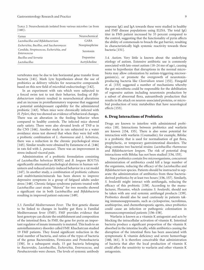
Recent studies have demonstrated the ability of probi-otics to influence psychological states. Mark Lyte talks of“Microbial Endocrinology,” a new interdisciplinary field,which addresses the ability of probiotics to both synthesizeand respond to neuroactive compounds as a mechanism bywhich biological processes of the host, both physiological andneurological, may be influenced [136–139]. Many probioticbacteria produce neurochemicals which are identical tothose produced by mammalian systems (Table 2) [140]. Thepresence of catecholamine biosynthetic pathways in bacteria[141] indicates the possibility that cell-to-cell signalling in
Gastroenterology Research and Practice 9
Table 2: Neurochemicals isolated from various microbes (as from[140]).
Genus Neurochemical
Lactobacillus and Bifidobacterium GABA
Escherichia, Bacillus, and Saccharomyces Norepinephrine
Candida, Streptococcus, Escherichia, andEnterococcus
Serotonin
Bacillus and Serratia Dopamine
Lactobacillus Acetylcholine
vertebrates may be due to late horizontal gene transfer frombacteria [141]. Mark Lyte hypothesizes about the use ofprobiotics as delivery vehicles for neuroactive compoundsbased on this new field of microbial endocrinology [142].
In an experiment with rats which were subjected toa forced swim test to test their behavior, feeding of Bifi-dobacterium infantis resulted in neurochemical alterationsand an increase in proinflammatory response that suggesteda potential antidepressant capability for the administeredprobiotic [143]. When mice were chronically infected withH. Pylori, they too showed an evidence of behavioral changes.There was an alteration in the feeding behavior whencompared to healthy controls. The infected mice showedearly satiety. There were also elevated levels of TNF-α inthe CNS [144]. Another study in rats subjected to a wateravoidance stress test showed that when they were fed witha probiotic combination of L. rhamnosus and L. helveticus,there was a reduction in the chronic psychological stress[145]. Similar results were obtained by Eutamene et al. [146]in rats fed with L. paracasei. There was an improvement instress-induced visceral pain.
Administration of a probiotic formulation consistingof Lactobacillus helveticus RO052 and B. longum RO175Asignificantly attenuated psychological distress in human vol-unteers and reduced anxiety-induced behavior in a rat model[147]. In another study, a combination of probiotic culturesand multivitamin/minerals has been shown to improvedepressive symptoms in a group of fatigued adults understress [148]. Chronic fatigue syndrome patients treated withLactobacillus casei strain “Shirota” for two months showeda significant rise in both Lactobacillus and Bifidobacteriaresulting in improved patient outcome [149].
5.3. Familial Mediterranean Fever. The first genetic diseaseto be linked to changes in healthy gut flora is FamilialMediterranean fever (FMF). FMF provides evidence thathost genotype can dictate the establishment and compositionof the intestinal flora. In FMF, the gene for pyrin an impor-tant regulator of innate immunity is mutated. This leads to anautoinflammatory disorder called FMF. Khachatryan studied19 FMF patients. They found significant reduction in thetotal number of bacteria, and ratios of the types of bacteriaof the genus Bacteroidetes, Firmicutes and Proteobacteria[150]. In a subsequent study, 15 gut bacteria belongingto Bacteroides, Lactobacillus, Escherichia, Enterococcus, andParabacteroides were chosen. The levels of systemic antibody
response IgG and IgA towards these were studied in healthyand FMF disease populations using ELISA. The total IgGtiter in FMS patient increased by 35 percent compared tothe control, suggesting that the functionality of pyrin affectsthe ability of commensals to breach the gut barrier, resultingin characteristically high systemic reactivity towards thesebacteria [151].
5.4. Autism. Very little is known about the underlyingetiology of autism. Extensive antibiotic use is commonlyassociated with late-onset autism (18–24 mo of age), causingsome to hypothesize that disruptions in the normal micro-biota may allow colonization by autism-triggering microor-ganism(s), or promote the overgrowth of neurotoxin-producing bacteria like Clostridium tetani [152]. Finegoldet al. [153] suggested a number of mechanisms wherebythe gut microbiota could be responsible for the debilitationof regressive autism including neurotoxin production bya subset of abnormal flora, autoantibody production thatresults in the attack on neuron-associated proteins, or micro-bial production of toxic metabolites that have neurologicaleffects [153].
6. Drug Interactions of Probiotics
Drugs are known to interfere with administered probi-otics [10]. Interactions between probiotics and warfarinare known [154, 155]. There is also some potential forinteraction with warfarin (Coumadin); for example, Bifolacis a probiotic that is used for normalisation of gut flora,prophylactic, or temporary gastrointestinal disorders. Thedrug contains two bacterial strains: Lactobacillus rhamnosusand Bifidobacterium longum. The risk of potential druginteractions with Bifolac has not been studied [154, 155].
Since probiotics contain live microorganisms, concurrentadministration of antibiotics could kill a large number ofthe organisms, reducing the efficacy of the Lactobacillus andBifidobacterium species. Patients should be instructed to sep-arate the administration of antibiotics from these bacteria-derived probiotics by at least two hours [156, 157]. Similarly,S. boulardii might interact with antifungals, reducing theefficacy of this probiotic [158]. According to the manu-facturer, Florastor, which contains S. boulardii, should notbe taken with any oral systemic antifungal products [159].Probiotics should also be used cautiously in patients tak-ing immunosuppressants, such as cyclosporine, tacrolimus,azathioprine, and chemotherapeutic agents, since probioticscould cause an infection or pathogenic colonisation inimmunocompromised patients [156–158].
Warfarin is known as a vitamin K antagonist and acts byblocking the intracellular activation of vitamin K. Intestinalbacteria produce a significant proportion of the vitamin Kabsorbed in the intestine locally, while antibiotics causing thedisruption of the intestinal flora has been associated withsymptomatic K vitamin deficiency and severe hemorrhage[160, 161]. It is therefore conceivable that administrationof bacteria that alter the local production of vitamin Kcould affect the sensitivity to warfarin and other vitamin Kantagonists.
10 Gastroenterology Research and Practice
Animal studies indicate that B. longum lacks the abilityto synthesize vitamin C [162], and in vitro studies using thebacterial strain showed a decrease in vitamin K levels of theculture medium. Vitamin C acts as a growth factor for manybacterial strains, and since B. longum only requires low levelsof the vitamin for its growth, we can interpret its ability tolower vitamin K concentrations in the surrounding area as ameans of competing with other strains that depend on highvitamin K levels [163].
While this may result in a theoretical risk of interactionswith warfarin based on the proposed mechanism, clinicalsignificance has yet to be shown. No information on L.rhamnosus’ possible role in the production and metabolismof vitamin K has been found. In view of the theoreticalinteraction potential, careful international normalized ratio(INR) monitoring of warfarin-treated patients starting Bifo-lac treatment is recommended [164].
Other than the influence of dietary vitamin K intake,there is essentially no experimental or clinical evidence thatany particular food or nutrient will interact with warfarinthrough modulation of CYP2C9 activity [164–167]. Furthercontrolled studies should be conducted to determine if actualinteraction potentials exist [10, 164, 165].
7. Safety of Administration of Probiotics
Consumers are increasingly using probiotics for their varioushealth benefits. In healthy individuals probiotics are safeto be used. Clinical evidence for their efficacy is strong incase of antibiotic-associated diarrhea management [168].However there are areas of uncertainty. Caution has to beexercised with certain patient groups like premature neonatesor immune deficiency. Paucity of information regardingthe mechanisms through which probiotics act, appropriateadministrative regimes, and probiotic interactions neces-sitate further investigations in these areas. Properties ofprobiotics are strain specific. Hence confirmation studiesneed to be performed, and effects cannot be generalized.A detailed assessment by NIH on the safety aspects hasbeen published [169]. The conclusion which has emergednecessitates the need for systematic reporting of adverseevents and better documentation of interventions.
8. Human Microbiome and Human Health
The Human Microbiome project launched in 2008 by theNational Institute of Health (NIH) aims to understandif changes in the human microbiome are associated withhuman health or disease. These studies have revealed thateven healthy individuals differ remarkably in the microbesthat occupy different body sites or habitats. Different groupsare studying the microbiology of five body sites oral,skin, nasal, gut and urogenital. A study with 242 healthyadults analysed 4,788 specimens from different body habitats[170]. Analysis showed that no taxa were observed to beuniversally present among all body habitats and individualsstudied. Inter-individual variation in the microbiome wasspecific and personalized. Even within the same speciesthere were strain level genomic variations. Jeffrey Gordon’s
group studied a wider and diverse population from differentgeographic, socioeconomic, and cultural settings. The studycomprised of healthy adults and children including mono-and dizygotic twins from the Amazons of Venezuela, ruralMalawi, and the US metropolitan areas [171]. A total of531 individuals were studied. Single fecal sample from eachindividual was analysed. 16S rRNA genes present in the feceswere analysed to define the phylogenetic types. The findingsare interesting and notable. Interpersonal variation wassignificantly greater among children. There were significantdifferences in the phylogenetic composition of fecal micro-biota between individuals living in the different countries.The fecal microbiota of US adults was the least diverse. Animportant observation was the degree of similarity amongfamily members regarding the microbial community struc-ture across the three populations studied. Another grouphas studied the carbohydrate metabolizing enzymes in thefive body sites [172]. Carbohydrate metabolizing enzymesare specific to bacterial taxa, and different body sites areinhabited by different bacterial communities. Despite this,complex carbohydrate cleaving enzyme (CAZymes) profileswere found to be very similar within a body site. This suggestsquote “the carbohydrate composition of each body site hasa profound influence and probably constitutes one of themajor driving forces that shapes the community compositionand therefore the CAZyme profile of the local microbialcommunities, which in turn reflects the microbiome fitnessto a body site.” Yet another study using new statistical analysisshowed that highest diversity was present in stools. Oral andskin habitats had variable diversity patterns while vaginalhabitats were the least diverse [173].
Such varied studies would enable us to understandthe complex relationship between human health and thehuman microbiome and might pave the way to bettermanagement of health using beneficial microbes for diseaseprevention/management.
9. Concluding Remarks
As the gut microbiota appears to contribute to nearly everyaspect of the host’s growth and development, it is notsurprising that a tremendous array of diseases and dys-functions have been associated with an imbalance in eithercomposition, numbers, or habitat of the gut microbiota.
Probiotics, prebiotics, and their combinations have beenfound to be clinically effective for a large number of gutbased disorders like IBD, digestion, travelers diarrhea, andfor improving/helping to maintain general health. Emergingareas of research have shown promise in cancer, brain,kidney, and obesity. It remains to be seen whether probioticsand prebiotics can be effective in combating diseases likeautism, pancreatitis, fibromyalgia, etc.,) where dysbiosis hasbeen observed. The future is going to be challenging butpromising, since tools for probiotic research are now avail-able. Much work has already been accomplished to helpus understand probiotics and the manner in which theyfunction. Therefore the field of probiotics, prebiotics andsynbiotics may potentially open a new branch of science,
Gastroenterology Research and Practice 11
paving a new way for personalized medicine, and maybe evenfuture biotherapeutics.
Conflict of Interests
Both the authors are associated with Kibow Biotech, Inc. afourteen-year old biotechnology company specifically tar-geted in the business of probiotics and prebiotics for RenalHealth.
Acknowledgment
The authors wish to thank Mary Galambert, Bohdan Pechen-yak, and Albert Ruggieri of Kibow Biotech Inc. for proofread-ing of the article.
References
[1] C. Kunz, S. Kuntz, and S. Rudloff, “Intestinal flora,” Advancesin Experimental Medicine and Biology, vol. 639, pp. 67–79,2009.
[2] L. Morelli, “Postnatal development of intestinal microflora asinfluenced by infant nutrition,” Journal of Nutrition, vol. 138,supplement 9, pp. S1791–S1795, 2008.
[3] A. S. Neish, “Microbes in gastrointestinal health and disease,”Gastroenterology, vol. 136, no. 1, pp. 65–80, 2009.
[4] R. E. Ley, D. A. Peterson, and J. I. Gordon, “Ecological andevolutionary forces shaping microbial diversity in the humanintestine,” Cell, vol. 124, no. 4, pp. 837–848, 2006.
[5] D. C. Savage, “Associations of indigenous microorganismswith gastrointestinal mucosal epithelia,” American Journal ofClinical Nutrition, vol. 23, no. 11, pp. 1495–1501, 1970.
[6] W. B. Whitman, D. C. Coleman, and W. J. Wiebe, “Prokary-otes: the unseen majority,” Proceedings of the National Acade-my of Sciences of the United States of America, vol. 95, no. 12,pp. 6578–6583, 1998.
[7] K. Chiller, B. A. Selkin, and G. J. Murakawa, “Skin microfloraand bacterial infections of the skin,” Journal of InvestigativeDermatology Symposium Proceedings, vol. 6, no. 3, pp. 170–174, 2001.
[8] M. W. Hull and A. W. Chow, “Indigenous microflora andinnate immunity of the head and neck,” Infectious DiseaseClinics of North America, vol. 21, no. 2, pp. 265–282, 2007.
[9] H. Verstraelen, “Cutting edge: the vaginal microflora andbacterial vaginosis,” Verhandelingen, vol. 70, no. 3, pp. 147–174, 2008.
[10] N. T. Williams, “Probiotics,” American Journal of Health-Sys-tem Pharmacy, vol. 67, no. 6, pp. 449–458, 2010.
[11] M. G. Dominguez-Bello, E. K. Costello, M. Contreras etal., “Delivery mode shapes the acquisition and structureof the initial microbiota across multiple body habitats innewborns,” Proceedings of the National Academy of Sciencesof the United States of America, vol. 107, no. 26, pp. 11971–11975, 2010.
[12] E. G. Zoetendal, A. D. L. Akkermans, and W. M. DeVos, “Temperature gradient gel electrophoresis analysis of16S rRNA from human fecal samples reveals stable andhost-specific communities of active bacteria,” Applied andEnvironmental Microbiology, vol. 64, no. 10, pp. 3854–3859,1998.
[13] D. Mariat, O. Firmesse, F. Levenez et al., “The firmicutes/bacteroidetes ratio of the human microbiota changes withage,” BMC Microbiology, vol. 9, article 123, 2009.
[14] B. Kleessen and M. Blaut, “Modulation of gut mucosalbiofilms,” British Journal of Nutrition, vol. 93, pp. S35–S40,2005.
[15] H. M. Hamer, D. Jonkers, K. Venema, S. Vanhoutvin, F.J. Troost, and R. J. Brummer, “Review article: the role ofbutyrate on colonic function,” Alimentary Pharmacology andTherapeutics, vol. 27, no. 2, pp. 104–119, 2008.
[16] S. Salminen, C. Bouley, M. C. Boutron-Ruault et al., “Func-tional food science and gastrointestinal physiology and func-tion,” British Journal of Nutrition, vol. 80, no. 1, pp. S147–S171, 1998.
[17] T. S. Stappenbeck, L. V. Hooper, and J. I. Gordon, “Devel-opmental regulation of intestinal angiogenesis by indigenousmicrobes via Paneth cells,” Proceedings of the NationalAcademy of Sciences of the United States of America, vol. 99,no. 24, pp. 15451–15455, 2002.
[18] P. Lefebvre, B. Cariou, F. Lien, F. Kuipers, and B. Staels, “Roleof bile acids and bile acid receptors in metabolic regulation,”Physiological Reviews, vol. 89, no. 1, pp. 147–191, 2009.
[19] J. M. W. Wong, R. de Souza, C. W. C. Kendall, A. Emam,and D. J. A. Jenkins, “Colonic health: fermentation and shortchain fatty acids,” Journal of Clinical Gastroenterology, vol. 40,no. 3, pp. 235–243, 2006.
[20] H. Tlaskova-Hogenova, R. Stepankova, T. Hudcovic et al.,“Commensal bacteria (normal microflora), mucosal immu-nity and chronic inflammatory and autoimmune diseases,”Immunology Letters, vol. 93, pp. 97–108, 2004.
[21] J. L. Round and S. K. Mazmanian, “The gut microbiotashapes intestinal immune responses during health anddisease,” Nature Reviews Immunology, vol. 9, no. 5, pp. 313–323, 2009.
[22] K. Haverson, Z. Rehakova, J. Sinkora, L. Sver, and M. Bailey,“Immune development in jejunal mucosa after colonizationwith selected commensal gut bacteria: a study in germ-free pigs,” Veterinary Immunology and Immunopathology, vol.119, no. 3-4, pp. 243–253, 2007.
[23] S. Hapfelmeier, M. A. E. Lawson, E. Slack et al., “Reversiblemicrobial colonization of germ-free mice reveals the dynam-ics of IgA immune responses,” Science, vol. 328, no. 5986, pp.1705–1709, 2010.
[24] K. Atarashi, T. Tanoue, T. Shima et al., “Induction of colonicregulatory T cells by indigenous Clostridium species,” Sci-ence, vol. 331, no. 6015, pp. 337–341, 2011.
[25] S. K. Mazmanian, H. L. Cui, A. O. Tzianabos, and D. L.Kasper, “An immunomodulatory molecule of symbiotic bac-teria directs maturation of the host immune system,” Cell,vol. 122, no. 1, pp. 107–118, 2005.
[26] I. I. Ivanov, K. Atarashi, N. Manel et al., “Induction ofintestinal Th17 cells by segmented filamentous bacteria,”Cell, vol. 139, no. 3, pp. 485–498, 2009.
[27] H. Luhrs, T. Gerke, J. G. Muller et al., “Butyrate inhibits NF-κB activation in lamina propria macrophages of patients withulcerative colitis,” Scandinavian Journal of Gastroenterology,vol. 37, no. 4, pp. 458–466, 2002.
[28] K. M. Maslowski, A. T. Vieira, A. Ng et al., “Regulation ofinflammatory responses by gut microbiota and chemoattrac-tant receptor GPR43,” Nature, vol. 461, no. 7268, pp. 1282–1286, 2009.
[29] U. G. Strauch, F. Obermeier, N. Grunwald et al., “Influence ofintestinal bacteria on induction of regulatory T cells: lessons

12 Gastroenterology Research and Practice
from a transfer model of colitis,” Gut, vol. 54, no. 11, pp.1546–1552, 2005.
[30] K. Honda and K. Takeda, “Regulatory mechanisms of im-mune responses to intestinal bacteria,” Mucosal Immunology,vol. 2, no. 3, pp. 187–196, 2009.
[31] R. B. Sartor, “Microbial influences in inflammatory boweldiseases,” Gastroenterology, vol. 134, no. 2, pp. 577–594, 2008.
[32] B. J. Lee and Y.-T. Bak, “Irritable bowel syndrome, gutmicrobiota and probiotics,” Journal of Neurogastroenterologyand Motility, vol. 17, no. 3, pp. 252–256, 2011.
[33] U. Hofer and R. F. Speck, “Disturbance of the gut-associatedlymphoid tissue is associated with disease progression inchronic HIV infection,” Seminars in Immunopathology, vol.31, no. 2, pp. 257–266, 2009.
[34] C. H. Liu, X. Q. Yang, C. H. Liu, Y. He, and L. J. Wang, “Aller-gic airway response associated with the intestinal microfloradisruption induced by antibiotic therapy,” Zhonghua Er Ke ZaZhi, vol. 45, no. 6, pp. 450–454, 2007.
[35] J. Penders, E. E. Stobberingh, P. A. V. D. Brandt, and C. Thijs,“The role of the intestinal microbiota in the developmentof atopic disorders,” Allergy, vol. 62, no. 11, pp. 1223–1236,2007.
[36] S. L. Verhulst, C. Vael, C. Beunckens, V. Nelen, H. Goossens,and K. Desager, “A longitudinal analysis on the associationbetween antibiotic use, intestinal microflora, and wheezingduring the first year of life,” Journal of Asthma, vol. 45, no. 9,pp. 828–832, 2008.
[37] Food and Agriculture Organization and World Health Orga-nization, Report of a Joint FAO/WHO Expert Consultation onEvaluation of Health and Nutritional Properties of Probioticsin Food Including Powder Milk with Live Lactic Acid Bacteria,2001.
[38] G. R. Gibson, K. P. Scott, R. A. Rastall et al., “Dietaryprebiotics: current status and new definition,” Food Scienceand Technology Bulletin, vol. 7, no. 1, pp. 1–19, 2010.
[39] World Gastroenterology Organization Global Guidelines,October 2011.
[40] P. D. Cani and N. M. Delzenne, “The role of the gutmicrobiota in energy metabolism and metabolic disease,”Current Pharmaceutical Design, vol. 15, no. 13, pp. 1546–1558, 2009.
[41] P. D. Cani and N. M. Delzenne, “Interplay between obesityand associated metabolic disorders: new insights into the gutmicrobiota,” Current Opinion in Pharmacology, vol. 9, no. 6,pp. 737–743, 2009.
[42] N. Larsen, F. K. Vogensen, F. W. J. Van Den Berg et al., “Gutmicrobiota in human adults with type 2 diabetes differs fromnon-diabetic adults,” PLoS ONE, vol. 5, no. 2, Article IDe9085, 2010.
[43] J. P. Furet, L. C. Kong, J. Tap et al., “Differential adaptation ofhuman gut microbiota to bariatric surgery-induced weightloss: links with metabolic and low-grade inflammationmarkers,” Diabetes, vol. 59, no. 12, pp. 3049–3057, 2010.
[44] R. E. Ley, “Obesity and the human microbiome,” CurrentOpinion in Gastroenterology, vol. 26, no. 1, pp. 5–11, 2010.
[45] A. Schwiertz, D. Taras, K. Schafer et al., “Microbiota andSCFA in lean and overweight healthy subjects,” Obesity, vol.18, no. 1, pp. 190–195, 2010.
[46] X. Wu, C. Ma, L. Han et al., “Molecular characterisation ofthe faecal microbiota in patients with type II diabetes,” Cur-rent Microbiology, vol. 61, no. 1, pp. 69–78, 2010.
[47] E. M. Dewulf, P. D. Cani, A. M. Neyrinck et al., “Inulin-type fructans with prebiotic properties counteract GPR43overexpression and PPARγ-related adipogenesis in the white
adipose tissue of high-fat diet-fed mice,” Journal of Nutri-tional Biochemistry, vol. 22, no. 8, pp. 712–722, 2011.
[48] P. D. Cani, A. M. Neyrinck, F. Fava et al., “Selective increasesof bifidobacteria in gut microflora improve high-fat-diet-induced diabetes in mice through a mechanism associatedwith endotoxaemia,” Diabetologia, vol. 50, no. 11, pp. 2374–2383, 2007.
[49] P. D. Cani, S. Possemiers, T. Van De Wiele et al., “Changes ingut microbiota control inflammation in obese mice througha mechanism involving GLP-2-driven improvement of gutpermeability,” Gut, vol. 58, no. 8, pp. 1091–1103, 2009.
[50] L. Aronsson, Y. Huang, P. Parini et al., “Decreased fat storageby Lactobacillus paracasei is associated with increased levelsof angiopoietin-like 4 protein (ANGPTL4),” PLoS ONE, vol.5, no. 9, Article ID e13087, 2010.
[51] A. S. Andreasen, N. Larsen, T. Pedersen-Skovsgaard et al.,“Effects of Lactobacillus acidophilus NCFM on insulin sen-sitivity and the systemic inflammatory response in humansubjects,” British Journal of Nutrition, vol. 104, no. 12, pp.1831–1838, 2010.
[52] Y. Kadooka, M. Sato, K. Imaizumi et al., “Regulationof abdominal adiposity by probiotics (Lactobacillus gasseriSBT2055) in adults with obese tendencies in a randomizedcontrolled trial,” European Journal of Clinical Nutrition, vol.64, no. 6, pp. 636–643, 2010.
[53] Z. X. Lu, K. Z. Walker, J. G. Muir, and K. O’Dea, “Arabinoxy-lan fibre improves metabolic control in people with type IIdiabetes,” European Journal of Clinical Nutrition, vol. 58, no.4, pp. 621–628, 2004.
[54] A. L. Garcia, J. Steiniger, S. C. Reich et al., “Arabinoxylanfibre consumption improved glucose metabolism, but didnot affect serum adipokines in subjects with impaired glucosetolerance,” Hormone and Metabolic Research, vol. 38, no. 11,pp. 761–766, 2006.
[55] A. L. Garcia, B. Otto, S. C. Reich et al., “Arabinoxylan con-sumption decreases postprandial serum glucose, seruminsulin and plasma total ghrelin response in subjects withimpaired glucose tolerance,” European Journal of ClinicalNutrition, vol. 61, no. 3, pp. 334–341, 2007.
[56] J. A. Parnell and R. A. Reimer, “Weight loss during oligofruc-tose supplementation is associated with decreased ghrelinand increased peptide YY in overweight and obese adults,”American Journal of Clinical Nutrition, vol. 89, no. 6, pp.1751–1759, 2009.
[57] J. Luo, M. Van Yperselle, S. W. Rizkalla, F. Rossi, F. R.Bornet, and G. Slama, “Chronic consumption of short-chainfructooligosaccharides does not affect basal hepatic glucoseproduction or insulin resistance in type 2 diabetics,” Journalof Nutrition, vol. 130, no. 6, pp. 1572–1577, 2000.
[58] C. A. Daubioul, Y. Horsmans, P. Lambert, E. Danse, andN. M. Delzenne, “Effects of oligofructose on glucose andlipid metabolism in patients with nonalcoholic steatohep-atitis: results of a pilot study,” European Journal of ClinicalNutrition, vol. 59, no. 5, pp. 723–726, 2005.
[59] G. Musso, R. Gambino, and M. Cassader, “Interactionsbetween gut microbiota and host metabolism predisposingto obesity and diabetes,” Annual Review of Medicine, vol. 62,pp. 361–380, 2011.
[60] C. Prussin and D. D. Metcalfe, “IgE, mast cells, basophils, andeosinophils,” Journal of Allergy and Clinical Immunology, vol.111, supplement 2, pp. S486–S494, 2003.
[61] W. Cookson, “Genetics and genomics of asthma and allergicdiseases,” Immunological Reviews, vol. 190, pp. 195–206,2002.
Gastroenterology Research and Practice 13
[62] J. W. Steinke, L. Borish, and L. J. Rosenwasser, “Genetics ofhypersensitivity,” Journal of Allergy and Clinical Immunology,vol. 111, supplement 2, pp. S495–S501, 2003.
[63] F. S. Larsen and J. M. Hanifin, “Epidemiology of atopic der-matitis,” Immunology and Allergy Clinics of North America,vol. 22, no. 1, pp. 1–24, 2002.
[64] B. Bjorksten, P. Naaber, E. Sepp, and M. Mikelsaar, “Theintestinal microflora in allergic Estonian and Swedish 2-year-old children,” Clinical and Experimental Allergy, vol. 29, no.3, pp. 342–346, 1999.
[65] E. Sepp, K. Julge, M. Vasar, P. Naaber, B. Bjorksten, and M.Mikelsaar, “Intestinal microflora of Estonian and Swedishinfants,” Acta Paediatrica, vol. 86, no. 9, pp. 956–961, 1997.
[66] M. Kalliomaki, S. Salminen, H. Arvilommi, P. Kero, P.Koskinen, and E. Isolauri, “Probiotics in primary preventionof atopic disease: a randomised placebo-controlled trial,” TheLancet, vol. 357, no. 9262, pp. 1076–1079, 2001.
[67] H. Majamaa and E. Isolauri, “Probiotics: a novel approachin the management of food allergy,” Journal of Allergy andClinical Immunology, vol. 99, no. 2, pp. 179–185, 1997.
[68] V. Rosenfeldt, E. Benfeldt, S. D. Nielsen et al., “Effect of probi-otic Lactobacillus strains in children with atopic dermatitis,”Journal of Allergy and Clinical Immunology, vol. 111, no. 2,pp. 389–395, 2003.
[69] S. Weston, A. Halbert, P. Richmond, and S. L. Prescott,“Effects of probiotics on atopic dermatitis: a randomisedcontrolled trial,” Archives of Disease in Childhood, vol. 90, no.9, pp. 892–897, 2005.
[70] K. Wickens, P. N. Black, T. V. Stanley et al., “A differentialeffect of 2 probiotics in the prevention of eczema and atopy: adouble-blind, randomized, placebo-controlled trial,” Journalof Allergy and Clinical Immunology, vol. 122, no. 4, pp. 788–794, 2008.
[71] L. Niers, R. Martın, G. Rijkers et al., “The effects of selectedprobiotic strains on the development of eczema (the PandAstudy),” Allergy, vol. 64, no. 9, pp. 1349–1358, 2009.
[72] R. Gøbel, N. Larsen, C. Mølgaard, M. Jakobsen, and K. F.Michaelsen, “Probiotics to young children with atopic der-matitis: a randomized placebo-controlled trial,” InternationalJournal of Probiotics and Prebiotics, vol. 5, no. 2, pp. 53–60,2010.
[73] V. Rosenfeldt, E. Benfeldt, N. H. Valerius, A. Pærregaard, andK. F. Michaelsen, “Effect of probiotics on gastrointestinalsymptoms and small intestinal permeability in children withatopic dermatitis,” Journal of Pediatrics, vol. 145, no. 5, pp.612–616, 2004.
[74] R. Williams, “Review article: bacterial flora and pathogenesisin hepatic encephalopathy,” Alimentary Pharmacology andTherapeutics, vol. 25, supplement 1, pp. 17–22, 2007.
[75] G. R. Gibson and X. Wang, “Regulatory effects of bifidobac-teria on the growth of other colonic bacteria,” Journal ofApplied Bacteriology, vol. 77, no. 4, pp. 412–420, 1994.
[76] K. Madsen, A. Cornish, P. Soper et al., “Probiotic bacteriaenhance murine and human intestinal epithelial barrierfunction,” Gastroenterology, vol. 121, no. 3, pp. 580–591,2001.
[77] C. Yurdaydin, T. J. Walsh, H. D. Engler et al., “Gut bacteriaprovide precursors of benzodiazepine receptor ligands in arat model of hepatic encephalopathy,” Brain Research, vol.679, no. 1, pp. 42–48, 1995.
[78] M. Malaguarnera, F. Greco, G. Barone, M. P. Gargante, M.Malaguarnera, and M. A. Toscano, “Bifidobacterium longumwith fructo-oligosaccharide (FOS) treatment in mini-mal hepatic encephalopathy: a randomized, double-blind,
placebo-controlled study,” Digestive Diseases and Sciences,vol. 52, no. 11, pp. 3259–3265, 2007.
[79] H. Y. Zhao, H. J. Wang, Z. Lu, and S. Z. Xu, “Intestinalmicroflora in patients with liver cirrhosis,” Chinese Journalof Digestive Diseases, vol. 5, no. 2, pp. 64–67, 2004.
[80] Q. Liu, Z. P. Duan, D. K. Ha, S. Bengmark, J. Kurtovic, andS. M. Riordan, “Synbiotic modulation of gut flora: effect ofminimal hepatic encephalopathy in patients with cirrhosis,”Hepatology, vol. 39, no. 5, pp. 1441–1449, 2004.
[81] R. G. McGee, A. Bakens, K. Wiley, S. M. Riordan, and A. C.Webster, “Probiotics for patients with hepatic encephalopa-thy,” Cochrane Database of Systematic Reviews, vol. 9, no. 11,Article ID CD008716, 2011.
[82] I. A. Abd El-Gawad, E. M. El-Sayed, S. A. Hafez, H. M. El-Zeini, and F. A. Saleh, “The hypocholesterolaemic effect ofmilk yoghurt and soy-yoghurt containing bifidobacteria inrats fed on a cholesterol-enriched diet,” International DairyJournal, vol. 15, no. 1, pp. 37–44, 2005.
[83] T. D. T. Nguyen, J. H. Kang, and M. S. Lee, “Characterizationof Lactobacillus plantarum PH04, a potential probioticbacterium with cholesterol-lowering effects,” InternationalJournal of Food Microbiology, vol. 113, no. 3, pp. 358–361,2007.
[84] M. Fukushima, A. Yamada, T. Endo, and M. Nakano, “Effectsof a mixture of organisms, Lactobacillus acidophilus orStreptococcus faecalis on δ6-desaturase activity in the livers ofrats fed a fat- and cholesterol-enriched diet,” Nutrition, vol.15, no. 5, pp. 373–378, 1999.
[85] J. W. Anderson and S. E. Gilliland, “Effect of fermented milk(yogurt) containing Lactobacillus acidophilus L1 on serumcholesterol in hypercholesterolemic humans,” Journal of theAmerican College of Nutrition, vol. 18, no. 1, pp. 43–50, 1999.
[86] J. Z. Xiao, S. Kondo, N. Takahashi et al., “Effects of milkproducts fermented by B longum on blood lipids in rats andhealthy adult male volunteers,” Journal of Dairy Science, vol.86, no. 7, pp. 2452–2461, 2003.
[87] E. A. Trautwein, D. Rieckhoff, and H. F. Erbersdobler,“Dietary inulin lowers plasma cholesterol and triacylglyceroland alters biliary bile acid profile in hamsters,” Journal ofNutrition, vol. 128, no. 11, pp. 1937–1943, 1998.
[88] C. K. Hsu, J. W. Liao, Y. C. Chung, C. P. Hsieh, and Y.C. Chan, “Xylooligosaccharides and fructooligosaccharidesaffect the intestinal microbiota and precancerous coloniclesion development in rats,” Journal of Nutrition, vol. 134, no.6, pp. 1523–1528, 2004.
[89] J. L. Causey, J. M. Feirtag, D. D. Gallaher, B. C. Tungland,and J. L. Slavin, “Effects of dietary inulin on serum lipids,blood glucose and the gastrointestinal environment in hyper-cholesterolemic men,” Nutrition Research, vol. 20, no. 2, pp.191–201, 2000.
[90] M. T. Liong, F. R. Dunshea, and N. P. Shah, “Effects ofa synbiotic containing Lactobacillus acidophilus ATCC 4962on plasma lipid profiles and morphology of erythrocytesin hypercholesterolaemic pigs on high- and low-fat diets,”British Journal of Nutrition, vol. 98, no. 4, pp. 736–744, 2007.
[91] G. Kießling, J. Schneider, and G. Jahreis, “Long-termconsumption of fermented dairy products over 6 monthsincreases HDL cholesterol,” European Journal of ClinicalNutrition, vol. 56, no. 9, pp. 843–849, 2002.
[92] G. Schaafsma, W. J. A. Meuling, W. Van Dokkum, and C.Bouley, “Effects of a milk product, fermented by Lactobacil-lus acidophilus and with fructo-oligosaccharides added, onblood lipids in male volunteers,” European Journal of ClinicalNutrition, vol. 52, no. 6, pp. 436–440, 1998.
14 Gastroenterology Research and Practice
[93] K. Hatakka, M. Mutanen, R. Holma, M. Saxelin, and R. Kor-pela, “L rhamnosus LC705 together with Propionibacteriumfreudenreichii ssp shermanii JS administered in capsules isineffective in lowering serum lipids,” Journal of the AmericanCollege of Nutrition, vol. 27, no. 4, pp. 441–447, 2008.
[94] L. A. Simons, S. G. Amansec, and P. Conway, “Effect ofL fermentum on serum lipids in subjects with elevatedserum cholesterol,” Nutrition, Metabolism and CardiovascularDiseases, vol. 16, no. 8, pp. 531–535, 2006.
[95] S. J. Lewis and S. Burmeister, “A double-blind placebo-controlled study of the effects of L acidophilus on plasmalipids,” European Journal of Clinical Nutrition, vol. 59, no. 6,pp. 776–780, 2005.
[96] S. Tarpila, A. Aro, I. Salminen et al., “The effect of flaxseedsupplementation in processed foods on serum fatty acids andenterolactone,” European Journal of Clinical Nutrition, vol.56, no. 2, pp. 157–165, 2002.
[97] M. H. Davidson, K. C. Maki, C. Synecki, S. A. Torri, and K.B. Drennan, “Effects of dietary inulin on serum lipids in menand women with hypercholesterolemia,” Nutrition Research,vol. 18, no. 3, pp. 503–517, 1998.
[98] K. A. Greany, M. J. L. Bonorden, J. M. Hamilton-Reeves etal., “Probiotic capsules do not lower plasma lipids in youngwomen and men,” European Journal of Clinical Nutrition, vol.62, no. 2, pp. 232–237, 2008.
[99] Y. Aso, H. Akaza, T. Kotake, T. Tsukamoto, K. Imai, and S.Naito, “Preventive effect of a Lactobacillus casei preparationon the recurrence of superficial bladder cancer in a double-blind trial,” European Urology, vol. 27, no. 2, pp. 104–109,1995.
[100] A. Klinder, A. Forster, G. Caderni, A. P. Femia, and B. L.Pool-Zobel, “Fecal water genotoxicity is predictive of tumor-preventive activities by inulin-like oligofructoses, probiotics(Lactobacillus rhamnosus and Bifidobacterium lactis), andtheir synbiotic combination,” Nutrition and Cancer, vol. 49,no. 2, pp. 144–155, 2004.
[101] F. Bolognani, C. J. Rumney, B. L. Pool-Zobel, and I. R.Rowland, “Effect of lactobacilli, bifidobacteria and inulinon the formation of aberrant crypt foci in rats,” EuropeanJournal of Nutrition, vol. 40, no. 6, pp. 293–300, 2001.
[102] B. Pool-Zobel, J. Van Loo, I. Rowland, and M. B. Roberfroid,“Experimental evidences on the potential of prebiotic fruc-tans to reduce the risk of colon cancer,” British Journal ofNutrition, vol. 87, supplement 2, pp. S273–S281, 2002.
[103] M. Roller, G. Caderni, G. Rechkemmer, and B. Watzl,“Long term treatment with a prebiotic modulates the gut-associated immune system of azomethane-treated F344 rats,”in Proceedings of the SKLM Symposium “Functional Food:Safety Aspects”, pp. 5–7, Karlsruhe, Germany, May 2002.
[104] M. Verghese, D. R. Rao, C. B. Chawan, L. L. Williams, andL. Shackelford, “Dietary inulin suppresses azoxymethane-induced aberrant crypt foci and colon tumors at the promo-tion stage in young Fisher 344 rats,” Journal of Nutrition, vol.132, no. 9, pp. 2809–2813, 2002.
[105] H. S. Taper and M. B. Roberfroid, “Inulin/oligofructose andanticancer therapy,” British Journal of Nutrition, vol. 87,supplement 2, pp. S283–S286, 2002.
[106] A. P. Femia, C. Luceri, P. Dolara et al., “Antitumorigenicactivity of the prebiotic inulin enriched with oligofructosein combination with the probiotics Lactobacillus rhamnosusand Bifidobacterium lactis on azoxymethane-induced coloncarcinogenesis in rats,” Carcinogenesis, vol. 23, no. 11, pp.1953–1960, 2002.
[107] J. Rafter, M. Bennett, G. Caderni et al., “Dietary synbioticsreduce cancer risk factors in polypectomized and coloncancer patients,” American Journal of Clinical Nutrition, vol.85, no. 2, pp. 488–496, 2007.
[108] Y. Aso, H. Akaza, T. Kotake, T. Tsukamoto, K. Imai, and S.Naito, “Preventive effect of a Lactobacillus casei preparationon the recurrence of superficial bladder cancer in a double-blind trial,” European Urology, vol. 27, no. 2, pp. 104–109,1995.
[109] Y. Ohashi, S. Nakai, T. Tsukamoto et al., “Habitual intakeof lactic acid bacteria and risk reduction of bladder cancer,”Urologia Internationalis, vol. 68, no. 4, pp. 273–280, 2002.
[110] T. Okawa, H. Niibe, T. Arai et al., “Effect of LC9018 combinedwith radiation therapy on carcinoma of the uterine cervix:a phase III, multicenter, randomized, controlled study,”Cancer, vol. 72, no. 6, pp. 1949–1954, 1993.
[111] Z. Liu, H. Qin, Z. Yang et al., “Randomised clinical trial:the effects of perioperative probiotic treatment on barrierfunction and post-operative infectious complications incolorectal cancer surgery—a double-blind study,” vol. 33, no.1, pp. 50–63, 2011.
[112] I. Chitapanarux, T. Chitapanarux, P. Traisathit, S. Kudumpee,E. Tharavichitkul, and V. Lorvidhaya, “Randomized con-trolled trial of live Lactobacillus acidophilus plus bifidobac-terium bifidum in prophylaxis of diarrhea during radiother-apy in cervical cancer patients,” Radiation Oncology, vol. 5,no. 1, article 31, 2010.
[113] P. Delia, G. Sansotta, and V. Donato, “Use of probiotics forprevention of radiation-induced diarrhea,” World Journal ofGastroenterology, vol. 13, no. 6, pp. 912–915, 2007.
[114] P. Osterlund, T. Ruotsalainen, R. Korpela et al., “Lactobacillussupplementation for diarrhoea related to chemotherapy ofcolorectal cancer: a randomised study,” British Journal ofCancer, vol. 97, no. 8, pp. 1028–1034, 2007.
[115] S. Prakash and T. M. S. Chang, “Microencapsulated geneti-cally engineered live E. coli DH5 cells administered orally tomaintain normal plasma urea level in uremic rats,” NatureMedicine, vol. 2, no. 8, pp. 883–887, 1996.
[116] N. Ranganathan, B. Patel, P. Ranganathan et al., “Probioticamelioration of azotemia in 5/6th nephrectomized Sprague-Dawley rats,” TheScientificWorldJournal, vol. 5, pp. 652–660,2005.
[117] N. Ranganathan, B. Patel, P. Ranganathan et al., “Probioticsreduces azotemia in gottingen mini-pigs,” in Proceedings ofthe 3rd World Congress of Nephrology Poster Presentation,Singapore, June 2005.
[118] R. Palmquist, “A preliminary clinical evaluation of kibowbiotics, a probiotic agent, on feline azotemia,” Journal of theAmerican Holistic Medical Association, vol. 24, no. 4, pp. 23–27, 2006.
[119] M. L. Simenhoff, S. R. Dunn, G. P. Zollner et al., “Biomod-ulation of the toxic and nutritional effects of small bowelovergrowth in end stage kidney disease using freeze dried Lacidophilus,” Mineral and Electrolyte Metabolism, vol. 22, no.1–3, pp. 92–96, 1996.
[120] S. R. Dunn, M. L. Simenhoff, K. E. Ahmed et al., “Effect oforal administration of freeze-dried Lactobacillus acidophiluson small bowel bacterial overgrowth in patients with endstage kidney disease: reducing uremic toxins and improvingnutrition,” International Dairy Journal, vol. 8, no. 5-6, pp.545–553, 1998.
[121] E. Altermann, W. M. Russell, M. A. Azcarate-Peril et al.,“Complete genome sequence of the probiotic lactic acidbacterium Lactobacillus acidophilus NCFM,” Proceedings of
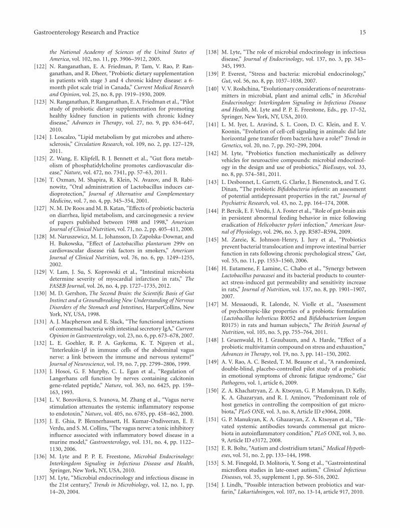
Gastroenterology Research and Practice 15
the National Academy of Sciences of the United States ofAmerica, vol. 102, no. 11, pp. 3906–3912, 2005.
[122] N. Ranganathan, E. A. Friedman, P. Tam, V. Rao, P. Ran-ganathan, and R. Dheer, “Probiotic dietary supplementationin patients with stage 3 and 4 chronic kidney disease: a 6-month pilot scale trial in Canada,” Current Medical Researchand Opinion, vol. 25, no. 8, pp. 1919–1930, 2009.
[123] N. Ranganathan, P. Ranganathan, E. A. Friedman et al., “Pilotstudy of probiotic dietary supplementation for promotinghealthy kidney function in patients with chronic kidneydisease,” Advances in Therapy, vol. 27, no. 9, pp. 634–647,2010.
[124] J. Loscalzo, “Lipid metabolism by gut microbes and athero-sclerosis,” Circulation Research, vol. 109, no. 2, pp. 127–129,2011.
[125] Z. Wang, E. Klipfell, B. J. Bennett et al., “Gut flora metab-olism of phosphatidylcholine promotes cardiovascular dis-ease,” Nature, vol. 472, no. 7341, pp. 57–63, 2011.
[126] T. Oxman, M. Shapira, R. Klein, N. Avazov, and B. Rabi-nowitz, “Oral administration of Lactobacillus induces car-dioprotection,” Journal of Alternative and ComplementaryMedicine, vol. 7, no. 4, pp. 345–354, 2001.
[127] N. M. De Roos and M. B. Katan, “Effects of probiotic bacteriaon diarrhea, lipid metabolism, and carcinogenesis: a reviewof papers published between 1988 and 1998,” AmericanJournal of Clinical Nutrition, vol. 71, no. 2, pp. 405–411, 2000.
[128] M. Naruszewicz, M. L. Johansson, D. Zapolska-Downar, andH. Bukowska, “Effect of Lactobacillus plantarum 299v oncardiovascular disease risk factors in smokers,” AmericanJournal of Clinical Nutrition, vol. 76, no. 6, pp. 1249–1255,2002.
[129] V. Lam, J. Su, S. Koprowski et al., “Intestinal microbiotadetermine severity of myocardial infarction in rats,” TheFASEB Journal, vol. 26, no. 4, pp. 1727–1735, 2012.
[130] M. D. Gershon, The Second Brain: the Scientific Basis of GutInstinct and a Groundbreaking New Understanding of NervousDisorders of the Stomach and Intestines, HarperCollins, NewYork, NY, USA, 1998.
[131] A. J. Macpherson and E. Slack, “The functional interactionsof commensal bacteria with intestinal secretory IgA,” CurrentOpinion in Gastroenterology, vol. 23, no. 6, pp. 673–678, 2007.
[132] L. E. Goehler, R. P. A. Gaykema, K. T. Nguyen et al.,“Interleukin-1β in immune cells of the abdominal vagusnerve: a link between the immune and nervous systems?”Journal of Neuroscience, vol. 19, no. 7, pp. 2799–2806, 1999.
[133] J. Hosoi, G. F. Murphy, C. L. Egan et al., “Regulation ofLangerhans cell function by nerves containing calcitoningene-related peptide,” Nature, vol. 363, no. 6425, pp. 159–163, 1993.
[134] L. V. Borovikova, S. Ivanova, M. Zhang et al., “Vagus nervestimulation attenuates the systemic inflammatory responseto endotoxin,” Nature, vol. 405, no. 6785, pp. 458–462, 2000.
[135] J. E. Ghia, P. Blennerhassett, H. Kumar-Ondiveeran, E. F.Verdu, and S. M. Collins, “The vagus nerve: a tonic inhibitoryinfluence associated with inflammatory bowel disease in amurine model,” Gastroenterology, vol. 131, no. 4, pp. 1122–1130, 2006.
[136] M. Lyte and P. P. E. Freestone, Microbial Endocrinology:Interkingdom Signaling in Infectious Disease and Health,Springer, New York, NY, USA, 2010.
[137] M. Lyte, “Microbial endocrinology and infectious disease inthe 21st century,” Trends in Microbiology, vol. 12, no. 1, pp.14–20, 2004.
[138] M. Lyte, “The role of microbial endocrinology in infectiousdisease,” Journal of Endocrinology, vol. 137, no. 3, pp. 343–345, 1993.
[139] P. Everest, “Stress and bacteria: microbial endocrinology,”Gut, vol. 56, no. 8, pp. 1037–1038, 2007.
[140] V. V. Roshchina, “Evolutionary considerations of neurotrans-mitters in microbial, plant and animal cells,” in MicrobialEndocrinology: Interkingdom Signaling in Infectious Diseaseand Health, M. Lyte and P. P. E. Freestone, Eds., pp. 17–52,Springer, New York, NY, USA, 2010.
[141] L. M. Iyer, L. Aravind, S. L. Coon, D. C. Klein, and E. V.Koonin, “Evolution of cell-cell signaling in animals: did latehorizontal gene transfer from bacteria have a role?” Trends inGenetics, vol. 20, no. 7, pp. 292–299, 2004.
[142] M. Lyte, “Probiotics function mechanistically as deliveryvehicles for neuroactive compounds: microbial endocrinol-ogy in the design and use of probiotics,” BioEssays, vol. 33,no. 8, pp. 574–581, 2011.
[143] L. Desbonnet, L. Garrett, G. Clarke, J. Bienenstock, and T. G.Dinan, “The probiotic Bifidobacteria infantis: an assessmentof potential antidepressant properties in the rat,” Journal ofPsychiatric Research, vol. 43, no. 2, pp. 164–174, 2008.
[144] P. Bercik, E. F. Verdu, J. A. Foster et al., “Role of gut-brain axisin persistent abnormal feeding behavior in mice followingeradication of Helicobacter pylori infection,” American Jour-nal of Physiology, vol. 296, no. 3, pp. R587–R594, 2009.
[145] M. Zareie, K. Johnson-Henry, J. Jury et al., “Probioticsprevent bacterial translocation and improve intestinal barrierfunction in rats following chronic psychological stress,” Gut,vol. 55, no. 11, pp. 1553–1560, 2006.
[146] H. Eutamene, F. Lamine, C. Chabo et al., “Synergy betweenLactobacillus paracasei and its bacterial products to counter-act stress-induced gut permeability and sensitivity increasein rats,” Journal of Nutrition, vol. 137, no. 8, pp. 1901–1907,2007.
[147] M. Messaoudi, R. Lalonde, N. Violle et al., “Assessmentof psychotropic-like properties of a probiotic formulation(Lactobacillus helveticus R0052 and Bifidobacterium longumR0175) in rats and human subjects,” The British Journal ofNutrition, vol. 105, no. 5, pp. 755–764, 2011.
[148] J. Gruenwald, H. J. Graubaum, and A. Harde, “Effect of aprobiotic multivitamin compound on stress and exhaustion,”Advances in Therapy, vol. 19, no. 3, pp. 141–150, 2002.
[149] A. V. Rao, A. C. Bested, T. M. Beaune et al., “A randomized,double-blind, placebo-controlled pilot study of a probioticin emotional symptoms of chronic fatigue syndrome,” GutPathogens, vol. 1, article 6, 2009.
[150] Z. A. Khachatryan, Z. A. Ktsoyan, G. P. Manukyan, D. Kelly,K. A. Ghazaryan, and R. I. Aminov, “Predominant role ofhost genetics in controlling the composition of gut micro-biota,” PLoS ONE, vol. 3, no. 8, Article ID e3064, 2008.
[151] G. P. Manukyan, K. A. Ghazaryan, Z. A. Ktsoyan et al., “Ele-vated systemic antibodies towards commensal gut micro-biota in autoinflammatory condition,” PLoS ONE, vol. 3, no.9, Article ID e3172, 2008.
[152] E. R. Bolte, “Autism and clostridium tetani,” Medical Hypoth-eses, vol. 51, no. 2, pp. 133–144, 1998.
[153] S. M. Finegold, D. Molitoris, Y. Song et al., “Gastrointestinalmicroflora studies in late-onset autism,” Clinical InfectiousDiseases, vol. 35, supplement 1, pp. S6–S16, 2002.
[154] J. Lindh, “Possible interaction between probiotics and war-farin,” Lakartidningen, vol. 107, no. 13-14, article 917, 2010.
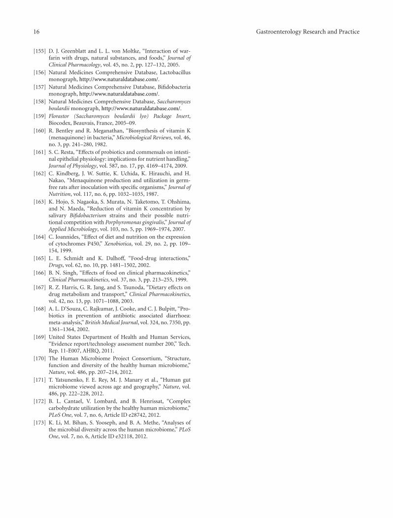
16 Gastroenterology Research and Practice
[155] D. J. Greenblatt and L. L. von Moltke, “Interaction of war-farin with drugs, natural substances, and foods,” Journal ofClinical Pharmacology, vol. 45, no. 2, pp. 127–132, 2005.
[156] Natural Medicines Comprehensive Database, Lactobacillusmonograph, http://www.naturaldatabase.com/.
[157] Natural Medicines Comprehensive Database, Bifidobacteriamonograph, http://www.naturaldatabase.com/.
[158] Natural Medicines Comprehensive Database, Saccharomycesboulardii monograph, http://www.naturaldatabase.com/.
[159] Florastor (Saccharomyces boulardii lyo) Package Insert,Biocodex, Beauvais, France, 2005–09.
[160] R. Bentley and R. Meganathan, “Biosynthesis of vitamin K(menaquinone) in bacteria,” Microbiological Reviews, vol. 46,no. 3, pp. 241–280, 1982.
[161] S. C. Resta, “Effects of probiotics and commensals on intesti-nal epithelial physiology: implications for nutrient handling,”Journal of Physiology, vol. 587, no. 17, pp. 4169–4174, 2009.
[162] C. Kindberg, J. W. Suttie, K. Uchida, K. Hirauchi, and H.Nakao, “Menaquinone production and utilization in germ-free rats after inoculation with specific organisms,” Journal ofNutrition, vol. 117, no. 6, pp. 1032–1035, 1987.
[163] K. Hojo, S. Nagaoka, S. Murata, N. Taketomo, T. Ohshima,and N. Maeda, “Reduction of vitamin K concentration bysalivary Bifidobacterium strains and their possible nutri-tional competition with Porphyromonas gingivalis,” Journal ofApplied Microbiology, vol. 103, no. 5, pp. 1969–1974, 2007.
[164] C. Ioannides, “Effect of diet and nutrition on the expressionof cytochromes P450,” Xenobiotica, vol. 29, no. 2, pp. 109–154, 1999.
[165] L. E. Schmidt and K. Dalhoff, “Food-drug interactions,”Drugs, vol. 62, no. 10, pp. 1481–1502, 2002.
[166] B. N. Singh, “Effects of food on clinical pharmacokinetics,”Clinical Pharmacokinetics, vol. 37, no. 3, pp. 213–255, 1999.
[167] R. Z. Harris, G. R. Jang, and S. Tsunoda, “Dietary effects ondrug metabolism and transport,” Clinical Pharmacokinetics,vol. 42, no. 13, pp. 1071–1088, 2003.
[168] A. L. D’Souza, C. Rajkumar, J. Cooke, and C. J. Bulpitt, “Pro-biotics in prevention of antibiotic associated diarrhoea:meta-analysis,” British Medical Journal, vol. 324, no. 7350, pp.1361–1364, 2002.
[169] United States Department of Health and Human Services,“Evidence report/technology assessment number 200,” Tech.Rep. 11-E007, AHRQ, 2011.
[170] The Human Microbiome Project Consortium, “Structure,function and diversity of the healthy human microbiome,”Nature, vol. 486, pp. 207–214, 2012.
[171] T. Yatsunenko, F. E. Rey, M. J. Manary et al., “Human gutmicrobiome viewed across age and geography,” Nature, vol.486, pp. 222–228, 2012.
[172] B. L. Cantael, V. Lombard, and B. Henrissat, “Complexcarbohydrate utilization by the healthy human microbiome,”PLoS One, vol. 7, no. 6, Article ID e28742, 2012.
[173] K. Li, M. Bihan, S. Yooseph, and B. A. Methe, “Analyses ofthe microbial diversity across the human microbiome,” PLoSOne, vol. 7, no. 6, Article ID e32118, 2012.


