

Sarcolemma in Health and DiseaseNoah Weisleder, Ph.D.
Department of Physiology and Cell Biology
Davis Heart and Lung Research Institute
The Ohio State University
[email protected](614) 292‐5321
M N il d B k C ll d Ti R h 2001

Kessel and Kardon. Tissues and Organs: a Text‐Atlas of Scanning Electron Microscopy. W.H.Freeman and Company, 1979
Sarcolemma is more than a cell membrane
• Lipid bilayer, integral proteins and associated basement membrane
• Specialized adaptations for physiological requirements of • skeletal muscle fibers
Quizlet.com

http://www.e‐heart.org/pages/01_cardiac_structure/01_Cardiac_Structure_Molecular_Anatomy_004.htmCummings, et al. Addison Wesley Longman
Specialized aspects of sarcolemma• Basement membrane• Electrical insulation• Neuromuscular junction• Link cytoskeleton to ECM• Endo‐ and Exocytosis• Propagation of action potential• Excitation/Contraction
coupling • Ion flux (calcium, etc)• Barrier function
– Sarcolemmal membrane repair

Bowman W. (1840) Philos. Trans. R. Soc. Lond. Biol. Sci. 130:457–494.
Skeletal muscle fiber basement membrane
• Extracellular matrix provides majority of the elastic properties of skeletal muscles to facilitate relaxation of muscle following contraction

Allamand et al. Skeletal Muscle 2011 1:30
Skeletal muscle fiber basement membraneBa
sal lam
ina Fibrillarlam
ina
• Highly elastic extracellular matrix• Electrical insulation• Muscle and nerve generation and regeneration
Collagen IV

Sanes, J. Journal of Biological Chemistry, 278, 12601‐12604. (2003)
Specialized extracellular matrix at the neuromuscular junction
• Relevant to a number of neuromuscular disorders
acetylcholinesterase

people.fmarion.edu/tbarbeau/physio_muscle_supplements.htmhttp://biology.clc.uc.edu/fankhauser/Labs/Anatomy_&_Physiology/A&P202/Nerve_Histology/
Neuromuscular junction in skeletal muscle
• Site of axon innervation into muscle fiber• Denervation a component of multiple diseases

Clark, et al. Annual Review of Cell and Developmental Biology Vol. 18: 637‐706 (2002)
Linking cytoskeleton to the extracellular matrix

Steele, at al. J Physiol July 1, 2007 vol. 582no. 1 17‐26
Sarcolemmal membrane as a site of endocytosis and exocytosis

2010 Pearson Education
Propagation of the action potential

Koeppen & Stanton. Berne and Levy Physiology, 6th edition (2005).
Excitation Contraction CouplingSarcolemma

Shinde, at al. Sci Signal. 2013 Mar 19;6(267):ra18.
Calcium influx through the sarcolemma

Dowling, et al. Biochem. J. (2004) 379 (479–488)
Calcium influx through the sarcolemma
• Excess Ca2+ contributes to myocyte death
• Multiple pathways contribute

Life Scicences Web Textbook CSLS/University of Tokyo
Barrier function of the sarcolemma
• Breakdown of the barrier function of the membrane following disruption can contribute to cell death

Sarcolemmal membrane repair maintains barrier function
Membrane disruption
Ca2+ entry through the injury site
Ca2+‐triggering vesicle exocytosis
Vesicle fusion
Patch formation; membrane resealing
Nature Reviews Molecular Cell Biology 6, 499-505 (June 2005)

Intracellular vesicles participate in membrane patching following damage
McNeil and Baker, Cell and Tissue Research, 2001
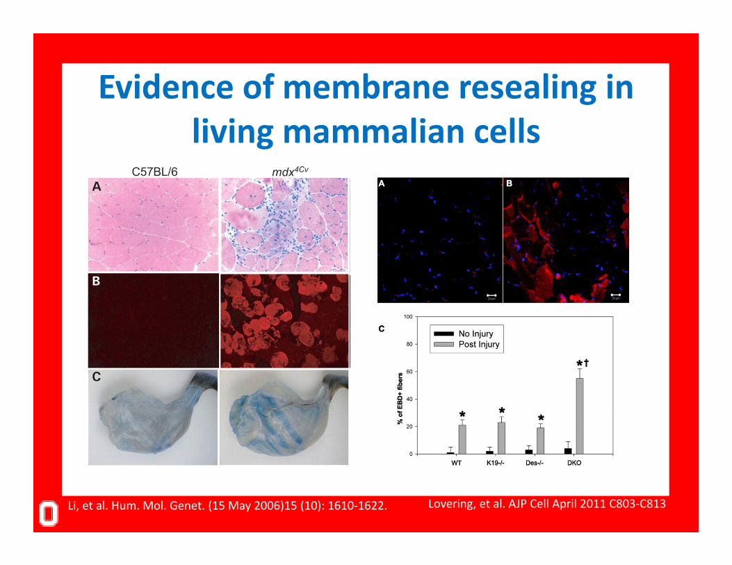
Evidence of membrane resealing in living mammalian cells
Li, et al. Hum. Mol. Genet. (15 May 2006)15 (10): 1610‐1622. Lovering, et al. AJP Cell April 2011 C803‐C813

Methods to measure membrane repair• Dye exclusion/entry into the cell
– UV laser damage– Physical disruption of the membrane– Histological analysis of tissues (Evans blue, IgG entry)
• Leak of a biomarker out of the cell– Creatine kinase– Troponins– Lactate dehydrogenase
• Changes in membrane biophysical conditions

Membrane repair is a conserved physiologic process in multiple tissues
Organ Type of mechanical force Principal cells stressed
Cell wounding (proportion of cells involved)
Reference
Skeletal muscle Aperiodic, highly variable intensity: shear, stretch
Skeletal muscle cells (myocytes) Yes (3-20%) McNeil and Khakee,
1992
Cardiac muscle Cyclic: shear, stretch Cardiac myocytes Yes (25%) Clarke et al., 1995
SkinAperiodic, highly variable intensity: shear, stretch, compression
Epidermal cells, Fibroblasts, etc. Yes (3-6%) McNeil and Ito, 1990
Gastrointestinal tract Cyclic: shear, stretch Epithelial cells, smooth
muscle cells
Yes, epithelial cells (% not measured)
McNeil and Ito, 1989
Vascular (conducting) Constant and cyclic:shear Endothelial cells,
smooth muscle cells
Yes, aortic endothelial cells
(6.5%)Yu and McNeil, 1992
Respiratory Cyclic: stretchEpithelial cells, endotherlial cells, smooth muscle cells
Alveolar cells (2-30% in
mechanically ventilated lung)
Gajic et al., 2003
Peripheral nervous Aperiodic, highly variable intensity: shear Inner ear cells Yes, hair cells (%
not measured) Mulroy et al., 1998
McNeil and Steinhardt, Annu Rev Cell Dev Biol. 2003

Necessity of membrane repair in eukaryotic cellsCellular insult
Cell Membrane
Membrane Patch = Cell Survival
MG53 drives membrane repair?

Resealing mechanism is dependent on size of membrane disruptionIncreasing size of membrane disruption
McNeil & Terasaki Nature Cell Biology 3, E124 - E129 (2001)

Membrane tension is a key determinate of cell membrane resealing
Steinhardt Annals of the New York Academy of Sciences Vol 1066 pages 152–165, March 2006

Evolution of fusion‐based resealing following development of endocytotic apparatus
McNeil & Terasaki Nature Cell Biology 3, E124 - E129 (2001)
A
B
C
D

Skeletal muscle displays a substantial amount of membrane resealing
Organ Type of mechanical force Principal cells stressed
Cell wounding (proportion of cells involved)
Reference
Skeletal muscle Aperiodic, highly variable intensity: shear, stretch
Skeletal muscle cells (myocytes) Yes (3-20%) McNeil and Khakee,
1992
Cardiac muscle Cyclic: shear, stretch Cardiac myocytes Yes (25%) Clarke et al., 1995
SkinAperiodic, highly variable intensity: shear, stretch, compression
Epidermal cells, Fibroblasts, etc. Yes (3-6%) McNeil and Ito, 1990
Gastrointestinal tract Cyclic: shear, stretch Epithelial cells, smooth
muscle cells
Yes, epithelial cells (% not measured)
McNeil and Ito, 1989
Vascular (conducting) Constant and cyclic:shear Endothelial cells,
smooth muscle cells
Yes, aortic endothelial cells
(6.5%)Yu and McNeil, 1992
Respiratory Cyclic: stretchEpithelial cells, endotherlial cells, smooth muscle cells
Alveolar cells (2-30% in
mechanically ventilated lung)
Gajic et al., 2003
Peripheral nervous Aperiodic, highly variable intensity: shear Inner ear cells Yes, hair cells (%
not measured) Mulroy et al., 1998
McNeil and Steinhardt, Annu Rev Cell Dev Biol. 2003

Contractile nature of skeletal muscles lead to extensive mechanical stress

How do cells repair the plasma membrane at the molecular level?
What are the pathologic consequences of defective membrane repair?

Cellular mechanism are better understood than the molecular mechanisms
McNeil & KirchhausenNature Reviews Molecular Cell Biology 6, 499‐505 (June 2005)

Dysferlin
McNeil & KirchhausenNature Reviews Molecular Cell Biology 6, 499‐505 (June 2005)
Originally determined to be a ferlin family protein that was known to be mutated inlimb girdle muscular dystrophy (type 2B) and Myoshi myopathy patients, (Nat Genet. 1998 Sep;20(1):31‐6).

Muscular dystrophy and compromised membrane repair in dysferlin null mice
Bansal ,et al. Nature 423, 168‐172 (May 2003)

Dysferlin function in membrane repair is not clear
Nat Clin Pract Cardiovascular Med (June 2005)
Molecular function of dysferlin in membrane repair is still not clear. Does it act asA fusogen for vesicles? Direct effects on remodeling of membrane? A platform to assemble other factors?

Annexin‐A5 assembled into two‐dimensional arrays promotes cell membrane repair
Bouterm et al. Nature Communications 2, Article number: 270 doi:10.1038/ncomms1270

AHNAK and other proteins interact with dysferlin and modulate membrane repair capacity
Huang, et al. March 2007 The FASEB Journal vol. 21 no. 3 732‐742
Multiple proteins have been shown to be important for the resealing of membranes, however the molecular function of these proteins is not clear.

Other dysferlin interacting proteins linked to membrane repair
Wallace and McNally Annual Review of Physiology Vol. 71: 37‐57 (2009)

Mitsugumin 53 (MG53) is a tripartite motif family protein linked to membrane trafficking during
membrane repair
82
40
MG53
Cai et al. Nature Cell Biology (2007)

MG53 knockout mice display myopathy and breakdown of plasma membrane integrity
Cai et al. Nature Cell Biology (2007)
wt mg53-/-
200 µm
wt (3m) 50 µm mg53-/- (10m)mg53-/- (3m)

MG53 is required for translocation of dysferlin to injury sites on the plasma membrane
Cai, Weisleder, et al. J Biol Chem. 2009 Jun 5;284(23):15894-902.
G

MG53 also associated with dysferlin to facilitate membrane repair
Han (2011) Skeletal Muscle Vol. 1 Issue 1

Proteins associated with vesicle fusion during endo/exocytosis are also involved in membrane repair
Andrews Sci. STKE, 3 May 2005 Vol. 2005, Issue 282, p. pe19

Reorganization of the cytoskeleton and organelles is required for effective membrane repair
Mellgren. J Biol Chem. 2010 November 19; 285(47): 36597–36607.

SummarySarcolemma mediates many aspects of cellular function, and loss of
these responses contributes to myocyte death in various ways
Membrane repair is a conserved physiologic process that allows resealing of larger membrane disruptions.
Loss of membrane integrity can lead to cell death unless he membrane is repaired.
Progressive loss of cells can lead to dysfunction of the tissue, in the case of skeletal muscle this can result in muscular dystrophy.
Increasing number of genes are linked to the cell repair process, however the molecular mechanism of their function will be areas of future study.

Sarcolemma in Health and DiseaseNoah Weisleder, Ph.D.
Department of Physiology and Cell Biology
Davis Heart and Lung Research Institute
The Ohio State University
[email protected](614) 292‐5321
M N il d B k C ll d Ti R h 2001


