

This article has been accepted for publication and undergone full peer review but has not been through the copyediting, typesetting, pagination and proofreading process, which may lead to differences between this version and the Version of Record. Please cite this article as doi: 10.1111/febs.12303 This article is protected by copyright. All rights reserved.
SERPINA3K induces apoptosis in human colorectal cancer cells via
activating the Fas/FasL/caspase-8 signaling pathway
Running title: SERPINA3K induces colon cancer cells apoptosis
Yachao Yao*1, Lei Li*1, Xuan Huang2, Xiaoqiong Gu3, Zumin Xu4, Yang Zhang5, Lijun
Huang1, Shuai Li1, Zhiyu Dai1, Cen Li1, Ti Zhou1, Weibin Cai1, ZhonghanYang1,Guoquan
Gao¶1,6, Xia Yang¶1,7
1Department of Biochemistry, Zhongshan School of Medicine, Sun Yat-sen University,
Guangzhou 510080, Guangdong Province, China.
2Department of Obstetrics and Gynecology, First Affiliated Hospital of Sun Yat-sen
University, Guangzhou, Guangdong Province, China.
3Department of Laboratory, Guangzhou Women and Children’s Medical Center, Guangzhou
510623, Guangdong Province, China.
4Cancer Center, Affiliated Hospital of Guangdong Medical College, Zhanjiang 524000,
Guangdong Province, China.
5Department of Medical Laboratory, Guangdong General Hospital, Guangdong Academy of
Medical Science, Guangzhou, China.
6China Key Laboratory of Tropical Disease Control (Sun Yat-sen University), Ministry of
Education, Guangzhou 510080, China.
7Key Laboratory of Functional Molecules from Marine Microorganisms (Sun Yat-sen
University), Department of Education of Guangdong Province, China.
*Y Yao and L Li contributed equally to this study.
¶ Address correspondence to: Xia Yang, Department of Biochemistry, Zhongshan School of
Medicine, Sun Yat-sen University, 74 Zhongshan 2nd Road, Guangzhou 510080, China. Acc
epte
d A
rtic
le
This article is protected by copyright. All rights reserved.
Phone: 86-20-87332020, Fax: 86-20-87332020, E-mail: [email protected];
Guoquan Gao, Department of Biochemistry, Zhongshan School of Medicine, Sun Yat-sen
University, 74 Zhongshan 2nd Road, Guangzhou 510080, China. Phone: 86-20-87332128,
Fax: 86-20-87332128, E-mail: [email protected].
Article type: Original Article
Abbreviations: CRC, colorectal cancer; FasL, Fas ligand; FADD, Fas-associated DD protein;
PPARγ, peroxisome proliferator-activated receptor γ; siRNA, small interfering RNA; MTT,
3-[4,5-dimethylthiazol-2-yl]-2,5-diphenyltetrazolium bromide; PI, propidium iodide; VEGF,
vascular endothelial growth factor; PEDF, pigment epithelium-derived factor; LRP, low
density lipoprotein receptor-related protein; sFRP, secreted frizzled-related protein.
1. Abstract
SERPINA3K, also known as kallikrein-binding protein (KBP), is a serine proteinase
inhibitor with anti-inflammatory and anti-angiogenic activities. Our previous studies showed
that SERPINA3K inhibited proliferation in a dose-dependent manner and induced apoptosis
of endothelial cells but had no influence on SGC-7901 gastric carcinoma cells or HepG2
hepatocarcinoma cells. However, it is unknown whether SERPINA3K has a direct impact on
other carcinoma cells and which mechanisms are involved. In this study, we reported for the
first time that SERPINA3K not only decreased cell viability but also induced apoptosis in the
colorectal carcinoma cell lines SW480 and HT-29. SERPINA3K-induced apoptosis of
SW480 and HT-29 was rescued by interference with FasL shRNA. Moreover, SERPINA3K
increased the expression of FasL and activated caspase-8. PPARγ,a transcription factor of
FasL, was also up-regulated by SERPINA3K in a dose-dependent manner. The up-regulation
effect of FasL induced by SERPINA3K was reversed after interference with PPARγ siRNA.
These results demonstrated that SERPINA3K-induced SW480 and HT-29 cell apoptosis was
mediated by the PPARγ/Fas/FasL signaling pathway. Therefore, our study provides additional
insight into the direct anti-tumor function by inducing tumor cell apoptosis of SERPINA3K
in colorectal tumors. Acc
epte
d A
rtic
le
This article is protected by copyright. All rights reserved.
Keywords: SERPINA3K; colorectal cancer; PPARγ; FasL; apoptosis
2. Introduction
Colorectal cancer (CRC), which includes cancer of the colon and rectum, is one of the
most common malignant tumors of the digestive tract. The mortality rate of CRC ranked third
of all cancers worldwide and second in the Western world [1]. Although treatment has
improved considerably, CRC affects approximately one million people each year with a
5-year survival rate of 62%. Traditional treatments, such as chemotherapy and surgery, have
limitations: Patients with stage IV cancer only have a median survival time of 15-18 months;
over 50% of the patients with CRC experience local recurrence or develop distant metastases
after surgery [2]. Therefore, identifying more effective and better-tolerated therapies is
critical.
Recent developments have focused on therapies that selectively target the pathways
involved in tumor growth. Inhibition of angiogenesis can be accomplished through several
strategies [3]. Angiogenesis, which forms a large nascent blood capillary at the base of an
existing blood capillary web, supplies nutrients and oxygen for tumor growth, migration and
invasion [4, 5]. The balance of pro-angiogenic factors, such as vascular endothelial growth
factor (VEGF), and angiogenesis inhibitors, such as endostatin, angiostatin, Kringle 5 and
pigment epithelium-derived factor (PEDF), is disrupted during this process. Combined with
5-fluorouacil (5-FU)-based chemotherapy in a phase III study of patients with metastatic
CRC, bevacizumab, a humanized anti-VEGF monoclonal antibody, showed significant
benefits [6]. Inhibition of angiogenesis can block the growth of endothelial cells or restore the
balance of angiogenesis effectively in various cancers including CRC.
SERPINA3K belongs to the serine proteinase inhibitor (serpin) superfamily. As with
other serpins, such as PEDF and Maspin, SERPINA3K can inhibit neovascularization.
SERPINA3K participates in a series of pathophysiological processes: hypertension [7-9], Acc
epte
d A
rtic
le
This article is protected by copyright. All rights reserved.
inflammation [10, 11], diabetes [12], optic nerve injury [13] and angiogenesis [14, 15]. Our
research found that SERPINA3K effectively inhibited retinal neovascularization and
decreased vascular leakage in an OIR model [15]. Moreover, SERPINA3K inhibited the
growth of the liver [16] and stomach xenograft tumors by anti-angiogenesis in nude mice
[17].
SERPINA3K is a multifunctional protein. However, whether SERPINA3K can suppress
the growth of tumors and induce apoptosis of tumor cells has not been concluded. Evidence
indicates that PEDF induces some tumor cell apoptosis via activating death receptor
pathways or mitochondria pathways [18]; we deduce that SERPINA3K may have the same
function in inducing apoptosis, which remains to be proven.
3. Results
3.1 SERPINA3K inhibits the proliferation of CRC cells SW480 and HT-29
To evaluate the effect of SERPINA3K on CRC cells, we separately treated CRC cells
with SERPINA3K for 72 hrs at doses of 160 nM, 320 nM, 640 nM, and 1280 nM. The MTT
assay was performed to detect the ratio of viable cells in each group (Fig. 1A). The ratio of
SW480 group compared with its own control was as follows: the ratio in 160 nM of
SERPINA3K was 83.28 ± 7.3 % (P<0.05); the ratio in 320 nM of SERPINA3K was 80.73 ±
5.2 % (P<0.05); the ratio in 640 nM of SERPINA3K was 77.35 ± 10.2 % (P<0.05); the
ratio in 1280 nM of SERPINA3K was 64.14 ± 3.2 % (P<0.01). Additionally, the ratio of the
HT-29 group compared with its own control was as follows: the ratio in 160 nM of
SERPINA3K was 94.80 ± 12.2 %; the ratio in 320 nM of SERPINA3K was 85.33 ± 7.3 % (P
<0.05); the ratio in 640 nM of SERPINA3K was 80.31 ± 9.3 % (P<0.05); the ratio in 1280
nM of SERPINA3K was 78.21 ± 4.1 % (P<0.01). These results indicated that SERPINA3K
inhibited the viability of CRC cells SW480 and HT-29 in a dose-dependent manner.
3.2 SERPINA3K induces apoptosis of CRC cells SW480 and HT-29
To investigate whether SERPINA3K can induce apoptosis of SW480 and HT-29 cells,
Hoechst 33258 and Annexin V/PI staining were employed. As shown in Fig. 1B, the number Acc
epte
d A
rtic
le

This article is protected by copyright. All rights reserved.
of apoptotic cells that presented with a strong bright blue color increased when the
SERPINA3K concentration increased from 160 nM to 1280 nM. Additionally, an Annexin
V/PI analysis showed that the apoptotic ratios of the control group and the
SERPINA3K-treated SW480 groups with 160 nM, 320 nM, 640 nM and 1280 nM of
SERPINA3K were 7.52 ± 1.2 %, 13.67 ± 2.2 %, 16.32 ± 2.2 %, 18.79 ± 1.3 % and 20.31 ±
3.2 %, respectively. The apoptotic ratio of the control group and the SERPINA3K-treated
HT-29 groups with160 nM, 320 nM, 640 nM and 1280 nM SERPINA3K were 10.08 ± 1.7 %,
12.36 ± 1.6 %, 14.37 ± 2.9 %, 15.84 ± 1.7 % and 18.33 ± 2.7 %, respectively (Fig. 1C). Our
data demonstrated that SERPINA3K induced CRC cell death via apoptosis in a
dose-dependent manner.
3.3 SERPINA3K induces SW480 and HT-29 cells apoptosis via extrinsic death pathways
To identify which signaling pathway was involved in SERPINA3K-induced CRC cell
apoptosis, we examined the cleavage of caspase-3/9/8 in SERPINA3K-treated SW480 and
HT-29 cells by Western blotting analysis. Our data showed that the cleaved caspase-3 and
caspase-8 significantly increased the caspase-8 levels in the SERPINA3K-treated group (Fig.
2A, 2B). However, caspase-9 was not activated by SERPINA3K. These results showed that
extrinsic death pathways participated in SERPINA3K-induced SW480 and HT-29 cell
apoptosis.
Fas ligand (FasL) is recognized as one of most important factors in initiating extrinsic
death pathways; it induces apoptotic cell death by binding to its receptor Fas. The protein
level of FasL was up-regulated with the presence of SERPINA3K in SW480 and HT-29 cells.
To confirm the influence of FasL on SERPINA3K-induced CRC cell apoptosis, we designed
a FasL RNAi-expressing plasmid. As shown in Fig. 4A, compared to the vector group, the
level of FasL protein was dramatically reduced in the FasL RNAi group. Subsequently, we
observed apoptosis with the treatment of SERPINA3K or FasL RNAi. Vector or the FasL
RNAi-expressing plasmid was transfected with Attractene reagent into CRC SW480 and
HT-29 cells. Twelve hours later, the medium was changed to PRMI 1640, and the cells were
treated with SERPINA3K for another 72 hrs. Using Annexin V/propidium iodide staining, we
Acc
epte
d A
rtic
le

This article is protected by copyright. All rights reserved.
analyzed the quantification of apoptotic cells by cytometry. In Figs. 4C and 4D, the
down-regulation of FasL significantly blocked SERPINA3K-induced apoptosis of SW480
and HT-29 cells, which suggested that SERPINA3K-induced apoptosis of SW480 and HT-29
cells was likely to be FasL dependent. Collectively, our results confirmed that the extrinsic
death pathway was involved in SERPINA3K-induced apoptosis.
3.4 Effect of PPARγ protein in SERPINA3K-induced apoptosis
From the time PPARγ was reported to be a transcription factor of FasL, we evaluated the
PPARγ protein level in our study. We incubated both cell types with 640 nM of SERPINA3K
for 6 hrs. Subsequently, we extracted cellular proteins for Western blotting analysis. In Fig. 5,
SERPINA3K increased the expression of PPARγ in SW480 and HT29 cells in a
dose-dependent manner. Our results demonstrated that SERPINA3K activated the Fas/FasL
pathway through PPARγ.
To claim that the PPARγ up-regulation mediates FasL up-regulation and apoptosis
induction, we employed RNAi experiments blocking PPARγ. The results showed that both
PPARγ siRNA-2 and siRNA-3 could effectively block PPARγ expression (Fig. 6A and 6B).
Then, we observed FasL expression upon treatment with SERPINA3K or PPARγ RNAi. NC
siRNA or PPARγ siRNA was transfected in CRC cells SW480 and HT-29. Twelve hours later,
the medium was changed to PRMI 1640, and the cells were treated with SERPINA3K for
another 18 h. In Fig. 6C and 6D, compared to only the PPARγ RNAi group, the PPARγ RNAi
and SERPINA3K groups did not increase the expression of FasL. The results showed that the
up-regulation effect of FasL induced by SERPINA3K was reversed after interference with
PPARγ siRNA.
4. Discussion
SERPINA3K, which is also called KBP (kallikrein-binding protein), is an important
component of the kallikrein-kinin system. It was the first special tissue kallikrein inhibitor
discovered and covalently binds to kallikrein [19], which forms a heat-stable
serpin-proteinase complex to inhibit kallikrein peptidase activity [20, 21]. The SERPINA3K Acc
epte
d A
rtic
le

This article is protected by copyright. All rights reserved.
gene is composed of five exons and four introns. It is widely expressed in tissues, including
the kidney, heart, testis, uterus, lung, salivary gland, liver, retina, vitreous and blood vessels
[22-24].
SERPINA3K is also known as kallistatin in humans. The concentration of kallistatin in
plasma was 22.1 ± 3.5 µg/ml (equivalent to 295 nM to 412 nM) in normal subjects. During
physiologic angiogenesis, kallistatin levels in the plasma of pregnant women were
significantly lower than those in non-pregnant individuals. Additionally, the protein level of
kallistatin was reduced in patients with liver disease, sepsis and inflammatory bowel disease
(IBD) [25, 26]. Furthermore, adenovirus-mediated kallistatin gene delivery markedly
suppressed the angiogenic response in human breast carcinoma xenografts of athymic mice
model [14]. These findings suggested that kallistatin levels might be decreased in some
pathological state and that kallistatin might play a role as a negative regulator of human
tumors. Although the detail about the regulation of SERPINA3K is not understood in colon
cancer in vivo, the present study provides valuable information for the treatment of colon
cancer by supplement of higher than physiological concentation of SERPINA3K.
Recent studies indicated that SERPINA3K, which inhibits vascular endothelial growth
factor or basic fibroblast growth factor-activated endothelial cells, is a potent inhibitor of
tumor angiogenesis. The vascular activities of SERPINA3K are independent of its
interactions with the kallikrein-kinin system. SERPINA3K increased apoptosis in RCEC in a
dose-dependent manner [15].
CRC showed high Wnt/β-catenin activity [27]. Wnt antagonists had two functional
classes: the sFRP class that binds directly to Wnts; and the Dickkopf class that inhibits Wnt
signaling by binding to the LRP5/LRP6 component of the Wnt receptor complex [28]. A
recent report showed that SERPINA3K was a high-affinity, endogenous Wnt antagonist of
LRP and that it blocked the Wnt pathway activation induced by Wnt ligands and diabetes
Acc
epte
d A
rtic
le

This article is protected by copyright. All rights reserved.
[12]. SERPINA3K treatment significantly inhibited the growth of gastric and hepatocellular
carcinoma by anti-angiogenesis. However, it had no effect on the proliferation and apoptosis
of gastric and hepatocellular carcinoma cells, which indicated that SERPINA3K had no direct
anti-tumor property by targeting cancer cells [16, 17]. Interestingly, SERPINA3K inhibited
the proliferation of SW480 and HT-29 cells in a dose-dependent manner. Moreover, the
Hoechst and Annexin V/PI assay showed that SERPINA3K increased the apoptotic cells. In
conclusion, we demonstrated for the first time that SERPINA3K directly inhibited the tumor
growth and induced the apoptosis of CRC cells SW480 and HT-29.
Apoptosis is triggered by the intrinsic pathway and extrinsic death pathway [29]. The
mitochondrial apoptosis pathway is initiated by signals that result from DNA damage, loss of
cell-survival factors, or other types of severe cell stress. Normally, pro-apoptotic protein
cytochrome C is released from the mitochondria to activate caspase-9 proteases and trigger
apoptosis. The intrinsic pathway hinges on the balance of activity between pro- and
anti-apoptotic signals of the Bcl-2 family. The extrinsic pathway is activated by extracellular
ligands capable of interacting with death receptors, such as Fas (also called CD95 or APO1)
or other members of the tumor necrosis factor receptor superfamily [42, 43]. Within a variety
of apoptotic stimuli, FasL induces apoptosis by binding to its receptor Fas. This type of
binding results in the recruitment of the Fas-associated death domain (FADD) protein and the
activation of caspase-8 cascade.
Research implied that PEDF also belongs to the serpin superfamily that activated
apoptosis in a number of malignancies, including prostatic [30-33], ovarian [34, 35] and
pancreatic carcinomas [36, 37], melanomas [38-40], gliomas [41-43], and osteosarcoma
[23–26]. PEDF reportedly induced the apoptosis of CT26 cells (mouse colon carcinoma cells)
but did not affect the survival of HCT116 cells (human colon carcinoma cells) [44, 45]. For
SW480 and HT-29 cells, whether PEDF works has not yet been reported. Our results
showed that Ad-PEDF distinctly inhibited the proliferation both of SW480 and HT-29 cells Acc
epte
d A
rtic
le

This article is protected by copyright. All rights reserved.
(data not shown). The results for the first time indicated that PEDF may have the same
influence on SW480 and HT-29 cells, just like SERPINA3K. Moreover, reports showed that
PEDF induces HUVECs apoptosis by up-regulation of PPARγ [46]. These results suggested
that PEDF may induce apoptosis of colon cancer cells through similar mechanism as
SERPINA3K.
Maspin, also known as Serpin B5, protease inhibitor-5a, was involved in embryonic
development, tumor invasion, metastasis and angiogenesis. In human carcinoma, Maspin
displayed no or decreased expression, including human breast carcinoma and prostate cancer
[46, 47]. Overexpressed Maspin reduced angiogenesis and increased apoptosis in a
transgenic mouse model [48]; the local delivery of Maspin to human prostate tumor cells in a
mouse model blocked tumor growth and dramatically reduced the density of
tumor-associated microvessels [49]. Furthermore, Cher et al demonstrated that Maspin
expression inhibited osteolysis, tumor growth and angiogenesis in a model of prostate cancer
bone metastasis [50, 51]. Therefore, the effects of maspin and other members of serpin on
colon cancer and the potential unique mechanism are worth to be investigated in future
study.
In our study, we found that SERPINA3K enhanced the expression of FasL in SW480
and HT-29 cells. The activity of caspase-8 was increased in the treatment of SERPINA3K.
The induction of apoptosis by PEDF was blocked by neutralizing antibodies against FasL in
melanoma cells and the MG63 osteosarcoma cell line. Our results showed that by using FasL
shRNA, knockdown of the FasL gene almost completely abolished SERPINA3K-triggered
apoptosis of CRC cells. Collectively, these data suggested that SERPINA3K induced CRC
cells SW480 and HT-29 apoptosis via FasL-dependent extrinsic apoptosis pathway. This
finding was supported by our further study which showed that SERPINA3K had no effect on
SW620 cells, another CRC cell line that is metastatic and known to become resistant to
Fas-induced apoptosis (data not shown).
It is well known that peroxisome proliferator-activated receptor gamma (PPARγ) is a
ligand-activated nuclear receptor and has key roles in the regulation of cell differentiation,
Acc
epte
d A
rtic
le

This article is protected by copyright. All rights reserved.
immune function, glucose and fatty acid metabolism [52]. PPARγ has recently been
implicated in tumor biology. The proliferation inhibition effects of PPARγ were characterized
in several types of malignant cells [53], including those derived from liposarcoma, breast
adenocarcinoma [54], prostate carcinoma, colorectal carcinoma, non-small-cell lung
carcinoma, pancreatic carcinoma, bladder cancer, gastric carcinoma, and glial tumors of the
brain [55, 56]. In the context of colon cancer, loss-of-function mutations in PPARγ were
reportedly associated with human colorectal tumorigenesis [57]. Additionally, a clinical study
published in 2009 showed that PPARγ was an independent marker for long-term survival and
good prognosis for patients [58]. These findings suggested that PPARγ played a role as a
tumor suppressor gene. However, studies on animal models and cultured cells have raised
questions because both the development and the suppression of colon cancer growth have
been observed with the activation of PPARγ. The ligands of PPARγ, such as troglitazone and
pioglitazone, inhibited the growth of colon cancer cells and colon tumor xenografts and
prevented the development of AOM- and AOM/DSS-induced aberrant crypt foci in rats [59,
60]. In APCMIN mice, an animal model of familial polyposis, PPARγ agonists promoted the
number and size of intestinal polyps [61, 62]. These contradictory results might be
attributable to the differential state of cells and tumors, such as APC mutation. Additionally,
the concentration of PPARγ agonists used is a factor that influences the effect of PPARγ [63].
Most of the studies support the anti-tumor effect of PPARγ in colon cancer. The induction of
colon cancer cell apoptosis is an important pathway that is involved in the PPARγ-triggered
inhibition of colon cancer cell growth [64].
More recently, PPARγ was reported to be the transcription factor of FasL and the
activated FasL gene promoter expression, which induced apoptosis in human MCF7 breast
cancer cells [42]. In our report, we found that SERPINA3K not only activated FasL-aroused
apoptosis but also increased the protein level of PPARγ. Moreover, the up-regulation effect of
FasL induced by SERPINA3K was reversed after interference with PPARγ siRNA. Therefore,
PPARγ/Fas/FasL signaling pathway is likely to participate in SERPINA3K-induced SW480
and HT-29 cell apoptosis. The up-regulation of PPARγ might be due to the blockade of the
Wnt pathway by SERPINA3K because PPARγ was repressed by the activated-Wnt pathway
Acc
epte
d A
rtic
le

This article is protected by copyright. All rights reserved.
[65, 66], and SERPINA3K has been identified as an antagonist of the Wnt pathway [12]. The
up-regulation of PPARγ likely resulted because SERPINA3K blocked the Wnt signaling
pathway in the current study.
In this study, we reported for the first time that SERPINA3K directly exerted anti-tumor
activity by suppressing the rate of proliferation and inducing CRC cell apoptosis. We
confirmed that SERPINA3K is the preliminary apoptosis mechanism involved in the extrinsic
pathway, which played a vital role in inducing cell death. We demonstrated that SERPINA3K
triggered apoptotic events in the CRC cell lines SW480 and HT-29 via the PPARγ/Fas/FasL
signaling pathway.
5. Materials and methods
5.1 Cell culture and transfection
SW480 and HT-29 cells were maintained at 37℃ and 5% CO2 in RPMI 1640 medium
(Hyclone, Logan, UT, USA) supplemented with 10% fetal bovine serum (FBS) (Invitrogen,
Carlsbad, CA, USA), 100 U/ml penicillin and 100 µg/ml streptomycin. Twenty-four hours
before transfection, SW480 and HT-29 cells were seeded in 6-well plates and cultured with
fresh medium to 50-70% confluence. For transient transfection of SW680 and HT-29 cells,
1.2 µg of shRNA expression vectors in a 1:3 ratio with Attractene (Qiagen, Valencia, CA)
was used as described by the manufacturer. The cells were transfected with pSilencer 1.0-U6
vector with scrambled shRNA as the control group; the cells that were not transfected also
served as a control. For siRNA transfection, scrambled siRNA and human PPARγ siRNA
were purchased from Ribobio (Guang Zhou, Guang Dong, China). Transfection of synthetic
siRNA (10 nM) was performed with HiPerFect (Qiagen, Valencia, CA, USA) according to
the manufacturer’s instructions.
5.2 Chemicals, antibodies, and protein
All of the chemicals were purchased from Sigma-Aldrich (St. Louis, MO, USA).
Antibodies against Fas and PPARγ were purchased from Santa Cruz Biotechnology (Santa
Cruz, CA, USA). Antibodies against caspase-8, FasL and β-actin were obtained separately
from Cell Signaling Technology (Beverly, MA, USA), BD Biosciences (San Jose, CA, USA)
Acc
epte
d A
rtic
le

This article is protected by copyright. All rights reserved.
and Sigma-Aldrich (St. Louis, MO, USA). The SERPINA3K/pET28 expression plasmid was
introduced into the E. coli strain BL-21/DE3 (Novagen, Madison, WI, USA). The expression
and purification of SERPINA3K were analyzed as described previously [36].
5.3 Quantification of viable cells
The cells were plated in 24-well plates (Corning, NY, USA) in triplicate and cultured
until they reached 60% confluence. The culture medium was replaced with RPMI 1640
medium and supplemented with gradient concentrations (0 nM, 160 nM, 320 nM, 640 nM or
1280 nM) of SERPINA3K. After incubation at 37℃ for 48 hrs, the growth inhibitory
function of SERPINA3K on cells was measured by the MTT
(3-[4,5-dimethylthiazol-2-yl]-2,5-dephenyl tetrazolium bromide) colorimetric assay (Sigma
Chemical Co., St. Louis, MO). Absorbance was measured at a wavelength of 570 nm. The
data represent the absorbance relative to the respective controls.
5.4 Analysis of apoptosis by Hoechst 33258 and Annexin V/propidium iodide staining
Apoptosis was assessed by Hoechst staining and Annexin V/propidium iodide (PI)
detection as described previously [12]. The CRC SW480 or HT-29 cells were plated at a
density of 1×10⁵ cells per well in 6-well plates; the next day, the cells were washed with PBS,
and the medium was subsequently replaced with RPMI 1640 medium in the presence of
SERPINA3K (160 nM to 1280 nM) for 48 h. For Hoechst staining, the cells were labeled
with 5 µg/ml Hoechst 33258 for 10–20 mins at 37℃ and examined by fluorescence
microscopy. Hoechst 33258 is a nuclear stain that labels nuclei blue and can be used as an
apoptotic marker. The apoptotic cells appear a strong bright blue in color because of the
chromatin condensation characteristic of apoptotic cells, whereas the normal healthy cells
appear a uniform blue in color. Alternatively, for Annexin V/PI analysis, after transfection of
vector or FasL shRNA for 12 h, SERPINA3K at 640 nM was added to the culture medium
RPMI 1640 for another 72 hrs.
Acc
epte
d A
rtic
le
This article is protected by copyright. All rights reserved.
5.5 FasL-targeted RNAi plasmid construction
FasL gene sequence in RNAi analysis:
Upstream primer:
5'-CGGAAGACACCTATGGAATTTTCAAGAGAAATTCCATAGGTGTCTTCC TTTTTT G-3'
Downstream primer:
5'-AATTCAAAAAAGGAAGACACCTATGGAATTTCTCTTGAAAATTCCATAGGTGTCTTCC GGCC-3'
The sequence of FasL CDS was found in GenBank, and segments of siRNA targeting
FasL mRNA were designed using siRNA-designing software
(http://www.sirna.cn/support_design.aspx). The sense strand containing 19 nucleotides was
followed by a short space (TTCAAGAGA), and the reverse complement of the sense strand
was followed by six thymidines as an RNA polymerase III transcriptional stop signal. The
oligonucleotides were annealed in the buffer [100 mM of potassium acetate, 30 mM of
Hepes/KOH (pH 7.4), 2 mM of magnesium acetate], and the mixture was incubated at 90 °C
for 3 min, and subsequently at 37 °C for 1 h. The double-stranded oligonucleotides were
cloned into an ApaI–EcoRI site in the pSilencer 1.0-U6 vector (Ambion, Austin, TX, USA) in
which shRNAs were expressed under the control of the U6 promoter. A negative control
scrambled shRNA, which had no gene sequences, was designed to detect any nonspecific
effect.
5.6 Western blotting analysis
SW480 and HT29 cells were seeded in 100-mm plates and cultured in the growth
medium until they reached 90% confluence. The culture medium was replaced with RPMI
1640 supplemented with SERPINA3K at different concentrations, and the cells were
incubated at 37 °C for the indicated time. The cells were harvested and lysed for total protein
extraction. The protein concentration was determined using a Bio-Rad protein assay kit
(Bio-Rad, Hercules, CA) according to the manufacturer’s protocol. Using an ECL detection
kit, we subjected equal amounts of protein (80 µg) from the cell lysates to Western blotting
analysis for the target protein: caspase-8, FasL and PPARγ expression. The same membrane Acc
epte
d A
rtic
le

This article is protected by copyright. All rights reserved.
was stripped and re-blotted with an antibody specific to β-actin. Target protein concentrations
were normalized using β-actin.
5.7 Statistical analysis
All of the data were expressed as the mean ± SD. All of the statistical analyses were
conducted using SPSS 13.0 software and Student’s t test. Flow Jo 7.6.1 software was used to
analyze the portion of apoptotic cells. Image J (http://rsbweb.nih.gov/ij) was used for
measuring semi-quantified densitometry. P less than 0.05 was considered statistically
significant.
6. Acknowledgments
We thank Dr. Herui Yao from Sun Yat-Sen Memorial Hospital, Sun Yat-Sen University,
for providing several CRC cell lines. We also thank the professional language editing services
of Elsevier Web Shop for proofreading the manuscript. This study was supported by National
Nature Science Foundation of China, Grants 30971208, 30973449, 81070746, 81172163,
81272338, 81272515, 81200706; Doctor innovative personnel training project of Sun Yat-sen
University; National Key Sci-Tech Special Project of China, Grants 2009ZX09103-642,
2013ZX09102-053; Program for Doctoral Station in University, Grants 20100171110049,
2011M501364; Key Project of Nature Science Foundation of Guangdong Province, China,
Grant 10251008901000009; Key Sci-tech Research Project of Guangdong Province, China,
Grant 2011B031200006; Guandong Natural Science Fund, Grants 10151008901000007,
S2012010009250, S2012040006986; Key Sci-tech Research Project of Guangzhou
Municipality, China, Grants 2011Y1-00017-8, 12A52061519; Program for Young Teacher in
University, Grants 09YKPY73, 10YKPY28; Changjiang Scholars and Innovative Research
Team in University, 985 project PCSIRT 0947. The funding sources had no role in the study
design, data collection and analysis, publishing decision, or manuscript preparation.
Acc
epte
d A
rtic
le

This article is protected by copyright. All rights reserved.
References:
1. Jemal A, Siegel R, Ward E, Hao Y, Xu J & Thun MJ (2009) Cancer statistics, 2009. CA Cancer J Clin 59,
225-249. 2. Punt CJ (2004) New options and old dilemmas in the treatment of patients with advanced colorectal cancer.
Ann Oncol 15, 1453-1459. 3. Mancuso A & Sternberg CN (2005) Colorectal cancer and antiangiogenic therapy: what can be expected in
clinical practice? Crit Rev Oncol Hematol 55, 67-81. 4. Folkman J (2002) Role of angiogenesis in tumor growth and metastasis. Semin Oncol 29, 15-18. 5. Hanahan D & Weinberg RA (2000) The hallmarks of cancer. Cell 100, 57-70. 6. Kubota Y (2012) Tumor angiogenesis and anti-angiogenic therapy. Keio J Med 61, 47-56. 7. Chao J, Stallone JN, Liang YM, Chen LM, Wang DZ & Chao L (1997) Kallistatin is a potent new
vasodilator. J Clin Invest 100, 11-17. 8. Madeddu P & Emanueli C & El-Dahr S (2007) Mechanisms of disease: the tissue kallikrein-kinin system
in hypertension and vascular remodeling. Nat Clin Pract Nephrol 3, 208-221. 9. Ma JX, Yang Z, Chao J & Chao L (1995) Intramuscular delivery of rat kallikrein-binding protein gene
reverses hypotension in transgenic mice expressing human tissue kallikrein. J Biol Chem 270, 451-455. 10. Chao J & Chai KX & Chao L (1996) Tissue kallikrein inhibitors in mammals. Immunopharmacology 32,
67-72. 11. Chai KX, Chen VC, Ni A, Lindpaintner K, Rubattu S, Chao L & Chao J (1997) Molecular cloning and
expression of rat kallistatin gene. Biochim Biophys Acta 1353, 277-286. 12. Zhang B, Abreu JG, Zhou K, Chen Y, Hu Y, Zhou T, He X & Ma JX (2010) Blocking the Wnt pathway, a
unifying mechanism for an angiogenic inhibitor in the serine proteinase inhibitor family. Proc Natl Acad Sci U S
A 107, 6900-6905. 13. Jin SX, Zeng Y, Wan J, Wan K, Li YY, Li LY, Wang TH, Feng ZT & Li Y (2010) [Kallikrein-binding
protein promotes axonal regeneration and protect rat retinal ganglion cells following optic nerve injury]. Nan
Fang Yi Ke Da Xue Xue Bao 30, 738-741. 14. Miao RQ, Agata J, Chao L & Chao J (2002) Kallistatin is a new inhibitor of angiogenesis and tumor
growth. Blood 100, 3245-3252. 15. Gao G, Shao C, Zhang SX, Dudley A, Fant J & Ma JX (2003) Kallikrein-binding protein inhibits retinal
neovascularization and decreases vascular leakage. Diabetologia 46, 689-698. 16. Lu L, Yang Z, Zhu B, Fang S, Yang X, Cai W, Li C, Ma JX & Gao G (2007) Kallikrein-binding protein
suppresses growth of hepatocellular carcinoma by anti-angiogenic activity. Cancer Lett 257, 97-106. 17. Zhu B, Lu L, Cai W, Yang X, Li C, Yang Z, Zhan W, Ma JX & Gao G (2007) Kallikrein-binding protein
inhibits growth of gastric carcinoma by reducing vascular endothelial growth factor production and angiogenesis.
Mol Cancer Ther 6, 3297-3306. 18. Fernandez-Garcia NI & Volpert OV & Jimenez B (2007) Pigment epithelium-derived factor as a
multifunctional antitumor factor. J Mol Med (Berl) 85, 15-22. 19. Chao J, Chai KX, Chen LM, Xiong W, Chao S, Woodley-Miller C, Wang LX, Lu HS & Chao L (1990)
Tissue kallikrein-binding protein is a serpin. I. Purification, characterization, and distribution in normotensive
and spontaneously hypertensive rats. J Biol Chem 265, 16394-16401. 20. Zhou GX & Chao L & Chao J (1992) Kallistatin: a novel human tissue kallikrein inhibitor. Purification,
characterization, and reactive center sequence. J Biol Chem 267, 25873-25880. 21. Chen VC & Chao L & Chao J (2000) Roles of the P1, P2, and P3 residues in determining inhibitory A
ccep
ted
Art
icle

This article is protected by copyright. All rights reserved.
specificity of kallistatin toward human tissue kallikrein. J Biol Chem 275, 38457-38466. 22. Liu YY, Nakatani T, Kogai T, Mody K & Brent GA (2011) Thyroid hormone and COUP-TF1 regulate
kallikrein-binding protein (KBP) gene expression. Endocrinology 152, 1143-1153. 23. Hatcher HC, Ma JX, Chao J, Chao L & Ottlecz A (1997) Kallikrein-binding protein levels are reduced in
the retinas of streptozotocin-induced diabetic rats. Invest Ophthalmol Vis Sci 38, 658-664. 24. Ma JX, King LP, Yang Z, Crouch RK, Chao L & Chao J (1996) Kallistatin in human ocular tissues:
reduced levels in vitreous fluids from patients with diabetic retinopathy. Curr Eye Res 15, 1117-1123. 25. Chao J, Schmaier A, Chen LM, Yang Z & Chao L (1996) Kallistatin, a novel human tissue kallikrein
inhibitor: levels in body fluids, blood cells, and tissues in health and disease. J Lab Clin Med 127, 612-620. 26. Stadnicki A, Mazurek U, Plewka D & Wilczok T (2003) Intestinal tissue kallikrein-kallistatin profile in
inflammatory bowel disease. Int Immunopharmacol 3, 939-944. 27. White BD & Chien AJ & Dawson DW (2012) Dysregulation of Wnt/beta-catenin signaling in
gastrointestinal cancers. Gastroenterology 142, 219-232. 28. Yang ZR, Dong WG, Lei XF, Liu M & Liu QS (2012) Overexpression of Dickkopf-3 induces apoptosis
through mitochondrial pathway in human colon cancer. World J Gastroenterol 18, 1590-1601. 29. Krysko DV, Vanden BT, D'Herde K & Vandenabeele P (2008) Apoptosis and necrosis: detection,
discrimination and phagocytosis. Methods 44, 205-221. 30. Doll JA, Stellmach VM, Bouck NP, Bergh AR, Lee C, Abramson LP, Cornwell ML, Pins MR, Borensztajn
J & Crawford SE (2003) Pigment epithelium-derived factor regulates the vasculature and mass of the prostate
and pancreas. Nat Med 9, 774-780. 31. Filleur S, Volz K, Nelius T, Mirochnik Y, Huang H, Zaichuk TA, Aymerich MS, Becerra SP, Yap R,
Veliceasa D, Shroff EH & Volpert OV (2005) Two functional epitopes of pigment epithelial-derived factor
block angiogenesis and induce differentiation in prostate cancer. Cancer Res 65, 5144-5152. 32. Guan M, Jiang H, Xu C, Xu R, Chen Z & Lu Y (2007) Adenovirus-mediated PEDF expression inhibits
prostate cancer cell growth and results in augmented expression of PAI-2. Cancer Biol Ther 6, 419-425. 33. Halin S, Wikstrom P, Rudolfsson SH, Stattin P, Doll JA, Crawford SE & Bergh A (2004) Decreased
pigment epithelium-derived factor is associated with metastatic phenotype in human and rat prostate tumors.
Cancer Res 64, 5664-5671. 34. Cheung LW, Au SC, Cheung AN, Ngan HY, Tombran-Tink J, Auersperg N & Wong AS (2006) Pigment
epithelium-derived factor is estrogen sensitive and inhibits the growth of human ovarian cancer and ovarian
surface epithelial cells. Endocrinology 147, 4179-4191. 35. Phillips NJ, Ziegler MR, Radford DM, Fair KL, Steinbrueck T, Xynos FP & Donis-Keller H (1996) Allelic
deletion on chromosome 17p13.3 in early ovarian cancer. Cancer Res 56, 606-611. 36. Doll JA, Stellmach VM, Bouck NP, Bergh AR, Lee C, Abramson LP, Cornwell ML, Pins MR, Borensztajn
J & Crawford SE (2003) Pigment epithelium-derived factor regulates the vasculature and mass of the prostate
and pancreas. Nat Med 9, 774-780. 37. Uehara H, Miyamoto M, Kato K, Ebihara Y, Kaneko H, Hashimoto H, Murakami Y, Hase R, Takahashi R,
Mega S, Shichinohe T, Kawarada Y, Itoh T, Okushiba S, Kondo S & Katoh H (2004) Expression of pigment
epithelium-derived factor decreases liver metastasis and correlates with favorable prognosis for patients with
ductal pancreatic adenocarcinoma. Cancer Res 64, 3533-3537. 38. Abe R, Shimizu T, Yamagishi S, Shibaki A, Amano S, Inagaki Y, Watanabe H, Sugawara H, Nakamura H,
Takeuchi M, Imaizumi T & Shimizu H (2004) Overexpression of pigment epithelium-derived factor decreases
angiogenesis and inhibits the growth of human malignant melanoma cells in vivo. Am J Pathol 164, 1225-1232. 39. Garcia M, Fernandez-Garcia NI, Rivas V, Carretero M, Escamez MJ, Gonzalez-Martin A, Medrano EE, A
ccep
ted
Art
icle

This article is protected by copyright. All rights reserved.
Volpert O, Jorcano JL, Jimenez B, Larcher F & Del RM (2004) Inhibition of xenografted human melanoma
growth and prevention of metastasis development by dual antiangiogenic/antitumor activities of pigment
epithelium-derived factor. Cancer Res 64, 5632-5642. 40. Yang H, Xu Z, Iuvone PM & Grossniklaus HE (2006) Angiostatin decreases cell migration and vascular
endothelium growth factor (VEGF) to pigment epithelium derived factor (PEDF) RNA ratio in vitro and in a
murine ocular melanoma model. Mol Vis 12, 511-517. 41. Guan M, Pang CP, Yam HF, Cheung KF, Liu WW & Lu Y (2004) Inhibition of glioma invasion by
overexpression of pigment epithelium-derived factor. Cancer Gene Ther 11, 325-332. 42. Zhang T, Guan M, Xu C, Chen Y & Lu Y (2007) Pigment epithelium-derived factor inhibits glioma cell
growth in vitro and in vivo. Life Sci 81, 1256-1263. 43. Guan M, Yam HF, Su B, Chan KP, Pang CP, Liu WW, Zhang WZ & Lu Y (2003) Loss of pigment
epithelium derived factor expression in glioma progression. J Clin Pathol 56, 277-282. 44. Cui FY, Song XR, Li ZY, Li SZ, Mu B, Mao YQ, Wei YQ & Yang L (2010) The pigment
epithelial-derived factor gene loaded in PLGA nanoparticles for therapy of colon carcinoma. Oncol Rep 24,
661-668. 45. Konson A & Pradeep S & Seger R (2010) Phosphomimetic mutants of pigment epithelium-derived factor
with enhanced antiangiogenic activity as potent anticancer agents. Cancer Res 70, 6247-6257. 46. Zou Z, Gao C, Nagaich AK, Connell T, Saito S, Moul JW, Seth P, Appella E & Srivastava S (2000) p53
regulates the expression of the tumor suppressor gene maspin. J Biol Chem 275, 6051-6054. 47. Machtens S, Serth J, Bokemeyer C, Bathke W, Minssen A, Kollmannsberger C, Hartmann J, Knuchel R,
Kondo M, Jonas U & Kuczyk M (2001) Expression of the p53 and Maspin protein in primary prostate cancer:
correlation with clinical features. Int J Cancer 95, 337-342. 48. Shi HY, Zhang W, Liang R, Kittrell F, Templeton NS, Medina D & Zhang M (2003) Modeling human
breast cancer metastasis in mice: maspin as a paradigm. Histol Histopathol 18, 201-206. 49. Schaefer JS & Zhang M (2003) Role of maspin in tumor metastasis and angiogenesis. Curr Mol Med 3,
653-658. 50. Cher ML, Biliran HJ, Bhagat S, Meng Y, Che M, Lockett J, Abrams J, Fridman R, Zachareas M & Sheng
S (2003) Maspin expression inhibits osteolysis, tumor growth, and angiogenesis in a model of prostate cancer
bone metastasis. Proc Natl Acad Sci U S A 100, 7847-7852. 51. Liu Y, Geng Y, Li K, Wang F, Zhou H, Wang W, Hou J & Liu W (2012) Comparative proteomic analysis
of the function and network mechanisms of MASPIN in human lung cells. Exp Ther Med 3, 470-474. 52. Bishop-Bailey D (2011) PPARs and angiogenesis. Biochem Soc Trans 39, 1601-1605. 53. Debril MB, Renaud JP, Fajas L & Auwerx J (2001) The pleiotropic functions of peroxisome
proliferator-activated receptor gamma. J Mol Med (Berl) 79, 30-47. 54. Rubin GL, Zhao Y, Kalus AM & Simpson ER (2000) Peroxisome proliferator-activated receptor gamma
ligands inhibit estrogen biosynthesis in human breast adipose tissue: possible implications for breast cancer
therapy. Cancer Res 60, 1604-1608. 55. Chattopadhyay N, Singh DP, Heese O, Godbole MM, Sinohara T, Black PM & Brown EM (2000)
Expression of peroxisome proliferator-activated receptors (PPARS) in human astrocytic cells: PPARgamma
agonists as inducers of apoptosis. J Neurosci Res 61, 67-74. 56. Tyagi S, Gupta P, Saini AS, Kaushal C & Sharma S (2011) The peroxisome proliferator-activated receptor:
A family of nuclear receptors role in various diseases. J Adv Pharm Technol Res 2, 236-240. 57. Sarraf P, Mueller E, Smith WM, Wright HM, Kum JB, Aaltonen LA, de la Chapelle A, Spiegelman BM &
Eng C (1999) Loss-of-function mutations in PPAR gamma associated with human colon cancer. Mol Cell 3, Acc
epte
d A
rtic
le

This article is protected by copyright. All rights reserved.
799-804. 58. Ogino S, Shima K, Baba Y, Nosho K, Irahara N, Kure S, Chen L, Toyoda S, Kirkner GJ, Wang YL,
Giovannucci EL & Fuchs CS (2009) Colorectal cancer expression of peroxisome proliferator-activated receptor
gamma (PPARG, PPARgamma) is associated with good prognosis. Gastroenterology 136, 1242-1250. 59. Sarraf P, Mueller E, Jones D, King FJ, DeAngelo DJ, Partridge JB, Holden SA, Chen LB, Singer S,
Fletcher C & Spiegelman BM (1998) Differentiation and reversal of malignant changes in colon cancer through
PPARgamma. Nat Med 4, 1046-1052. 60. Tanaka T, Kohno H, Yoshitani S, Takashima S, Okumura A, Murakami A & Hosokawa M (2001) Ligands
for peroxisome proliferator-activated receptors alpha and gamma inhibit chemically induced colitis and
formation of aberrant crypt foci in rats. Cancer Res 61, 2424-2428. 61. Saez E, Tontonoz P, Nelson MC, Alvarez JG, Ming UT, Baird SM, Thomazy VA & Evans RM (1998)
Activators of the nuclear receptor PPARgamma enhance colon polyp formation. Nat Med 4, 1058-1061. 62. Lefebvre AM, Chen I, Desreumaux P, Najib J, Fruchart JC, Geboes K, Briggs M, Heyman R & Auwerx J
(1998) Activation of the peroxisome proliferator-activated receptor gamma promotes the development of colon
tumors in C57BL/6J-APCMin/+ mice. Nat Med 4, 1053-1057. 63. Auwerx J (2002) Nuclear receptors. I. PPAR gamma in the gastrointestinal tract: gain or pain? Am J
Physiol Gastrointest Liver Physiol 282, G581-G585. 64. Lin MS, Chen WC, Bai X & Wang YD (2007) Activation of peroxisome proliferator-activated receptor
gamma inhibits cell growth via apoptosis and arrest of the cell cycle in human colorectal cancer. J Dig Dis 8,
82-88. 65. Ross SE, Hemati N, Longo KA, Bennett CN, Lucas PC, Erickson RL & MacDougald OA (2000)
Inhibition of adipogenesis by Wnt signaling. Science 289, 950-953. 66. Perobner I, Karow M, Jochum M & Neth P (2012) LRP6 mediates Wnt/beta-catenin signaling and
regulates adipogenic differentiation in human mesenchymal stem cells. Int J Biochem Cell Biol 44, 1970-1982. 67. Ho TC, Chen SL, Yang YC, Liao CL, Cheng HC & Tsao YP (2007) PEDF induces p53-mediated
apoptosis through PPAR gamma signaling in human umbilical vein endothelial cells. Cardiovasc Res 76,
213-223.
Acc
epte
d A
rtic
le

This article is protected by copyright. All rights reserved.
Figure 1. Effect of SERPINA3K on growth and apoptosis in colon cancer cell lines.
SW480 and HT29 cells were treated with increasing concentrations of SERPINA3K for 72 h.
(A) Cell proliferation was determined by the MTT assay as described, and the results are
expressed as the percentage of untreated control. *P < 0.05; **P < 0.01 compared with
untreated controls. (B) In parallel experiments, the samples were harvested for Hoechst Acc
epte
d A
rtic
le

This article is protected by copyright. All rights reserved.
staining. I, control cells; II-V, cells treated with different SERPINA3K concentrations: 160
nM, 320 nM, 640 nM, 1280 nM; VI, colchicine positive control cells. Shown are
representative pictures from three independent experiments photographed at 400×
magnification. (C) Apoptotic cells were analyzed using an Annexin V/PI assay. SERPINA3K
was used to treat SW480 and HT-29 cells at concentrations of 160 nM, 320 nM, 640 nM or
1280 nM, respectively. The data are presented as the mean ± SD. n = 3. *P < 0.05; **P <
0.01compared with controls.
Figure 2. SERPINA3K induces the cleavage of procaspase-8 in SW480 (A) and HT29
cells (B) after exposure to 640 nM of SERPINA3K for 72 hrs. The protein levels of
procaspase-3/9/8 and the cleaved caspase-3/9/8 in cell lysates were measured by Western
blotting analysis, semi-quantified densitometry and normalized by β-actin. All of the
experiments were repeated at least 3 times.
Acc
epte
d A
rtic
le

This article is protected by copyright. All rights reserved.
Figure 3. SERPINA3K increases the expression of FasL but not Fas in SW480 (A) and
HT29 cells (B). Both cell types were treated with 640 nM SERPINA3K for 18 hrs. The
protein levels of Fas and FasL were detected by Western blotting analysis, semi-quantified
densitometry and normalized by Image J software and β-actin. The data are shown as the
mean ± SD. n = 3. **P < 0.01 compared with controls.
Acc
epte
d A
rtic
le

This article is protected by copyright. All rights reserved.
Figure 4. SERPINA3K-induced apoptosis of SW480 and HT29 cells is mediated via
Fas/FasL signaling pathway. The Western blotting analysis was performed in SW480 (A) and
HT29 cells (B) transfected with FasL RNAi-expressing plasmid. SW480 (C) and HT29 (D)
cells were incubated with 640 nM of SERPINA3K for 72 hrs after transfection of the control
vector or the FasL RNAi-expressing plasmid. The apoptotic cells were evaluated by flow
cytometry. The diagrams of FITC-Annexin V/propidium iodide flow cytometry in a
representative experiment are presented above the graphs. Quantification of the apoptotic
cells is shown as the mean ± SD of triplicate analyses. *P < 0.05; **P < 0.01 versus vector
control. Acc
epte
d A
rtic
le

This article is protected by copyright. All rights reserved.
Figure 5. SERPINA3K enhances the expression of PPARγ in SW480 (A) and HT29
cells (B) in a dose-dependent manner. Both cell types were incubated with 640 nM
SERPINA3K for 6 h. The cellular proteins were subsequently extracted for Western blotting
analysis. The PPARγ protein level was quantified by densitometry and normalized relative to
the β-actin levels. The data are presented as the mean ± SD. n = 3. **P < 0.01 compared with
control.
Acc
epte
d A
rtic
le
This article is protected by copyright. All rights reserved.
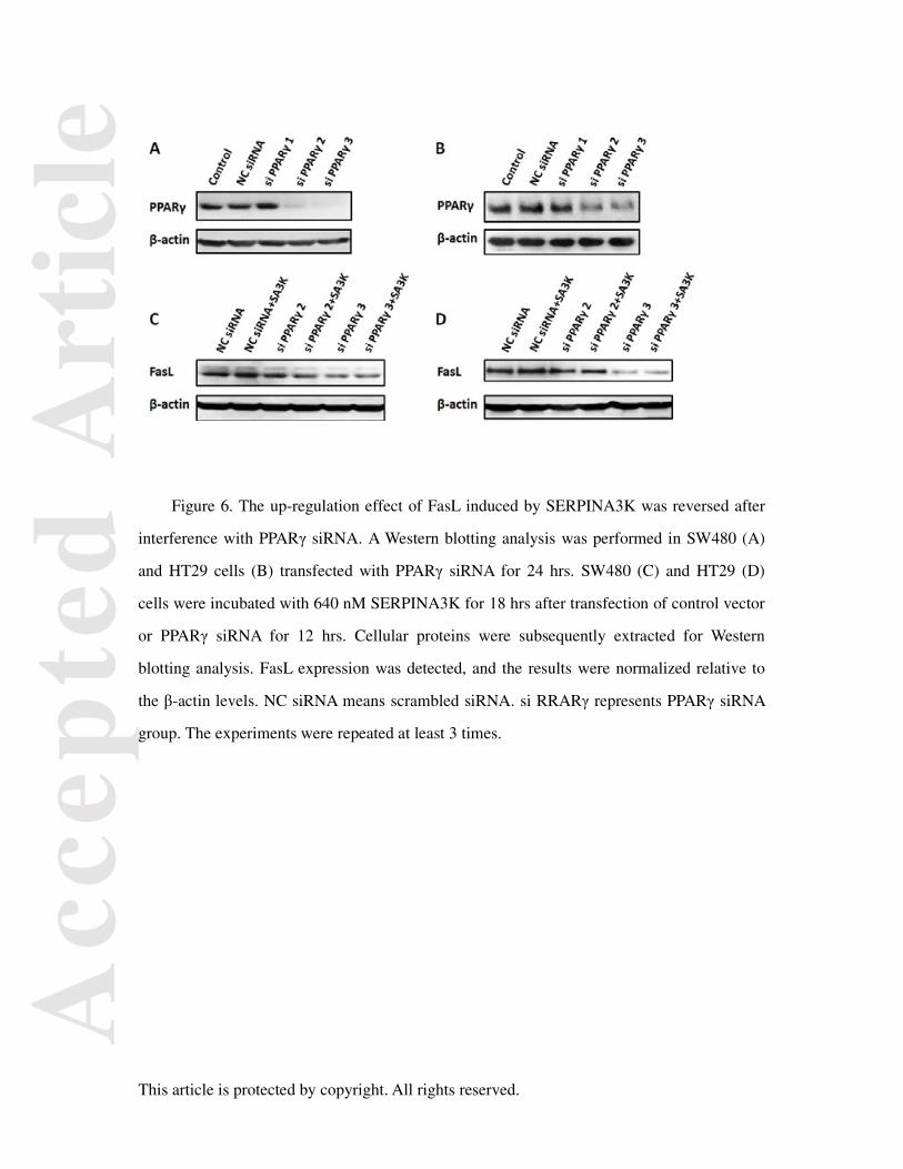
Figure 6. The up-regulation effect of FasL induced by SERPINA3K was reversed after
interference with PPARγ siRNA. A Western blotting analysis was performed in SW480 (A)
and HT29 cells (B) transfected with PPARγ siRNA for 24 hrs. SW480 (C) and HT29 (D)
cells were incubated with 640 nM SERPINA3K for 18 hrs after transfection of control vector
or PPARγ siRNA for 12 hrs. Cellular proteins were subsequently extracted for Western
blotting analysis. FasL expression was detected, and the results were normalized relative to
the β-actin levels. NC siRNA means scrambled siRNA. si RRARγ represents PPARγ siRNA
group. The experiments were repeated at least 3 times.
Acc
epte
d A
rtic
le


![1 Pensions (FAS 87); Post Retirement Benefits (FAS 106); Post Employment Benefits (FAS 112); Disclosure about Pensions, etc. (FAS 132 [R]) – amendment.](https://static.documents.pub/doc/80x56/56649d1f5503460f949f3b1c/1-pensions-fas-87-post-retirement-benefits-fas-106-post-employment-benefits.jpg)


